HAL Id: hal-02353654
https://hal.archives-ouvertes.fr/hal-02353654
Submitted on 6 May 2020
HAL is a multi-disciplinary open access
archive for the deposit and dissemination of
sci-entific research documents, whether they are
pub-lished or not. The documents may come from
teaching and research institutions in France or
abroad, or from public or private research centers.
L’archive ouverte pluridisciplinaire HAL, est
destinée au dépôt et à la diffusion de documents
scientifiques de niveau recherche, publiés ou non,
émanant des établissements d’enseignement et de
recherche français ou étrangers, des laboratoires
publics ou privés.
NKX3.1 is a direct TAL1 target gene that mediates
proliferation of TAL1-expressing human T cell acute
lymphoblastic leukemia
Sophie Kusy, Bastien Gerby, Nicolas Goardon, Nathalie Gault, Federica Ferri,
Delphine Gérard, Florence Armstrong, Paola Ballerini, Jean-Michel Cayuela,
Andre Baruchel, et al.
To cite this version:
Sophie Kusy, Bastien Gerby, Nicolas Goardon, Nathalie Gault, Federica Ferri, et al.. NKX3.1 is
a direct TAL1 target gene that mediates proliferation of TAL1-expressing human T cell acute
lym-phoblastic leukemia. Journal of Experimental Medicine, Rockefeller University Press, 2010, 207 (10),
pp.2141-2156. �10.1084/jem.20100745�. �hal-02353654�
The Rockefeller University Press $30.00
T cell acute lymphoblastic leukemia (T-ALL) is
a neoplastic disorder that occurs in 15% of
pediatric and 25% of adult acute lymphoblastic
leukemias. The TAL1/SCL gene (hereafter
referred to as TAL1) is activated by
chromo-somal translocation, interstitial deletion, or
un-known mechanisms in >40% of human T-ALLs
(Bash et al., 1995; Ferrando et al., 2002, 2004;
Armstrong and Look, 2005). This activation
re-sults in the production of a normal TAL1 protein
ectopically expressed from the double-negative
stage onwards during T-lymphopoiesis, whereas
during normal T cell differentiation, TAL1
ex-pression is restricted to the DN1-DN2 subset of
immature CD4
/CD8
thymocytes (Herblot
et al., 2000).
TAL1 encodes a class II basic
helix-loop-helix (HLH [bHLH]) transcription factor that
can activate or repress genes by forming E-box
CORRESPONDENCE Paul-Henri Romeo: paul-henri.romeo@cea.fr Abbreviations used: bHLH, basic HLH; ChIP, chromatin immuno-precipitation; HLH, helix-loop-helix; miRNA, microRNA; mRNA, messenger RNA; shRNA, short hairpin RNA; T-ALL, T cell acute lympho-blastic leukemia.
B. Gerby and N. Goardon contributed equally to this paper.
NKX3.1
is a direct TAL1 target gene that
mediates proliferation of TAL1-expressing
human T cell acute lymphoblastic leukemia
Sophie Kusy,
1,3,8,9Bastien Gerby,
2,3,8,9Nicolas Goardon,
4Nathalie Gault,
1,3,8,9Federica Ferri,
1,3,8,9Delphine Gérard,
5Florence Armstrong,
2,3,8,9Paola Ballerini,
6Jean-Michel Cayuela,
7André Baruchel,
5,7Françoise Pflumio,
2,3,8,9and Paul-Henri Roméo
1,3,8,91Laboratoire de recherche sur la Réparation et la Transcription dans les cellules Souches and 2Laboratoire de recherche
sur les cellules Souches Hématopoïétiques et Leucémiques, Institut de Radiobiologie Cellulaire et Moléculaire, Direction des Sciences du Vivant, Commissariat à l’Energie Atomique et aux Energies Alternatives, 92265 Fontenay-aux-Roses, France
3Institut National de la Santé et de la Recherche Médicale Unité 967, 92265 Fontenay-aux-Roses, France
4Medical Research Council Molecular Haematology Unit, Weatherall Institute of Molecular Medicine, University of Oxford,
Oxford OX3 9DS, England, UK
5Service Pédiatrie Hématologie et Immunologie, Hôpital Robert-Debré, 75019 Paris, France 6Service d’Hématologie et d’Oncologie Pédiatrique, Hôpital Armand Trousseau, 75012 Paris, France 7Service d’Hématologie et d’Oncologie Adulte et Pédiatrique, Hôpital Saint-Louis, 75010 Paris, France 8Université Paris-Diderot, Paris 7, 92265 Fontenay-aux-Roses, Paris, France
9Université Paris-Sud, Paris 11, 92265 Fontenay-aux-Roses, Paris, France
TAL1 (also known as SCL) is expressed in >40% of human T cell acute lymphoblastic
leukemias (T-ALLs). TAL1 encodes a basic helix-loop-helix transcription factor that can
interfere with the transcriptional activity of E2A and HEB during T cell leukemogenesis;
however, the oncogenic pathways directly activated by TAL1 are not characterized. In this
study, we show that, in human TAL1–expressing T-ALL cell lines, TAL1 directly activates
NKX3.1, a tumor suppressor gene required for prostate stem cell maintenance. In human
T-ALL cell lines, NKX3.1 gene activation is mediated by a TAL1–LMO–Ldb1 complex that is
recruited by GATA-3 bound to an NKX3.1 gene promoter regulatory sequence.
TAL1-induced NKX3.1 activation is associated with suppression of HP1- (heterochromatin
protein 1 ) binding and opening of chromatin on the NKX3.1 gene promoter. NKX3.1 is
necessary for T-ALL proliferation, can partially restore proliferation in TAL1 knockdown
cells, and directly regulates miR-17-92. In primary human TAL1–expressing leukemic cells,
the NKX3.1 gene is expressed independently of the Notch pathway, and its inactivation
impairs proliferation. Finally, TAL1 or NKX3.1 knockdown abrogates the ability of human
T-ALL cells to efficiently induce leukemia development in mice. These results suggest that
tumor suppressor or oncogenic activity of NKX3.1 depends on tissue expression.
© 2010 Kusy et al. This article is distributed under the terms of an Attribution– Noncommercial–Share Alike–No Mirror Sites license for the first six months after the publication date (see http://www.rupress.org/terms). After six months it is available under a Creative Commons License (Attribution–Noncommercial–Share Alike 3.0 Unported license, as described at http://creativecommons.org/licenses/ by-nc-sa/3.0/).
The Journal of Experimental Medicine
1998). A thorough understanding of additional genes directly
activated by TAL1 in T-ALL will provide greater insight into
the mechanisms by which TAL1 induces or maintains a
leu-kemic phenotype.
To characterize genes directly or indirectly regulated by
TAL1 in human T-ALL, we combined TAL1 knockdown
by RNA interference and gene expression profiling in human
T cell lines that express high or low levels of TAL1 protein.
In addition to already known TAL1 target genes, we have
identified the NKX3.1 gene as a potential TAL1 target gene
in human T-ALL. NKX3.1 is a homeobox gene switched on
during embryonic development of prostate tissue and then
specifically expressed in the adult prostate epithelium (Shen
and Abate-Shen, 2003). NKX3.1 is crucial for prostate stem
cell maintenance (Wang et al., 2009), and its inactivation is
one of the earliest events that occurs in prostate cancer
initia-tion, thus defining NKX3.1 as a major tumor suppressor gene
of prostate cancer (He et al., 1997; Abdulkadir et al., 2002;
Magee et al., 2003). We show that TAL1 directly activates the
transcription of the human NKX3.1 gene by binding to
reg-ulatory sequences that are not conserved in the mouse Nkx3.1
gene, suggesting differences between human TAL1-expressing
T-ALL and mouse models of TAL1-mediated T-ALL. Finally,
we document the role of NKX3.1 in human T-ALL both
in vitro and in vivo.
RESULTS
TAL1 knockdown results in decreased proliferation
of human T-ALL cell lines
We decreased TAL1 protein level using a lentiviral delivery
of shTAL1 in four human T-ALL cell lines (Jurkat, HSB2,
CEM, and RPMI) that express high (Jurkat and RPMI) or
low (HSB2 and CEM) levels of TAL1 protein (Fig. 1 a and
Fig. S1 a
). Growth curves of the four T-ALL cell lines
showed that TAL1 knockdown impaired proliferation of
these cell lines, but only when the cells were grown in low
(<2.5%) concentrations of FCS (Fig. 1 a, bottom; Fig. S1 b;
and not depicted for RPMI and CEM). These effects were
obtained with another shTAL1, indicating little or no
off-targets effects (unpublished data). To determine on which
cellular functions TAL1 was acting, doubling time,
apopto-sis, and cell cycle of Jurkat and HSB2 cell lines expressing
shCTL or shTAL1 were analyzed during the first 3 d of
cul-ture to avoid side effects caused by over-growth of
leuke-mic cells. TAL1 knockdown lengthened the doubling time
of cells (Fig. 1 a, bottom) but affected neither apoptosis nor
necrosis (not depicted). Cell cycle analyses showed fewer
cells in G1 phase and more cells in G2/M phase (Jurkat) or
S phase (HSB2; Fig. 1 b). BrdU pulse chase labeling of Jurkat
cells showed that TAL1 knockdown induced a partial
block-age of the G2/M to G1 transition (Fig. 1 c, left), and diminished
uptake of BrdU in HSB2 cells indicated a slow progression
in S phase (Fig. 1 c, right). Thus, TAL1 knockdown
re-sulted in an impaired proliferation under low serum
condi-tions as the result of modificacondi-tions of different phases of the
cell cycle.
(CANNTG)–binding heterodimers with ubiquitous class I
bHLH partners known as E-proteins, which include products
of the E2A gene (E12 and E47), HEB, and E2-2 (Hsu et al.,
1991, 1994a; Voronova and Lee, 1994). In hematopoietic
cells, TAL1 regulates the transcriptional activities of its target
genes through binding to an E-box/GATA motif and
nucle-ation of a large complex that includes an E-protein, the
LIM-only protein LMO2, GATA-1/2, Ldb1, and other associated
transcription factors (Wadman et al., 1997; Lécuyer et al.,
2002; Xu et al., 2003) or through its recruitment to
regula-tory sequences after interaction with GATA factors bound
to DNA (Lahlil et al., 2004; Fujiwara et al., 2009; Yu et al.,
2009). Functionally, TAL1 is a master regulatory protein of
primitive and definitive hematopoiesis and in the
develop-ment and maintenance of immature hematopoietic
progen-itors (Porcher et al., 1996; Robb et al., 1996; Brunet de la
Grange et al., 2006; Souroullas et al., 2009; Lacombe et al.,
2010). These TAL1 functions suggest that this transcription
factor might have an important role during initiation or
main-tenance of T cell leukemia, but the transcriptional
pro-grams directly regulated by TAL1 in human T-ALL are still
being elucidated.
The prevailing model of TAL1-induced leukemogenesis
describes TAL1 as a transcriptional repressor through its
het-erodimerization with E-proteins and blocking the E2A,
HEB, and/or E2-2 transcriptional activities (Park and Sun,
1998; O’Neil et al., 2004) and/or through a DNA binding–
independent sequestration mechanism similar to that
pro-posed for the Id family of HLH proteins (Benezra et al., 1990;
Engel and Murre, 2001). This view is supported by the fact
that E2A
/, HEB
/, and Id2–overexpressing transgenic
mice exhibit a defect in T cell differentiation, characterized
by a block at the transition of CD4/CD8 double-negative to
double-positive thymocytes and by the emergence of T-ALL
in surviving E2A-deficient mice or Id-overexpressing mice
(Bain et al., 1997; Yan et al., 1997; Morrow et al., 1999). At
the molecular level, TAL1 might exert its inhibitory effects
on E-protein target genes by interfering with E2A-mediated
recruitment of chromatin-remodeling complexes that
facili-tate transcription activation (Bradney et al., 2003) and/or by
recruitment of corepressors such as mSin3A and HDAC1
(Huang and Brandt, 2000; O’Neil et al., 2004) or Brg1 and
HDAC2 (Xu et al., 2006) on E2A, HEB, or E2-2 target genes.
In addition to the inhibition of class I bHLH functions,
TAL1 might also exert its oncogenic effects through
inap-propriate gene activation. This possibility has been suggested
by Ono et al. (1998), who have shown that TAL1 can induce
the expression of the RALDH2 gene and is strengthened by the
recent observation of a transcriptional regulatory network
downstream of TAL1 in human T-ALL, showing that TAL1
might act as a repressor or an activator of target genes
(Palomero et al., 2006). Interestingly, rather than requiring
an E-box motif, activation of the RALDH2 gene by TAL1
occurred through its recruitment to a cryptic intronic
pro-moter by DNA-bound GATA-3, although the relevance of this
target gene for leukemogenesis remains unknown (Ono et al.,
both cell lines was found (
Fig. S2
; deposited
in ArrayExpress under accession no.
E-MEXP-2197). Among these genes, we
found already known TAL1 target genes
such as RAG1, TM4SF2, or RALDH2
(Ito et al., 2003; Ono et al., 1998; Soulier
et al., 2005), thus validating the cell lines
used to identify TAL1 target genes in
human T-ALL.
Among potential TAL1 target genes,
we focused our study on the NKX3.1
gene as (a) this gene is not expressed in
normal human thymocytes but is
ectopi-cally expressed in primary T-ALL samples
expressing TAL1 (Soulier et al., 2005),
(b) NKX2.5, another member of the NKX
homeobox protein family, is activated
by chromosomal translocations in a small
number of primary human T-ALL and T cell lines (Nagel
et al., 2003; Przybylski et al., 2006), and (c) NKX3.1 is
in-volved in the maintenance of prostate stem cells and is a
tumor suppressor gene of prostate cancer (Magee et al., 2003;
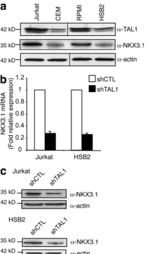
Wang et al., 2009). NKX3.1 protein level correlated with
TAL1 protein level in the four T-ALL cell lines studied
(Fig. 2 a), and NKX3.1 messenger RNA (mRNA) and
NKX3.1 protein levels were three- to fourfold decreased
when TAL1 expression was decreased in Jurkat and HSB2
cells using shTAL1 constructs (Fig. 2, b and c). These results
indicated that NKX3.1 gene expression was dependent on
Reduced TAL1 expression in Jurkat or HSB2 T cell lines
is associated with up- or down-regulation of a common set
of genes that includes the NKX3.1 gene
To identify TAL1 target genes involved in the proliferation
of T-ALL cell lines, we performed gene expression
profil-ing of Jurkat and HSB2 cell lines expressprofil-ing shTAL1 or
shCTL. We isolated RNA from Jurkat and HSB2 cells after
2 or 3 d of culture and used them to interrogate Affymetrix
DNA microarrays representing 30,000 known human genes.
Transcription profiles were compared, and a common set of
genes for which expressions were dependent on TAL1 in
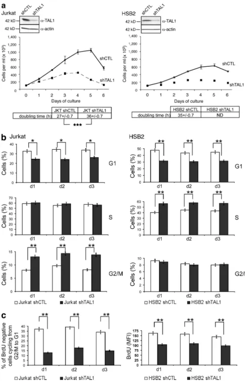
Figure 1. TAL1 regulates proliferation of Jurkat and HSB2 T-ALL cell lines. (a, top) TAL1
protein in Jurkat and HSB2 cells that stably ex-pressed shRNAs directed against human TAL1 (shTAL1) or hepatitis B virus (shCTL) was mea-sured by immunoblotting. Actin is shown as a loading control. The data shown correspond to a representative experiment out of two performed. (middle) Growth curves of Jurkat and HSB2 cells expressing shTAL1 or shCTL. Graphs show num-ber of cells ± SEM (n = 4 experiments). (bottom)
doubling times of Jurkat and HSB2 cells express-ing shCTL or shTAL1 analyzed durexpress-ing the expo-nential phase of the growth curves (***, P < 0.001). (b) Percentages of Jurkat (left) and HSB2 (right) cells expressing shCTL or shTAL1 in G1, S, and G2/M phases were determined at days 1, 2, and 3 of culture. Graphs show percentages of cells ± SEM (n = 4 experiments; *, P < 0.05; **, P < 0.01).
(c, left) Percentages of BrdU-negative Jurkat cells expressing shCTL or shTAL1 cycling from G2/M to G1 were determined at days 1, 2, and 3 of cul-ture. (right) BrdU mean fluorescence of HSB2 cells expressing shCTL or shTAL1 labeled with BrdU at days 1, 2, and 3 of culture. Graphs show percentages of cells ± SEM (n = 4 experiments;
**, P < 0.01). MFI, mean fluorescence intensity.
This region contains multiple E-boxes and GATA binding
sites (
Fig. S3 a
), and using oligonucleotides that cover all
E-boxes and GATA binding sites present in the 875/570
human NKX3.1 promoter region, we found that TAL1 and
GATA-3 can bind an E-box located at 738 and/or two
GATA binding sites at 748 and 697 (Fig. 3 c). To
iden-tify the relative contributions of these three regulatory motifs to
the transcriptional activity of the NKX3.1 gene promoter, the
977/482 sequence and associated mutants at positions
748/738 and/or 697 were cloned upstream of a minimal
promoter that drives luciferase expression and used in
tran-sient transfection assays. This promoter region could activate
transcription in Jurkat cells (Fig. 3 d) but not in NIH3T3 cells
(not depicted), and this activation was dependent on TAL1
expression (Fig. 3 d). Transcriptional activities of the different
mutants of the 977/482 promoter region pinpointed the
GATA-3–TAL1 binding site at 697 as a major regulatory
site that mediated the transcriptional activity of this NKX3.1
promoter region in Jurkat cells (Fig. 3 d). Interestingly, the
two regulatory sequences that can bind TAL1 in the human
NKX3.1 gene are not conserved in the mouse gene (Fig. S3 a),
and as a consequence, no transcriptional activity of the
corre-sponding mouse Nkx3.1 promoter region could be detected
in Jurkat cells (Fig. 3 d). These results suggest why increased
Nkx3.1 mRNA levels are not found in gene expression
pro-filing of mouse TAL1–LMO1 leukemic cells (Fig. S3 b).
At the chromatin level, GATA-3 was bound to the
NKX3.1 promoter in T cell lines expressing or not NKX3.1
(Fig. 4 a, top left). Expression of TAL1 led to its recruitment
by GATA-3 (Fig. 4 a, top right) and to the formation of a
transcriptional complex containing LMO1/2 and Ldb1
pro-teins on the NKX3.1 promoter (Fig. 4 a, bottom).
Recruit-ment (respectively suppression) of this complex on the NKX3.1
promoter was associated with suppression (respectively
recruit-ment) of HP1- (heterochromatin protein 1 ) binding and
histone H3 trimethylation on lysine 9, which are hallmarks of
gene repression (Fig. 4 b, left). The effects of TAL1
knock-down on activating (H3K14ac or H3K4me3) or repressive
(H3K27me3) histone marks further strengthened a
TAL1-dependent chromatin structure of the NKX3.1 gene promoter
in the Jurkat cell line (Fig. 4 b, right). The NKX3.1 gene
acti-vation is similar to the TAL1-mediated RALDH2 gene
activation in human T-ALL (Ono et al., 1998). We found
TAL1-dependent chromatin modifications of the RALDH2
gene similar to the ones found on the NKX3.1 promoter
(
Fig. S4
), indicating that TAL1 might regulate the chromatin
structure of a subset of genes already marked by GATA-3. As
the GATA-3–TAL1 interaction nucleates a transcriptional
complex containing Ldb1 and LMO1 in Jurkat cells, the
ex-pression of GATA-3, Ldb1, and LMO1 was knocked down
after transduction of Jurkat cells by lentiviruses that could
express short hairpin RNAs (shRNAs) targeting the
cor-responding mRNAs (Fig. 5 a), and the NKX3.1 mRNA level
was quantified in the transduced cell line. NKX3.1 mRNA
level was not affected by overexpression of E12, indicating that
NKX3.1 gene expression was not dependent on E2A protein
the presence of TAL1 in T cell lines and suggested a possible
function of the NKX3.1 protein in TAL1-mediated
prolifer-ation of human T-ALL.
The human NKX3.1 gene is directly activated by TAL1
in T-ALL cell lines through regulatory sequences
that are not conserved in the mouse Nkx3.1 gene
To determine whether TAL1 could directly activate
transcrip-tion of the NKX3.1 gene, we first expressed TAL1 in a human
T cell line, Peer, which expresses neither TAL1 nor NKX3.1.
Ectopic expression of TAL1 in Peer cells resulted in the
tran-scriptional activation of the endogenous NKX3.1 gene and
expression of the NKX3.1 protein (Fig. 3 a), indicating that
the NKX3.1 gene is a potential direct target of TAL1.
Chro-matin immunoprecipitation (ChIP) showed that TAL1 and
GATA-3, the only member of the GATA family expressed in
T cells, were specifically bound in vivo to the 875/570
region of the human NKX3.1 gene promoter (Fig. 3 b).
Figure 2. NKX3.1 mRNA and protein levels are dependent on TAL1 expression. (a) TAL1 and NKX3.1 protein in high (Jurkat and RPMI) and
low (CEM and HSB2) TAL1-expressing T cell lines was measured by immuno-blotting. Actin is shown as a loading control. The data shown correspond to one representative experiment out of two performed. Vertical black lines indicate that intervening lanes have been spliced out. (b) NKX3.1
mRNA was determined by quantitative RT-PCR normalized to GAPDH
mRNA in shCTL- or shTAL1-expressing T cell lines. Error bars indicate SEM (n = 4 experiments). (c) NKX3.1 protein was measured by Western blotting
using nuclear extracts from the indicated T cell lines. Actin is shown as a loading control. The data shown correspond to one representative experi-ment out of three performed.
increased doubling time, and did not affect
apoptosis/necro-sis of the cells (Fig. 6 a, bottom; and not depicted). NKX3.1
protein level was more reduced with shNKX3.1 than with
shTAL1 (Fig. S5 b), indicating why the effects on cell growth
of NKX3.1 knockdown could be more dramatic than TAL1
knockdown. In Jurkat and HSB2 cells, NKX3.1 knockdown
also had effects on the cell cycle similar to those of TAL1;
i.e., it diminished the percentage of cells in G1 phase and
in-creased the percentage of cells in G2/M phase (Jurkat) or
in S phase (HSB2; Fig. 6 b). Finally, constitutive expression
of NKX3.1 in shTAL1-expressing Jurkat and HSB2 cells
(Fig. 6 c, top) rescued the proliferative deficiency associated
with low TAL1 expression but did not prevent cell death
(Fig. 6 c, bottom).
To identify common target genes regulated by TAL1 and
NKX3.1 knockdown, we performed gene expression
profil-ing of Jurkat and HSB2 T cell lines expressprofil-ing shNKX3.1,
shTAL1, or shCTL. We isolated RNAs from cells after 2 d
of culture and used them to interrogate Affymetrix DNA
microarrays. Transcription profiles were compared, and we
characterized a common set of genes possibly involved in
leukemic cell proliferation and for which expressions were
level but decreased when the protein level of any member of
the TAL1-containing complex decreased (Fig. 5 b).
Alto-gether, these results indicated that ectopic expression of TAL1
in human T-lymphoid cells could activate the transcription of
the human gene NKX3.1 at its chromosomal locus via TAL1
recruitment by GATA-3 already bound to the NKX3.1
pro-moter and formation of a nuclear complex that can eliminate
repression of the NKX3.1 gene.
NKX3.1 is required for the proliferation
of TAL1-expressing T cell lines
NKX3.1-deficient mice develop prostatic hyperplasia, and
NKX3.1 has been shown to regulate cell cycle exit during
luminal cell regeneration, indicating that NKX3.1 has an
antiproliferative effect in the prostate (Lei et al., 2006). As the
NKX3.1 gene is activated by TAL1 in human T cell lines, we
studied the role of NKX3.1 in the cellular proliferation of
Jurkat and HSB2 cell lines expressing shNKX3.1. NKX3.1
pro-tein level was decreased using two different shNKX3.1
lenti-viral constructs (Fig. 6 a, top; and not depicted). NKX3.1
knockdown impaired proliferation of Jurkat and HSB2 cells
cultured in low (<2.5%) FCS concentrations (
Fig. S5 a
),
Figure 3. TAL1 directly activates NKX3.1 gene expression in human T cell lines. (a) Peer, a T-ALL cell line which expresses neither TAL1 nor
NKX3.1, was transduced with lentiviral particles containing TAL1 cDNA. Expression of TAL1 (left) and NKX3.1 (right) mRNA was studied in Peer cells (Peer NT)
or in Peer cells expressing TAL1 (Peer TAL1) by quantitative PCR normalized to GAPDH mRNA. Error bars indicate SEM (n = 4 experiments). (b) The
human 875/570 NKX3.1 promoter region (+1 is the ATG start codon) contains E-boxes (open boxes) and GATA binding sites (gray boxes). Arrows
indi-cate positions of oligonucleotides used for PCR amplification. Jurkat cell chromatin extract was immunoprecipitated with GATA-3, TAL1 antibodies, or control-matched Ig (m., mouse; r., rabbit). Immunoprecipitated DNA was amplified to detect the 790/570 NKX3.1 promoter or exon 2 sequences. Input
is amplification of Jurkat cell DNA. The results shown are representative of four independent experiments. (c) Jurkat cell nuclear extract was incubated with biotin-labeled oligonucleotides that covered E-boxes and/or GATA binding sites present in the 875/570 NKX3.1 promoter region. Proteins bound
to the different oligonucleotides were analyzed by Western blot using GATA-3 or TAL1 antibodies. The data shown correspond to one representative experiment out of three performed. (d) Jurkat cells expressing shCTL or shTAL1 were transfected with plasmids containing wild-type or mutated 977/482 sequence of the NKX3.1 gene promoter cloned upstream of a minimal promoter that drives the firefly luciferase (LUC) gene. The mutated
sequences, indicated by crosses, abolished GATA-3 and/or TAL1 binding sites. The corresponding mouse Nkx3.1 gene promoter region was cloned
up-stream of the same reporter gene and assayed in Jurkat cells. Transfections were normalized using the dual-luciferase reporter assay system, and expres-sion of Renilla luciferase provides an internal control. Data are the means ± SEM (n = 4 experiments).
decreased E2F1 protein level without any significant
de-crease of E2F1 mRNA level (Fig. 7 a). NKX3.1 could bind
a miR-17-92 gene region located 4 kb 5 from the transcription
start site (Fig. 7 b). This binding is associated with increased
(vs. decreased) binding of histone H3 marked with repressive
label (vs. activating label; H3K27me3 vs. H3K14ac) on this
regulatory sequence (Fig. 7 c), and NKX3.1 could repress
tran-scriptional activity of the 4-kb regulatory sequence of
the miR-17-92 gene (Fig. 7 d). Finally, quantitative PCR
analysis of pri-miR-17-92 showed that its expression level
inversely correlated with TAL1 and NKX3.1 expressions
(Fig. 7 e). These results identified miR-17-92 as a direct
tar-get of NKX3.1 in the Jurkat cell line. Altotar-gether, these results
showed that NKX3.1 could mediate the proliferative effects
of TAL1 in T cell lines through a direct regulation of a
spe-cific set of genes.
dependent on TAL1 and NKX3.1 expressions (
Fig. S6 a
;
de-posited in ArrayExpress under accession no. E-MEXP-2197).
Most genes have an expression dependent on TAL1 and/or
NKX3.1, whereas few genes have an expression dependent
only on NKX3.1, indicating NKX3.1 as a mediator of
TAL1-regulated pathways (Fig. S6 b).
MicroRNAs (miRNAs) are expressed in human T-ALL
(Chen et al., 2010), and the oncogenic 17-92 cluster of miRNAs
is of high importance in human hematopoietic cancers
(Mavrakis et al., 2010). Furthermore, miR-17-92 regulates
expression of E2F1 and thus of the cell cycle (Nagel et al.,
2009), and miR-19, a member of the miR-17-92 cluster, is
sufficient to promote leukemogenesis in Notch1-induced T-ALL
(Mavrakis et al., 2010). As NKX3.1 knockdown results in
growth inhibition, we studied E2F1 expression in Jurkat cells
expressing shCTL, shTAL1, or shNKX3.1 and showed a
Figure 4. TAL1 expression modifies the chromatin structure of the NKX3.1 gene promoter. (a) Jurkat shCTL, Jurkat shTAL1, Peer NT, and Peer
TAL1 cell chromatin extracts were immunoprecipitated with GATA-3 (top left), TAL1 (top right), LMO1/2 (bottom left), and Ldb1 (bottom right) antibodies, control-matched Ig, or no antibody (no Ab). Immunoprecipitated DNA was amplified to detect the 790/570 NKX3.1 promoter region as in Fig. 3 b. The
data shown correspond to one representative experiment out of two performed. (b, left) HP1-, me3H3K9 antibodies, or control Ig was used to immuno-precipitate the same chromatin extracts as in panel a. NKX3.1, Pax6 (repressed gene), and Bmi (activated gene) promoter sequences were detected by
quantitative PCR on the immunoprecipitated DNA. (right) H3K14ac, H3K4me3, and H3K27me3 antibodies or control Ig was used to immunoprecipitate chromatin extracts from Jurkat shCTL and Jurkat shTAL1. NKX3.1 promoter sequence was detected by quantitative PCR on the immunoprecipitated DNA.
Data are the means ± SEM (n = 3 experiments; **, P < 0.01).
DISCUSSION
The class II bHLH factor TAL1 is aberrantly expressed in
60% of children and in 45% of adults with T-ALL, but the
mechanisms that mediate its oncogenic potential remain
un-clear (O’Neil and Look, 2007). Using RNA interference to
knock down TAL1 expression in human T-ALL cell lines
that express low or high levels of TAL1 protein, we showed
that TAL1 regulates proliferation in these cell lines. These
results are in accordance with a previously described role for
TAL1 in promoting the aberrant growth of Jurkat cells
with-out any effect on apoptosis (Palomero et al., 2006). The
ef-fects observed on Jurkat and HSB2 cell proliferation shown
in this article are particularly drastic as we used lentiviruses
that transduce 100% of cells and consequently eliminate any
potential side effects, such as affecting clonal selection.
More-over, the cell culture conditions used (i.e., 1% FCS) allowed
us to detect this drastic effect of TAL1 knockdown on
prolif-eration, indicating that growth factors present in the serum
can complement some of the oncogenic effects of TAL1.
To study the TAL1 network that regulates the
prolifera-tion of T-ALL cell lines, we performed gene expression
pro-filing using oligonucleotide microarrays after 2 or 3 d of
culture of Jurkat or HSB2 cell lines expressing or not TAL1.
We focused our study on a set of genes whose transcriptional
activity was dependent on TAL1 expression in both cell
lines. Some of these genes, like Six6, RAG2, or RALDH2,
have been shown to be specifically expressed in TAL1-
expressing primary T-ALL blasts (Soulier et al., 2005),
indi-cating that our screening might be more relevant to human
T-ALL biology than to cell line–associated secondary events.
No obvious metabolic pathway regulated by TAL1
expres-sion could be deduced from the analysis of the set of genes
characterized in the two cell lines, and our results indicated
that TAL1 might act as an activator or a repressor in human
T-ALL, a feature previously suggested (Hsu et al., 1994b;
Palomero et al., 2006).
Among the genes whose expression depends on TAL1, we
focused our study on NKX3.1, a member of the NKX family of
homeobox genes that is specifically expressed in the prostate
NKX3.1 expression, regulation, and function in primary
human T-ALL
As cell lines may not reliably reflect patient samples, TAL1 and
NKX3.1 mRNA levels were quantified in human primary
cir-culating T-ALL blasts, and these quantifications showed a
correlation between TAL1 and NKX3.1 expressions (Fig. 8 a).
T-ALLs proliferate in bone marrow and other organs, but 90%
of the circulating blasts are in the G0 phase and do not proliferate
(
Fig. S7 a
, left). Therefore, to study any role of TAL1 and
NKX3.1 in proliferation of leukemic cells, we used the
previ-ously described MS5-DL1 mouse stromal cell line, which allows
activation, maintenance, and proliferation of primary T-ALL
leukemic cells in vitro (Fig. S7 a [right panel] and b; Armstrong
et al., 2009). Four human T-ALL primary samples that express
(M18, M61, and M69) or do not express (M30) TAL1 were
grown on MS5-DL1. TAL1 and NKX3.1 mRNA levels were
10 times higher in M18 and M61 leukemic proliferating cells
than in M18 or M61 circulating blasts, and these mRNAs were
not detected in M30 proliferating cells (Fig. 8 b, top). These
in-creased mRNA levels were mostly related to proliferation and
not to activation of the Notch signaling pathway, as shown by
the effect of DAPT, an inhibitor of this pathway, on TAL1 and
NKX3.1 mRNA levels (Fig. S7 c) and on Ki67-positive
leuke-mic cells (Fig. S7 d). Decreased TAL1 or NKX3.1 expression
abrogated proliferation of M18, M61, and M69 but not of M30
leukemic cells, indicating that these two proteins were necessary
for proliferation of primary TAL1-expressing T-ALL leukemic
cells (Fig. 8 b, bottom). Decreased TAL1 or NKX3.1 expression
also impaired leukemia development in vivo as leukemia
devel-opment from shTAL1- or shNKX3.1-expressing M18 leukemic
cells was limited compared with control cells after
transplanta-tion into immunodeficient mice (Fig. 8 c, top). Furthermore,
NKX3.1 or TAL1 expression was detected in the leukemias that
developed in mice, indicating that only blasts with little
inhibi-tion of TAL1 or NKX3.1 could escape from the proliferainhibi-tion
defect and were selected and responsible for T-ALL leukemia
development (Fig. 8 c, bottom). These results show that TAL1
and NKX3.1 are involved in efficient development of human
T-ALL in recipient immunodeficient mice.
Figure 5. LMO1, Ldb1, and GATA-3 pro-teins are necessary for NKX3.1 expression in Jurkat cells. (a) shRNAs directed against
LMO1, Ldb1, and GATA-3 decreased the cor-responding protein level compared with cells expressing an shCTL. Jurkat cells were also transduced with a lentivirus that allowed overexpression of the E12 or GFP protein. Nuclear extracts from transduced Jurkat cells were analyzed by Western blotting using the indicated antibodies. Actin is shown as a load-ing control. The data shown correspond to one representative experiment out of two performed. Vertical black lines indicate that intervening lanes have been spliced out. (b) NKX3.1 mRNA was monitored by quantitative RT-PCR normalized to GAPDH mRNA in Jurkat cells transduced
with lentiviral vectors that expressed GFP, E12 cDNA, or control shRNA or shRNA directed against LMO1, Ldb1, or GATA-3. Data are the means ± SEM (n = 4 experiments).
Figure 6. NKX3.1 mediates proliferating effects of TAL1 in human T cell lines. (a, top) NKX3.1
pro-tein in Jurkat and HSB2 cells that stably expressed shRNAs directed against human NKX3.1 (shNKX3.1) or human hepatitis B virus (shCTL) was measured by immunoblotting. Actin is shown as a loading control. The data shown correspond to one representative experiment out of two performed. (middle) Growth curves of Jurkat and HSB2 cells expressing shNKX3.1 or shCTL. Graphs show number of cells ± SEM (n = 4
experiments). (bottom) Doubling times of Jurkat and HSB2 cells expressing shCTL or shNKX3.1 analyzed during the exponential phase of the growth curves. (b) Percentages of Jurkat (left) and HSB2 (right) cells expressing shCTL or shNKX3.1 in G1, S, and G2/M phases were determined at days 1, 2, and 3 of culture. Graphs show percentages of cells ± SEM (n = 3
ex-periments; *, P < 0.05; **, P < 0.01). (c, top left) Jurkat and HSB2 cells were transduced with viral particles containing human NKX3.1 cDNA followed by an IRES/ EGFP under the control of the MND promoter. These cells were then transduced with viral particles con-taining the EF1-–EGFP construct and shTAL1 under the control of the H1 promoter. Each transduction was 100% efficient as shown by FACS analysis of EGFP expression. Dashed lines and arrows determine the fluorescent population studied. (top right) Western blot analysis of TAL1 and NKX3.1 expression in shCTL- and shTAL1-expressing Jurkat cells. “NKX3.1 + shTAL1” defined the NKX3.1-expressing Jurkat cells transduced with the shTAL1 lentivirus. Actin is shown as a loading control. The data shown correspond to one represen-tative experiment out of two performed. (bottom) Growth curves of Jurkat and HSB2 cells expressing shCTL, shTAL1, or shTAL1 and NKX3.1. Graphs show number of cells ± SEM (n = 4 experiments).
epithelium during prostate development and
in adulthood. Only one member of the NKX
family, NKX2.5, has already been
charac-terized as a master oncogene in human T-ALL.
NKX2.5 is activated only by T-ALL–specific
rearrangements (Przybylski et al., 2006) and is
expressed in two T cell lines, Peer and
CCFR-CEM (Nagel et al., 2003). In accordance with
previous studies, we did not find any
expres-sion of NKX2-5 in Jurkat, HSB2, or RPMI
T cell lines but found NKX2.5 expression
in CCFR-CEM cells (Nagel et al., 2003;
Przybylski et al., 2006). As TAL1
knock-down (respectively expression) did not affect
NKX2.5 expression in CCFR-CEM cells
(re-spectively Peer; unpublished data), we did not
study any further NKX2.5 roles in T cell lines
and focused our research on NKX3.1. We
showed that the NKX3.1 gene is directly
activated by TAL1 in T-ALL through a
GATA-3–mediated recruitment of a TAL1–
LMO–Ldb1 complex on regulatory sequences
TAL1 functions in
erythropoiesis and
T cell
leukemogen-esis with a
repres-sive or activating
function of TAL1
depending on its recruitment to DNA. Finally, as for the
RALDH2 gene, the regulatory sequences that mediated TAL1
recruitment on the NKX3.1 gene promoter are not conserved
between human and mouse, which suggests why the Nkx3.1
gene is not activated in mouse models of TAL1-mediated
T-ALL. Tissue-specific transcriptional regulation is markedly
different between human and mouse. A recent study has shown
that >50% of four tissue-specific transcription factor binding
sites are species specific, and it is thus not surprising to find
spe-cific gene activations in human T-ALL that cannot be mimicked
by mouse models (Odom et al., 2007). Together with works on
of the NKX3.1 gene. This recruitment is associated with
sup-pression of the NKX3.1 gene resup-pression at the chromatin level.
This peculiar gene activation in human T-ALL is similar to the
TAL1-mediated RALDH2 gene activation in T-ALL (Ono
et al., 1998) and pinpoints GATA-3 as an important
compo-nent of TAL1 function in human T cell leukemogenesis.
Dur-ing erythropoiesis, a TAL1–LMO2–Ldb1–E2A complex can
distinguish active from repressive GATA-1 binding sites as this
complex is only present at activating elements and is not
as-sociated with repressive GATA-1 binding sites. In human
T-ALL, TAL1–E2A direct binding to DNA is often associated
with repression of target genes after recruitment of
corepres-sors such as LSD1 or HP1- (Wen et al., 2005; Hu et al., 2009),
but we now showed that the recruitment of the TAL1–LMO–
Ldb1 complex by GATA-3 could relieve the repression of
NKX3.1 and RALDH2 genes by antagonizing HP1-
func-tion. Altogether, these results indicated a similarity between
Figure 7. NKX3.1 directly regulates miR-17-92 promoter activity. (a, left) E2F1 protein
in shCTL-, shTAL1-, and shNKX3.1-expressing Jurkat cells at days 3 and 4 of culture was measured by immunoblotting. ERK1/2 is shown as a loading control. The data shown correspond to one representative experiment out of three performed. (right) E2F1 mRNA
was determined by quantitative RT-PCR nor-malized to GAPDH mRNA in the same T cell
lines at days 3 and 4 of culture. Error bars indicate SEM (n = 4 experiments). (b, top)
Schematic organization of the miR-17-92
promoter region. The transcriptional start site (+1), TATA box (32), and exon 1 and E2F binding sites (+50 and +60) are indicated. The cluster of NKX3.1 consensus binding sites lo-cated 4 kb upstream of the transcription start site is shown by blue boxes. Location of the primer set used in the ChIP analysis is shown by arrows. (bottom) Jurkat cell chromatin ex-tract was immunoprecipitated with NKX3.1 antibody or control Ig. Immunoprecipitated DNA was amplified to detect a 300-bp region located at 4 kb in the miR-17-92 promoter
region. Input is amplification of Jurkat cell DNA. The data shown correspond to one repre-sentative experiment out of two performed. (c) H3K27me3 and H3K14ac antibodies or control Ig was used to immunoprecipitate chromatin extracts from Jurkat shCTL or Jurkat shNKX3.1. The 4-kb miR-17-92 regulatory sequence was detected by quantitative PCR on the immunoprecipitated DNA. Data are the means ± SEM (n = 3 experiments; **, P < 0.01).
(d) Jurkat cells were transfected with a plasmid containing the 4-kb miR-17-92 regulatory
sequence that binds NKX3.1 cloned upstream of a promoter that drives the firefly luciferase gene with or without an NKX3.1 cDNA se-quence. Transfections were normalized using the dual-luciferase reporter assay system. Data are the means ± SEM (n = 4 experiments).
(e) miR-17-92 RNA was determined by
quanti-tative RT-PCR normalized to 36B4 RNA in
shCTL, shTAL1, or shNKX3.1 Jurkat cells at days 3 and 4 of culture. Error bars indicate the SEM (n = 4 experiments).
rodent cells compared with human cells (Rangarajan et al.,
2004). Rodent cells seemed to need fewer events to be
trans-formed as two signaling pathways are required for tumorigenic
conversion of normal mouse fibroblasts, whereas
perturba-tion of six pathways is required for the transformaperturba-tion of
the transcriptional networks downstream of TAL1 in human
T-ALL (Rangarajan et al., 2004; Soulier et al., 2005), our study
indicates that mouse models of human T-ALL might
recapitu-late only part of human T cell leukemogenesis. Species-specific
differences have already been described for transformation of
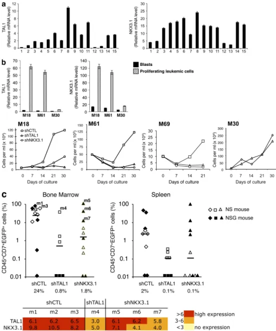
Figure 8. NKX3.1 is expressed in human TAL1-expressing T-ALL cells and is necessary for in vitro proliferation and in vivo development of human T-ALL. (a) TAL1 and NKX3.1 mRNA levels in human T-ALL blasts at diagnosis. Quantitative RT-PCR analysis of TAL1 and NKX3.1 mRNA was
per-formed using RNA prepared from blasts obtained from 15 human T-ALL. Relative expressions were normalized to 2m mRNA. Error bars represent means ±
SEM (n = 4 experiments). (b, top) TAL1 and NKX3.1 mRNA levels were analyzed by quantitative RT-PCR using RNA extracted from TAL1-expressing (M18
and M61) or non-TAL1–expressing (M30) T-ALL leukemic cells at diagnosis (black bars) or after 3 d of culture on the MS-5 cell line expressing the DL1 Notch ligand (MS5-DL1; hatched bars). Relative expressions were normalized to 2m mRNA level. Error bars indicate SEM (n = 4 experiments).
(bottom) Growth curves of M18, M61, M69, and M30 primary T-ALL expressing shCTL, shTAL1, or shNKX3.1 co-cultured with MS5-DL1 for 21 d (M69) or 1 mo. (c) M18 leukemic cells expressing shCTL, shTAL1, or shNKX3.1 were injected into NS and NSG mice. (top) percentages of human leukemic transduced (CD45+CD7+EGFP+) cells detected in bone marrow and spleen of mice at 6 (NSG) and 12 (NS) wk after transplantation. Median engraftment levels
(hori-zontal bars) are indicated under the diagram. (bottom) Quantitative RT-PCR analyses of TAL1 and NKX3.1 mRNA were performed using RNA from
CD45+CD7+EGFP+ cells isolated from mice that developed leukemia (m1–m7). Relative expression levels were normalized to 2m mRNA level and
repre-sented by a color scale (n = 3 experiments).
TAL1 and NKX3.1 in leukemia development, we used
pa-tient samples. We first studied TAL1 and NKX3.1 expression
levels in primary samples from T-ALL patients and showed a
correlation between NKX3.1 and TAL1 gene expressions, a
result which is in accordance with previous data (Soulier et al.,
2005). However, as we observed that blood-derived T-ALL
blasts are mostly quiescent cells, we switched to a cell culture
system we recently showed to allow activation and/or
prolif-eration of T-ALL leukemic cells as well as preservation of
T-ALL leukemia–initiating cell activity (Armstrong et al.,
2009). In this context, we found that TAL1 and NKX3.1
gene expressions were increased >10 times in leukemic cells
recovered after culture in comparison with uncultivated T-ALL
blasts. These results suggested that caution should be exercised
when using gene expression profiling of blood-derived T-ALL
blasts, especially when studying genes involved in the
ac-tivation or proliferation of T-ALL cells. In the context of
culture-activated leukemic cells, we found that the Notch
pathway did not significantly regulate TAL1 and NKX3.1
gene expression, indicating that the TAL1–NKX3.1 and
Notch pathways might cooperate in the development of
human T-ALL. We finally showed that NKX3.1 expression
is an important component of the development of human
T-ALL in vivo as NKX3.1 knockdown in human T-ALL
samples limited leukemia burden in immunodeficient mice.
These results strengthen the importance of NKX3.1 gene
ac-tivation by TAL1 during human T cell leukemogenesis.
Fur-ther experiments are needed to establish wheFur-ther NKX3.1
and TAL1 participate in the maintenance and/or
prolifera-tion of T-ALL leukemia–initiating cell, as we have recently
demonstrated for Notch (Armstrong et al., 2009).
In conclusion, the results described in this study
charac-terize NKX3.1 as the first gene directly activated by TAL1 in
human T-ALL and involved in the proliferation of human
T-ALL in vitro and in vivo. These findings extend the current
view of TAL1-mediated T leukemogenesis, e.g., interference
with the transcriptional regulation elicited by the tumor
sup-pressor gene E2A, and may define new pathways that could
be used for the treatment of T-ALL.
MATERIALS AND METHODS
Cell lines, T-ALL samples, and culture conditions. Jurkat, HSB2, and
Peer cells were maintained in RPMI 1640 containing 10% FCS (Invitrogen). Cells were grown in 10, 5, 2.5, and 1% FCS for the growth curve analysis, and viable cells were counted every day by trypan blue exclusion. Primary T-ALL samples were obtained and characterized (Armstrong et al., 2009) at diagnosis after informed consent of all patients’ parents, in accordance with national ethics rules, and cultured with mouse stromal MS5 cells as described previously (Armstrong et al., 2009).
Lentiviral vectors and cell transduction of cell lines and T-ALL pri-mary samples. Oligonucleotides 5-GTTCAGCCATCAGAAGTAC-3
(respectively 5-TGTGTCACTGAATATCAAC-3) were designed in the coding region of the human NKX3.1 gene and used for PCR amplification. The 230-bp DNA fragment obtained was purified and cloned in the pSuper plasmid 3 of the polIII H1 promoter. H1-shRNANKX3.1 DNA fragment was then subcloned in pTRIP/U3–EF1-–EGFP, where the GFP coding sequence is under the control of the EF1- promoter (TRIP/U3–EF1-– EGFP; Sirven et al., 2001). The lentiviral vectors pTRIP/U3–EF1-–EGFP
human fibroblasts (Rangarajan et al., 2004). Our results
indi-cated that TAL1-mediated leukemogenesis might also require
less signaling pathways in mouse than in human and shows
the importance of paralleled experiments in mouse and
human leukemia and of mouse models of human leukemia.
What could be the role of NKX3.1, a tumor suppressor
gene in human prostate cancer, in human T-ALL? To address
this question, we first inhibited NKX3.1 gene expression in
the different TAL1
+/T-ALL cell lines studied and found a
near failure of proliferation when the NKX3.1 protein was
down-regulated. These results indicated that NKX3.1 could
mediate some of the proliferative effects of TAL1 in T-ALL
and complementation experiments of TAL1 knockdown by
overexpression of NKX3.1 confirmed this hypothesis.
Dur-ing the prostate cancer initiation, NKX3.1 blocks some of
the effects of PTEN loss and inhibits AKT phosphorylation
(Lei et al., 2006). In human T-ALL, PTEN is frequently
de-leted (Maser et al., 2007), and thus the TAL1-mediated
acti-vation of NKX3.1 might block some of the effects of PTEN
loss during TAL1-mediated T cell leukemogenesis.
How-ever, in Jurkat cells, which harbor a mutated PTEN protein,
NKX3.1 knockdown did not result in a diminished AKT
phosphorylation (unpublished data), indicating that NKX3.1
regulated the proliferation of these cells using other
path-ways. The oncogenic function of NKX3.1 in human T-ALL
might also be related to the role of NKX3.1 in cellular
prolif-eration of LNCaP, a hormone-sensitive model of prostate
cancer (Possner et al., 2008). As NKX3.1 might have
differ-ent roles during prostate cancer progression, including a role
in proliferation at the early steps of prostate carcinogenesis,
our results indicated that NKX3.1 might only have
prolifera-tive functions in human T-ALL. To determine the metabolic
pathways regulated by NKX3.1 and TAL1 in human T-ALL,
we performed and analyzed gene expression profiling and
found a common set of genes for which transcription was
de-pendent on TAL1 and NKX3.1 expression in Jurkat and
HSB2. Among these genes, two subsets could be related to
the growth curves previously obtained. The first one contains
genes known to be involved in the T cell signaling and/or
cellular proliferation (Fli1, Vav3, or EiF4E3), and the second
one contains genes involved in calcium metabolism (calnexin
and calumenin) that regulates T cell proliferation (Michalak
et al., 2002; Medyouf et al., 2007). In addition to mRNAs
whose levels were dependent on NKX3.1 expression, we found
that miR-17-92 was directly regulated by NKX3.1. miR-19,
a member of the miR-17-92 cluster, has been recently shown
to promote leukemogenesis in a mouse Notch1-induced
T-ALL (Mavrakis et al., 2010), and activation of miR-17-92
by NKX2-5 suppresses apoptosis via reduction of E2F1 (Nagel
et al., 2009). Our results indicated that NKX3.1 might be a
possible link between TAL1 and the miR-17-92 transcriptional
regulation in human T-ALL, but further studies are needed to
precisely define the role of NKX3.1 on mature miRNA
ex-pression (particularly miR-19, derived from miR-17-92).
Patient-derived T-ALL cell lines may not totally reflect
the original patient samples. To further explore the roles of
Supersignal West Pico chemiluminescent substrate (Thermo Fisher Scientific). Hybridization signals were quantified by using the GS-800 calibrated densi-tometer with Quantity One 1D analysis software (Bio-Rad Laboratories).
ChIP. Immunoprecipitation conditions were determined to ensure that the
amount of antibody and percentage of cross-linking agent did not limit the ChIP yields. Cells (40 × 106) from the indicated cell lines were cross-linked
for 10 min at 37°C with 1% formaldehyde in RPMI medium containing 10% FCS, washed twice with ice-cold PBS, and lysed in 2 ml of ChIP lysis buffer for 10 min (50 mM Tris, pH 8.0, 10 mM EDTA, and 1% SDS). Chromatin fragments between 250 and 3,000 bp were obtained by sonica-tion with a Sonifier 450 (Branson).
5 µg of antibodies against HP1- (Euromedex), H3K9me3 (Millipore), H3K14ac (Millipore), H3K4me3 (Millipore), and H3K27me3 (Millipore) was used to precipitate 1 µg of fragmented chromatin. Immunoprecipitations were performed in buffer containing 0.1% SDS, 1% Triton X-100, 2 mM EDTA, 20 mM Tris-HCl, pH 8.0, 150 mM NaCl, and a protease inhibitor cocktail (Roche) at 4°C overnight. A negative control without antibody or with control Ig was run simultaneously with the samples. Immunocom-plexes were collected by adsorption to protein G agarose/Salmon Sperm DNA beads (Millipore). Washing and elution of immunocomplexes were performed, cross-linking was reversed for 5 h at 65°C, and samples were then treated with proteinase K (Invitrogen). Immunoprecipitated DNA was extracted with phenol-chloroform-isoamyl alcohol. The Bmi, Pax6, NKX3.1,
RALDH2, and miR-17-92 promoters were assessed for the presence of
his-tone modifications using quantitative PCR. Amplification was performed using power SYBR green PCR Master mix (Applied Biosystems) supple-mented with 0.5 µM of specific primer pairs and 100 ng DNA in a final vol-ume of 20 µl. Amplification conditions included an initial denaturation at 95°C for 10 min, followed by 35 cycles at 95°C for 15 s and 60°C for 60 s. Data were normalized to input signal and reported to IgG values ± SD.
4 µg -LMO1 (Abcam), 2 µg -LMO2, 2 µg -Ldb1, 5 µg -TAL1, 5 µg –GATA-3, and 5 µg -NKX3.1 antibodies (Santa Cruz Biotechnology, Inc.) were used to precipitate 30 µg of fragmented chromatin. Immunopre-cipitations were performed as described in the previous paragraphs, and PCR amplifications of eluted DNA from immunocomplexes were per-formed in 20 µl of reaction mixture using the specific primers for the
NKX3.1, RALDH2, and miR-17-92 promoters. Amplification conditions
included an initial denaturation at 95°C for 5 min, followed by 35 cycles at 94°C for 30 s, 55°C for 30 s, and 72°C for 30 s. The amplified DNA was separated on 2% agarose gels and visualized with ethidium bromide.
The sequences of the primers used were as follows: NKX3.1 promoter forward, 5-AACGCCTTCCATCCGTCTGG-3; NKX3.1 promoter re-verse, 5-TGGTGCAAACTCAGATCTGC-3; NKX3.1 exon 2 forward, 5-CCTCCCTGGTCTCCGTGTA-3; NKX3.1 exon 2 reverse, 5-TGT-CACCTGAGCTGGCATTAC-3; 4-kb miR-17-92 regulatory sequence forward, 5-CCAGATAGCAAAGATATAACAG-3; 4-kb miR-17-92 regulatory sequence reverse, 5-CTTATATGAATCACGACTGAC-3; RALDH2 intron 2 forward, 5-AGAGTGTTCCCTGTCTATAATC-CAGCC-3; RALDH2 intron 2 reverse, 5-TGTTAGAGAGGAAGAG-GCACAACTGA-3; RALDH2 exon 6 forward, 5-AACTTCCCCCT-GCTGATGTTTGCC-3; and RALDH2 exon 6 reverse, 5-CTTGAT-GAGGGCTCCCATGTAGAGTGC-3.
For quantitative PCR, the primers used were as follows: NKX3.1 moter forward, 5-ATCATCTCTGTTCCACTTTCG-3; NKX3.1 pro-moter reverse, 5-TAACCTGTTAACTTTCCTTCC-3; Pax6 propro-moter forward, 5-GTCCGGTGCCTTGAACCAT-3; Pax6 promoter re-verse, 5-GCGCAACTACCGCCTCTAAA-3; Bmi promoter forward, 5-CCAGTCTAGTGCATGCCTTCTTAA-3; Bmi promoter reverse, 5-CAAGCCAGCGACGCAGT-3; miR-17-92 promoter forward, 5-CCA-GATAGCAAAGATATAACAG-3; and miR-17-92 promoter reverse, 5-GTAACCTTGTTTGGGAAGACAC-3.
Real-time quantitative RT-PCR. Total RNA was isolated from 5 × 10⁵
cells using 1 ml TRIZOL reagent (Invitrogen) or the mirVana miRNA isolation kit (Applied Biosystems). RNA was reverse transcribed using encoding an shTAL1 or a control shCTL directed against the human
hepa-titis B virus have been described previously (Lazrak et al., 2004; Brunet de la Grange et al., 2006), and we also used another shTAL1 (5-GCAACATT-GTTCACCTTTG-3) to avoid off-target effects. The human TAL1 coding sequence cloned under the EF1- promoter (TRIP/U3–EF1-–TAL1) has been described previously (Ravet et al., 2004). The human NKX3.1 coding sequence was cloned into the pTRIPU3-Mnd-IRES-EGFP after PCR amplification using primers (forward) 5-GCGGGTGCATTCAG-GCCAAGG-3 and (reverse) 5-GGTGTGCACACCTCTTGCTCC-3 and sequenced. Concentrated VSV/G pseudotyped shRNA and cDNA len-tiviral vectors were produced as described previously (Ravet et al., 2004) and added to T-ALL cell lines cultured at a P24 concentration of 0.5 µg/ml twice at 24-h intervals. T-ALL samples were transduced with unconcentrated G/IL-7SUx envelope pseudotyped vectors (Verhoeyen et al., 2003) accord-ing to Gerby et al. (2010). In brief, T-ALL samples were spinoculated for 2 h in the presence of plastic-bound human DL1, 5 µg/ml protamine sulfate, and G/IL-7SUx pseudotyped lentiviral vectors, and transduction was continued for a further 48 h. EGFP+ T-ALL primary cells were sorted (FACSAria; BD)
and used in functional assays.
Cell cycle assay. Cells (106) were labeled with 10 µM BrdU for 45 min at
days 1, 2, and 3 of culture. Cells were washed to remove BrdU and put in chase medium (RPMI 1640 and 1% FCS) or immediately fixed. After a chase period of 2 h, cells were fixed with 70% ethanol and stored overnight at 20°C. After washing and centrifugation, cells were treated with 10 µg/ml RNase A for 20 min at 37°C. DNA was denatured with 2 N HCL/0.5% Triton X-100 for 30 min at 37°C to recover BrdU epitope and rinsed in PBS. Cells were incubated with FITC-conjugated mouse BrdU anti-body (1:10; BD) for 1 h at room temperature, washed in PBS, and then in-cubated with 20 µg/ml propidium iodide before acquisition on the flow cytometer (Mow Flow Cytometer [Beckman Coulter] and FACSCalibur [BD]). Data were analyzed with FlowJo software (Tree Star, Inc.) for bivari-ate analysis of DNA content versus BrdU.
Oligonucleotide pull-down. Cells were washed with PBS and incubated
for 10 min at 4°C in buffer A (10 mM Hepes, pH 7.6, 3 mM MgCl2, 10 mM
KCl, 5% glycerol, and 0.5% NP-40) containing proteinase inhibitor cocktail (Roche). After centrifugation, nuclear pellets were resuspended in buffer B (10 mM Hepes, pH 7.6, 3 mM MgCl2, 300 mM KCl, 5% glycerol, and 0.5%
NP-40) and incubated for 30 min at 4°C. For oligonucleotide pull-down assays, nuclear extracts from Jurkat cells were incubated with 1 µg of the indi-cated double-strand biotin-labeled oligonucleotide at 4°C overnight in buffer containing 10 mM Hepes, pH 7.6, 3 mM MgCl2, 150 mM KCl, 5% glycerol,
and 0.5% NP-40. DNA–protein complexes were pelleted using streptavidin beads (GE Healthcare), and the beads were then washed three times with buf-fer A, resuspended in Laemmli bufbuf-fer, and subjected to immunoblotting analysis using antibodies against TAL1 or GATA-3 (Santa Cruz Biotechnology, Inc.).
Biotin-labeled oligonucleotides used for the oligonucleotide pull-down were as follows: E867,
5-biotin-TACATGTTAACAGTTGGTAATCT-GCA-3; E800, 5-biotin-GCAGTTTTTGCATTTGTCCTGGCCTA-3;
G748/E738, 5-biotin-GATGGTATGTCATGTGTCTGGGGAGG-3;
G697, 5-biotin-GGTTATTGCGGATAAAGGAACCAC-3; and G577,
5-biotin-TGAGGTCGTAGATATTGCAGATCT-3.
Immunoblotting analysis. Total proteins from 1–2 × 10⁶ T-ALL cell lines
were extracted with 1× Laemmli buffer. Proteins were separated by 12% SDS-PAGE and transferred onto nitrocellulose membrane (Schleicher & Schuell). Membranes were incubated in Tris-buffered saline with 5% nonfat milk con-taining antibodies against TAL1 (3BTL73 mouse monoclonal antibody pro-vided by D. Mathieu [Centre National de la Recherche Scientifique Unité Mixte de Recherche 5535, Montpellier, France] or Santa Cruz Biotechnol-ogy, Inc.), LMO1 (Abcam), Ldb1, GATA-3, E2F1, or NKX3.1 (Santa Cruz Biotechnology, Inc.). Normalization was performed using -actin (clone AC-15; Sigma-Aldrich) or ERK1/2 antibodies (Cell Signaling Technology). After staining with secondary antibodies, the proteins were detected with
Microarray analyses were performed at the Institut de Génétique et de Biologie Moléculaire et Cellulaire French resource and by PartnerChip. HG-U133 Plus 2.0 array (Affymetrix) contains 54,600 human probe sets corresponding to 22,400 UniGene clusters. CEL files containing raw intensities were produced using GCOS (GeneChip operating software; Affymetrix). Data were normalized using GeneChip robust multi-array average, which generates intra- and interchip normalizations in a single step. Microarray data were submitted to ArrayExpress (deposited under accession no. E-MEXP-2197).
Promoter in silico analysis. NKX3.1 promoter sequence alignments
were generated using ClustalW2 (http://www.ebi.ac.uk/Tools/clustalw2/ index.html). Mouse (promoter ID 55970) and human (promoter ID 41156) sequences were obtained from the Transcriptional Regulator Element Data-base (http://rulai.cshl.edu/cgi-bin/TRED/tred.cgi?process=searchPromForm). The c13orf25 gene contains a polycistronic cluster of miRNAs (miR-17–92), and its promoter has been previously described (Ota et al., 2004; Woods et al., 2007).
Promoter reporter constructs, transient transfections, and luciferase assays. A 494-bp DNA fragment of the human NKX3.1 promoter spanning
nt 977 to 482 relative to the start codon was amplified using the prim-ers 5-ATGCCTCGAGATCATCTCTGTTCCACTTTCG-3 (forward) and 5-GCATTCTAGAGATGAGCACGCAGTCACTGC-3 (reverse). A 434-bp DNA fragment of the mouse NKX3.1 promoter spanning nt 875 to 441 relative to the start codon was amplified using the primers 5-ATGCCTCGAGACCTGGCTGTCCAAGAAATCC-3 (forward) and 5-GCATTCTAGAGAAGGTCCAAGTAGTATGAC-3 (reverse). The fragments were sequenced and cloned upstream from a TATA box sequence (5-TCTAGAGGGTATATAATGGATCTAAGTAAGCTT-3), and the resulting DNA fragments were cloned upstream from the firefly luciferase-coding sequence in pGL2b (Promega). Mutations (base substitution) of the E-box E738 and the GATA motifs G748 and G697 in the human promoter
were performed using the QuikChange II site-directed mutagenesis kit (Agilent Technologies). The binding site sequences 5-GATG-3 (G748),
5-GATA-3 (G697), and 5-CATGTG-3 (E738) were, respectively,
mutated to 5-GCCC-3, 5-GCCC-3, and 5-CCTCGG-3 sequences. Jurkat cells or Jurkat shTAL1 (107) were electroporated in a volume of 150 µl
of PBS-Hepes (10 mM, pH 7.4) by a single pulse of 200 V at 960 µF with a Gene Pulser apparatus (Bio-Rad Laboratories). 10 µg of reporter gene plas-mid and 2 µg of pRL-TK plasplas-mid (Promega) for normalization of transfec-tion efficiency were used. Cells were stimulated with 1 µM ionomycin and 50 ng/ml PMA (Sigma-Aldrich) and harvested 24 h after transfection. Lucif-erase activities were determined using the dual-lucifLucif-erase reporter assay sys-tem as indicated by the manufacturer (Promega).
A 1612-bp DNA fragment of the human miR-17-92 promoter spanning nt 4840 to 3228 relative to the transcriptional start site was amplified using the primers 5-TACTGAGGTACCAAGTGCCCCCAGAAAGGCAA-3 (forward) and 5-TACTGACTCGAGCAAAGACCATAATCATTTA-ACCTG-3 (reverse). The resulting DNA fragment was cloned into KpnI and XhoI sites of the pGL3-control vector (Promega) upstream from the firefly luciferase–coding sequence, sequenced, and cotransfected with 100 ng pcDNA3-NKX3.1 and Renilla luciferase vector (Promega) into HEK 293 cells. Luciferase activities were determined using the dual-luciferase reporter assay system as indicated by the manufacturer (Promega).
Animals. NOD.CB17-Prkdc(scid) (abbreviated NOD/scid; NS) and
NOD.Cg-Prkdc(scid)Il2rg(tm1Wjll)/SzJ (abbreviated NOD/scid/IL-2R null; NSG; both from The Jackson Laboratory) were housed in pathogen-free animal facilities at the Commissariat à l’Energie Atomique et aux Energies Alter-natives, Fontenay-aux-Roses, France. Mice were sublethally irradiated at 3 Gy (IBL 637 CisBio International, France; dose rate 0.61 Gy/min) and anesthetized with isoflurane before i.v. injection of human leukemic trans-duced (CD45+CD7+EGFP+; 5 × 10⁴/mouse) cells. All experimental procedures
were performed in compliance with French Ministry of Agriculture regulations (animal facility registration number A920322) for animal experimentation. random hexamers and the Superscript RT kit (Invitrogen) according to the
manufacturer’s instructions. The cDNA reactions were diluted 1:10 in water and used as template in real-time PCR reactions using the power SYBR green PCR Master mix or the TaqMan Universal PCR MasterMix No AmpErase UNG (Applied Biosystems; supplemented with 0.5 µM specific probe for the specific primer pairs if necessary) in the 7300 fast real-time PCR system (Applied Biosystems). The cycle at which a particular sample reaches an arbitrary threshold fluorescent level (Ct) is indicative of the input quantity of the template. The raw data were obtained in terms of Ct values that refer to the PCR cycle number during exponential amplification at which the product crosses an arbitrary threshold. To adjust for variations in the amount of RNA, the Ct values for each gene were normalized against the Ct values for the housekeeping genes, GAPDH, 2m, or 36B4. The primer sequences used for real-time quantitative PCR were the following: GAPDH forward, 5-GGGAAACTGTGGCGTGAT-3; GAPDH reverse, 5-GGAGGAGTGGGTGTCGCTGTT-3; 2m forward, 5-CACAG-CCCAAGATAGTTAAGT-3; 2m reverse, 5-CCAGCCCTCCTAG-AGC-3; NKX3.1 forward, 5-CCTCCCTGGTCTCCGTGTA-3; NKX3.1 reverse, 5-TGTCACCTGAGCTGGCATTAC-3; TAL1 forward, 5-TCT-GAAGCAAGGCGGTGGAC-3; TAL1 reverse, 5-GGAAGACCGTGCC-GTCTTCA-3; Hes1 forward, 5-CAACACGACACCGGATAAACC-3; Hes1 reverse, 5-CCAGAATGTCCGCCTTCC-3; Deltex forward, 5-TTC-TGACTTCAGGAGCGAAAG-3; Deltex reverse, 5-TGCCCACT-CCCAACGA-3; E2F1 forward, 5-CCCAACTCCCTCTACCCTTGA-3; E2F1 reverse, 5-TCTGTCTCCCTCCCTCACTTTC-3; miR-17-92 forward, 5-CAGTAAAGGTAAGGAGAGCTCAATCTC-3; miR-17-92 reverse, 5-CATACAACCACTAAGCTAAAGAATAATCTGA-3; miR-17-92 probe (200 nM final), FAM-TGGAAATAAGATCATCATGCC-CACTTGAGAC-TAMRA; 36B4 forward 5- GATGCCCAGGGAAG-ACAG-3; 36B4 reverse 5-TCTGCTCCCACAATGAAACAT-3; and 36B4 probe (50 nM final), FAM-GACCTGGA-TAMRA.
Microarray processing. Quality of RNA samples was assessed using RNA
6000 Nano chips (Agilent Technologies), and quantity was assessed by mea-suring the absorbance at 260 nm with ND-1000 (NanoDrop). cRNA was prepared by the PartnerChip Company according to the protocols of the manufacturer (Affymetrix). In brief, 2 µg of RNA samples was used to gen-erate first-strand cDNA using a T7–oligo (dT) primer and the Superscript II RT (Invitrogen). Second-strand synthesis was achieved using a cocktail of enzymes from Escherichia coli (DNA ligase and DNA polymerase I), RNase H, and T4 DNA polymerase. Clean-up of double-stranded cDNA was per-formed using the GeneChip Sample Cleanup Module (Affymetrix). In vitro transcription was performed in the presence of T7 RNA polymerase and a biotinylated nucleotide analogue/ribonucleotide mix for cRNA amplifica-tion and biotin labeling (GeneChip IVT Labeling kit; Affymetrix). Resulting biotinylated cRNAs were cleaned up using the GeneChip Sample Cleanup Module and quantified by absorbance measurement at 260 nm (NanoDrop). Starting with 5 µg of total RNA yielded between 50 and 80 µg of purified cRNA. Then, 20 µg of cRNA from samples was incubated at 94°C for 35 min in a fragmentation buffer to be reduced to a mean size of 35–200 nt and finally added to the hybridization buffer. Fragmented cRNAs were hy-bridized on HG-U133 Plus 2.0 array (Affymetrix) for 16 h at 45°C together with internal hybridization controls (bioB, bioC, bioD, cre, and oligonucle-otide B2). The washing and staining procedure was performed in the Fluid-ics Station 450 (Affymetrix). Probe arrays were exposed to 10 washes in nonstringent wash buffer A (6× saline–Na phosphate–EDTA and 0.01% Tween 20) at 30°C, followed by 6 washes in stringent buffer B (100 mM 2-[4-morpholino]ethanesulfonic acid, 0.1 M [Na+], and 0.01% Tween 20) at
50°C. The biotinylated cRNAs were stained with 10 µg/ml of a streptavi-din-phycoerythrin conjugate (SAPE) for 5 min at 35°C and washed again with nonstringent buffer A (10 washes at 30°C). An antibody amplification step was added using 0.1 mg/ml goat IgG and 3 µg/ml biotinylated anti-streptavidin antibody followed by an additional SAPE stain (5 min at 35°C for each step). Finally, arrays were washed 15 times in nonstringent buffer A at 35°C before scanning in GeneChip Scanner 3000 (Affymetrix).