HAL Id: tel-01231223
https://tel.archives-ouvertes.fr/tel-01231223
Submitted on 19 Nov 2015HAL is a multi-disciplinary open access archive for the deposit and dissemination of sci-entific research documents, whether they are pub-lished or not. The documents may come from teaching and research institutions in France or abroad, or from public or private research centers.
L’archive ouverte pluridisciplinaire HAL, est destinée au dépôt et à la diffusion de documents scientifiques de niveau recherche, publiés ou non, émanant des établissements d’enseignement et de recherche français ou étrangers, des laboratoires publics ou privés.
Crystal Engineering of Anisotropic Gold Nanoparticles
through Modulation of Seed Size and Crystal Structure
Zeliha Cansu Canbek
To cite this version:
Zeliha Cansu Canbek. Crystal Engineering of Anisotropic Gold Nanoparticles through Modulation of Seed Size and Crystal Structure. Theoretical and/or physical chemistry. Université de Versailles-Saint Quentin en Yvelines, 2014. English. �NNT : 2014VERS0052�. �tel-01231223�
Chantal Larpent
Laurence Motte
Francois Ribot
Suzanne Giorgio
Hynd Remita
Mona Treguer-Delapierre
Fabienne Testard
Nicolas Menguy
Professeur-UVSQ, Versaille
Professeur-Université Paris-Nord
Chargé de Recherche-LCMCP, Paris
Professeur-AMU, Marseille
Directrice de Recherche-Université Paris-Sud
Maitre de Conférence, Université de Bordeaux
Chercheuse, CEA,Saclay
Professeur-UPMC, Paris
Présidente
Rapporteuse
Rapporteur
Examinateur
Examinatrice
Examinatrice
Directrice de thèse
Co-Directeur de thèse
THESE DE DOCTORAT DE
L’UNIVERSITE DE VERSAILLES
SAINT-QUENTIN-EN-YVELINES
Présentée par
Zeliha Cansu Canbek
Crystal Engineering of Anisotropic Gold Nanoparticles through
Modulation of Seed Size and Crystal Structure
« Influence de taille et de la structure des germes dans la formation de
nanoparticules d’or Anisotropes »
Chantal Larpent Laurence Motte Francois Ribot Suzanne Giorgio Hynd Remita Mona Treguer-Delapierre Fabienne Testard Nicolas Menguy Professeur-UVSQ, Versaille Professeur-Université Paris-Nord Chargé de Recherche-LCMCP, Paris Professeur-AMU, Marseille
Directrice de Recherche-Université Paris-Sud Maitre de Conférence, Université de Bordeaux Chercheuse, CEA,Saclay Professeur-UPMC, Paris Présidente Rapporteuse Rapporteur Examinateur Examinatrice Examinatrice Directrice de thèse Co-Directeur de thèse
THESE DE DOCTORAT DE
L’UNIVERSITE DE VERSAILLES SAINT-QUENTIN-EN-YVELINES
Présentée par
Zeliha Cansu Canbek
pour obtenir le grade de
DOCTEUR de l’UNIVERSITE de VERSAILLES SAINT-QUENTIN-En-YVELINES
Domaine: Physico-Chimie
Crystal Engineering of Anisotropic Gold Nanoparticles through
Modulation of Seed Size and Structure
Influence de taille et de la structure des germes dans la formation de
nanoparticules d’or Anisotropes
i
Acknowledgements
People who know me would agree that my journey in Paris during my PhD thesis have been quite colorful. So now I would like to use this opportunity to express my gratitude to everyone who supported me throughout this time and participated to this colorful journey with me.To begin with, I would like to express my special appreciation and thanks to the thesis committee members, Laurence Motte and Francois Ribot for accepting to be reporters on this dissertation. I also want to thank to Suzanne Giorgio, Hynd Remita and Mona Treguer-Delapierre for their participation to my thesis as jury members and Chantal Larpent for directing as president.
During my PhD thesis I was lucky because instead of having one I had three incredible scientists supporting me all this time as my supervisors. Fabienne Testard, you have been a tremendous mentor for me during this time. Thank you for allowing me to grow as a research scientist with your strong guidance, priceless advices and end full patience. Nicolas Menguy, this project would not be possible without your valuable contributions. You enthusiasm and immense knowledge helped me all this time to improve myself and become better every day. And lastly Olivier Spalla, your inspiring ideas were most valuable during this research and making it possible me to accomplish this PhD.
In this ANR project I had the honor to work with great scientist whom I am truly grateful. Luc Belloni, Florent Malloggi, Claudine Noguera, Jacek Goniakowski, Ali Abou Hassan and Veronique Peyre thank you for your insightful comments and precious discussions. I am so glad to meet both Robinson Cortes-Huerto and Blaise Fleury great young scientists and sincere friends.
If you have read my thesis you would understand that this work has been achieved mostly with the help of our valuable collaborators: Damien Alloyeau, Guillaume Yangshu Wang and Christian Ricolleau thank you for your great help and patience during in-situ STEM experiments. Ovidiu Ersen, Simona Moldovan and Andreas Wisnet thank you for your contributions to this thesis with the tomography experiments. Hynd Remita, thank you for letting us this opportunity to use gamma source for radiolysis experiments and obtaining the most beautiful nanorods sample that we have ever produced. Lastly Pierre-Eugene Coluon, thank you for your helps in microscopy experiments.
ii
I was cherished with great colloquies during this journey who motivate me every day and helped me to pass such a memorable time in LIONS, CEA. So thank you so much Elodie, Valerie, Olivier Tache, Michelle, Stephanie, Debasish, Sergio and all the other members of LIONS team.
And lastly, I am incredibly grateful to my two families: to my mother, aunt and brothers for supporting me all my life and to my surrogate family Brigitte Barraud and Dominique Coudignac for making my life easier here 3000 km away from my home town. I will always be there for you when you need me as you were for me.
Sebastien Monteagudo, thank you for telling me that everything will be perfect. In fact you were right, it did. I hope we meet again one day.
iii
Abstract
Between the ongoing research on various type of nanomaterials to tune the particle size and crystal design in nanoscale for their potential applications, anisotropic gold nanoparticles has attracted the most intention not only because of their divine color but also their enhanced catalytic activities, optical properties and electrical conductivities. Event though, many efforts have been already made in the field of synthesis of anisotropic gold nanoparticles, with defined sizes and structures, growth mechanism of many unique anisotropic shapes is still a controversial subject.Overall objective of this thesis is to understand the origin of anisotropy during the formation of anisotropic gold nanoparticles, especially gold nanorods, in liquid phase. For our envisaged aim, between numerous synthetic methods developed for production of nanoparticles, seed mediated approach is chosen for the fabrication of final anisotropic gold nanoparticles from small seeds which is grown into final nanoparticle later on. During the synthesis of nanoparticles, those seeds play critical role as precursors to control the yield of and the crystal structure of final anisotropic nanoparticle. Here we offer a systematical study on the origin of anisotropy with respect to “seed size” and “crystal morphology”. Since these small particles are the genesis of anisotropic metal nanoparticle synthesis, in this thesis we answer following questions to explain the origin of anisotropy;
i. How to control the crystal structure and the size of the seeds?
ii. What are the influences of controlled seed size and structure on the kinetics of nanoparticle growth?
Résume
De par leurs nombreuses applications potentielles, de nombreux efforts de recherche ont été poursuivis dans le domaine de la synthèse de nanoparticules. Cependant le mécanisme à l'origine des formes anisotropes de nanoparticules d'or, pour une taille et une structure bien définies, reste encore un sujet controverse.L'objectif général de cette thèse est de comprendre l'origine de cette anisotropie, lors de la formation de nanoparticules d'or, en particulier sous la forme de nano-bâtonnets d'or, en phase liquide. Parmi les nombreux procédés de synthèse existants, la "synthèse de particules anisotropes par croissance à partir de germes" a été retenu, car il permet un contrôle précis de la taille et de la structure des nanoparticules. Lors de la synthèse de nanoparticules, les germes jouent un rôle de précurseur et permettent ainsi de maitriser la structure cristalline des nanoparticules finales. Si le rôle crucial des germes a déjà pu être étudié par différents groupes de recherche, une étude systématique sur la genèse de l'anisotropie par rapport à la taille et la structure initale des germes restait à réaliser. Ce travail a ainsi pour objectif de répondrre aux deux questions :
i. Comment contrôler la structure cristalline et de la taille des germes?
ii. Quelles sont les influences de la taille des germes et de leur structure sur la cinétique de la croissance?
v
Table of Contents
ABSTRACT ... iii TABLE OF CONTENTS ... v ABBREVIATIONS ... ix INTRODUCTION ... xiCHAPTER I General Introduction ... 1
1.1 Brief Overwiew on Metallic Nanoparticles ... 5
1.2 Gold Nanoparticles: Why they are more valuable than bulk gold? ... 6
1.2.1 Anisotropic Au Nanoparticles: Properties and Synthesis ... 8
1.2.1.1 Milestones in the Seed Mediated Synthesis of Gold Nanorods ... 10
1.2.1.2 Shape Control in Seed Mediated Synthesis of Gold Nanorods ... 12
i) Role of Growth Solution During Formation of Anisotropy ... 12
ii) Role of Seeds During Formation of Anisotropy ... 16
iii) Crystal Structure of Aniostropic Nanoparticles ... 19
1.3 Charactherisation Methods ... 21
1.4 References ... 24
CHAPTER II Role of Seed on the Development of Anisotropy ... 29
2.1 Materials and Methods ... 33
2.1.1 Synthesis of Different Type of Seeds ... 33
2.1.1.1 Preperation of CTAB Capped Seed ... 33
2.1.1.2 Preperation of Citrate Capped Seeds ... 34
2.1.1.3 Preperation of Uncapped Capped Seeds ... 34
2.1.2 Growth of As-Prepared Seeds ... 36
2.2 Results ... 37
2.2.1 Full Charactherisation of Seeds ... 37
2.2.1.1 Size Analysis of Seeds ... 37
vi
2.2.2 Charactherisation of Final Nanoparticles After Growth ... 47
2.2.3 Kinetics Experiments ... 49
2.2.3.1 Kinetics of Growth from Type 1 Seeds ... 49
2.2.3.2 Kinetics of Growth from Type 2 Seeds ... 53
2.2.3.3 Kinetics of Growth from Type 3 Seeds ... 55
2.2.3.4 Kinetics of Growth from Type 4 Seeds ... 56
2.2.4 Size Tunning of Anisotropic Gold Nanoparticles ... 57
2.2.4.1 Tunning the Aspect Ratio by Varying Seed Volume ... 57
2.2.4.2 Tunning the Aspect Ratio by Varying Silver Nitrate Concentration ... 60
2.3 Discussion ... 64
2.4 Conclusion ... 67
2.5 References ... 68
CHAPTER III Competitive Growth of Different Type of Seeds ... 71
3.1 Methodology ... 75
3.1.1 Competitive Approach ... 75
3.2 Results ... 78
3.2.1 Competitive Growth Between Type 1 and Type 2 Seeds ... 78
3.2.1.1 Charactherisation of Final States by UV-Vis Spectroscopy and TEM ... 80
3.2.1.2 Kinetic Studies on Competitive Growth ... 86
3.2.2 Competitive Growth Between Type 3 and Type 4 Seeds ... 91
3.3 Discussion ... 94
3.4 Conclusion ... 96
3.5 References ... 96
CHAPTER IV Synthesis and Charactherisation of Gold Nanorods and
Bipyramids ... 99
PART A: Gold Nanorods ... 104
A.4.1 State of Art ... 104
A.4.1 Materials and Methods ... 105
A.4.1.1 Batch Synthesis of Gold Nanorods ... 105
vii
A.4.2 Results ... 109
A.4.2.1 Batch Synthesis of Gold Nanorods with Variable Aspect Ratios ... 109
A.4.2.1.1 Morphology of Gold Nanorods ... 117
A.4.2.2 Charactherisation of Nanorods Obtained via Microfluidic Device ... 120
A.4.3 Discussion ... 123
PART B: Gold Bipyramids ... 126
B.4.1 State of Art ... 126
B.4.2 Materials and Methods ... 127
B.4.2.1 Syntheis of Different Aspect Ratio Bipyramids ... 127
B.4.3 Results and Discussion ... 128
B.4.3.1 TEM Observations on Different Aspect Ratio Bipyramids ... 128
B.4.3.2 Morphology of Bipyramids ... 130
B.4.3.3 Charactherisation of Seeds and Small Size Nanoparticles ... 133
B.4.3.4 Selection of Morphology Depending on Surface Energies ... 135
B.4.4 Discussion ... 137
Conclusion ... 140
References ... 141
CHAPTER V In-situ electron microscopy observations on gold
nanoparticles ... 145
5.1 State of Art ... 150
5.1.1 Radiolysis of Water under Electron Beam ... 151
5.1.2 High Angle Annular Dark Field STEM ... 151
5.1.2 Application of in-situ STEM to the System of Interest: Anisotorpic GNPs .... 154
5.2 Materials and Methods ... 155
5.2.1 Preperation of Growth Solution ... 155
5.2.1 Loading Growth Solution into Liquid Cell ... 156
5.2.1 Observation Conditions & Calculation of Dose Rate ... 157
5.3 Results and Discussion ... 158
5.3.1 In-situ Growth of Gold Nanoparticles under Electron Beam ... 158
5.3.1.1 Aerated Growth ... 159
5.3.1.2 Deaerated Growth ... 168
viii
5.4 Discussion ... 177 5.4 Conclusion ... 180 5.5 References ... 181GENERAL CONCLUSIONS ... 185
APPENDICES ... 191
ix
Abbreviations
AuNPS Gold nanoparticles
GNR Gold Nanorods
BIPY Bipyramid
UV Ultra-Violet Visible Spectroscopy
SAXS Small Angle X-Ray Scattering
DLS Dynamic Light Scattering
PdI Polydispersity Index
TEM Transmission Electron Microscopy
HRTEM High Resolution Transmission Electron Microscopy
FFT Fast Fourier Transformation
AR Aspect Ratio
CTAB Cetyl-trimethyl ammonium bromide
CTAC Cetyl-trimethyl ammonium chloride
DTAB Dodecyl- ammonium bromide
WAXS Wide Angle X-Ray Scattering
UPD Under Potential Deposition
STEM Scanning Transmission Electron Microcopy
HAADF-STEM High angle annular dark field STEM
SC Single Crystal
PTC Penta-twinned Crystal
MTC Multi-twinned Crystal
AA L-Ascorbic Acid
SPR Surface Plasmon Resonance
L-SPR Longitudinal Surface Plasmon Resonance
xi
Introduction
Creating stronger, lighter, more reactive or more conductive materials to be used in technological innovations, biomedicine or energy sectors, is one of the strong reason that pushes scientists to produce nanoparticles. Indeed, the bottom-up approach of nanotechnology will make use of nanoparticles as first building units and aims to assemble them into the materials of future. So the first challenge before any application is to produce nanoparticles with desired properties, to benefit from the strong size and morphology dependence of the properties of materials with one nano scale dimension. By controlling these two parameters, the properties of a chosen nano system can be tuned for the desired application. Further success of the targeted application is generally defined by the homogeneity of the size and the shape of the nanoparticles employed and the means (physical, chemical) to tune their properties.Among various types of nanomaterials, gold has attracted the most of the intention because of its optical properties, enhanced catalytic activities at very small scale and electrical conductivity. Listed improved properties can be ameliorated even more (especially the optical
ones) when the shape of the gold nanoparticle is controlled to produce anisotropic
morphologies. Such unusual shapes are the source of the optical response in near infrared region (> 800 nm), which makes them important candidates for biological applications. Even though many efforts have already been made in the field of anisotropic gold nanoparticle synthesis in water, with defined sizes and structures, the knowledge about the mechanism at the origin of the growth of many different anisotropic shapes (rods, stars, bipyramids, beans) is still incomplete.
Anisotropic gold nanoparticles in water are generally obtained through the seeded growth method in the presence of a mild reducing agent and an excess of surfactant, typically the bromide cetyl-trimethyl ammonium cationic ones. The specific role of this surfactant has been described both as a stabilizer for nanoparticles through 1) the formation of a double layer around the nanoparticles, which favors alimentation at the extremity of the nanorods 2) a stabilization of some facets (due to its adsorption) leading to their preferential formation and a micellar carrier of the reactants during the reaction. The promotion of anisotropy is strongly related to the nature and the concentration of different reactants and particularly the presence
xii
of some added salt in the growth media. These salts can strongly modulate the adsorption properties of the surfactant, through a complex formation, leading to the stabilization of different facets of the particle. The kinetic of monomer alimentation is also an important parameter to preserve the anisotropy during the growth since faster kinetic unfavors the development of anisotropy. The temperature has also been shown to be a crucial parameter, hence, the temperature closer to Kraft point works in favor of the anisotropy. Finally the nature, size and the internal structure of seeds can modify the nature of the final anisotropic shapes obtained during the growth stage. As a consequence, the development of the anisotropy results from a complex interplay between the nature of the seeds and the conditions of the growth. That’s why, obtaining a final states with homogeneous size and shape distribution, is quite difficult to achieve. Experimentally, the syntheses of anisotropic shapes are always polluted by other isotropic shapes and the post-synthetic purification steps are always needed. If some tendencies for the internal mechanism have been obtained from kinetic studies, the particular role of the seeds is not fully described until now [1,2].
To date there is no systematic study on the genesis of anisotropy with respect to “seed size” and “crystal morphology”, analyzing the complete structural characterization of the initial and the final objects. With the general aim of understanding the origin of anisotropy, the driving’s questions along the present thesis were:
i. How to control the crystal structure and the size of seeds?
ii. What are the influences of controlled seed size and structure on the kinetics of growth?
We have related the initial seeds to final nanoparticles by employing traditional techniques used for nanoparticle research (UV-Vis Spectroscopy, Transmission Electron
Microcopy, Dynamic Light Scattering and Small Angle X-Ray Scattering) and a recent
innovative technique in-situ Transmission Electron Microcopy designed to visualize the crystal growth in liquid state. Additionally, we have introduced an original competitive approach to emphasize the critical role played by the precursors, via simultaneous growth of different as-prepared seeds (with non-identical size and crystal structure) in the same reaction container.
The results of these studies unravel a general picture of the source of the anisotropy in gold nanoparticle systems and are reported here in five chapters with their key conclusions.
xiii
The opening chapter describes the brief history of the synthesis of anisotropic gold nanoparticles with the evaluation of seeds throughout the time.
In the second chapter, we discuss the growth kinetics of a set of well-characterized seeds (with controlled crystal structure and size) in a chosen growth media, using UV-Vis spectroscopy and transmission electron microscopy to quantify the growth kinetics.
For further understanding on the importance of the crystalline structure of seeds over the growth kinetics, an original competitive approach (simultaneous growth of as prepared
seeds in the same reaction container) is used in chapter 3. Again, quantification of growth
kinetics is achieved by spectroscopic and microscopic techniques.
Chapter 4 is dedicated to reveal the role of the growth solution during the formation of anisotropic nanoparticles, obtained in chapter 3 for a given seed (nanorods and bipyramids). The crystalline structures of both types of nanoparticles were analyzed by microscopy techniques as well as 3D re-constructed volume analysis obtained by electron tomography. Additionally, we examine the growth mechanism of bipyramids from initial seeds. The question regarding their symmetry is the main focus of the HRTEM and the 3D reconstruction experiments.
Finally in Chapter 5, results on the following the nucleation and growth of NPs in a confined cell, by using recent development in microscopy, in-situ STEM are presented, showing that other different shapes such as stars can also be produced in a reproducible manner. The general conclusion offers a perspective on the implications of the crucial importance of the seeds structure and the challenges still opened after this work regarding their control and use.
References
[1] A. Gole and C. J. Murphy, Chem Mater 16 (2004) 3633.
[2] Susanne Köppl, Seed-Mediated Synthesis of High Aspect Ratio Nanorods and Nanowires of Gold and Silver, Univ., Technische Universität München, 2011.
CHAPTER I
C ha pter I : Ge n era l Int ro duc ti on
2
C ha pter I : Ge n era l Int ro duc ti on
3
CHAPTER I
1.1 Brief Overwiev on Metallic Nanoparticles ... 5 1.2 Gold Nanoparticles: Why They are More Valuable than Bulk Gold? ... 6
1.2.1 Anisotropic Gold Nanoparticles: Properties and Synthesis ...8 1.2.1.1. Milestones in Seed Mediated Synthesis of Gold Nanorods ...10 1.2.1.2. Shape Control in Seed Mediated Synthesis of Gold Nanorods...12 i) Role of Growth Solution in the Formation of Anisotropy ...12 ii) Role of Seeds in the Formation of Anisotropy ...16 iii) Crystal Structure of Anisotropic Gold Nanoparticles Obtained by Seed Mediated Synthesis Single Crystalline Nanorods ... 19 Gold Bipyramids ...20
1.3 Charactherisation Methods ... 21
1.3.1 UV-Vis Spectroscopy (UV-Vis) ...21 1.3.2 Transmission Electron Microscopy (TEM) ...21 1.3.3 In-Situ Scanning Transmission Electron Microscopy (in-situ STEM) ...22 1.3.3 Electron Tomography ...22 1.3.4 Dynamic Light Scattering (DLS) ...23 1.3.5 Small Angle X-Ray Scattering (SAXS) ...23
C ha pter I : Ge n era l Int ro duc ti on
4
C ha pter I : Ge n era l Int ro duc ti on
5
Chapter 1
General Introduction
1.1 Brief Overview on Metallic Nanoparticles
Production of unique metallic nanoparticles has been initiated by medieval artisans, through the discovery of alchemical experimentation. For instance, by adding gold chloride and silver nitrate to molten glass one can obtain red tint and yellow tint, respectively [1]. Although the reason of such optical property was not really known, the technique has been continuously used for coloring various artistic objects (e.g. Lycurgus cup) and windows of medieval churches (e.g. Notre Dame Cathedral in Paris, Figure I.1) through the history [1,2].
Figure I. 1 South rose window of Notre Dame Cathedral from 13th century [1]
Later on, growing technologies on imaging techniques helped scientist to understand that the reason of such coloration is due to the presence of different shaped colloidal metallic nanoparticles trapped in glass. Along the way, it has been discovered that when the matter is organized in nano scale, it gains various properties that are not available for bulk size, such as; higher strength, increased control of light spectrum, large surface area to volume ratio (helping to improve the surface reactivity of the material) [2], etc. Dependence of given properties to size, shape and composition of the metallic nanoparticles let them to find place in many different application fields ranging from catalysis, sensing, electronics, magnetics, photonics and biomedicine [3–7]. Since, all kind of applications of any nanomaterial require
C ha pter I : Ge n era l Int ro duc ti on
6
particles with well-defined sizes and shapes, the control over given parameters became fundamental goal of material scientists. Nowadays, shape controlled synthesis of unique metallic nano structures possessing characteristic sizes can be realized by two different methods: top-down to bottom-up synthesis. In case of top down method, which is also known as break down, an external force is applied on solid metal to divide it into multiple smaller pieces. The bottom-up approach includes use of chemical or physical forces, operating at nanoscale, to assemble basic units into larger structures.
Among all metallic nanoparticles, gold has specific place and has been widely studied by material scientists.
1.2 Gold Nanoparticles: Why they are more valuable than bulk
gold?
The case of gold has drawn strong interest in nanoscale materials; hence the physical and the chemical properties in nanoscale may considerably be different than the respected bulk metal itself. Change of the physical state of bulk gold bar into liquid ruby colored (spherical) gold nanoparticle solution is a good illustration of such variation (Figure I.2).
Figure I. 2 Variation of properties between bulk gold and isotropic gold nanoparticles
Optical response of such nanoparticle solution shown in Figure I.2, results from the collective oscillation of free conduction electrons in the metal surface under light irradiation at a resonance frequency, giving rise to significant surface plasmons resonance (SPR) [7]. This specific interaction with light has been first described by Mie theory, with a solution to Maxwell equations, explaining the scattering of electromagnetic radiation by sphere [8]. In his expression Mie defined the wavelength-dependent extinction cross section (Cext(λ)) of a single particle as;
C ha pter I : Ge n era l Int ro duc ti on
7
in which, R is the radius of the particle and ε is the dielectric constant of the medium. Basically, this equation defines the energy loss in the direction of propagation of the incident light due to scattering and absorption of particle. As the size of the nanoparticle varies, the surface geometry changes resulting in a shift in the electric field density on the surface of the material. The frequency of this oscillation for spherical gold nanoparticles is found generally in the visible region of UV-Vis spectra (~520 nm). By changing the size of spherical nanoparticles, one can manipulate slightly the length of the plasmon peak to alter the optical properties of material [9] (Figure I. 3).Figure I. 3 Schematic representation of surface plasmon absorbance in gold spheres
Such an optical advantage makes gold nanoparticles more valuable than bulk gold itself for various applications, like plasmonics where gold nanoparticles are used as biomedical labels and sensors [7,10]. Also gold plasmon resonance is the basis for enhanced spectroscopy techniques such as Surface Enhanced Raman Spectroscopy (SERS) [11] and Surface Enhanced Fluorescence Spectroscopy [12] which can be used to detect analytes with ultrahigh sensitivity. Given reasons are the origins of high interest on gold nanoparticles.
Cext(λ) =24π 2R3ε m 3 2⁄ λ ε˶(λ) (ε′(λ)+2εm)+ε˶(λ)2 (1.1) λSP A bs o rba nc e λ(nm) Amax
C ha pter I : Ge n era l Int ro duc ti on
8
1.2.1 Anisotropic Gold Nanoparticles: Properties and Synthesis
When spherical gold nanoparticles are replaced with anisotropic analogues, like gold nanorods, the optical properties become much size dependent since single SPR in this case splits into two different modes; a transverse surface plasmon resonance (T-SPR), corresponding to the light absorption and scattering along the short axis of the particle, and a longitudinal surface plasmon resonance (L-SPR), corresponding to light absorption and scattering along the long axis of the particles as represented in Figure I.4 [9,13–15].
Figure I. 4 Surface plasmon response of Gold nanorods showing distinct TSPR and LSPR
The optical absorption spectrum of randomly oriented elongated plasmonic nanoparticles with defined aspect ratios (AR) has been first modelled by Gans in 1912 using the extension of the Mie theory [8,16]. In his expression it has been demonstrated that the position of the LSPR varies widely according to the aspect ratio of the particle (length to
diameter ratio, L/D) [5] rather than absolute dimensions.
Apart from the optical features, electronical and magnetic properties of the anisotropic nanoparticles may also be superior to those obtained for spherical nanoparticles [17]. This kind of amelioration make them more desirable than the isotropic morphologies for variable applications like biomedicine, sensing, photo thermal therapy and photonic [5,6,15,18–20]. Since each type of application demands specific properties (e.g. biomedical applications
demanding a longitudinal absorption band located in the near infrared region), shape control
is strongly needed for anisotropic nanoparticles.
λTSPR λLSPR λ(nm) A bs o rba nc e TSPR LSPR
C ha pter I : Ge n era l Int ro duc ti on
9
Figure I. 5Variety of Anisotropic AuNPs can be synthesized via seed mediated synthesis a) Nanorod b) Branched NPs with elongated pyramid like structures c) Bipyramids d) Nanowires [21]
e) Nanotriangles [22] f) Nanoflowers [20] g) Au Nanodentrites [23]
The figure I.5 shows some exotic examples of different shaped nanoparticles that can be obtained for gold in solution, such as; nanorods, branched nanoparticles, triangle shapes, gold nanodentrites, etc. Between different shapes, first dedicated researches on anisotropy basically focus on nanorods (Figure I.5a). Today, the interest for bipyramids (Figure I.5c) is increasing due to higher plasmon enhancement properties related to their sharp tips [24]. Because of their potential applications in bio-sensing, imaging and potential vivo implementation due to their high tissue penetration, another remarkable and highly desired morphology is branched gold nanoparticles (Figure I.5b) [10,17,25].
Multiple methods can be used for the synthesis of given structures, in liquid phase, like; chemical reduction (via strong reducing agent) , irradiation (reduction by radicals and
aqueous electrons) [26,27], photochemical reduction etc. [28]. Whatever the way chosen to
produce the nano material, there are crucial requirements that the final particles should exhibit: well defined sizes, shapes, crystal structures and compositions, minimum amount of
a)
b)
c)
d)
e)
f)
C ha pter I : Ge n era l Int ro duc ti on
10
pollution from other shapes, high stability of the final dispersion, high reproducibility of synthesis and finally, low cost with high mass production [29].
Among different synthetic processes known to produce high yield of anisotropic gold nanoparticles, wet chemical synthesis via seed mediated approach is a well-known cost effective method for small scale production and easy handle of protocol. In the next section, we will focus on background, brief history and the mechanism of seed mediated synthesis of gold nanorods in liquid state. Mechanism of particle formation will be extended to other anisotropic analogues (bipyramids and branched nanoparticles) in the following chapters.
1.2.1.1.
Milestones in Seed Mediated Synthesis of Gold Nanorods (GNRs)
Seed mediated synthesis, also known as “two pot” synthesis of nanorods in water solution, involves the preparation of small sized nanoparticles called seeds in one reaction pot and their growth into anisotropic structures in a second reaction pot. The chemical composition of the growth solution as well as the structure and the size of the seeds have great importance in the production of gold nanorods.Nature of the seeds used for nanorod production has been modified over time, with respect to the type of reducing agent and the ligand used during preparation, to synthesize monocrystalline or multi-twinned nanorods with different aspect ratios and other additional shapes.
Today’s version of seed mediated synthesis, known to obtain high yield nanorods, is developed in the early 2000’s [28, 29]. Initially, the technique involved the use of sub-5 nm citrate capped seeds (produced by the reduction of a gold salt, i.e. HAuCl4, via a strong reducing agent, i.e. NaBH4) in a growth media containing high concentration of Cetyl-trimethyl ammonium bromide (CTAB) surfactant, gold precursor (HAuCl4) and L-Ascorbic
Acid (Vitamin C) as mild reducing agent. After being isolated, obtained gold nanorods can be grown further by following the same protocol, i.e. by replacing the initial seeds with obtained nanorod solution to attain higher aspect ratio gold nanorods (~20) with a typical penta-twinned crystal structures. The major drawback of this synthesis was the production of very low yield of nanorods during the first step of the growth (less than 5%).
The yield of nanorods later has been ameliorated by using small size CTAB seeds (1.5 nm) and introducing small amount of AgNO3 into the growth solution [7, 29, 32]. However,
C ha pter I : Ge n era l Int ro duc ti on
11
in this case the obtained nanorods exhibit single crystalline structures with smaller aspect ratios [9, 32]. It was established that by varying AgNO3 concentration, one can control theaspect ratio of final nanorods and increase the yield up to 95%. Up to now the precise role played by each chemical compounds in the mechanism of production of gold nanorods is still not fully understood [15].
Later on, Liu and Guyot-Sionnest [33] have proven that pentagonal gold bipyramids, with multiple twining faults, can be prepared similarly in the presence of AgNO3 via citrate
capped seeds.
Lately, Murray et al. [34] have shown that the polydispersity in nanorod aspect ratio (AR) can be reduced by the use of organic additives in a growth solution (e.g.
5-Bromosalicylic acid) prepared with lower amount of surfactant, CTAB, (0.05 M) than known
seed mediated methods (0.1 M). Systematical study, to understand the effect of salicylic acid on the synthesis of gold nanorods, revealed that this compound acts as both stabilizing factor by penetrating into the hydrophobic tail of CTAB molecules anchored on nanorod surface [34] and as a pre-reducing agent during the reduction of Au(I) from gold precursor, Au(III) [35].
Then, in opposition to seed mediated synthesis, Jana et al. [36] has developed a seedless synthesis based on the direct injection of a few amount of strong reducing agent into the growth solution with a similar composition as the seeded growth procedure. Here, the formation of small nuclei and their growth into nanorods arise in the same reaction container. The separation between the nucleation and the growth process is ensured by the difference in the kinetic of formation of seeds (fast process) and the growth of the formed seeds into final nanoparticles (slow process).
In conclusion, in case of seed mediated synthesis even though the technique has great deal of benefits to control the size and the structure of seeds to be grown in final nanoparticles, it lacks of reproducibility of seeds with similar sizes and crystal structures. Small fluctuations in the size of mentioned precursors can drastically change the yield of final nanoparticles. Therefore, it is important to emphasize the critical role of the seeds during the formation of anisotropy. Additionally, monodisperse GNRs are generally obtained after a long purification procedure involving high speed centrifugation. Such procedure may result with the modification of nanoparticle surface, sometimes ending up with an irreversible
C ha pter I : Ge n era l Int ro duc ti on
12
aggregation. Yet, there is still needs for improving the synthetic scheme allowing the production of high yield of monodisperse nanorods without any purification procedure. The complexity of used solutions, the role of pH and the ionic strength render difficulties for full understanding of the mechanism and finally the optimization of the procedure. Understanding the accurate role of the seed and the growth solution would help researchers to produce high yield of anisotropic nanoparticles without the need for further purification.
1.2.1.2.
Shape Control in Seed Mediated Synthesis of GNRs
For classical seed mediated growth of gold nananorods in liquid phase, there is a need to control the shape and the structure of the initial seeds as well as the composition of the growth solution used to propagate anisotropy on as-synthesized precursors since both factors play crucial role for development of anisotropy.
The effect of different parameters over the growth of gold nanorods can be followed by
in-situ techniques (UV-Vis Spectroscopy [37], Small Angle X-Ray Scattering (SAXS) [38] and X-ray Absorption Near Edge Structure (XANES)[39]) as well as ex-situ techniques (TEM, HRTEM, UV, SAXS [15,20,38,39] etc.).
Role of Growth Solution in the Formation of Anisotropy
o In seed mediated synthesis, developed to obtain GNRs, the cationic surfactant CTAB, consisting of a hydrophilic cationic head group and a hydrophobic tail (Figure I.6a), has been employed traditionally as stabilizing agent to control the growth of anisotropic shape [40].
DTAB - Dodecyl-trimethyl-ammonium bromide CTAC - Cetyl-trimethylammonium chloride CTAB - Cetyl-trimethylammonium bromide
a)
c)
b)
Figure I. 6 Various surfactants used for the preparation of gold nanoparticles with different shapes a) CTAB b) CTAC and c) DTAB
C ha pter I : Ge n era l Int ro duc ti on
13
The first important role of CTAB is to form strong complexes with Au(III), Au(I) and Au(0) with a direct effect on the oxydo-reduction potential of the gold species. After the formation of initial CTA-Au(III) complex, a first reduction is achieved by the mild reducing agent, ascorbic acid (AA), via following chemical reaction (Eq. 1.2) [41];The further reduction cannot be obtained directly by the ascorbic acid without the presence of seeds. This is because the [AuBr2]--CTA+ complex has a standard potential lower
than the gold ion itself, making it quite stable and harder to reduce by AA with pH less than 4 [28,41,42]. As soon as seeds are added in the growth solution, the second step of the reduction is catalyzed by their surface [15,43]:
The second important role of CTAB is to solubilize the metal precursor (the precursor
is not soluble in water without the presence of micelles), and to stabilize the obtained
nanorods by forming a bilayer around the particle [15,44] (Figure I.7). First reduction: Au3+ + 2 e- Au1+
(1.2)
[AuBr4]--CTA++C6H8O6 [AuBr2]--CTA++2Br-+2H++ C6H6O6
Second reduction: Au1+ + e- Au0
(1.3) 2[AuBr4]--CTA++3 C6H8O 2Au+ 2CTA++8Br-+6H++ 3C6H6O6
Highest curvature points
C ha pter I : Ge n era l Int ro duc ti on
14
It is remarkable that in similar chemical conditions, the replacement of CTAB by CTAC (cetyl-trimethyl ammonium chloride), given in Figure I.6b, or DTAB (dodecyltrimethylammonium bromide), given in Figure I.6c, prevents the formation of nanorods. This experimental behavior can be related to the phase diagram of these surfactants shown in Figure I.8. For each of them the spherical micelles (L1), formed above the critical
micelle concentration, are transformed into elongated micelles which is then organized into hexagonal phase (H1) at higher concentrations. While DTAB and CTAC form spherical
micelles on a large concentration range, CTAB spherical micelles transit towards elongated micelles well below the transition between L1 and H1. This is due to the strong reduction of
the surface per polar head of the surfactant when bromide is replaced with the chloride counter ions of cetyl-trimethyl ammonium cations. For DTAB, this reduction is not strong enough to allow this transition.
Figure I. 8 Phase diagrams of (a) CTAB-water and (b) CTAC-water (d) DTAB-water highlighting in red the temperature used during the synthesis of anisotropic nanoparticles (~30 °C)
The surface per polar head can be also modified by some external additives, such as 5-Bromo Salicylic acid, to promote the formation of elongated threadlike micelles in CTAC and DTAB [45–47]. Recently, Yoo et al. [48] have demonstrated that the anisotropic bipyramids can be obtained by using CTAC and salicylic acid mixture in classical seed mediated growth. Additionally, Murray et al. [34] have shown that addition of derivative of salicylic acid into CTAB solution improve the formation of nanorods.
It is thus remarkable that the efficiency of any surfactant used to produce anisotropic nanoparticles may be related to its ability to form elongated micelles at low concentrations
20 150 10 0 50 0 10 20 30 40 50 60 70 80 T/°C CTAC (wt. %) CTAB (wt. %) T/°C 0 25 50 75 100 150 10 0 50
a)
b)
c)
DTAB (wt. %) 0 20 40 60 80 90 100 80 60 40 20 L1 H1C ha pter I : Ge n era l Int ro duc ti on
15
and temperatures in water solution. This is an indication of the importance of the curvature points induced by the elongated micelles of the surfactant, used to produce the nanoparticles (Figure I.7). These curvature points can be quantified by the packing of the surfactant (packing parameter = surfactant tail volume/ area of the surfactant head group X the lengthof the tail) [49]. The main importance of this curvature property is the induction of strong
effect on the feeding of monomers during the growth. Namely, the feeding proceeds by the tip of the particle due to the electrostatic interactions between the micelles and the bilayer [50] and also because of the less dense packing layer at the tip of the nanorods because of high curvature. These correlation underlines one important question still unsolved today in the development of anisotropy in gold nanoparticles: Is it the stabilization of the bilayer which promotes the anisotropy or an epitaxy of the surfactant head on chosen plane of the nanoparticles?
o Effectively, the complex role of CTAB also includes specific adsorption on certain facets of gold nanorods. In particular, when AgNO3 is used as shape directing agent, in the
form of complex CTA-AgBr2- (named CTASB) which blocks the growth along transversal
axis [43]. The role of silver nitrate during formation of gold nanorods is to promote the anisotropy and to increase the yield of NRs. However the mechanism to achieve such complex task is still under debate [15]. The other proposed mechanism is the under potential deposition (UPD) mechanism which states the deposition of metallic silver sub-monolayer on the longitudinal axis of nanorods to favor the growth in the opposite direction of the deposition [13,20].
o Apart from CTAB and AgNO3, another important feature of the producing nanorods is
the use of a smooth reducing agent for the growth step. Even though many reducing agent is commercially possible such as hydroquinone, salicylic acid, etc. [5,35], general tendency is the use of Ascorbic Acid [15,30,42].
Even though all these mechanisms, explained in relation to each chemical reagent, may have been responsible to have a control on aspect ratio and the yield of gold NRs, yet they fail to explain the symmetry breaking between the initial seed which assumed to be isotropic and the final anisotropic particle.
C ha pter I : Ge n era l Int ro duc ti on
16
Role of Seeds in the Formation of Anisotropy
In all represented techniques to prepare anisotropic gold nanoparticles, a strategy of symmetry breaking must be developed to alter the crystallographic isotropy inherited to the face centered cubic (fcc) structure of gold [28]. Namely, if the formation of nanoparticles is divided into three stages; (i) nucleation, (ii) the evolution of nuclei into seeds and (iii) the growth of anisotropic structures from isotropic seeds [51], the most important step during the formation of anisotropy would be the breaking of the symmetry of isotropic seeds. In 2009, Xia and coworkers have defined the pathways for fcc metal crystals leading to anisotropic nanoparticles [51] and summarized the results in the following Figure I. 9.
Figure I. 9 Reaction pathways to obtain nanocrystals with different shapes via seed mediated technique [51]
C ha pter I : Ge n era l Int ro duc ti on
17
In this representation, the very first stage of the crystallization process (nucleation) starts with the reduction of a metal precursor to generate zero-valent atoms to form building blocks of crystal called nuclei. However, the mechanism of formation of such nuclei from the metal precursor is still an unclear subject, since all detection methods stay insufficient to capture the crystal at these small sizes [51]. The nucleation theory developed by Lamer et al in the 1950’s [52] and largely improved later by Kashchiev et al. [49] and Sugimoto et al. [53] can explain the distribution in size but do not allow any insight in the internal structure of the nanoparticles and particularly the numbers of defects.As soon as the nuclei are formed, they continue their growth which causes structural fluctuations forcing crystal to form well-defined structures (seeds) to minimize the energy cost [51]. Depending on various factors, the crystal structure of seeds may vary from single crystalline to singly twinned or multiply twinned structures. Some of the predicted Wulff conditions for small size particles are given in Figure I.10. Even though the equilibrium shape for a small size NP is estimated as single crystalline truncated octahedron (TOh) by the Wulff constructions (due to its spherical profile yielding the minimum number of surface atoms per
volume and the minimum surface energy) [54,55], deviations from such structure may result
in reconfiguration in the crystal to induce various defects and twining faults to generate more complex structures (Dh & IOh).
Figure I. 10 Structure of a) TOh nanoparticle enclosed by low Miller index facets {100} and {111} b) MDh composed of five tetrahedron bounded by common {111} twin planes and oriented along single
common twining axis [011] with additional small truncation on the side edges to form small {100} facets c) IOh structured formed by 20 tetrahedral subunits joining each other via 30 twin boundaries
resulting in a surface enclosed by 20 {111} facets (reproduced from [56])
Truncated Octahedron (Toh) Mark’s Decahedron (MDh)
Icosahedron (IOh)
a)
b)
C ha pter I : Ge n era l Int ro duc ti on
18
In solution, to access the internal structure of small sized nanoparticles and to relate the morphology to the estimated Wulff shapes is a challenging task. Due to such difficulties, Guyot-Sionnest [33] have performed HRTEM analysis (with limited resolution) on dried samples and deduced a monocrystalline structure for seeds prepared in presence of CTAB and multi-twinned structures for seeds prepared with citrate.
As described previously, GNRs can only be obtained via the growth from CTAB capped seeds, assumed to be monocrystalline and initially isotropic, even though the precise structure is not yet supported by any further experimental observations. And the transition from isotropic monocrystalline seeds to final anisotropic nanorods still remains unclear. All the proposed mechanisms involve the explanation of the elongation of nanoparticle after symmetry is already broken [15].
In 2010 Hubert et al. [57] observed a bifurcation point (BP) in the L, W representation of gold nanorods (Figure I.11), hence the genesis of anisotropy below this size is still under debate. This bifurcation point around 5 nm has been retrieved in the seeded growth method in the work of Gomez at al. [44].
Figure I. 11 Size analysis of TEM images represented as length vs diameter diagram revealing BP mechanism in NR formation [57] W id th ( n m )
C ha pter I : Ge n era l Int ro duc ti on
19
Crystal Structure of Anisotropic Gold Nanoparticles Obtained by Seed Mediated
Synthesis
Single Crystalline Nanorods
Numerous descriptions on GNRs structure can be found in literature [58–61]. A classical description about the crystal structure of final nanorods prepared by electrochemical methods has been published by Wang et al. [58] in 1999, stating that the particle is enclosed by low index {100} and {110} facets with a [001] growth axis. Later on, Guyot-Sionnest et al. [33] have supported this seminal results by analyzing nanorods prepared via seed mediated synthesis with CTAB capped seeds in presence of AgNO3.
Figure I. 12 Reconstructed tomogram of single gold NR from 2D tilt series images a) at low resolution revealing {0 5 12} high index external facets for the surface of NR b) at atomic resolution
exhibiting low Miller index faces for CTAB nanorods, high index {2 5 0} for Gemini nanorod
With the latest development on electron tomography, two different groups similarly stated that the surface of NRs, prepared by seed mediated growth, are bound by higher index facets [60,61] on contrary to previous observations of Wang et al. [58] on gold nanorods, obtained by electrochemical methods. The first description, from Katz-Boon et al. [60], reports that NRs having octagonal sections are mainly packed by {0 5 12} side facets terminating with pyramidal facets on the tips enclosed {0 1 3} surfaces (Figure 12a). The
C ha pter I : Ge n era l Int ro duc ti on
20
second description, from Goris et al. [61], for gold NRs prepared with Gemini surfactant (bis(hexa-decyl-dimethyl-ammonium)diethyl ether bromide), states that the particles bear also octagonal cross sections and they are formed by 8 high index {2 5 0} facets (Figure
12b-right). Same group, additionally performed similar type of analysis on NRs prepared with
CTAB and show that when CTAB is used during preparation, low index facets {100} and {110} (Figure I. 12b-left) continuous its existence supporting the analysis of Wang [58] on single crystalline nanorods.
Gold Bipyramids
It is known that gold bipyramids can only be obtained by growth from citrate capped seeds in a growth media containing AgNO3 [33,62]. The mechanism of formation of
bipyramids, is generally explained by the silver under potential deposition (UPD) along the lateral facets of the decahedral seeds, leading to faster growth along the twinning axis of the nanoparticle [33], even though there is limited experimental information supporting the role of silver during growth.
Crystal structures of final bipyramids have also become a question of debate between researchers due to the complex internal structure of the particles. Initial explanation came from Guyot-Sionnest et al. [33], who was inspired by the work of Lisiecki et al. [63] on copper nanorods, to use Selected Area Diffraction Pattern (SAED) of single particle to reveal the crystal structure. They have shown that particle exhibits various twinning faults as in the case of classical decahedra with an elongation on common twining axis. These results later on have been supported by various groups and the pentagonal structure is accepted as the morphology of bipyramid [62,64,65].
However, recently an irregular six fold cross section (Figure I.13a-c), with highly stepped {151} side facets (Figure I.13b), has been proposed by Burgin et al. [66] to explain the crystal structure of single bipyramid by the use of HRTEM and electron tomography analysis.
Further structural analysis on such type of nanoparticles is needed to explain the complex internal structure of the bipyramid in correlation to the initial precursor.
C ha pter I : Ge n era l Int ro duc ti on
21
Figure I. 13 a) 3D reconstructed model of single bipyramid showing irregular 6 fold twinning b)Atomic resolution image of the edge of the bipyramid with high index {151} facets c) Schematic
model of the particle oriented along 110 direction exhibiting {151} stepped surface [66]
1.3 Characterization Methods
1.3.1 UV-Vis Spectroscopy
Extinction spectra’s of final nanoparticles were measured by using Shimadzu UV-2550 UV-Vis Spectrophotometer with UV Probe Software. Spectra’s were collected over the wavelength range from 200-900 nm. For static experiments, UV spectra’s were taken the day after the injection of seeds to make sure that the growth is completed.
Kinetic spectra’s are collected directly after the addition of seed into the growth media.
Basically, 300 µL of this resulting mixture, after injection of seed, was added directly in a 1 mm UV cell and 20 spectra’s were recorded with time interval of 90 s at controlled temperature of 30°C.
1.3.2 Transmission Electron Microscopy (TEM)
The morphology and the mean size of the nanoparticles were examined by transmission electron microscopy (TEM). Low resolution TEM images were taken by Philips CM 30 operating at 300 kV (Ecole Polytechnique/LSI). An 8 µL drop of concentrated gold
a)
C ha pter I : Ge n era l Int ro duc ti on
22
nanoparticles solution was dried on a carbon coated cupper grid the night before TEM analysis. For high resolution images, diluted solution is used for observation. HRTEM observations were achieved by using two different microscopes: JEOL 2100F microscope equipped with Gatan US4000 CCD Camera operating at 200 kV (installed at IMPMC, Université Pierre et Marie Curie, Paris, France) and JEOL ARM 200 F Cold FEG equipped with a CEOS Cs-image corrector operating at 80 kV or 200 kV (installed at MPQ-Université Paris Diderot, Paris France).
1.3.3 In-Situ Scanning Transmission Electron Microscopy (in-situ STEM)
In-situ growth experiments were performed by using JEOL ARM 200 F Cold FEG equipped with Posedion TEM holder operating at 200 kV (installed at MPQ). Sample preparation as well as operating conditions for in-situ STEM experiments will be explained in detail in Chapter V.
1.3.4 Electron Tomography
The experimental data for tomography were acquired in a JEOL 2100F transmission electron microscope with a field emission gun, equipped with a spherical aberration (Cs) probe corrector and operating at 200 kV. The acquisition of BF and DF tilt series was carried out simultaneously in the scanning mode (STEM), by using a circular detector for the BF and a high angle annular detector for the HAADF. A camera length of 10 cm was chosen for this experiment. It corresponds to inner and outer semi-angles of 60 and 160 mrad, respectively for the HAADF detector. A 100 µm condenser aperture was employed, allowing one to reach a probe diameter of about 0.12 nm with a current density of 0.5 pA.Å-2. Under these conditions, the tomography series have been acquired by using the Digital Micrograph software (tomography plugin).
It gives access to an automatic increment of the tilt angles and a sharp control of the specimen drift and defocusing. A high tilt specimen holder from Gatan has been employed for a tilting range from -65° to 65°, with an equal angular step of 2.5°. By using analytical relations from Midgley et al [67] the tomogram resolution has been estimated at about 0.16 nm in the direction perpendicular to the electron beam. For the direction of the electron beam, the resolution diminishes with a factor of 1.22, due to the missing wedge from data acquisition.
C ha pter I : Ge n era l Int ro duc ti on
23
Once the series acquisition completed and given the high contrast achieved in the HAADF mode, a cross-correlation algorithm was sufficient to properly align the images from the series. The volume reconstruction has been carried out by discrete algebraic reconstruction techniques (DART) using the Tomo J plugin under the Fiji (ImageJ) software. The very same software has been used for the volume visualization and analysis.1.3.5 Dynamic Light Scattering (DLS)
The size distribution of isotropic gold NPs was monitored using dynamic light scattering (DLS). Analysis was carried out using a Zetasizer (MALVERN Instruments) at 25oC. The size analysis of seeds was carried out immediately after the preparation. The volume size distribution, the Z-average diameter (Z-ave) and the polydispersity index (PdI) were obtained from the autocorrelation function using general purpose mode for all samples.
1.3.6 Small Angle X-Ray Scattering (SAXS)
SAXS experiments have been performed on a home-made apparatus with a wave vector range 0.02 Å-1 < q < 0.35Å-1. The X-ray source (rotating copper anode, λ=1.542 Å) is collimated via an Osmic mirror. The scattered beam is collected on a Pilatus3R 200K detector of 83.8 mm2 x 70.0 mm2 dimension. Calibration (i.e. normalized intensity) is ensured with 3 mm Lupolen (Imax = 6 cm-1) and 1.5 mm water (I=0.016 cm-1) after a classical radial averaging
procedure. The average flux is 2.6 106 ph/s. Samples are measured in 1.5 mm glass capillaries.
Wide angle X-ray scattering (WAXS) have been performed on a homemade apparatus in the wave vector 0.02 A-1< q < 3 A-1. The X-ray source (rotating molybdenum anode, λ=0.712 Å) is collimated via an Osmic mirror through two hybrid slits (1x1 mm2
). The scattered beam is collected on a Mar 345 image plate. 3 mm Lupolen and 3 mm water are used for calibration after classical averaging procedure. The incoming flux is found to be 9 107 photons per second. Samples are measured in a 3 mm glass capillary.
C ha pter I : Ge n era l Int ro duc ti on
24
1.4 References
[1] C. Chi, Chem. Herit. Mag. (2008). [2] Nano.gov (2014).
[3] J. Xiao, L. Qi, Nanoscale 3 (2011) 1383. [4] D.T. Thompson, Nano Today 2 (2007) 40.
[5] L. Vigderman , B. P. Khanal , and E.R. Zubarev, Adv. Mater. (2012).
[6] G. Maltzahn, J. Park, A. Agrawal, N. K. Bandaru, S. K. Das, M. J. Sailor and S. N. Bhatia, Cancer Res. 69 (2009) 3982.
[7] M. Hu, J. Chen, Z.-Y. Li, L. Au, G.V. Hartland, X. Li, M. Marquez, Y. Xia, Chem. Soc. Rev. 35 (2006) 1084.
[8] G. Mie, Ann Phy 25 (1908) 377.
[9] K.L. Kelly, E. Coronado, L.L. Zhao, G.C. Schatz, J. Phys. Chem. B 107 (2002) 668. [10] S.K. Dondapati, T.K. Sau, C. Hrelescu, T.A. Klar, F.D. Stefani, J. Feldmann, ACS
Nano 4 (2010) 6318.
[11] P. Sajanlal, T. Pradeep, Nano Res. 2 (2009) 306.
[12] Y. Li, J. Sun, L. Wu, J. Ji, X. Sun, Y. Qian, Biosens. Bioelectron. 62 (2014) 255.
[13] C.J. Murphy, T.K. Sau, A.M. Gole, C.J. Orendorff, J. Gao, L. Gou, S.E. Hunyadi, T. Li, J. Phys. Chem. B 109 (2005) 13857.
[14] I.O. Sosa, C. Noguez, R.G. Barrera, J. Phys. Chem. B 107 (2003) 6269. [15] S.E. Lohse, C.J. Murphy, Chem. Mater. 25 (2013) 1250.
[16] R. Gans, Ann. Phys. 342 (1912) 881.
[17] N. Li, P. Zhao, D. Astruc, Angew. Chem. Int. Ed. (2014) 1756. [18] X. Huang, S. Neretina, and M. A. El-Sayed, Adv. Mater. 21 (2009) 1.
[19] A. V. Alekseeva, V. A. Bogatyrev, B. N. Khlebtsov, A. G. Mel’nikov, L. A. Dykman and N. G. Khlebtsov, Colloid Journal. 68 (2006) 661.
[20] M. Grzelczak, J. Perez-Juste, P. Mulvaney, L.M. Liz-Marzan, Chem Soc Rev 37 (2008) 1783.
[21] F. Kim, K. Sohn, J. Wu, J. Huang, J. Am. Chem. Soc. 130 (2008) 14442.
[22] L. Scarabelli, M. Coronado-Puchau, J.J. Giner-Casares, J. Langer, L.M. Liz-Marzán, ACS Nano 8 (2014) 5833.
C ha pter I : Ge n era l Int ro duc ti on
25
[23] T. Huang, F. Meng, L. Qi, Langmuir 26 (2009) 7582.[24] J. Burgin, M. Liu, P. Guyot-Sionnest, J. Phys. Chem. C 112 (2008) 19279.
[25] H. Yuan, K.G. Khoury, H. Hwang, C.M. Wilson, G.A. Grant, T. Vo-Dinh, Nanotechnology 23 (2012) 075102.
[26] E. Gachard, H. Remita, J. Khatouri, B. Keita, L. Nadjo, and Jacqueline Belloni, New J Chem 22 (1998) 1257.
[27] W. Abidi, P.R. Selvakannan, Y. Guillet, I. Lampre, P. Beaunier, B. Pansu, B. Palpant, H. Remita, J. Phys. Chem. C 114 (2010) 14794.
[28] C. Louis, O. Pluchery, Gold Nanoparticles for Physics, Chemistry and Biology, Imperial College Press, London, 2012.
[29] S. Horikoshi, N. Serpone, in:, Microw. Nanoparticle Synth., Wiley-VCH Verlag GmbH & Co. KGaA, 2013, pp. 1–24.
[30] N.R. Jana, L. Gearheart, C.J. Murphy, Adv. Mater. 13 (2001) 1389.
[31] J. Rodríguez-Fernández, J. Pérez-Juste, P. Mulvaney, L.M. Liz-Marzán, J. Phys. Chem. B 109 (2005) 14257.
[32] N. Babak, M.A. El-Sayed, Chem. Mater. 15 (2003) 1957. [33] M. Liu, P. Guyot-Sionnest, J. Phys. Chem. B 109 (2005) 22192.
[34] X. Ye, L. Jin, H. Caglayan, J. Chen, G. Xing, C. Zheng, V. Doan-Nguyen, Y. Kang, N. Engheta, C. R. Kagan and C. B. Murray, ACS Nano 6 (2012) 2804.
[35] L. Scarabelli, M. Grzelczak, L.M. Liz-Marzán, Chem. Mater. 25 (2013) 4232. [36] N.R. Jana, L. Gearheart, C.J. Murphy, J. Phys. Chem. B 105 (2001) 4065.
[37] C. Bullen, P. Zijlstra, E. Bakker, M. Gu, C. Raston, Cryst. Growth Des. 11 (2011) 3375. [38] H. Koerner, R.I. MacCuspie, K. Park, R.A. Vaia, Chem. Mater. 24 (2012) 981.
[39] F. Hubert, F. Testard, A. Thill, Q. Kong, O. Tache, and O. Spalla, Cryst. Growth Des. 12 (2012) 1548.
[40] J. Xiao, L. Qi, Nanoscale 3 (2011) 1383.
[41] K. Park, Synthesis, Characterization, and Self –Assembly of Size Tunable Gold Nanorods, Georgia Institute of Technology, 2006.
[42] C. J. Johnson, E. Dujardin, S. A. Davis, C. J. Murphy and S. Mann, J Mater Chem 12 (2002) 1765.
C ha pter I : Ge n era l Int ro duc ti on
26
[44] S. Gómez-Graña, F. Hubert, F. Testard, A. Guerrero-Martínez, I. Grillo, L.M. Liz-Marzán, O. Spalla, Langmuir 28 (2011) 1453.
[45] T.M. Clausen, P.K. Vinson, J.R. Minter, H.T. Davis, Y. Talmon, W.G. Miller, J. Phys. Chem. 96 (1992) 474.
[46] Z. Wang, R.G. Larson, J. Phys. Chem. B 113 (2009) 13697. [47] T. Imae, S. Ikeda, Colloid Polym. Sci. 265 (1987) 1090. [48] H. Yoo and M. H. Jang, Nanoscale 5 (2013) 6708. [49] D. Kashchiev, J. Chem. Phys. 125 (2006).
[50] N. Almora-Barrios, G. Novell-Leruth, P. Whiting, L.M. Liz-Marzán, N. López, Nano Lett. 14 (2014) 871.
[51] Y. Xia, Y. Xiong, B. Lim, S.E. Skrabalak, Angew. Chem. Int. Ed. 48 (2009) 60. [52] V.K. LaMer, R.H. Dinegar, J. Am. Chem. Soc. 72 (1950) 4847.
[53] T. Sugimoto, Adv. Colloid Interface Sci. 28 (1987) 65. [54] G. Wulff, Z.Kristallogr. 34 (1901) 449.
[55] E. Ringe, R.P. Van Duyne, L.D. Marks, J. Phys. Chem. C 117 (2013) 15859. [56] R. Cortes-Huerto, J. Goniakowski, C. Noguera, J. Chem. Phys. 138 (2013) 244706. [57] F. Hubert, F. Testard, G. Rizza, O. Spalla, Langmuir 26 (2010) 6887.
[58] Z.L. Wang, M.B. Mohamed, S. Link, M.A. El-Sayed, Surf. Sci. 440 (1999) 809.
[59] E. Carbó-Argibay, B. Rodríguez-González, S. Gómez-Graña, A. Guerrero-Martínez, I. Pastoriza-Santos, J. Pérez-Juste, L.M. Liz-Marzán, Angew. Chem. Int. Ed. 49 (2010) 9397.
[60] H. Katz-Boon, C.J. Rossouw, M. Weyland, A.M. Funston, P. Mulvaney, J. Etheridge, Nano Lett. 11 (2010) 273.
[61] B. Goris, S. Bals, W. Van den Broek, E. Carbó-Argibay, S. Gómez-Graña, L. M. Liz-Marzán and G. Van Tendeloo, Nat. Mater. 11 (2012) 930.
[62] X. Kou, S. Zhang, C.-K. Tsung, M.H. Yeung, Q. Shi, G.D. Stucky, L. Sun, J. Wang, C. Yan, J. Phys. Chem. B 110 (2006) 16377.
[63] I. Lisiecki, A. Filankembo, H. Sack-Kongehl, K. Weiss, M.-P. Pileni, J. Urban, Phys. Rev. B 61 (2000) 4968.
[64] X. Kou, W. Ni, C.-K. Tsung, K. Chan, H.-Q. Lin, G.D. Stucky, J. Wang, Small 3 (2007) 2103.
C ha pter I : Ge n era l Int ro duc ti on
27
[65] G. Zhou, Y. Yang, S. Han, W. Chen, Y. Fu, C. Zou, L. Zhang, and S. Huang, ACS ApplMater Interfaces 5 (2013) 13340.
[66] J. Burgin, I. Florea, J. Majimel, A. Dobri, O. Ersen, M. Treguer-Delapierre, Nanoscale 4 (2012) 1299.
[67] P.A. Midgley, M. Weyland, Proc. Int. Workshop Strateg. Adv. At. Level Spectrosc. Anal. 96 (2003) 413.
C ha pter I : Ge n era l Int ro duc ti on
28
CHAPTER II
Role of Seed on the
C ha pter I I : R ole of S ee d on the D eve lopm ent of Anisot ropy
![Figure I. 9 Reaction pathways to obtain nanocrystals with different shapes via seed mediated technique [51]](https://thumb-eu.123doks.com/thumbv2/123doknet/12852030.367956/35.892.110.759.420.1063/figure-reaction-pathways-obtain-nanocrystals-different-mediated-technique.webp)
![Figure I. 11 Size analysis of TEM images represented as length vs diameter diagram revealing BP mechanism in NR formation [57]](https://thumb-eu.123doks.com/thumbv2/123doknet/12852030.367956/37.892.233.703.631.852/figure-analysis-represented-diameter-diagram-revealing-mechanism-formation.webp)
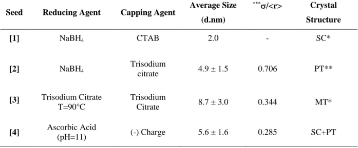
![Table II. 1 Summary of the different synthetic conditions used to prepare four classes of seeds with same final gold concentration ([Au 3+ ] final =0.25 mM)](https://thumb-eu.123doks.com/thumbv2/123doknet/12852030.367956/54.1262.103.1126.225.583/table-summary-different-synthetic-conditions-prepare-classes-concentration.webp)


![Figure II. 7 UV-Vis Spectrum of growth solution right after addition of 120 µL of Type 1 seed in growth solution ([CTAB] = 0.1M, [HAuCl 4 ] = 0.5mM, [AgNO3] =0.045mM, [AA] =0.75mM)](https://thumb-eu.123doks.com/thumbv2/123doknet/12852030.367956/69.892.186.753.107.544/figure-spectrum-growth-solution-addition-growth-solution-haucl.webp)


