Vol. 16, 1997 Letters 549
community were not different from cluster sizes of the drug-resistant isolates, suggesting that the drug-resistant isolates are transmitted as easily as the drug-susceptible isolates. In addition, the well-documented spread of the multidrug-resist- ant New York strain W is further evidence for transmissibility (as a virulence measure) of mul- tidrug-resistant tuberculosis (7). Our data from South Africa and the New York study demonstrate transmissibility of highly resistant strains and these strains have the ability to cause disease within a defined time interval. This may be inter- preted as a strong indicator of strain virulence, where virulence is defined as the ability to be transmitted and cause disease. Thus, we believe that resistance to high concentrations of isoni- azid does not (in general) imply lower strain viru- lence in the human as a function of impaired cat- alase activity. Virulence in these strains may be compensated for by the upregulation of addition- al virulence factors. Transmission of these resist- ant strains may create a large reservoir of individ- uals infected by resistant strains, who in the near future could develop multidrug-resistant disease. Therefore, an increased understanding of the mo- lecular mechanisms associated with mycobacteri- al virulence in the human and efficient procedures for the quick diagnosis of drug resistance in My- cobacterium tuberculosis are important for preven- tion of transmission of these highly pathogenic strains.
Acknowledgement
This work was supported by the Glaxo Wellcome Action TB Initiative.
T.C. Victor 1., R. Warren 1, N. Beyers 2,
RD. van Helden I
1 Department of Medical Biochemistry and 2Department of Pediatrics and Child Health, University of Stellen- bosch/Medical Research Council Centre for Molecular and Cellular Biology, PO Box 19063, University of Stellenbosch, Tygerberg, 7505 South Africa.
References
1. Mitchison DA, Wallace JG, Bhatia AL, Selkon JB, Sub- baiah TV, Lancaster MC: A comparison of the virulence in guinea pigs of South India and British tubercle bacil- li. Tubercle 1960, 41: 1-22.
2. Sherman DR, Mdluli K, Hickey M J, Arain TM, Morris SL, Barry CE, Stover CK: Compensatory ahpC gene expres- sion in isoniazid-resistant Mycobacterium tuberculosis. Science 1996, 272: 1641-1643.
3. Ordway D J, Sonnenberg MG, Donahue SA, Belisle JT, Orme IM: Drug-resistant strains of Mycobacterium tuber- culosis exhibit a range of virulence for mice. Infection and Immunity 1995, 63: 741-743.
4. Ausina V, Riutort N, Vifiado B, Manteroia JM, Ruiz Man- zano J, Rodrigo C, Matas L, Gim6nez M, Tor J, Roca J: Prospective study of drug-resistant tuberculosis in a Spanish urban population including patients at risk for HIV infection. European Journal of Clinical Microbiology & Infectious Diseases 1995, 14:105-110.
5. Warren R, Hauman J, Beyers N, Richardson M, Schaaf HS, Donald P, Van Helden PD: Unexpectedly high strain diversity of Mycobacterium tuberculosis in a high inci- dence community. South African Medical Journal 1996, 86: 45-49.
6. Warren R, Richardson M, Sampson S, Hauman JH, Beyers N, Donald PR, Van Helden PD: Genotyping of My- cobacterium tuberculosis with additional markers en- hances accuracy in epidemiological studies. Journal of Clinical Microbiology 1996, 34:2219-2224.
7. Bifani P J, Plikaytis BB, Kapur V, Stockbauer K, Pan X, Lutfey ML, Moghazeh SL, Eisner W, Daniel TM, Kaplan MH, Crawford JT, Musser JM, Kreisworth BN: Origin and interstate spread of a New York city multidrug-resistant Mycobacterium tuberculosis clone family. Journal of the American Medical Association 1996, 275: 452-457.
Detection of Mycobacterium avium- intracellulare in the Blood of HIV-Infected Patients by a Commercial Polymerase Chain Reaction Kit
In North America and Europe, disseminated in- fections with mycobacteria other than tuberculo- sis occur frequently in the late stages of AIDS (1). In a prospective study of patients with positive my- cobacterial blood cultures in Switzerland, Myco- bacterium genavense was responsible for 12.8% of the infections (2), and Mycobacterium avium ac- counted for 82%.
Until recently, the only method available to diag- nose these infections was culture in liquid medi- um (Bactec; Becton Dickinson, USA). Such cul- tures require a long incubation time (up to 14 weeks) to become positive, which delays diagno- sis and treatment. Amplification methods such as the polymerase chain reaction (PCR) applied di- rectly to blood specimens may accelerate diagno- sis. The few studies that have attempted to detect mycobacteria directly in blood have used nested PCR methods to increase sensitivity (3-5). How- ever, PCR performed on blood presents particu- lar methodologic problems because of the neces-
550 Letters Eur. J. Clin. Microbiol. Infect. Dis.
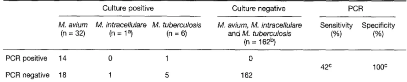
Table 1: Detection of Mycobacterium avium, Mycobacterium intracellulare, and Mycobacterium tuberculosis in blood from HIV-infected patients by the Amplicor PCR test and culture.*
Culture positive Culture negative PCR
M. aviurn M. intracellulare M. tuberculosis M. avium, M. intracellulare Sensitivity Specificity (n = 32) (n = 1 a) (n = 6) and M. tuberculosis (%) (%)
(n = 162 b)
PCR positive 14 0 1 0
PCR negative 18 1 5 162
42 c 100 c
*The positive culture specimens were hybridized with the M.avium, M. intracellulare, and M. tuberculosis probes according to their identifi- cation by biochemical tests. The negative culture specimens were final hybridized with the M. avium probe.
a Also positive for M. avium.
b Four blood cultures positive for M. genavense, 1 positive for M. haemophilum; 157 blood cultures negative.
c Sensitivity and specificity calculated for M. avium/M, intracellulare Amplicor test (values of the M. tuberculosis-positive cultures excluded).
sity to eliminate inhibitors of the PCR, such as the heine compound, during preparation of the D N A sample (6).
We report here the results of a retrospective study in which 43 of 200 Ficoll gradients from the blood of HIV-infected patients were positive for mycobacteria by culture in Bactec liquid medium. Samples were analyzed by a commercial PCR kit (Amplicor MAI; Roche Molecular Systems, Swit- zerland) that uses a simple PCR method to target the first variable region of the 16S rRNA gene. When used previously for the detection of Myco- bacterium tuberculosis complex in respiratory specimens, this PCR method had an overall sen- sitivity of approximately 70% and a specificity ex- ceeding 98% (7, 8).
Blood samples were collected from patients in our hospital during a two-year period. Five ml of whole blood was cultured in Bactec 13A medium, and 5 ml was used to isolate the polynuclear and mononuclear blood cells in Ficoll/Hypaque solu- tion. These Ficoll samples were frozen and then analyzed in batches using the Amplicor MAI PCR assay according to the standard protocol provid- ed by the manufacturer. Briefly, the samples were prepared by washing the sample pellet three times with mycobacteria blood wash solution, lysing mycobacteria with mycobacteria blood lysis reagent and incubating at 60 ~ for 45 min, and adding a neutralization reagent to provide the magnesium necessary for amplification. The am- plification target is a 582 bp of the mycobacterial 16S rRNA gene, and the amplicons are detected using a probe specific for Mycobacterium avium or Mycobacterium intracellulare.
Of the 157 negative blood cultures, 43 were ob- tained from patients harboring mycobacteria in other specimens such as sputum, bronchoalveolar lavage fluid, or biopsies; 17 negative blood cultures
were contaminated by bacteria other than myco- bacteria. Of the 43 positive blood cultures, 31 were positive for Mycobacterium avium, one was mixed ( Mycobacterium avium and Mycobacterium intracellulare), four were positive for Mycobacte- rium genavense, six were positive for Mycobacte- rium tuberculosis, and one was positive for Myco- bacterium haemophilum. The six cultures positive for Mycobacterium tuberculosis were amplified by the Amplicor test, and the PCR products were hy- bridized with the Mycobacterium tuberculosis complex probe available in a commercial respir- atory kit (Amplicor M. tuberculosis complex; Roche Diagnostic System, Switzerland).
The results of PCR compared with the results of Bactec culture, which is considered the gold stan- dard, are presented in Table 1. The sensitivity of the Amplicor Mycobacterium avium/Mycobacteri- um intracellulare test on blood specimens was low (42%), whereas the specificity was excellent (100 %). Another technique to extract D N A from blood (QIAamp blood kit; Qiagen, Switzerland) was also performed, but no additional mycobac- teria-positive sample was detected by PCR. The weak sensitivity was not due to PCR inhibition, since an internal control was successfully co- amplified in each sample tested. The only blood sample positive for Mycobacterium avium and My- cobacterium intracellulare was tested by hybridi- zation with the Mycobacterium avium and the Mycobacterium intracellulare probes. These two hybridization results were negative. The amplifi- cation products of the six blood cultures positive for Mycobacterium tuberculosis were hybridized with the Mycobacterium tuberculosis complex probe supplied by the manufacturer (9). Only one blood culture was positive by PCR.
The PCR results for Mycobacterium avium did not correlate with the time to positivity in the Bactec
Vol. 16, 1997 Letters 551
system. The mean Bactec detection time was 16 days and 14 days for positive and negative PCR results, respectively (p > 0.2). The time to positiv- ity in Bactec is correlated with the inoculum. Therefore, the lack of correlation we observed sug- gests that a low concentration of mycobacteria in the samples is not the only explanation for PCR negativity. This result differs from results with the Amplicor kit for respiratory specimens, in which sensitivity is higher for samples that are positive by microscopy. It also differs from an internal study conducted by Roche Diagnostic Systems (V. Tevere, Benchmark, Journal of Amplicor PCR Di- agnostics 1996, 3:6), which compared Amplicor MAI to quantitative culture to determine the de- tection limit of the test. Depending on the patho- gen load, a range of sensitivities was observed: 36.4% when < 1 cfu/ml was tested; 66.7% with 1-100 cfu/ml; and 100% with > 100 cfu/ml. In our study no correlation was seen between the PCR results and the clinical characteristics of the patients (CD4+ cell counts, levels of p24 antigen and 132 microglobulin, presence of antimycobac- terial treatment). For the same patient during the same period of hospitalization, some blood spec- imens were clearly positive by PCR, while others were devoid of positive signals. Our results are in accordance with those of Schneider et al. (36th ICAAC, 1996, Abstract no. 1165), who found the sensitivity of the Amplicor test to be 50% and the specificity 98 %.
In our opinion the MAI Amplicor test is not sen- sitive enough to be used prospectively to detect Mycobacteriurn avium or Mycobacteriurn intracel- lulare directly in the blood samples of all HIV- infected patients. The development of a nested PCR, along with improved methods for D N A preparation, may be essential to detect mycobac- terial DNA in more culture-positive blood samples.
B. Ninet*, R. Auckenthaler, R
Rohner,
O. Delaspre, B. Hirschel
Division of Infectious Diseases, H6pital Cantonal Universi- taire, CH-1211 Geneva, Switzerland.
References
Chan ISF, Neaton JD, Saravolatz LD, Crane LR, Oster- berger J: Frequencies of opportunistic diseases prior to death among HIV-infected persons. AIDS 1995, 9:
1145-1151.
2. Pechere M, Opravil M, Wald A, Chave JP, Bessesen M, Sievers A, Hein R, Von Overbeck JP, Clark RA, Tortoli E: Clinical and epidemiologic features of infection with My- cobacterium genavense. Swiss HIV cohort study. Ar- chives of Internal Medicine 1995, 155: 400-404. 3. Schluger NW, Condos R, Lewis S, Rom WN: Amplifica-
tion of DNA Mycobacterium tuberculosis from peripher- al blood of patients with pulmonary tuberculosis. Lancet 1994, 344: 232-233.
4. Emler S, B6ttger EC, Broers B, Cassis I, Perrin L, Hir- schel B: Growth-deficient mycobacteria in patients with AIDS: diagnosis by analysis of DNA amplified from blood or tissue. Clinical Infectious Diseases 1995, 20: 772-775.
5. Iralu JV, Shritharan VK, Pieciak WS, Wirth DF, Maguire JH, Barker RH Jr: Diagnosis of Mycobacterium avium bacteremia by polymerase chain reaction. Journal of Clin- ical Microbiology 1993, 31:1811-1814.
6. Akane A, Matsubara K, Nakamura H, Takahashi S, Kimu- ra K: Identification of the heme compound copurified with deoxyribonucleic acid (DNA) from bloodstains, a major inhibitor of polymerase chain reaction (PCR) amplifica- tion. Journal of Forensic Science 1994, 39: 362-372. 7. Chin DP, Yajko DM, Keith Hadley W, Sander CA, Nassos
PS, Madej JJ, Hopewell PC: Clinical utility of a commer- cial test based on the polymerase chain reaction for de- tecting Mycobacterium tuberculosis in respiratory spec- imens. American Journal of Respiratory and Critical Care Medicine 1995, 151 : 1872-1877.
8. D'Amato RF, Wallman AA, Hochstein LH, Colaninno PM, Scardamaglia M, Ardila E, Ghouri M, Kim K, Patel RC, Miller A: Rapid diagnosis of pulmonary tuberculosis by using Roche AMPLICOR Mycobacterium tuberculo- sis PCR test. Journal of Clinical Microbiology 1995, 33: 1832-1834.
9. Stauffer F, Mutschlechner R, Hasenberger P, Stadlbauer S, Schinko H: Detection of Mycobacterium tuberculosis complex in clinical specimens by a commercial poly- merase chain reaction kit. European Journal of Clinical Microbiology & Infectious Diseases 1995, 14: 1046-1051.
Edwardsiella tarda Septicemia with Ceilulitis in a Patient with A I D S
A 39-year-old woman was admitted to hospital in August 1994 with fever, rigors and confusion. She was known to be HIV-positive since 1990, and at the time of admission had a CD4+ cell count of 6/mm 3. She had a history of pulmonary tubercu- losis in 1990, cerebral toxoplasmosis in 1991, cryptococcal meningitis in 1993, sclerosing cholan- gitis since 1994 and chronic hepatitis B (HBsAg positive, H B V D N A negative). A venous access device was implanted in September 1993 for ad- ministration of prolonged i.v. amphotericin B treatment, but removed in March 1994 due to cen-