ORIGINAL ARTICLE
Evaluation of quality indicators following implementation
of total mesorectal excision in primarily resected rectal
cancer changed future management
Paul M. Schneider&Daniel Vallbohmer&
Yvonne Ploenes&Georg Lurje&Ralf Metzger&
Frederike C. Ling&Jan Brabender&Uta Drebber&
Arnulf H. Hoelscher
Accepted: 1 February 2011 / Published online: 22 February 2011 # Springer-Verlag 2011
Abstract
Background and aims We evaluated the outcome of primarily resected rectal cancer patients immediately after the implementation of total meserectal excision (TME) based on potential quality indicators.
Patients and methods Following initial teaching of two staff surgeons (PMS and AHH) by RJ Heald, 164 consecutive patients were analyzed. The following quality indicators were evaluated: (a) frequency of local recurrence, (b) number of resected lymph nodes, (c) selection of operative technique depending on tumor localization, (d) use of a protective loop ileostomy, and (e) frequency and type of adjuvant therapy. Results Local recurrence rate was 8.5% after a minimum follow-up of 5 years. An increasing pT category (p<0.02) and the presence of lymph node metastases (pN+, p<0.05) were significantly associated with local recurrence rates. The number of resected lymph nodes was significantly associated with nodal metastases rate (p<0.02). Patients with distal third rectal cancer underwent significantly more often an abdominoperineal amputation (p<0.0001). Clinical
course, but not the rate of anastomotic leakage (9.5%) itself was influenced by using a protective loop ileostomy. Forty-two (29.7%) patients received adjuvant therapy; however, local recurrence rate was higher in patients with adjuvant chemo-/radiotherapy (14.2% vs. 6.1%).
Conclusions The local recurrence rate of 8.5% demonstrates that through consequent implementation of TME excellent onclogical results can be achieved. The number of resected lymph nodes significantly influenced the pN category. The primary construction of a protective loop ileostomy after TME became standard. Neoadjuvant chemoradiation was system-atically introduced in order to improve local tumor control and prevent abdominoperineal amputations. No conclusions can be drawn concerning adjuvant therapy.
Keywords Rectal cancer . Surgical therapy . Total mesorectal excision
Introduction
Quality assurance in oncologic surgery is increasingly accepted as an important factor that affects the clinical outcome of patients [1, 2]. This applies particularly for rectal cancer that requires a demanding, meticulous surgical technique. Heald et al. [3] proposed the method of total mesorectal excision (TME) which necessitates complete excision of the mesorectal tissue to the level of the levators with simultaneous preservation of autonomic nerve func-tion. This technique has certainly improved the outcome of patients with rectal cancer by significantly reducing the local recurrence rate and became the standard surgical procedure in rectal cancer treatment [4,5].
P. M. Schneider
:
D. Vallbohmer:
Y. Ploenes:
G. Lurje:
R. Metzger
:
F. C. Ling:
J. Brabender:
A. H. HoelscherDepartment of General, Visceral and Tumor Surgery, University of Cologne,
Cologne, Germany
P. M. Schneider (*)
:
G. LurjeDepartment of Surgery, University Hospital Zurich, Raemistrasse 100,
8091, Zurich, Switzerland e-mail: [email protected] U. Drebber
Institute of Pathology, University of Cologne, Cologne, Germany
In addition, multiple studies assesed the relationsship between surgeon/hospital volume and clinical outcome of patients with rectal cancer. For example, Harmon et al. examined the association of surgeon and hospital case volumes with the outcome of patients with colorectal cancer. They showed that “medium-volume” surgeons achieved excellent outcomes similar to “high-volume” surgeons when operating in “medium-volume” or “high-volume” hospitals, while the results of “low-“high-volume” surgeons improved with increasing hospital volume but never reached those of the“high-volume” surgeons [6]. In contrast, Marusch et al. [7] failed to show significant associations between hospital caseload and postoperative outcome in 2,293 colorectal cancer patients undergoing surgery.
Even though controversial data are available about the volume–quality relationship, a limitation on the perfor-mance of rectal cancer surgery to highly specialized surgeons is impractical in view of the prevalence of rectal cancer. As evidence suggests that training is a prerequisite to obtain good results in surgeons’ hands; in the last years, many countries have focused on improvements in the training of rectal surgery [8–10]. For instance, Wibe et al. [10] performed a prospective study to assess the influence of educational programs and training courses in Norway. They verified that the prognosis of rectal cancer patients was significantly improved by the increased organizational focus on rectal cancer treatment with a significant reduction of local recurrence [10].
To evaluate and improve this training in rectal surgery and also to improve the overall quality assurance in rectal cancer treatment, a data set with defined quality indicators has to be established. The purpose of this study was evaluate the outcome of primary resected rectal cancer patients after the implementation of partial mesorectal excision (PME) and TME by the following potential quality indicators: (1) number of lymph nodes analyzed in the surgical specimen, (2) selection of operative technique depending on tumor localization, (3) primary construction of a protective loop ileostomy to prevent complications due to an anastomotic leakage, (4) frequency of local recurrence rate, and (5) frequency of adjuvant therapy.
Patients and methods
On February 20, 1997, Professor Heald from Basingstoke, England, introduced the TME technique in the Department of General, Visceral and Cancer Surgery, University of Cologne, Cologne, Germany by teaching PMS and AHH. In addition, a prospective database was set up immediately for all rectal cancer patients treated newly in our institution.
The presented analysis includes 164 primarily resected patients (intention to treat) between February 1997 and December 2004 out of 234 patients. Forty-eight patients receiving neoadjuvant chemoradiation and 22 patients undergoing local resection by transanal endoscopic micro-surgery were excluded. The last follow-up was December 2009 ensuring a minimum follow-up of 5 years after resection for surviving patients.
This study was performed in accordance with the guidelines of the local Institutional Review Board.
Diagnostic and staging procedures
TNM staging was performed according to the criteria of the International Union Against Cancer [11]. Clinical staging consisted of endoscopy, endoscopic ultrasound, CT scanning of the abdomen, and thorax. Classification of localization was performed using rigid rectoscopy using the distance of the anocutaneous line to the lower border of the tumor according to TNM supplement, 3rd edition, as follows:
Upper rectal third 12–16 cm Middle rectal third 6≤12cm Lower rectal third <6 cm
Surgical resection and adjuvant therapy
The patients underwent three surgical procedures depending on the tumor localization: (1) anterior resec-tion with partial PME, (2) low anterior rectal resecresec-tion with TME, and (3) abdominoperineal rectum amputation with TME.
Adjuvant therapy was not delivered by protocol and was mainly offered to patients with Union for International Cancer Control (UICC) stage III, generally using chemo-therapy, next to radio- and radiochemotherapy.
Follow up
Patients were under regular follow-up, undergoing colono-scopy, CT scan of chest and abdomen, and ultrasound of the liver up to 5 years.
Statistical analysis
Data were collected prospectively according to a standardized protocol. Chi-square statistics were calcu-lated for frequencies of factors. Kaplan–Meier plots were used to describe survival distribution and the log-rank test was used to evaluate for survival differences. Postoperative mortality was included in the calculation
of prognosis. The level of significance was set to p < 0.05 in all tests.
All statistical analyses were performed using the statistic program SPSS for Windows version 17.0.
Results
Demographic and clinical parameters of study patients
There were 61 women (37.2) and 103 men (62.8%) with a median age of 64.4 years (range, 34–87 years). Of these, 160 patients underwent primary surgical resection, while four patients received palliative chemotherapy because of diffuse metastatic disease confirmed by explorative laparotomy without resection. In 156 (97.5%) patients, an R0 resection; in two, (1.25%) an R1 resection; and in two (1.25%) patients, an R2 resection of the primary tumor was performed.
Distant metastases were detected in 49 (29.8%) patients. From these, 30 (18.3%) patients had synchronous and 19 (11.5%) metachronous distant metastases. Thirteen (7.9%) patients with synchronous metastases underwent surgical resection of the metastases.
Analysis of potential quality indicators
Number of analyzed lymph nodes in the surgical specimen
According to the UICC guidelines, patients were classified into two groups: (a) patients with 12 or more resected
lymph nodes (n=91; 56.9%) and (b) patients with less than 12 lymph nodes analyzed in the surgical specimen lymph nodes (n=69; 43.1%) [11]. Higher lymph node retrieval was significantly associated with a higher histopathologic nodal metastases rate (p<0.02; Table1).
No significant difference (p = 0.71, log-rank test) in terms of survival was observed between the two groups. Patients with ≥12 lymph nodes analyzed in the surgical specimen had a median survival of 79.21 months (95% CI, 73.34–85.07 months) and patients with <12 had a median survival of 81.11 months (95% CI, 74.55– 87.66 months).
Selection of the operative technique depending on tumor localization
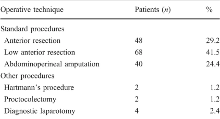
Distribution of tumor localization was as follows: 58 patients had an upper third, 64 a middle third, and 42 a lower third rectal cancer. The patients underwent predominantly three surgical procedures: (a) anterior resection (n = 48, 29.2%), (b) low anterior resection (n = 68, 41.5%), and (c) abdominoperineal amputation (n = 40, 24.4%; Table2).
A significant association was detected between tumor localization and surgical procedure. Patients with lower third rectal cancer underwent significantly more often an abdominoperineal amputation compared to patients with upper or middle third rectal cancer (p < 0.0001, Table3).
Tumor localization was also significantly associated with the pT category: patients with a lower third rectal cancer undergoing an abdominoperineal amputation had signifi-cantly more often a pT3/4 category than patients undergo-ing low anterior resection.
Construction of a protective loop ileostomy
In 28 (24.1%) patients, a protective ileostomy was constructed. The overall anastomotic leakage rate was 9.5% (11 patients). There was no difference in overall leak rates between patients with and without a protective loop
Table 1 Correlation between the number of analyzed lymph nodes in the surgical specimenand pN category
Resected lymph nodes pN0 stage (%) pN1–2 stage Total
<12 51 (32.1) 18 (11.9) 69
≥12 46 (28.9) 43 (27.7) 91
p<0.02;χ² test
Table 2 Surgical procedure and tumor localization; distribution of operative techniques
Operative technique Patients (n) %
Standard procedures
Anterior resection 48 29.2
Low anterior resection 68 41.5
Abdominoperineal amputation 40 24.4
Other procedures
Hartmann’s procedure 2 1.2
Proctocolectomy 2 1.2
Diagnostic laparotomy 4 2.4
Table 3 Surgical procedure and tumor localization; operative tech-nique depending on tumor localization
Localization Anterior resection (%) Low anterior resection (%) Abdominoperineal amputation (%) Total Upper third 41 (75.9) 13 (24.1) 0 (0) 54 Middle third 7 (11.3) 49 (79) 6 (9.7) 62 Lower third 0 (0) 6 (15) 34 (85) 40
ileostomy (Table 4). However, a protective ileostomy significantly reduced the incidence of anastomotic leakage that became symptomatic and required surgical interven-tion. Six out of seven patients without a protective ileostomy became symptomatic due to a leakage and required surgical revision, where as none of the patients with a protective stoma needed a relaparotomy due to anastomotic leackage.
In addition, a significant correlation was observed between the leakage rate in patients without a protective stoma and surgical procedure: patients undergoing anterior resection had significant fewer leaks compared to patients undergoing a low anterior resection (three vs. eight patients; p<0.05, Table5).
Frequency of local recurrence
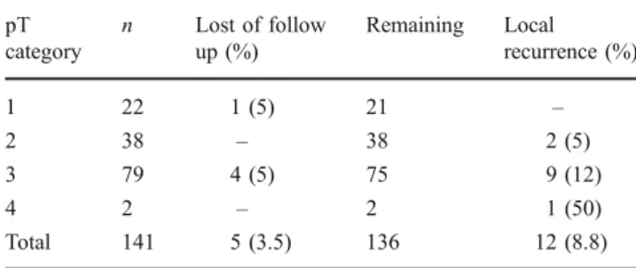
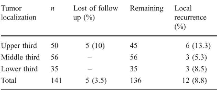
With a minimum follow-up of the surviving patients of 60 months, 12 from 136 (8.8%) patients (five patients were lost to follow-up) patients developed local recur-rence after R0 resection. Analysis of risk factors for local recurrence revealed that an advanced pT category and the pN category was a risk factor for local recurrence (see Table 5, 6, and 7). Tumor localization merely showed a tendency towards frequency of local recurrences.
Frequency of adjuvant therapy
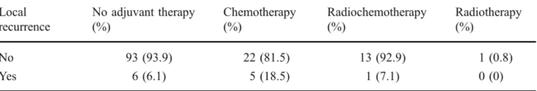
Adjuvant therapy was not standardized and left to the discretion of the consultant surgeon. Forty-two (29.7%) patients with R0 resection received an adjuvant therapy, most of them (n=26, 61.9%) with an histopathologic UICC stage III (Table8). Six of the 42 (14.2%) patients developed a local recurrence. From the patients receiving no adjuvant therapy, six patients (6.1%) only developed local recurrence (Table 9).
Discussion
The purpose of this study was to evaluate a data set of quality indicators in primarily resected rectal cancer patients after the implementation of TME. The following potential quality indicators were analyzed: (1) number of resected lymph nodes, (2) selection of operative technique depending on tumor localization, (3) primary construction of a protective loop ileostomy to prevent complications due to an anastomotic leakage, (4) frequency of local recur-rence, and (5) frequency of adjuvant therapy.
For a precise determination of the lymph node category, the UICC recommend that a minimum of 12 lymph nodes should be resected and recovered in colorectal cancer patients undergoing surgical therapy [11]. This goal was achieved in 56.9% of our study patients only and does not meet the standard as suggested by Bittner et al. [12]. Moreover, we observed that tumors with a lymph node
Anastomotic leackage
No (%) Yes (%) Total (%)
Frequency of anastomotic leckage with and without a protective loop ileostomy Protective stoma
No 81 (92) 7 (8) 88 (100)
Yes 24 (85.7) 4 (14.3) 28 (100)
Correlation between anastomotic leakage and type of surgical procedure Surgical procedure
Anterior resection 45 (93.8) 3 (6.3) 48 (100)
Low anterior resection 60 (88.2) 8 (11.8) 68 (100)
Table 4 Frequency of anasto-motic leckage with and without a protective loop ileostomy and correlation between anastomotic leakage and type of surgical procedure
Table 5 Local recurrence rate based on pT/pN categories and tumor localization; pT category pT category n Lost of follow up (%) Remaining Local recurrence (%) 1 22 1 (5) 21 – 2 38 – 38 2 (5) 3 79 4 (5) 75 9 (12) 4 2 – 2 1 (50) Total 141 5 (3.5) 136 12 (8.8) p<0.02,χ² test
Table 6 Local recurrence rate based on pT/pN categories and tumor localization; pN category pN category n Lost of follow up (%) Remaining Local recurrence (%) 0 93 2 (2.1) 91 5 (5.5) 1/2 47 2 (4.2) 45 7 (15.5) Total 140 4 (2.8) 136 12 (8.8) p<0.05,χ² test
retrieval number≥12 were significantly associated with a higher nodal metastases rate (pN category). We could however not demonstrate that the number of resected lymph nodes had a prognostic impact. These findings are consistent with recent studies evaluating the prognostic influence of lymph node harvest in colorectal cancer patients [13–15]. Kim et al. [14] demonstrated in a retrospective study of 151 patients with colorectal cancer that a higher number of lymph nodes resected/examined were associated with a higher nodal metastasis rate. On the other hand, Prandi et al. reported on 3,648 patients with stage B colon cancer that patients with <7 nodes in the specimen had both a shorter overall survival and relapse free survival [13]. More recently, Tsai et al. [15] showed in 366 patients with T2-4N0M0 colorectal cancer undergoing radical tumor resection that patients with 18 or more examined lymph nodes had a significantly lower postoper-ative relapse and a higher 5-year overall survival than patients who had less than 18 nodes examined. Despite the fact that the minimum number of lymph nodes examined per surgical specimen is still under discussion, lymph node retrieval should be used as a quality indicator in rectal surgery, for both, surgeons and pathologists because the number of resected/retrieved lymph nodes is important for the detection of nodal metastases. In addition, nodal metastases are significantly associated with local recurrence rates and this again could influence whether postoperative adjuvant therapy should be recommended.
Our study showed that patients with lower third rectal cancer underwent significantly more often an
abdomino-perineal rectum amputation compared to patients having a tumor in the upper or middle third. However, mainly locally advanced tumors were resected by an amputation and through the consequent implementation of neoadjuvant chemoradiation, more sphincter-preserving operations should be expected in our institution. This is in line with observations from other studies that showed that through improvement of surgical techniques and especially by administering neoadjuvant chemoradiation, permanent colostomy rates in patients with lower third rectal cancer can be substantially reduced [16, 17]. Since evidence suggests that the quality of life after anterior or low anterior resection is usually improved compared with abdominoper-ineal amputation, the selection of the operative techniques depending on tumor localization might be used as good quality indicator in rectal surgery [12,18,19].
We could also show that clinical symptoms but not the very frequency of anastomotic leakage was influenced by the primary construction of a protective loop ileostomy: six of seven patients without a protective ileostomy became symptomatic due to a leakage and surgical revision was needed, whereas none of the patients with a primary ileostomy needed a relaparotomy. Similar findings were reported by Gastinger et al. [20] in a prospective multicen-ter study that evaluated early outcome afmulticen-ter low anmulticen-terior resection in patients with and without a protective stoma. They showed that the overall anastomotic leakage rates were similar in patients with or without a protective stoma but the incidence of symptomatic leaks that required surgical intervention was significantly lower in those with a protective stoma. In addition, Matthiessen et al. [21] assessed in a randomized multicenter trial the rate of symptomatic anastomotic leakage in patients operated by low anterior resection and were able to demonstrate that a defunctioning loop stoma significantly decreased the leakage rate. These data suggest that a protective stoma should be recommended in low anterior resection for rectal cancer to avoid serious complications of anastomotic leackage.
In our study the local recurrence rate after a minimum follow-up of 5 years was 8.5%. These data are in agreement with the majority of studies reported in the literature after
Table 7 Tumor localization and local recurrence rates Tumor localization n Lost of follow up (%) Remaining Local recurrence (%) Upper third 50 5 (10) 45 6 (13.3) Middle third 56 – 56 3 (5.3) Lower third 35 – 35 3 (8.5) Total 141 5 (3.5) 136 12 (8.8) p=0.06,χ² test Adjuvant therapy UICC stage No adjuvant therapy (%) Chemotherapy (%) Radiochemotherapy (%) Radiotherapy (%) I 52 (100) 0 (0) 0 (0) 0 (0) II 31 (83.8) 4 (10.5) 3 (7.9) 0 (0) III 14 (35) 15 (37.5) 10 (25) 1 (2.5) IV 2 (18.2) 8 (72.7) 1 (9.1) 0 (0) Total 99 (70.2) 27 (19.1) 14 (9.9) 1 (0.7)
Table 8 Adjuvant therapy in study patients; adjuvant therapy depending on UICC stage
the implementation of TME with recurrence rates between 5% and 10% [9, 22–25]. In fact, the radical surgical approach with TME is the major determinant for the significant decrease of local recurrence rates in the last years and was shown to be an effective surrogate indicator of treatment outcome quality [26]. Also, the minimum requirement of 5 years follow-up for patients with surgery alone as suggested by Merkel et al. [27] was fulfilled in our study. We did however, not routinely evaluate the quality of PME or TME by histopathological criteria as suggested by Quirke and Morris [28] and therefore cannot rule out technical surgical problems in our patients with observed local recurrences. Tumor stage and lymph node involve-ment and the use of (neo-) adjuvant therapy were also described in the literature as prognostic factors influencing local recurrence in rectal cancer. In our study, both the T and N categories significantly predicted for a higher recurrence rate [22–25]. In conclusion, the local recurrence rate should be used as an important quality indicator in rectal cancer surgery because it is a very sensitive parameter determining the surgeons’ accuracy, i.e., the correct performance of the TME technique [10,12,27,28]. Our study results did not show that adjuvant therapy is an effective treatment in rectal cancer patients following TME. Actually, the local recurrence rate was higher in patients receiving adjuvant therapy compared with patients obtaining no postoperative treatment. Indeed, randomized studies from the late 1980s and early 1990s established postoperative radiochemotherapy as the standard of care for patients with resected stage II/III rectal cancer [29–33] when conventional blunt dissection with higher recurrence rates were performed.
Since adjuvant therapy was not administered by a standardized protocol, no conclusions can be drawn from those results besides that it is important not only to standardize operative techniques but also implement guide-lines for adjuvant and neoadjuvant treatment modalities.
In 2004, the German Rectal Cancer Study demonstrated that preoperative radiochemotherapy doubled the rate of sphincter-sparing operations and significantly lowered the rates of local recurrence compared to postoperative treat-ment. Based on these convincing data, we implemented neoadjuvant chemoradiation as standard treatment for locally advanced middle and lower third rectal cancer in 2004 in our institution [34].
In summary, the following conclusions can be made: (1) the number of analyzed lymph nodes in the surgical specimen influences the pN category which is important regarding the indication for adjuvant therapy. (2) The primary construction of a protective loop ileostomy significantly decreased the need for a surgical revision if an anastomotic leakage occurred following low anterior rescetion. (3) In contrast to neoadjuvant chemoradiation, the value of adjuvant therapy after the implementation of TME remains highly controversial, because it is at least questionable if postoperative therapy additionally reduces the local recurrence rates. (4) The local recurrence rate of 8.5% after a minimum follow-up of 5 years demonstrates that through the consequent implementation of TME local recurrence rates similar to the reported series from internationally renowned groups can be achieved.
References
1. Kapiteijn E, van de Velde CJ (2002) Developments and quality
assurance in rectal cancer surgery. Eur J Cancer 38:919–36
2. Peeters KC, van de Velde CJ (2005) Surgical quality assurance in rectal cancer treatment: the key to improved outcome. Eur J Surg Oncol 31:630–5
3. Heald RJ, Husband EM, Ryall RD (1982) The mesorectum in rectal cancer surgery—the clue to pelvic recurrence? Br J Surg 69:613–616
4. Heald RJ, Ryall RD (1986) Recurrence and survival after total
mesorectal excision for rectal cancer. Lancet i:1479–1482
5. MacFarlane JK, Ryall RD, Heald RJ (1993) Mesorectal excision
for rectal cancer. Lancet 341:457–60
6. Harmon JW, Tang DG, Gordon TA, Bowman HM, Choti MA, Kaufman HS, Bender JS, Duncan MD, Magnuson TH, Lillemoe KD, Cameron JL (1999) Hospital volume can serve as a surrogate for surgeon volume for achieving excellent outcomes in colorectal
resection. Ann Surg 230:404–11
7. Marusch F, Koch A, Schmidt U, Zippel R, Lehmann M, Czarnetzki HD, Knoop M, Geissler S, Pross M, Gastinger I, Lippert H (2001) Effect of caseload on the short-term outcome of colon surgery: results of a multicenter study. Int J Colorectal Dis
16:362–9
8. Mack LA, Temple WJ (2005) Education is the key to quality of surgery for rectal cancer. Eur J Surg Oncol 31:636–44
9. Martling A, Holm T, Rutquist L-E, Moran BJ, Heald RJ, Cedermark B (2000) Effect of a surgical training programme on outcome of rectal cancer in the County of Stockholm. Lancet
356:93–6
10. Wibe A, Carlsen E, Dahl O, Tveit KM, Weedon-Fekjaer H, Hestvik UE, Wiig JN, The Norwegian Rectal Cancer Group Adjuvant therapy Local recurrence No adjuvant therapy (%) Chemotherapy (%) Radiochemotherapy (%) Radiotherapy (%) No 93 (93.9) 22 (81.5) 13 (92.9) 1 (0.8) Yes 6 (6.1) 5 (18.5) 1 (7.1) 0 (0)
Table 9 Adjuvant therapy and local recurrence
(2006) Nationwide quality assurance of rectal cancer treatment.
Colorectal Dis 8:224–9
11. UICC (2002) TNM classification of malignant tumours, 6th edn. Sobin LH,Wittekind C (eds) Wiley-Liss, NewYork.
12. Bittner R, Burghardt J, Gross E, Grundmann RT, Hermanek P, Isbert C, Junginger T, Köckeling F, Merkel S, Möslein G, Raab HR, Roder J, Ruf G, Schwenk W, Strassburg J, Tannapfel A, de Vries A, Zühlke H (2007) Quality indicators for diagnosis and therapy of rectal carcinoma. Zentralblatt Chir 132:85–94 13. Prandi M, Lionetto R, Bini A, Francioni G, Accarpio G, Anfossi
A, Ballario E, Becchi G, Bonilauri S, Carobbi A, Cavaliere P, Garcea D, Giuliani L, Morziani E, Mosca F, Mussa A, Pasqualini M, Poddie D, Tonetti F, Zardo L, Rosso R (2002) Prognostic evaluation of stage B colon cancer patients is improved by an adequate lymphadenectomy: results of a secondary analysis of a
large scale adjuvant trial. Ann Surg 235:458–63
14. Kim J, Huynh R, Abraham I, Kim E, Kumar RR (2006) Number of lymph nodes examined and its impact on colorectal cancer
staging. Am Surg 72:902–5
15. Tsai HL, Lu CY, Hsieh JS, Wu DC, Jan CM, Chai CY, Chu KS, Chan HM, Wang JY (2007) The prognostic significance of total lymph node harvest in patients with T2-4N0M0 colorectal cancer. J Gastrointest Surg 11:660–5
16. Schiessel R, Novi G, Holzer B, Rosen HR, Renner K, Hölbling N, Feil W, Urban M (2005) Technique and long-term results of intersphinc-teric resection for low rectal cancer. Dis Colon Rectum 48:1858–65 17. Wallace MH, Glynne-Jones R (2007) Saving the sphincter in rectal
cancer: are we prepared to change practice? Colorectal Dis 9:302–8
18. Zolciak A, Bujko K, Kepka L, Oledzki J, Rutkowski A, Nowacki MP (2006) Abdominoperineal resection or anterior resection for rectal cancer: patient preferences before and after treatment.
Colorectal Dis 8:575–80
19. Bossema E, Stiggelbout A, Baas-Thijssen M, van de Velde C,
Marijnen C (2008) Patients’ preferences for low rectal cancer
surgery. Eur J Surg Oncol 34(1):42–48
20. Gastinger I, Marusch F, Steinert R, Wolff S, Koeckerling F,
Lippert H (2005) Working Group ‘Colon/Rectum Carcinoma’.
Protective defunctioning stoma in low anterior resection for rectal
carcinoma. Br J Surg 92:1137–42
21. Matthiessen P, Hallböök O, Rutegård J, Simert G, Sjödahl R (2007) Defunctioning stoma reduces symptomatic anastomotic leakage after low anterior resection of the rectum for cancer: a randomized multicenter trial. Ann Surg 246:207–14
22. Arenas RB, Fichera A, Mhoon D et al (1998) Total mesenteric excision in the surgical treatment of rectal cancer: a prospective
study. Arch Surg 133:608–611
23. Tocchi A, Mazzoni G, Lepre L et al (2001) Total mesorectal excision and low rectal anastomosis for the treatment of rectal cancer and prevention of pelvic recurrences. Arch Surg 136:216– 220
24. Law WL, Chu KW (2004) Anterior resection for rectal cancer with mesorectal excision: a prospective evaluation of 622 patients.
Ann Surg 240:260–8
25. Bernardshaw SV, Øvrebø K, Eide GE, Skarstein A, Røkke O (2006) Treatment of rectal cancer: reduction of local recurrence
after the introduction of TME—experience from one University
Hospital. Dig Surg 23:51–9
26. Nagtegaal ID, van de Velde, Marijnen CA (2005) Low rectal cancer: a call for a change of approach in abdominoperineal
resection. J Clin Oncol 23:9257–64
27. Merkel S, Mannsmann U, Hohenberger W, Hermanek P (2009) Time to locoregional recurrence after curative resection of rectal carcinoma is prolonged after neoadjuvant treatment. A systematic
review and meta-analysis. Colorectal Disease 13(2):123–131
28. Quirke P, Morris E (2007) Reporting colorectal cancer. Histopa-thology 50:103–112
29. Engel J, Kerr J, Eckel R, Günther B, Heiss M, Heitland W, Jauch KW, Siewert JR, Hölzel D (2005) Quality of treatment in routine care in a population sample of rectal cancer patients. Acta Oncol
44:65–74
30. Douglass HO Jr, Moertel CG, Mayer RJ et al (1986) Survival after postoperative combination treatment of rectal cancer. N Engl J
Med 315:1294–1295
31. Krook JE, Moertel CG, Gunderson LL et al (1991) Effective surgical adjuvant therapy for high-risk rectal carcinoma. N Engl J
Med 324:709–715
32. Gastrointestinal Tumor Study Group (1985) Prolongation of the disease-free interval in surgically treated rectal carcinoma. N Engl
J Med 312:1465–1472
33. O’Connell MJ, Martenson JA, Wieand HS et al (1994) Improving
adjuvant therapy for rectal cancer by combining protracted-infusion fluorouracil with radiation therapy after curative surgery. N Engl J Med 331:502–507
34. Sauer R, Becker H, Hohenberger W et al (2004) Preoperative versus postoperative chemoradiotherapy for rectal cancer. N Engl J Med 351:1731–1740