Invasive Group B Streptococcal
Disease in Non-pregnant Adults
A Review with Emphasis on Skin and Soft-tissue Infections
P. Sendi, L. Johansson, A. Norrby-Teglund
Abstract
Streptococcus agalactiae, commonly referred as group B Streptococcus (GBS), is a major cause of neonatal sepsis and infections in pregnant women. However, the number of invasive infections in non-pregnant adults is growing. Elderly patients and those with chronic underlying condi-tions, such as diabetes mellitus or compromised immune defence, are at increased risk of invasion. The spectrum of clinical manifestations is broad and includes necrotizing fasciitis and toxic shock syndrome. Although, primary bacteremia and skin and soft-tissue infections are the most frequently reported diagnosis. This article reviews the epidemiology, pathogenesis and treatment of invasive GBS disease in non-pregnant adults, with an emphasis on skin and soft-tissue infections.
Infection 2008; 36: 100–111 DOI 10.1007/s15010-007-7251-0
Introduction
Streptococcus agalactiae is the only streptococcal species that carries the Lancefield group B antigen, hence its alternative designation as group B Streptococcus (GBS). It is a leading cause of morbidity and mortality in neonates and pregnant women. In many industrialized countries, recommendations for diagnosing maternal GBS colonization and administering intrapartum anti-microbial prophylaxis have been introduced, leading to a significance decrease in these infections [1]. However, the rate of invasive GBS disease in non-pregnant adults continues to climb [2]. Elderly persons and those with underlying diseases – two expanding segments of the population – are at increased risk. This article reviews the epidemiology of invasive GBS disease in non-preg-nant adults, describes clinical, immunological and bac-terial perspectives of its pathogenesis, and discusses treatment concepts for this condition. Emphasis is placed on skin and soft-tissue infections, which are among the most frequently reported clinical problems.
Epidemiology
Incidence of Invasive Group B Streptococcal Disease
In the early 1990s, three large surveillance studies in the United States reported a significant increase over the previous two decades in the annual incidence of invasive GBS disease (defined as isolation of S. agalactiae from a normally sterile site [3]) among non-pregnant adults. The rates had reached 2.4–4.4 cases per 100,000 population [4–6]. Thereafter, several reports revealed an increase in incidence of both invasive diseases and bacteremia per 1000 hospital admissions [2, 7]. Recent studies have re-ported an annual incidence of invasive GBS disease of 0.96 (Spain) or an incidence of GBS bacteremia of 0.136 (Hong Kong), 0.30 (Taiwan) and 0.42 (Spain) cases per 1000 hospital admissions, which also illustrate regional differences [7–9]. This trend can be partly explained by a growing number of patients with predisposing factors, in particular, chronic medical conditions.
Relapsing Infections
Approximately 5% of non-pregnant adults will have at least a second episode of GBS disease. In the study by Harrison et al. [10] the interval between the first and second episode averaged 13 weeks if the bacteremia was caused by the same strain and 43 weeks if it was caused by a different strain. In this study, all infections occurred in patients with predisposing conditions, and cellulitis was the most fre-quent recurrent clinical manifestation.
P. Sendi, L. Johansson, A. Norrby-Teglund
Center for Infectious Medicine, F59, Department of Medicine, Karolinska Institutet, Karolinska University Hospital, Huddinge, Sweden
P. Sendi
Unit of Infectious Diseases, Basel University Medical Clinic, Liestal, Switzerland
P. Sendi (corresponding author)
Center for Infectious Medicine, F59, Department of Medicine, Karolinska Institutet, Karolinska University Hospital, Huddinge, Sweden; e-mail: sendi-pa@magnet.ch
Received: June 5, 2007 Æ Revision accepted: September 19, 2007 Published online: January 12, 2008
Predisposing Conditions and Risk Factors
The vast majority of patients with GBS infections have at least one predisposing condition. Important risk factors and their calculated incidence rates are presented in ta-ble 1. Older age (> 65 years), diabetes mellitus, cancer and compromised immunity are recognized to increase the risk of acquiring invasive GBS disease [2, 4, 6, 11]. Venous oedema and/or lymphoedema have been deter-mined as risk factors for relapsing erysipelas/cellulitis of the lower leg [12]. However, in a retrospective study analysing 71 patients suffering from GBS skin and soft-tissue infections [13], 17 (24%) had no obvious underlying disease. This percentage is higher than those previously reported, ranging between 0–8% [2, 4, 6, 7, 14–17]. Nev-ertheless, it emphasizes that GBS is not exclusively affecting risk groups.
Since, like all patients, GBS-colonized individuals are exposed to various instrumentation techniques (e.g. uri-nary catheter, sigmoidoscopy), this iatrogenic factor may be contributing to the incidence rate [18, 19]. In one-quarter (17–30%) of the cases, invasive GBS disease oc-curs ‡ 48 hours after admission to a hospital [6, 11, 20]. Nosocomial infection may therefore arise from pre-exist-ing colonized skin or mucosal surfaces.
Prevalence of GBS Colonization
In colonized individuals, GBS is generally isolated from cultures of the rectum, perianal area, vagina, cervix and urethra or, less commonly, from cultures of the skin and pharynx. The colonization rate ranges from 20 to 35% [21–26]. The prevalence is higher among sexually active
persons and those who have multiple sex partners, sug-gesting that acquisition of this microorganism is caused by intimate contact [23, 27]. This is also supported by a study showing identical strains in 86% of co-colonized sex partners [21].
Serotype Distribution
GBS expresses a polysaccharide antigen on its surface that is used for serotype identification. To date, nine serotypes have been identified: Ia, Ib and II–VIII [28]. Character-ization of a GBS strain also includes the presence of surface proteins. The C-protein complex, which consists of two independently expressed components (the a- and b-antigen), is among the best-studied of these surface proteins.
Most of the epidemiological data on GBS serotypes have been collected from strains causing invasive disease. In non-pregnant adults, almost 70% of the invasive cases have been attributed to serotypes Ia, III and V [29–31]. These three serotypes also dominated in a study analysing colonizing strains in healthy elderly persons [26]. C-protein antigens are found in 40–60% of clinical isolates, mostly in strains with serotype Ia, Ib and II [28, 32]. However, the serotype distribution has shifted somewhat during the past few years [33]. Pregnant Japanese women were found to be predominantly colonized with serotypes VI and VIII [34], whereas invasive disease was being increasingly caused by serotype VIII in a Danish study [35]. Whether this differ-ence is due to geographic predominance or due to the type of infection remains to be determined. However, the serotype distribution of both invasive and colonizing
Table 1
Underlying conditions associated with an increased risk for acquiring GBS infections in non-pregnant adults.
Study design, study population, number of patients
Predisposing condition Incidence of invasive infectiona
Risk calculation (95% CI) Relative risk Odds ratio Retrospective population-based surveillance,
non-pregnant adults, 56 [4]
Diabetes mellitus 13.7 10.5 (7.8–14.4) – Cancer 21.3 16.4 (11.5–23.3) – Case-control study, non-pregnant adults, 219 cases
and 645 hospital-matched controls [11]
Diabetes mellitusb – – 3.0 (1.9–4.7) Breast cancerb – – 4.0 (1.6–9.8) Liver cirrhosisb – – 9.7 (3.5–26.9) Neurogenic bladderb – – 4.6 (1.4–15.1) Decubitus ulcerb – – 4.0 (1.6–9.8) Strokeb – – 3.5 (1.9–6.4)
Prospective population-based surveillance, non-pregnant adults, 137 [6]
Diabetes mellitus (age 20–40) 20 30 (11–79) – HIV (age 30–49) 54 30 (11–78) – Active bacterial core surveillancec Age > 65 25.6 – – The surveillance areas represented 27,350,255 persons [3]
Active surveillance, non-pregnant adults, 867 cases including 84 nursing home residents [134]
Nursing home residents age‡ 65 – 4.1 (2.6–6.7)d a
Annual incidence of invasive GBS infection per 100,000 population;bMultivariate analysis [11] adjusted for age, diabetes mellitus, breast cancer, liver cirrhosis, neurogenic bladder, decubitus ulcer, stroke, congestive heart failure, dementia, incontinence, gastrointestinal bleeding, alcoholism and prostate cancer;cCDC = Centers for Disease Control (USA);dCompared to selected community residents (age‡ 65)
strains is continuously evolving and demonstrates not only regional but also temporal variation.
Macrolide- and Clindamycin-resistant Strains There is significant and rising resistance to erythromycin and clindamycin in both invasive and colonizing strains [36]. Reported frequencies range from 7 to 16% and 3 to 9%, respectively [36, 37], although there are geographic variations in resistance rates and prevalence of resistance mechanisms. High rates of erythromycin and clindamycin resistance have, however, only been rarely reported: in one study testing 200 GBS isolates collected from vaginal/ rectal specimens [38], the resistance rate was 54% and 33%, respectively.
Among erythromycin-resistant isolates, several stud-ies have reported serotype V as the most frequent sero-type [39–43].
Challenges in Detecting GBS
Detailed routine diagnostic methods have been described elsewhere and are beyond the scope of this review [44, 45]. However, clinicians should be aware that apart from the commonly known phenotype with a narrow zone of haemolysis on a blood agar plate, an unknown percentage of the strains are either non-hemolytic or hyperhemolytic. Non-haemolytic strains are non-pigmented, while hyper-hemolytic strains are hyperpigmented [46]. Non-hyper-hemolytic strains can be missed when strains are not screened for the group B antigen. Furthermore, in screening procedures for carrier status, low colonization levels, the presence of other organisms (e.g. in a vaginal culture) and low test sensitivity of the commercial assays pose problems in identifying GBS. Therefore, the detection of GBS isolates and the interpretation of culture results, both from colo-nized individuals and patients suffering from invasive disease, require a good collaboration between clinicians, microbiologists and laboratory technicians.
Pathogenesis — the Clinical Perspective
According to the traditional concept, a clone of GBS is entering normally sterile sites such as blood or cerebro-spinal fluid after pre-existing or new colonization of the host [47]. This model is supported by two recently pub-lished case reports, showing identical strains isolated from the vagina and the blood [48, 49].
The integrity and functionality of the mucosa and skin represent the first line of host defence. Alteration of these barriers allows the pathogen to escape from its reservoir and cause invasive disease. Chronic medical conditions typically known to be associated with skin alterations (e.g. diabetes mellitus, peripheral vascular diseases, pressure ulcers) or mucosal lesions (e.g. ulcer disease, HIV) enhance the risk of invasive disease. Lymphatic or vas-cular insufficiency, a history of radiation therapy or chronic dermatological diseases are often present in skin and soft-tissue infections [12, 49, 50]. As a typical example
of such a predisposing constellation to GBS infection, arm and chest wall cellulitis have been reported in patients with a history of breast cancer that has included surgical and radiation therapy [6, 11, 48].
In relapsing diseases, focal infections such as endo-carditis or osteomyelitis that may have been unrecognized during the fist episode must be excluded [10]. Persistent gastrointestinal or genitourinary carriage of a certain GBS strain to which the host is susceptible (due to the patho-gen’s virulence, or to the host’s immune status, or both) may serve as a reservoir for relapsing invasive diseases [48, 49]. Another possibility includes repetitive acquisition of matching strains from intimate contact with a GBS carrier. Bacterial persistence at the local site of infection has been also speculated as a cause of recurrent skin and soft-tissue infections [49]. In relapsing cellulitis, often observed as a chronic complication after surgery, lym-phatic tissue is altered and may lead to locally impaired immune responses and insufficient bacterial clearance [12, 48, 51]. The observation of intracellular persistence in host cells could also help to explain relapsing infections; through this strategy, bacteria may escape common extracellular immunological mechanisms as well as bac-tericidal concentrations of antibiotic. Intracellular persis-tence in non-professional phagocytes and in macrophages has been shown in patient material with Staphylococcus aureus [52] and Group A Streptococcus (GAS) [53] infections, respectively.
Pathogenesis — the Immunological Perspective The role of type-specific anti-GBS antibodies has been primarily investigated in neonates and pregnant women. Serum analyses of neonates (and their mothers) suffering from systemic GBS serotype III infection have shown low titres of type-specific antibodies directed against the cap-sular polysaccharide (CPS) [54]. Clinical data to confirm the hypothesis that type-specific antibodies may play an important role in protecting non-pregnant adults from invasive disease are sparse. Several studies have tested GBS conjugate vaccines in healthy young adults and investigated the functional activity and the concentration of the induced antibodies [55–57]. They showed a significant positive cor-relation between the concentration of CPS-specific anti-body in the serum and opsonophagocytic activity.
Functional assays in elderly persons may partially explain why this group is more susceptible to invasive GBS disease. In a cross-sectional study with healthy el-derly people [58], impaired GBS killing was associated with a low concentration of CPS-specific antibodies [58]. The addition of pooled sera from young adults vaccinated against type V GBS improved impaired neutrophil-med-iated phagocytosis. Similarly, type V vaccination of heal-thy elderly adults induced normal, functional antibodies that promoted opsonophagocytic killing in vitro [59]. Ta-ken together, these data suggest that CPS-specific anti-bodies may play an important role in the immune defence
of non-pregnant adults, although the clinical significance of these results is difficult to assess. They also indicate that due to limitations of factors other than immunoglobulins in the host defence of the elderly, higher concentrations of CPS-specific antibodies might be required to confer pro-tection [47].
Similar conclusions can be drawn from studies in diabetics, who are generally more prone to bacterial infections. In assays using serum samples from persons with insulin-dependent diabetes, poor opsonophagocyto-sis to type II GBS improved following the addition of CPS-specific antibodies [60]. Furthermore, neutrophils obtained from elderly adults with type 2 diabetes showed diminished superoxide production during hyperglycaemia when stimulated with GBS type III. Improvement of the oxidative burst was shown in the presence of a higher CPS-specific IgG concentration [61].
Pathogenesis — the Bacterial Perspective
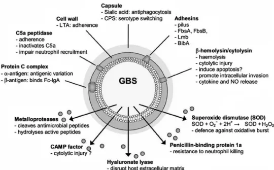
Detailed elucidation of the bacterial pathogenesis in hu-mans poses several problems. GBS adapts to its host during the process of colonization and expresses host-specific surface proteins, some of which are only expressed in the host and not in laboratory cultures. Nevertheless, much research effort has been successfully devoted to pathomechanisms of GBS diseases and has identified a large number of important bacterial compo-nents involved in adhesion, invasion, avoiding immune clearance and causing inflammation (schematically in figure 1). Several of these bacterial features occur via a ‘two-component regulatory system’. One component (e.g.
a sensor protein) is responsible for detecting and com-municating the external stimuli, while the other compo-nent (e.g. regulator protein) will react to those signals by activating or repressing specific genes, leading to the appropriate bacterial functions [62]. This system has been recognized as a bacterial strategy to overcome environ-mental stress factors, such as pH, temperature and osmolarity [63], but also to adhere to host cells [64, 65] and to express virulence factors [66].
Adhesion and Invasion
GBS interacts with extracellular matrix components of eukaryotic host cells, such as fibronectin, fibrinogen, lam-inin and cytokeratin 8. Several GBS surface-associated factors have been implicated in the adhesion process (Fig-ure 1). Once adhered, GBS is able to invade eukaryotic cells, such as fibroblasts, endothelial and epithelial cells, thus promoting bacterial penetration of host cellular bar-riers, such as the blood-brain barrier [67], chorioamniotic membranes [68] or barriers in the respiratory tract [69].
Avoidance of Immune Clearance
Apart from adaptation to environmental stress factors, GBS displays several functions that provide protection against killing by the host’s immune system. On mucosal surfaces, GBS binds the Fc fragment of IgA via a surface-associated protein complex (b-antigen of protein-C) [70] and thus inhibits interaction of IgA with Fc receptors ex-pressed on neutrophils, eosinophils or macrophages [71].
GBS possesses also several mechanisms to resist op-sonophagocytosis. Sialic acid, a component of the capsule, Figure 1. Schematic overview of GBS factors involved in adhesion, invasion, avoidance of immune clearance and virulence. The list of bacterial features shown is not exhaustive and additional findings are being continuously reported in the literature. LTA: lipoteichoic acid; CPS: capsular polysaccharide antigen, Fbs: fibrinogen-binding protein; Lmb: laminin-binding protein; BibA: B Streptococcus immunogenic bacterial adhesion.
prevents deposition of active C3b, and thus its function as an opsonin [72]. C5a, another important factor of the complement system that acts as a chemotaxin for neutro-phils, can also be inactivated by a surface-bound strepto-coccal protease, namely C5a peptidase [73]. Other factors that endow GBS with resistance to phagocytic killing by neutrophils includes its penicillin-binding protein 1a, me-talloproteases and superoxide dismutases (Figure 1) [74, 75]. Further strategies that have been suggested for how the microorganism avoids opsonization by antibodies include antigenic variation of a surface-associated protein complex (the a-antigen of the protein C) [76] and capsular serotype switching [77]. Also, intracellular survival in macrophages has been demonstrated in vitro [78], by a mechanism that presumably impairs activation of the host cell.
An offensive mode used by GBS to evade the host immune system is its ability to induce apoptosis in monocytes and macrophages [79]. Experimental data suggest that apoptosis is induced by b-haemolysin/cyto-lysin (b-h/c), a surface-associated toxin [80], although the detailed pathogenesis that triggers apoptosis is not yet fully understood.
Virulence
Several virulence factors have been identified in GBS, which contribute significantly to tissue damage at the local site of infection, as well as to a systemic inflammatory response by the host.
The capsule, a relatively unstructured network of high-molecular-weight polymers, is considered to be a major virulence factor. An overwhelming majority of GBS isolates from invasive disease are encapsulated. In animal experiments, non-encapsulated isogenic mutants demonstrated significantly reduced virulence compared to the corresponding wild-type strain [81].
The already mentioned surface-associated b-h/c is a potent, oxygen-stable, non-immunogenic GBS toxin, comparable to streptolysin S of GAS [82]. In addition to its hemolytic activity, b-h/c is responsible for pore for-mation and cytolytic injury to fibroblasts, endothelial and epithelial cells, as well as for promoting intracellular invasion, cytokine and nitric oxide release and, conse-quently, for triggering the sepsis cascade [80, 83]. Its expression has therefore been correlated with the severity of disease and tissue damage [84].
The CAMP factor has also been shown to have pore-forming activity, but this function is mainly seen on red blood cells pre-treated with sphingomyelinase (produced by S. aureus) [85]. Whether this factor plays a role in polymicrobial infections involving both GBS and S. aur-eus has not yet been assessed.
GBS is equipped with multiple oligopeptidases that promote the hydrolysis of several active peptides (or their fragments), such as bradykinin, the neuropeptides neurotensin and substance P, and adrenocorticotropin [74]. The biological function of these pathomechanisms is
not known, but peptide degradation in the tissue sur-roundings of GBS has been speculated in meningitis cases.
In GAS, superantigens have been implicated as major mediators of systemic toxicity and tissue damages. In GBS, though, most studies have failed to demonstrate exotoxins with superantigen activities. However, a recent case report described a GBS toxic shock-like syndrome, in which further experiments with human peripheral mono-nuclear cells and the isolated strain demonstrated super-antigenic stimulation [86]. Also, Schlievert et al. [87] purified a protein from a GBS strain that had the bio-logical activities of a pyrogenic toxin. Whether a hori-zontal transfer of DNA encoding such a virulence factor is occurring between different GBS strains, as has been shown for GAS [88], remains to be proven; there are, nevertheless, convincing indications for this hypothesis, in particular for genes encoding certain GBS surface pro-teins (a-antigen of protein C, C5a peptidase, Lmb protein) [89]. It is therefore plausible that such a mechanism is contributing to an increased incidence of severe diseases [90–92].
Macrolide Resistance
The mechanisms are mainly based on efflux pumps transporting macrolides out of the bacterial cytoplasm or on ribosome modifications that alter the antibiotic target site. The efflux pump is encoded by the mefA gene, whereas the acquisition of ermB and/or ermTR (ery-thromycin ribosome methylase) genes confers a ribo-somal modification. erm genes are associated with macrolide-lincosamide-streptogramin B (MLSB) resis-tance and can be expressed constitutively (cMLSB) or upon induction (iMLSB), although there is a heterogenic distribution of erm genes among these phenotypes [39]. Several studies have indicated how macrolide resistance might be acquired. A comparative pulsed-field gel elec-trophoresis analysis showed genetic clustering among macrolide-resistant GBS strains, with the predominance of a single-clone family within an otherwise heteroge-neous serotype V population [40]. Based on these data, the emergence of a specific macrolide-resistant clone family that possibly acquired resistance at a certain point of evolution and then subsequently increased in numbers was suggested. Recently, Puopolo et al. [93] found that the ermB gene is present on the chromosome of GBS within a transposon similar to the one identified in Streptococcus pneumoniae, which possibly indicates the horizontal transfer of such a mobile transposable ele-ment among streptococci.
Clinical Manifestations
GBS may occur in a large variety of clinical manifestation, as presented in table 2. Bacteremia without identified source and skin and soft-tissue infections are the most frequently reported expressions of invasive GBS disease
in non-pregnant adults. In up to one-third of the cases, in particular in skin and soft-tissue diseases, the pathogen is involved in a polymicrobial infection [2, 6, 7, 11, 13, 15, 16, 94]. In such situations, S. aureus is the most frequently isolated co-microorganism.
Skin diseases typically present as erysipelas/cellulitis, infected wounds or ulcers. However, GBS can also cause myositis, necrotizing fasciitis and toxic shock syndrome.
Infected Skin Ulcers
Physical defence factors and mechanical barriers are impoverished in ulcers and wounds, facilitating the
inva-sion of GBS. Not surprisingly, the presence of decubitus ulcers has been associated with an increased risk for invasive GBS disease (Table 1) [11]. In patients with diabetes mellitus and foot ulcers, apart from Staphylo-coccus spp., GBS is the most frequently involved patho-gen among isolated Gram-positive bacteria [95]. In diabetic foot infections, it is important to rule out osteo-myelitis and to distinguish between colonizing and infecting pathogens by culturing biopsies.
Erysipelas and Cellulites
In both entities, skin lesions are frequently considered to be a portal of entry. Cellulitis is the most reported diag-nosis among GBS skin and soft-tissue infections (25% in one study) [13, 47]. However, since aetiologic organisms are only rarely isolated (positive blood cultures in 2–5% of cases) [96, 97], the true incidence might be underesti-mated. Thus, in patients suffering from cellulitis by an unidentified pathogen and with known risk factors and/or carrier status, GBS should be considered.
Necrotizing Fasciitis
This severe infection has been categorized into two types. Type I is usually a polymicrobial infection (including anaerobic bacteria) that is associated with antecedent surgery, diabetes mellitus and vascular dis-eases, and manifests clinically with destruction of fat and fascia. Type II is monomicrobial, typically but not exclusively caused by GAS, and occurs commonly in patients without underlying diseases; it often presents with severe local pain, rapidly extending necrosis of the subcutaneous tissue and systemic toxicity. GBS as the Table 2
Relative frequency of different clinical manifestations of invasive GBS infections in non-pregnant adults.
Disease Median % (IQR)
Bacteremia with unknown source 24 (4–40) Skin and soft-tissue infections 20 (12–36) Respiratory infections 12 (3–19) Genitourinary infections 10 (0–20) Joint and bone infections 8 (4–19) Abdominal infections 5 (0–10) Endocarditis 4 (0–13) Infections of the central nervous system 4 (0–7) Miscellaneous < 1 (0–12)
-Intravascular-device infections -Ear, nose and throat infections -Endophthalmitis
-Iatrogenic (e.g. post-endoscopy) -Others
Analyses are based on 20 studies including 1167 episodes of invasive GBS infections [2, 4, 6, 7, 9, 11, 14–17, 20, 94, 131–133, 135–139]; IQR: interquartile range
Table 3
Streptococcal toxic shock syndrome case definition.
Community-acquired case: An illness with the following clinical manifestations occurring within the first 48 hours of hospitalization Nosocomial case: An illness with the following clinical manifestations occurring within the first 48 hours of illness
Clinical criteria:
• Hypotension : Systolic blood pressure £ 90 mmHg
• Multiorgan involvement characterized by two or more of the following: 1. Coagulopathy: Platelets £ 100,000/mm3
(£ 100 x 106
/L) or DIC 2. Renal impairment: Creatinine‡ 2 mg/dL (‡ 177 lmol/L)
3. Liver involvement: ALAT, ASAT, total bilirubin levels‡ twofold the upper limit of normal for the patient’s age. In patients with preexisting liver disease, a greater than twofold increase over the baseline level
4. Lung: ARDS
5. Skin: A generalized erythematous macular rash that may desquamate 6. Soft-tissue: Necrosis, including necrotizing fasciitis or myositis, or gangrene Laboratory criteria:
• Isolation of GAS Diagnosis:
Probable STSS: clinical case definition + isolation of GAS from a non-sterile sitea
Confirmed STSS: clinical case definition + isolation of GAS from a normally sterile site Adapted from CDC case definition 1996 [107].
a
In the absence of another identified aetiology for the illness; GAS: Group A Streptococcus; DIC: disseminated intravasal coagulation; ASAT: aspartate aminotransferase; ALAT: alanine aminotransferase; ARDS: acute respiratory distress syndrome
causative pathogen has been rarely reported, but the number of cases in the literature, both in type I and II fasciitis, is steadily increasing [7, 9, 13, 50, 90–92, 98–104]. It has been suggested that GBS necrotizing fasciitis in non-pregnant adults tends to occur in patients with significant underlying diseases [90], but this rela-tionship is difficult to support by data due to the rarity of the disease. Moreover, cases in patients without risk factors have been reported [91, 100, 101].
Analyses of GBS serotypes and several virulence factors in a case series have not elucidated why some patients develop necrotizing fasciitis and others develop both necrotizing fasciitis and toxic shock syndrome [92]. Similarly, host genetic factors that may contribute to the development of GBS toxic shock syndrome or necro-tizing fasciitis are as yet undetermined. Genetic predispo-sition has been demonstrated in invasive GAS infections: specific haplotypes of class II human leukocyte antigens (HLA) conferred protection against severe systemic dis-ease, while certain haplotypes were found to be associated with the development of necrotizing fasciitis and others with that of toxic shock syndrome [105].
Toxic Shock Syndrome
Streptococcal toxic shock syndrome (STSS) is defined as an acute and febrile illness that begins with mild viral prodromi but rapidly progresses to shock and multiorgan failure (Table 3) [106, 107]. Although STSS is classically associated with GAS, there is an increasing recognition that other streptococci, i.e. Groups B, C and G, can be the causative pathogen [108–111].
Group B STSS cases are often associated with severe skin infections [90–92, 99, 100, 112]. Comparison of the clinical characteristics of STSS caused by GAS and GBS in non-pregnant adults is statistically difficult, mainly be-cause the distribution of incidence numbers is unequal. However, there is tendency for a lower GBS frequency in adults below 50 years of age, irrespective of the presence of underlying disease [87, 91, 92, 100, 103].
Medical Treatment
Clinical isolates of GBS are susceptible to penicillin, the antimicrobial agent of choice for treating these invasive diseases [36]. In a European inter-country comparison, the average MIC was 0.06 mg/L (broth microdilution as-say) and 0.09 mg/L (Etest) [113]. These values are two- to ninefold higher than those for GAS. The occurrence of penicillin tolerance in clinical GBS isolates varies among different studies, ranging from 5% to 15% [114]. This in vitro phenomenon is observed when bacteria are inhibited by low concentrations of penicillin but are killed at much higher concentrations (MBC/MIC-ratio ‡ 1/32).
GBS strains are also susceptible to ampicillin, ceftri-axone, cefotaxime, meropenem, levofloxacin and vanco-mycin [36, 41]. As mentioned above, erythrovanco-mycin and clindamycin resistance is rising. Strains demonstrating
resistance to macrolides should be tested for MLSB
resistance, in particular, when considering clindamycin treatment [115].
Aminoglycosides alone have little or no effect on GBS, but synergistic killing with penicillin has been shown in vitro [116]. These results have been extrapolated in therapeutic strategies for severe sepsis cases and for iso-lates with penicillin tolerance or with high MIC values (e.g. > 0.1 mg/L). However, there is increasing solid evi-dence, though more for negative than for Gram-positive pathogens, that the addition of an aminoglycoside to a b-lactam is not beneficial in sepsis and carries the risk of nephrotoxicity [117]. The clinical significance of a penicillin-aminoglycoside combination for penicillin-tol-erant strains has not yet been established. Moreover, GBS high-level gentamicin resistance, leading to a lack of synergistic effect, has been also described [118]. Never-theless, the addition of an aminoglycoside to penicillin might be considered in selected cases.
There are no uniform treatment recommendations for GBS skin and soft-tissue infections. Suggestions are mostly based on case reports, case series, clinical diagnosis or in vitro experiments. Practice guidelines published by the IDSA give a thorough overview for the management of skin and soft-tissue infections [119] and are likely also applicable to those caused by GBS. Most regimens for uncomplicated erysipelas/cellulitis include a 5- to 10-day course of antimicrobial treatment. However, based on the above-mentioned host risk factors for acquiring invasive disease, the ability of the pathogen to avoid immune clearance and the higher MICs compared to GAS, ther-apy for 10–14 days in GBS erysipelas/cellulitis might be considered [33]. Whether oral or intravenous therapy is adequate, and at what time parenteral antibiotics can be changed to oral therapy, should always be discussed.
In erysipelas, treatment with penicillin G (12–18 million units IV per day divided into four to six doses) or with a cephalosporin (e.g. ceftriaxone 2 g IV once daily) or vancomycin (15 mg/kg IV every 12 hours in penicillin-allergic patients with normal renal function) is commonly administered. Thereafter, systemic inflammatory response signs often resolve within 1–2 days, and treatment can be promptly switched to oral antibiotics. However, in a Swedish study including 60 episodes of erysipelas without previous treatment [120], oral therapy (with penicillin or clindamycin) had no inferiority in clinical course and outcome compared to intravenous treatment. Further-more, penicillin V concentrations measured in punch biopsies from 45 orally treated patients with erysipelas exceeded the MIC of isolated streptococci [121]. Never-theless, since penicillin MIC values of GBS are higher than those of GAS, and macrolide and clindamycin resistance is increasing, MIC testing can be helpful to evaluate treatment options.
In acute cellulitis, clinical presentation may require hospitalization and treatment with intravenous antibiotics.
Once GBS has been identified as the causative pathogen, antibiotic therapy should be streamlined accordingly. In clinical practice, intravenous treatment is generally swit-ched to oral agents after the infected area has begun to improve (e.g. within 3–5 days). There are numerous agents available for oral therapy, as reviewed by Jacobs et al. [122]. Amoxicillin, among others, is effective in treating GBS cellulitis (2–3 g per day divided into two to three doses). Clindamycin (300 mg every 6–8 hours), or levofloxacin (500 mg once daily) are possible alternatives for patients with penicillin allergy.
Treatment of necrotizing fasciitis requires early and aggressive surgical exploration (debridement of necrotic tissue), antibiotic therapy and intensive care support. The duration of antimicrobial treatment is generally based upon clinical judgment [119]. In GBS necrotizing fasciitis, re-ported treatment durations from case series include 14–21 days intravenous antibiotics, followed by 14–21 days oral treatment [91]. As a part of the treatment concept, some authors have advocated the use of clindamycin (600–900 mg IV every 8 hours) in combination with penicillin (20–30 million units IV per day divided into four to six doses) in cases of GBS necrotizing fasciitis and/or STSS [90, 91, 99]. The efficacy of clindamycin is likely related to its ability to suppress the synthesis of GAS proteins, including M-pro-tein and streptococcal pyrogenic exotoxins A and B [123]. Furthermore, the bactericidal effect of clindamycin is not dependent on bacterial growth stage or inoculum size; in contrast, these factors have been proposed as responsible for a decreasing killing rate of GAS at higher penicillin concentrations (‘‘Eagle effect’’) [124]. A population-based study of invasive GAS infections reported that clindamycin reduced mortality in patients who had necrotizing fasciitis [125]. On the other hand, in an in vitro time-kill study using clinically relevant antimicrobial concentrations, neither an antagonistic nor a synergistic effect of the penicillin-clin-damycin combination was shown [126]. However, such data are largely lacking for GBS, and therefore recommenda-tions for clindamycin in GAS necrotizing fasciitis cannot be simply applied to those of GBS.
The use of intravenous immunoglobulins (IVIG) in necrotizing fasciitis and STSS caused by GBS has not been evaluated, even though it has been administered in anal-ogy to Group A STSS in selected cases [90, 91, 99]. Given the reports of a beneficial effect in STSS and in necro-tizing fasciitis caused by GAS [127–129], together with the identification of a purified GBS toxin isolated from a patient suffering from STSS [86, 87], it is theoretically plausible to regard IVIG as an adjunctive treatment. However, no recommendation about the use of IVIG in GBS cases can be made at this time.
Patients with relapsing skin and soft-tissue infections and no identifiable source should be evaluated for a prophylactic course of antibiotics or for eradication of GBS carriage [119]. However, the most appropriate antibiotic regimen and the efficacy of such an approach
are unknown for GBS. Options proposed for recurrent erysipelas/cellulitis [119] may be insufficient for those caused by GBS [48, 49, 130].
Outcome
During the last two decades, the mortality from invasive GBS disease has declined steadily, and is currently esti-mated at between 15–20%, with an attributable mortality of approximately 10% [2, 4, 6, 7, 9, 14, 15, 17, 94, 131–133]. As in other bacterial diseases, critical illness, in particular septic shock, at admission is strongly associated with in-creased mortality. Other factors associated with mortality include GBS bacteremia, diabetes mellitus, lung cancer, corticosteroid therapy, chronic renal insufficiency and congestive heart failure [7]. In GBS skin and soft-tissue infections, advanced age, skin ulcers and polymicrobial infections have been evaluated as risk factors for limb amputation [13]. The mortality rate of necrotizing fasciitis remains high, irrespective of the pathogen. In a series of 89 patients treated at a specialized center, the mortality was still 21.3% [98]. In these infections, rapid recognition and adequate surgical treatment have a significant influ-ence on outcome. Group B STSS seems to have a similar outcome to STSS caused by GAS and other pathogens, even though the disease incidence is still too low for solid comparative studies [92].
Acknowledgment
We thank Dr. Christian Garzoni from the Institute for Infectious Diseases, University of Berne, Dr. Nicolas Vuilleumier from the Central Clinical Chemistry Laboratory, Department of Clinical Pathology, Geneva University Hospital, Switzerland and Docent Bengt Ga˚rdlund from the Department of Infectious Diseases, Karolinska University Hospital Huddinge, Stockholm, Sweden for their comments and suggestions.
Financial support: P.S. is supported by the Swiss National Science Foundation (PBBSB 113145) and the Margarete and Walter Lichtenstein-Stiftung. L.J. is supported by grants from the AFA Sjukfo¨rsa¨kring, the Magnus Bergvalls Foundation, the A˚ ke Wibergs foundation, Karolinska Institutet, the Lars Hiertas Foundation, Stiftelsen La¨ngmanska Kulturfonden and the Swedish Society of Medicine.
References
1. Schrag SJ, Zywicki S, Farley MM, et al. Group B streptococcal disease in the era of intrapartum antibiotic prophylaxis. N Engl J Med 2000; 342: 15–20.
2. Munoz P, Llancaqueo A, Rodriguez-Creixems M, Pelaez T, Martin L, Bouza E: Group B streptococcus bacteremia in nonpregnant adults. Arch Intern Med 1997; 157: 213–216.
3. Active Bacterial Core Surveillance (ABCs) Report, Emerging Infections Program Network, group B streptococcus, 2005: Centers for Disease Control and Prevention, 2007. 4. Schwartz B, Schuchat A, Oxtoby MJ, Cochi SL, Hightower A,
Broome CV: Invasive group B streptococcal disease in adults. A population-based study in metropolitan Atlanta. Jama 1991; 266: 1112–1114.
5. Zangwill KM, Schuchat A, Wenger JD: Group B streptococcal disease in the United States, 1990: report from a multistate active surveillance system. MMWR CDC Surveill Summ 1992; 41: 25–32. 6. Farley MM, Harvey RC, Stull T, et al. A population-based
assessment of invasive disease due to group B Streptococcus in nonpregnant adults. N Engl J Med 1993; 328: 1807–1811. 7. Blancas D, Santin M, Olmo M, Alcaide F, Carratala J, Gudiol F:
Group B streptococcal disease in nonpregnant adults: incidence, clinical characteristics, and outcome. Eur J Clin Microbiol Infect Dis 2004; 23: 168–173.
8. Huang PY, Lee MH, Yang CC, Leu HS: Group B streptococcal bacteremia in non-pregnant adults. J Microbiol Immunol Infect 2006; 39: 237–241.
9. Wang TK, Fung AM, Woo PC, Yuen KY, Wong SS: Streptococcus agalactiae (Lancefield group B) bacteraemia in nonpregnant adults. Eur J Clin Microbiol Infect Dis 2002; 21: 140–142. 10. Harrison LH, Ali A, Dwyer DM, et al. Relapsing invasive group B
streptococcal infection in adults. Ann Intern Med 1995; 123: 421–427. 11. Jackson LA, Hilsdon R, Farley MM, et al. Risk factors for group B streptococcal disease in adults. Ann Intern Med 1995; 123: 415– 420.
12. Cox NH: Oedema as a risk factor for multiple episodes of cel-lulitis/erysipelas of the lower leg: a series with community follow-up. Br J Dermatol 2006; 155: 947–950.
13. Lee NY, Yan JJ, Wu JJ, Lee HC, Liu KH, Ko WC: Group B strep-tococcal soft tissue infections in non-pregnant adults. Clin Microbiol Infect 2005; 11: 577–579.
14. Opal SM, Cross A, Palmer M, Almazan R: Group B streptococcal sepsis in adults and infants. Contrasts and comparisons. Arch Intern Med 1988; 148: 641–645.
15. Ho CM, Chi CY, Ho MW, et al. Clinical characteristics of group B streptococcus bacteremia in non-pregnant adults. J Microbiol Immunol Infect 2006; 39: 396–401.
16. Kim BN, Bae IG, Kim MN, Woo JH, Ryu J, Kim YS: Group B streptococcal bacteremia in nonpregnant adults with hepatic disease in Korea. Eur J Clin Microbiol Infect Dis 2001; 20: 639–642.
17. Colford JM Jr., Mohle-Boetani J, Vosti KL: Group B streptococcal bacteremia in adults. Five years’ experience and a review of the literature. Medicine (Baltimore) 1995; 74: 176–190.
18. Baddour LM, Cox JW Jr: Group B streptococcal infection of a pacemaker wire following sigmoidoscopy. Clin Infect Dis 1992; 15: 1069.
19. Triesenberg SN, Clark NM, Kauffman CA: Group B streptococcal prosthetic joint infection following sigmoidoscopy. Clin Infect Dis 1992; 15: 374–375.
20. Trivalle C, Martin E, Martel P, Jacque B, Menard JF, Lemeland JF: Group B streptococcal bacteraemia in the elderly. J Med Microbiol 1998; 47: 649–652.
21. Manning SD, Tallman P, Baker CJ, Gillespie B, Marrs CF, Foxman B: Determinants of co-colonization with group B streptococcus among heterosexual college couples. Epidemiology 2002; 13: 533–539.
22. Easmon CS, Tanna A, Munday P, Dawson S: Group B strepto-cocci–gastrointestinal organisms? J Clin Pathol 1981; 34: 921–3. 23. Meyn LA, Moore DM, Hillier SL, Krohn MA: Association of sexual
activity with colonization and vaginal acquisition of group B Streptococcus in nonpregnant women. Am J Epidemiol 2002; 155: 949–957.
24. Manning SD, Neighbors K, Tallman PA, et al. Prevalence of group B streptococcus colonization and potential for transmis-sion by casual contact in healthy young men and women. Clin Infect Dis 2004; 39: 380–388.
25. Brimil N, Barthell E, Heindrichs U, Kuhn M, Lutticken R, Spell-erberg B: Epidemiology of Streptococcus agalactiae colonization in Germany. Int J Med Microbiol 2006; 296: 39–44.
26. Edwards MS, Rench MA, Palazzi DL, Baker CJ: Group B strepto-coccal colonization and serotype-specific immunity in healthy elderly persons. Clin Infect Dis 2005; 40: 352–357.
27. Manning S, Tallman P, Foxman B: Prevalence and co-coloniza-tion with group b streptococcus (Gbs) Among heterosexual college couples. Ann Epidemiol 2000; 10: 472.
28. Lindahl G, Stalhammar-Carlemalm M, Areschoug T: Surface proteins of Streptococcus agalactiae and related proteins in other bacterial pathogens. Clin Microbiol Rev 2005; 18: 102– 127.
29. Tyrrell GJ, Senzilet LD, Spika JS, et al. Invasive disease due to group B streptococcal infection in adults: results from a Cana-dian, population-based, active laboratory surveillance study– 1996. Sentinel Health Unit Surveillance System Site Coordina-tors. J Infect Dis 2000; 182: 168–173.
30. Harrison LH, Elliott JA, Dwyer DM, et al. Serotype distribution of invasive group B streptococcal isolates in Maryland: implica-tions for vaccine formulation. Maryland Emerging Infecimplica-tions Program. J Infect Dis 1998; 177: 998–1002.
31. Blumberg HM, Stephens DS, Modansky M, et al. Invasive group B streptococcal disease: the emergence of serotype V. J Infect Dis 1996; 173: 365–373.
32. Hickman ME, Rench MA, Ferrieri P, Baker CJ: Changing epide-miology of group B streptococcal colonization. Pediatrics 1999; 104: 203–209.
33. Farley MM: Group B streptococcal disease in nonpregnant adults. Clin Infect Dis 2001; 33: 556–561.
34. Lachenauer CS, Kasper DL, Shimada J, et al. Serotypes VI and VIII predominate among group B streptococci isolated from preg-nant Japanese women. J Infect Dis 1999; 179: 1030–1033. 35. Ekelund K, Slotved HC, Nielsen HU, Kaltoft MS, Konradsen HB:
Emergence of invasive serotype VIII group B streptococcal infections in Denmark. J Clin Microbiol 2003;
41: 4442–4444.
36. Murdoch DR, Reller LB: Antimicrobial susceptibilities of group B streptococci isolated from patients with invasive disease: 10-year perspective. Antimicrob Agents Chemother 2001; 45: 3623–3634.
37. Fernandez M, Hickman ME, Baker CJ: Antimicrobial suscepti-bilities of group B streptococci isolated between 1992 and 1996 from patients with bacteremia or meningitis. Antimicrob Agents Chemother 1998; 42: 1517–1519.
38. DiPersio LP, DiPersio JR: High rates of erythromycin and clin-damycin resistance among OBGYN isolates of group B Strep-tococcus. Diagn Microbiol Infect Dis 2006; 54: 79–82. 39. von Both U, Buerckstuemmer A, Fluegge K, Berner R:
Hetero-geneity of genotype-phenotype correlation among macrolide-resistant Streptococcus agalactiae isolates. Antimicrob Agents Chemother 2005; 49: 3080–3082.
40. von Both U, Ruess M, Mueller U, Fluegge K, Sander A, Berner R: A serotype V clone is predominant among erythromycin-resis-tant Streptococcus agalactiae isolates in a southwestern region of Germany. J Clin Microbiol 2003; 41: 2166–2169.
41. Lin FY, Azimi PH, Weisman LE, et al. Antibiotic susceptibility profiles for group B streptococci isolated from neonates, 1995– 1998. Clin Infect Dis 2000; 31: 76–79.
42. Manning SD, Foxman B, Pierson CL, Tallman P, Baker CJ, Pearl-man MD: Correlates of antibiotic-resistant group B streptococ-cus isolated from pregnant women. Obstet Gynecol 2003; 101: 74–79.
43. Uh Y, Kim HY, Jang IH, Hwang GY, Yoon KJ: Correlation of ser-otypes and genser-otypes of macrolide-resistant Streptococcus agalactiae. Yonsei Med J 2005; 46: 480–483.
44. Murray P, Rosenthal K, Pfaller M. Medical Microbiology. 5th ed. Philadelphia, 2005; 247–250.
45. Picard FJ, Bergeron MG: Laboratory detection of group B Streptococcus for prevention of perinatal disease. Eur J Clin Microbiol Infect Dis 2004; 23: 665–671.
46. Nizet V, Gibson RL, Chi EY, Framson PE, Hulse M, Rubens CE: Group B streptococcal beta-hemolysin expression is associated with injury of lung epithelial cells. Infect Immun 1996; 64: 3818–3826.
47. Edwards MS, Baker CJ: Group B streptococcal infections in el-derly adults. Clin Infect Dis 2005; 41: 839–847.
48. Del Giudice P, van der Mee-Marquet N, David-Rubin F, et al. Se-vere relapsing erysipelas associated with chronic Streptococcus agalactiae vaginal colonization. Clin Infect Dis 2006; 43: e67–e70. 49. Sendi P, Graber P, Johansson L, Norrby-Teglund A, Zimmerli W: Streptococcus agalactiae in relapsing cellulitis. Clin Infect Dis 2007; 44: 1141–1142.
50. Bero SM, Brady MS: Streptococcal septic shock after inguinal lymphadenectomy. Surg Infect (Larchmt) 2006; 7: 547–550. 51. Binnick AN, Klein RB, Baughman RD: Recurrent erysipelas
caused by group B streptococcus organisms. Arch Dermatol 1980; 116: 798–799.
52. Clement S, Vaudaux P, Francois P, et al. Evidence of an intra-cellular reservoir in the nasal mucosa of patients with recurrent Staphylococcus aureus rhinosinusitis. J Infect Dis 2005; 192: 1023–1028.
53. Thulin P, Johansson L, Low DE, et al. Viable group A streptococci in macrophages during acute soft tissue infection. PLoS Med 2006; 3: e53.
54. Baker CJ, Edwards MS, Kasper DL: Role of antibody to native type III polysaccharide of group B Streptococcus in infant infection. Pediatrics 1981; 68: 544–549.
55. Kasper DL, Paoletti LC, Wessels MR, et al. Immune response to type III group B streptococcal polysaccharide-tetanus toxoid conjugate vaccine. J Clin Invest 1996; 98: 2308–2314. 56. Baker CJ, Paoletti LC, Wessels MR, et al. Safety and
immuno-genicity of capsular polysaccharide-tetanus toxoid conjugate vaccines for group B streptococcal types Ia and Ib. J Infect Dis 1999; 179: 142–150.
57. Baker CJ, Paoletti LC, Rench MA, et al. Use of capsular polysac-charide-tetanus toxoid conjugate vaccine for type II group B Streptococcus in healthy women. J Infect Dis 2000; 182: 1129–1138. 58. Amaya RA, Baker CJ, Keitel WA, Edwards MS: Healthy elderly
people lack neutrophil-mediated functional activity to type V group B Streptococcus. J Am Geriatr Soc 2004; 52: 46–50. 59. Palazzi DL, Rench MA, Edwards MS, Baker CJ: Use of type V
group B streptococcal conjugate vaccine in adults 65–85 years old. J Infect Dis 2004; 190: 558–564.
60. Baker CJ, Webb BJ, Kasper DL, Edwards MS: The role of com-plement and antibody in opsonophagocytosis of type II group B streptococci. J Infect Dis 1986; 154: 47–54.
61. Mazade MA, Edwards MS: Impairment of type III group B Streptococcus-stimulated superoxide production and opsono-phagocytosis by neutrophils in diabetes. Mol Genet Metab 2001; 73: 259–267.
62. Cotter PA, Miller JF: In vivo and ex vivo regulation of bacterial virulence gene expression. Curr Opin Microbiol 1998; 1: 17–26. 63. Tamura GS, Kuypers JM, Smith S, Raff H, Rubens CE: Adherence
of group B streptococci to cultured epithelial cells: roles of
environmental factors and bacterial surface components. Infect Immun 1994; 62: 2450–2458.
64. Spellerberg B, Rozdzinski E, Martin S, Weber-Heynemann J, Lutticken R: rgf encodes a novel two-component signal trans-duction system of Streptococcus agalactiae. Infect Immun 2002; 70: 2434–2440.
65. Poyart C, Lamy MC, Boumaila C, Fiedler F, Trieu-Cuot P: Regu-lation of D-alanyl-lipoteichoic acid biosynthesis in Streptococ-cus agalactiae involves a novel two-component regulatory system. J Bacteriol 2001; 183: 6324–6334.
66. Jiang SM, Cieslewicz MJ, Kasper DL, Wessels MR: Regulation of virulence by a two-component system in group B streptococcus. J Bacteriol 2005; 187: 1105–1113.
67. Nizet V, Kim KS, Stins M, et al. Invasion of brain microvascular endothelial cells by group B streptococci. Infect Immun 1997; 65: 5074–5081.
68. Winram SB, Jonas M, Chi E, Rubens CE: Characterization of group B streptococcal invasion of human chorion and amnion epithelial cells In vitro. Infect Immun 1998; 66: 4932–4941. 69. Rubens CE, Smith S, Hulse M, Chi EY, van Belle G: Respiratory
epithelial cell invasion by group B streptococci. Infect Immun 1992; 60: 5157–5163.
70. Lindahl G, Akerstrom B, Vaerman JP, Stenberg L: Characteriza-tion of an IgA receptor from group B streptococci: specificity for serum IgA. Eur J Immunol 1990; 20: 2241–2247.
71. Pleass RJ, Areschoug T, Lindahl G, Woof JM: Streptococcal IgA-binding proteins bind in the Calpha 2-Calpha 3 interdomain region and inhibit binding of IgA to human CD89. J Biol Chem 2001; 276: 8197–8204.
72. Marques MB, Kasper DL, Pangburn MK, Wessels MR: Prevention of C3 deposition by capsular polysaccharide is a virulence mechanism of type III group B streptococci. Infect Immun 1992; 60: 3986–3993.
73. Cheng Q, Stafslien D, Purushothaman SS, Cleary P: The group B streptococcal C5a peptidase is both a specific protease and an invasin. Infect Immun 2002; 70: 2408–2413.
74. Lin B, Averett WF, Novak J, et al. Characterization of PepB, a group B streptococcal oligopeptidase. Infect Immun 1996; 64: 3401–3406.
75. Poyart C, Pellegrini E, Gaillot O, Boumaila C, Baptista M, Trieu-Cuot P: Contribution of Mn-cofactored superoxide dismutase (SodA) to the virulence of Streptococcus agalactiae. Infect Im-mun 2001; 69: 5098–5106.
76. Madoff LC, Michel JL, Gong EW, Kling DE, Kasper DL: Group B streptococci escape host immunity by deletion of tandem re-peat elements of the alpha C protein. Proc Natl Acad Sci U S A 1996; 93: 4131–4136.
77. Luan SL, Granlund M, Sellin M, Lagergard T, Spratt BG, Norgren M: Multilocus sequence typing of Swedish invasive group B streptococcus isolates indicates a neonatally associated genetic lineage and capsule switching. J Clin Microbiol 2005; 43: 3727–3733.
78. Cornacchione P, Scaringi L, Fettucciari K, et al. Group B strep-tococci persist inside macrophages. Immunology 1998; 93: 86–95.
79. Fettucciari K, Rosati E, Scaringi L, et al. Group B Streptococcus induces apoptosis in macrophages. J Immunol 2000; 165: 3923–3933.
80. Liu GY, Doran KS, Lawrence T, et al. Sword and shield: linked group B streptococcal beta-hemolysin/cytolysin and carotenoid pigment function to subvert host phagocyte defense. Proc Natl Acad Sci U S A 2004; 101: 14491–14496.
81. Wessels MR, Rubens CE, Benedi VJ, Kasper DL: Definition of a bacterial virulence factor: sialylation of the group B strepto-coccal capsule. Proc Natl Acad Sci U S A 1989; 86: 8983–89837. 82. Nizet V: Streptococcal beta-hemolysins: genetics and role in
disease pathogenesis. Trends Microbiol 2002; 10: 575–580. 83. Doran KS, Chang JC, Benoit VM, Eckmann L, Nizet V: Group B
streptococcal beta-hemolysin/cytolysin promotes invasion of human lung epithelial cells and the release of interleukin-8. J Infect Dis 2002; 185: 196–203.
84. Puliti M, Nizet V, von Hunolstein C, et al. Severity of group B streptococcal arthritis is correlated with beta-hemolysin expression. J Infect Dis 2000; 182: 824–832.
85. Lang S, Palmer M: Characterization of Streptococcus agalactiae CAMP factor as a pore-forming toxin. J Biol Chem 2003; 278: 38167–38173.
86. Begly JS, Barnes RC: Group B streptococcus toxic shock-like syndrome in a healthy woman: a case report. J Reprod Med 2007; 52: 323–325.
87. Schlievert PM, Gocke JE, Deringer JR: Group B streptococcal toxic shock-like syndrome: report of a case and purification of an associated pyrogenic toxin. Clin Infect Dis 1993; 17: 26–31.
88. Bessen DE, Hollingshead SK: Allelic polymorphism of emm loci provides evidence for horizontal gene spread in group A streptococci. Proc Natl Acad Sci U S A 1994; 91: 3280–3284. 89. Broker G, Spellerberg B: Surface proteins of Streptococcus
agalactiae and horizontal gene transfer. Int J Med Microbiol 2004; 294: 169–175.
90. Gardam MA, Low DE, Saginur R, Miller MA: Group B strepto-coccal necrotizing fasciitis and streptostrepto-coccal toxic shock-like syndrome in adults. Arch Intern Med 1998; 158: 1704–1708. 91. Wong CH, Kurup A, Tan KC: Group B Streptococcus necrotizing
fasciitis: an emerging disease? Eur J Clin Microbiol Infect Dis 2004; 23: 573–575.
92. Chang B, Ikebe T, Wada A, et al. Surveillance of group B strep-tococcal toxic shock-like syndrome in nonpregnant adults and characterization of the strains in Japan. Jpn J Infect Dis 2006; 59: 182–185.
93. Puopolo KM, Klinzing DC, Lin MP, Yesucevitz DL, Cieslewicz MJ: A composite transposon associated with erythromycin and clindamycin resistance in group B Streptococcus. J Med Micro-biol 2007; 56: 947–955.
94. Verghese A, Mireault K, Arbeit RD: Group B streptococcal bac-teremia in men. Rev Infect Dis 1986; 8: 912–917.
95. Raja NS: Microbiology of diabetic foot infections in a teaching hospital in Malaysia: a retrospective study of 194 cases. J Microbiol Immunol Infect 2007; 40: 39–44.
96. Eriksson B, Jorup-Ronstrom C, Karkkonen K, Sjoblom AC, Holm SE: Erysipelas: clinical and bacteriologic spectrum and serolog-ical aspects. Clin Infect Dis 1996; 23: 1091–1098.
97. Perl B, Gottehrer NP, Raveh D, Schlesinger Y, Rudensky B, Yinnon AM: Cost-effectiveness of blood cultures for adult patients with cellulitis. Clin Infect Dis 1999; 29: 1483–1488.
98. Wong CH, Chang HC, Pasupathy S, Khin LW, Tan JL, Low CO. Necrotizing fasciitis: clinical presentation, microbiology, and determinants of mortality. J Bone Joint Surg Am 2003; 85–A: 1454–1460.
99. Crum NF, Wallace MR: Group B streptococcal necrotizing fas-ciitis and toxic shock-like syndrome: a case report and review of the literature. Scand J Infect Dis 2003; 35: 878–881.
100. Holmstrom B, Grimsley EW: Necrotizing fasciitis and toxic shock-like syndrome caused by group B streptococcus. South Med J 2000; 93: 1096–1098.
101. Akita S, Tanaka K, Hirano A: Lower extremity reconstruction after necrotising fasciitis and necrotic skin lesions using a porcine-derived skin substitute. J Plast Reconstr Aesthet Surg 2006; 59: 759–763.
102. Wong CH, Song C, Ong YS, Tan BK, Tan KC, Foo CL: Abdominal wall necrotizing fasciitis: it is still ‘‘Meleney’s Minefield’’. Plast Reconstr Surg 2006; 117: 147e–150e.
103. Tang WM, Ho PL, Yau WP, Wong JW, Yip DK: Report of 2 fatal cases of adult necrotizing fasciitis and toxic shock syndrome caused by Streptococcus agalactiae. Clin Infect Dis 2000; 31: E15–E17.
104. Riefler J 3rd, Molavi A, Schwartz D, DiNubile M: Necrotizing fasciitis in adults due to group B streptococcus. Report of a case and review of the literature. Arch Intern Med 1988; 148: 727–729.
105. Kotb M, Norrby-Teglund A, McGeer A, et al. An immunogenetic and molecular basis for differences in outcomes of invasive group A streptococcal infections. Nat Med 2002; 8: 1398–1404. 106. Defining the group A streptococcal toxic shock syndrome.
Rationale and consensus definition. The Working Group on Se-vere Streptococcal Infections. Jama 1993; 269: 390–391. 107. CDC. Streptococcal Toxic-Shock Syndrome (STSS); 1996 Case
Definition. http://www.cdc.gov/epo/dphsi/casedef/streptococ-calcurrent.htm.
108. Ekelund K, Skinhoj P, Madsen J, Konradsen HB: Invasive group A, B, C and G streptococcal infections in Denmark 1999–2002: epidemiological and clinical aspects. Clin Microbiol Infect 2005; 11: 569–576.
109. Sharma M, Khatib R, Fakih M: Clinical characteristics of necro-tizing fasciitis caused by group G Streptococcus: case report and review of the literature. Scand J Infect Dis 2002; 34: 468–471. 110. Tee WS, Lieu PK, Ngan CC: Epidemiology of beta-haemolytic
group G streptococcal bacteraemia in Singapore (1996 to 1998). Ann Acad Med Singapore 2002; 31: 86–91.
111. Hashikawa S, Iinuma Y, Furushita M, et al. Characterization of group C and G streptococcal strains that cause streptococcal toxic shock syndrome. J Clin Microbiol 2004; 42: 186–192. 112. Reich HL, Crawford GH, Pelle MT, James WD: Group B
strepto-coccal toxic shock-like syndrome. Arch Dermatol 2004; 140: 163–166.
113. Gemmell CG: Susceptibility of a variety of clinical isolates to linezolid: a European inter-country comparison. J Antimicrob Chemother 2001; 48: 47–52.
114. Betriu C, Gomez M, Sanchez A, Cruceyra A, Romero J, Picazo JJ: Antibiotic resistance and penicillin tolerance in clinical isolates of group B streptococci. Antimicrob Agents Chemother 1994; 38: 2183–2186.
115. Schoening TE, Wagner J, Arvand M: Prevalence of erythromycin and clindamycin resistance among Streptococcus agalactiae isolates in Germany. Clin Microbiol Infect 2005; 11: 579–582. 116. Baker CN, Thornsberry C, Facklam RR: Synergism, killing kinetics,
and antimicrobial susceptibility of group A and B streptococci. Antimicrob Agents Chemother 1981; 19: 716–725.
117. Paul M, Silbiger I, Grozinsky S, Soares-Weiser K, Leibovici L. Beta lactam antibiotic monotherapy versus beta lactam-aminogly-coside antibiotic combination therapy for sepsis. Cochrane Database Syst Rev 2006: CD003344.
118. Buu-Hoi A, Le Bouguenec C, Horaud T: High-level chromosomal gentamicin resistance in Streptococcus agalactiae (group B). Antimicrob Agents Chemother 1990; 34: 985–988.
119. Stevens DL, Bisno AL, Chambers HF, et al. Practice guidelines for the diagnosis and management of skin and soft-tissue infec-tions. Clin Infect Dis 2005; 41: 1373–1406.
120. Jorup-Ronstrom C, Britton S, Gavlevik A, Gunnarsson K, Redman AC: The course, costs and complications of oral versus intrave-nous penicillin therapy of erysipelas. Infection 1984; 12: 390–394. 121. Sjoblom AC, Bruchfeld J, Eriksson B, et al. Skin concentrations of phenoxymethylpenicillin in patients with erysipelas. Infection 1992; 20: 30–33.
122. Jacobs MR, Jones RN, Giordano PA: Oral beta-lactams applied to uncomplicated infections of skin and skin structures. Diagn Microbiol Infect Dis 2007; 57: S55–65.
123. Mascini EM, Jansze M, Schouls LM, Verhoef J, Van Dijk H: Pen-icillin and clindamycin differentially inhibit the production of pyrogenic exotoxins A and B by group A streptococci. Int J An-timicrob Agents 2001; 18: 395–398.
124. Stevens DL, Gibbons AE, Bergstrom R, Winn V: The Eagle effect revisited: efficacy of clindamycin, erythromycin, and penicillin in the treatment of streptococcal myositis. J Infect Dis 1988; 158: 23–28.
125. Mulla ZD, Leaverton PE, Wiersma ST: Invasive group A streptococcal infections in Florida. South Med J 2003; 96: 968–973.
126. Stevens DL, Madaras-Kelly KJ, Richards DM: In vitro antimicro-bial effects of various combinations of penicillin and clinda-mycin against four strains of Streptococcus pyogenes. Antimicrob Agents Chemother 1998; 42: 1266–1268. 127. Norrby-Teglund A, Muller MP, McGeer A, et al. Successful
management of severe group A streptococcal soft tissue infections using an aggressive medical regimen including intravenous polyspecific immunoglobulin together with a conservative surgical approach. Scand J Infect Dis 2005; 37: 166–172.
128. Darenberg J, Ihendyane N, Sjolin J, et al. Intravenous immuno-globulin G therapy in streptococcal toxic shock syndrome: a European randomized, double-blind, placebo-controlled trial. Clin Infect Dis 2003; 37: 333–340.
129. Kaul R, McGeer A, Norrby-Teglund A, et al. Intravenous immu-noglobulin therapy for streptococcal toxic shock syndrome–a
comparative observational study. The Canadian Streptococcal Study Group. Clin Infect Dis 1999; 28: 800–807.
130. Moylett EH, Fernandez M, Rench MA, Hickman ME, Baker CJ: A 5-year review of recurrent group B streptococcal disease: les-sons from twin infants. Clin Infect Dis 2000; 30: 282–287. 131. Gallagher PG, Watanakunakorn C: Group B streptococcal
bac-teremia in a community teaching hospital. Am J Med 1985; 78: 795–800.
132. Larppanichpoonphol P, Watanakunakorn C: Group B strepto-coccal bacteremia in nonpregnant adults at a community teaching hospital. South Med J 2001; 94: 1206–1211.
133. Schugk J, Harjola VP, Sivonen A, Vuopio-Varkila J, Valtonen M: A clinical study of beta-haemolytic groups A, B, C and G strepto-coccal bacteremia in adults over an 8-year period. Scand J Infect Dis 1997; 29: 233–238.
134. Henning KJ, Hall EL, Dwyer DM, et al. Invasive group B strep-tococcal disease in Maryland nursing home residents. J Infect Dis 2001; 183: 1138–1142.
135. Falagas ME, Rosmarakis ES, Avramopoulos I, Vakalis N: Strep-tococcus agalactiae infections in non-pregnant adults: Single center experience of a growing clinical problem. Med Sci Monit 2006; 12: CR447–CR451.
136. Skogberg K, Simonen H, Renkonen OV, Valtonen VV: Beta-hae-molytic group A, B, C and G streptococcal septicaemia: a clinical study. Scand J Infect Dis 1988; 20: 119–125.
137. Lerner PI, Gopalakrishna KV, Wolinsky E, McHenry MC, Tan JS, Rosenthal M: Group B streptococcus (S. agalactiae) bacteremia in adults: analysis of 32 cases and review of the literature. Medicine (Baltimore) 1977; 56: 457–473.
138. Bayer AS, Chow AW, Anthony BF, Guze LB: Serious infections in adults due to group B streptococci. Clinical and serotypic char-acterization. Am J Med 1976; 61: 498–503.
139. Bolanos M, Canas A, Santana OE, Perez-Arellano JL, de Miguel I, Martin-Sanchez AM: Invasive group B streptococcal disease in nonpregnant adults. Eur J Clin Microbiol Infect Dis 2001; 20: 837–839.