INVERTEBRATE MICROBIOLOGY
Bacterial Communities in Central European Bumblebees:
Low Diversity and High Specificity
Hauke Koch&Paul Schmid-Hempel
Received: 9 December 2010 / Accepted: 27 March 2011 / Published online: 10 May 2011 # Springer Science+Business Media, LLC 2011
Abstract Recent studies on the microbial flora of the honeybee gut have revealed an apparently highly specific community of resident bacteria that might play a role in immune defence and food preservation for their hosts. However, at present, very little is known about the diversity and ecology of bacteria occurring in non-domesticated bees like bumblebees, which are of similar importance as honeybees for the pollination of agricultural and wild flowers. To fill this gap in knowledge, we examined six of the most common bumblebee species in Central Europe from three locations in Germany and Switzerland for their bacterial communities. We used a culture-independent molecular approach based on sequencing the 16S rRNA gene from a selection of individuals and examining a larger number of samples by terminal restriction fragment length polymorphism profiles. The gut flora was dominated by very few and mostly undescribed groups of bacteria belonging to the Proteobacteria, Bacteroidetes, Firmicutes and Actinobacteria. This core set of bacteria was present in all of the examined bumblebee species. These bacteria are similar to, but distinct from, bacteria previously described
from the honeybee gut. Significant differences were
observed between the communities of bacteria in the different bumblebee species; the effect of sampling location was less strong. A novel group of Betaproteobacteria additionally shows evidence for host species-specific genotypes. The gut flora of bumblebees therefore is apparently composed of relatively few highly specialized
bacteria, indicating a strong interaction and possibly important functions with their hosts.
Introduction
Bacterial communities can be important for many vital functions of the host organism, such as digestive efficiency [20,26,33], for example in herbivorous insects [7,38]; for the defence against major enemies [67]; or through
interactions with infecting pathogens [13, 14]. We are,
however, still lacking a good understanding of the diversity and distribution of bacterial communities in most naturally occurring organisms. For example, wild bees provide important pollination services and are known to host a number of potentially important bacteria in their gut. But so far, the existing studies have only been looking at a few individual hosts of few species [44, 51,54] or limited the
sampling to the genus Bifidobacterium [42, 43]. We here
enlarge this database for a prominent group of large pollinators—the bumblebees, Bombus spp.
Bumblebees are a group of eusocial hymenoptera, most abundant in temperate and cold regions of the world [69]. Pollinating a variety of wild and agricultural flowering plants, they provide important ecosystem services [23]. They are also commercially bred for the pollination of greenhouse plants, most notably for the pollination of tomatoes [66]. Over recent years, a decline in the abundance and range of bumblebee species has been noted in several parts of the world [24,70]. Whereas man-made changes of natural ecosystems are likely playing a major role in this decline, the spillover of new parasites into wild bumblebee populations caused by the international trade with commercially bred bumblebee colonies has been discussed as a further important factor as well [11, 24]. H. Koch (*)
:
P. Schmid-HempelETH Zürich, Institute of Integrative Biology (IBZ), Universitätsstrasse 16,
CH-8092 Zürich, Switzerland e-mail: hauke.koch@env.ethz.ch Microb Ecol (2011) 62:121–133 DOI 10.1007/s00248-011-9854-3
So far, the ecology and effect of eukaryotic bumblebee parasites, such as the microsporidian Nosema bombi and the trypanosomatid Crithidia bombi, have been studied intensively (e.g.16,55, 60]. In contrast, the diversity and ecology of prokaryotic organisms associated with bumble-bees, such as bacteria, is much less known. As a yardstick, recent studies have shown a specialized community of
bacteria inhabiting the honeybee gut [4, 12, 53]. This
included lactic acid bacteria protecting the host against infections of the bacterium Paenibacillus larvae, the pathogenic agent of the American foulbrood [19]. Similar lactic acid bacteria and other members of the honeybee microbiota have been found in the bumblebee gut [44,51, 54]: these might potentially play an important role in defence against parasites of bumblebees as well.
We here examined the gut bacterial community of six common bumblebee species from three locations in Central Europe. Since methods that depend on cultur-ing the bacteria are likely to miss a considerable number of bacterial species in environmental samples [36], we used a culture-independent molecular approach that can be applied to field data. Specifically, we cloned and sequenced 16S rDNA, the most commonly used molecular marker for bacterial identification [63], for the gut bacteria of a sample of individuals. Terminal restriction fragment length polymorphism (T-RFLP) analysis [48] was then used for the characterization of a larger number of specimens. We identified the phyloge-netic position of the most common bumblebee gut bacteria, especially in relation to the bacteria found in the honeybee gut. In addition, we examined the effects of host species and sampling location on the bacterial community composition.
Methods
Collection and Preparation of Samples
Bumblebee workers were collected in three regions [1]: (1) Northern Germany in June 2008 (location: Celle, 52° 38′0.96″ N, 10°3′9.71″ E; sample sizes: Bombus terrestris, N = 39; Bombus lapidaries, N = 10; Bombus pascuorum, N = 20) [2]; (2) Northwestern Switzerland in the Jura range near Basel in August 2007 (location: Röschenz, 47°25′ 32.66″ N, 7°28′31.41″ E; sample sizes: B. terrestris, N= 18; B. lapidaries, N = 22; B. pascuorum, N= 24; Bombus hortorum, N = 20) [3]; (3) Swiss Alps in the Swiss National Park in July 2007 (location: Stabelchod, 46°39′ 40.02″ N, 10°14′25.47″ E; sample sizes: Bombus lucorum/ Bombus cryptorum, N = 24; Bombus soroeensis, N = 22). Because of a lack of reliable traits to distinguish workers of B. lucorum from B. cryptarum, samples resembling
these species from the Swiss Alps were not assigned to either of the two species and will be referred to as B. lucorum/B. cryptarum in the following.
Field-caught bumblebee workers were stored in pure
ethanol at −20°C. Whole guts were dissected out by
separating the abdomen from the thorax, cutting open the abdomen with a micro scissor along both sides, removing the ventral cuticula and transferring the gut to a 1.5-ml Eppendorf tube. All instruments used in the dissection process were flame-sterilized between each individual.
DNA Extraction and PCR
Whole guts were ground in a 1.5-ml Eppendorf tube in DNA lysis buffer (consisting of 20 mM Tris–Cl, 2 mM sodium EDTA and 1.2% Triton X-100) with a sterile plastic pestle until yielding a homogenous suspension. To digest cell walls of Gram-positive bacteria, 20 mg/ml lysozyme was added to the lysis buffer and the samples were incubated for 30 min at 37°C. DNA was then extracted with the Qiagen DNeasy kit for 96-well plates following the protocol for blood and tissue samples.
An approximately 1.5-kb-long fragment of the bacterial 16S rRNA gene was PCR-amplified with the universal eubacterial primers 27f (AGA GTT TGA TCM TGG CTC AG) and 1492r (ACG GYT ACC TTG TTA CGA CTT), annealing to Escherichia coli positions 8–27 and 1492– 1512 [68]. These primers are amongst the most widely used to generate surveys of diverse microbial communities (e.g.
[47,50]). Primer 27f was FAM-labelled for samples used in
the T-RFLP analysis. The PCR protocol consisted of an initial denaturation at 95°C for 5 min followed by 30 cycles of 94°C (30 s), 52°C (30 s) and 72°C (1.5 min) and a final extension step of 72°C for 5 min [47,50]. The success of the PCR reaction was verified by running samples on a 1.5% agarose gel.
T-RFLP Analysis, Cloning and Sequencing
For the T-RFLP analysis, PCR products were purified with Sephadex™G-50 (GE Healthcare, Glattbrugg, Switzerland) in 96-well filtration plates [37] and 10 μl of the purified PCR product was digested overnight at 37°C with the restriction enzyme HaeIII, followed by a heat deactivation at 80°C for 20 min. Restriction digests were desalted by
Sephadex™ G-50 purification, as described above, and
resuspended in 10μl ddH2O. Of this preparation, 2μl was
mixed with 0.15 μl MegaBACE™ ET900-R size standard
and 2.85 μl MegaBACE™ loading solution containing
formamide, denatured for 2 min at 95°C, put on ice and
subsequently run on a MegaBACE™ 1000 capillary
sequencer (GE Healthcare; injection time, 45 s; voltage, 8 kV; run time, 200 min).
Based on the T-RFLP profiles, 14 bumblebee individuals were selected for cloning to cover the majority of the frequently observed T-RFLP peaks and to represent the different bumblebee species. PCR-amplified 16S rRNA gene fragments were generated for each of the selected bumblebees individually using the procedure described above. The products were purified with Wizard SV Gel and PCR Clean-Up columns and ligated into the pGEM-T Easy Vector (both Promega, Madison, WI, USA) following instructions by the manufacturer. E. coli cells of the electrocompetent strain DH5 alpha were transformed with the ligation product in an electroporator. Cells were allowed to recover in SOC medium for 1 h at 37°C and plated out on Luria–Bertani (LB) agar plates substituted with 100 μg/ ml ampicillin. For a blue/white screening of successful
transformants, 100 μl of 100 mM IPTG and 20 μl of
50 mg/ml X-Gal were spread on the agar surface. The plates were incubated overnight at 37°C; for each sample, 48 clones were picked. The clones were grown overnight in
200 μl LB broth, and 10 μl of the overnight culture was
diluted in 90 μl Milli-Q water (Millipore, Billerica, MA, USA), heated for 5 min at 95°C and used as PCR template. Inserts of all clones were PCR-amplified with the primer pair SP6 (CTA TTT AGG TGA CAC TAT AG) and T7 (TAA TAC GAC TCA CTA TAG GG). PCR products were run on a 1.5% agarose gel to check for inserts of the right
size, and 3 μl of the PCR product of those matching the
expected insert length was digested with HaeIII for 2 h at 37°C. Digestion products were again run on a 1.5% agarose gel, and for each clone library, inserts with different restriction digest banding patterns were selected for subsequent sequencing. An incubation with exonuclease I and shrimp alkaline phosphatase removed unincorporated primers and dNTPs from the undigested PCR product of the selected clones. Cycle sequencing was conducted in a volume of 10μl with 0.8 μl BigDye 3.1, 1.6 μl sequencing
buffer (ABI, Foster City, CA, USA), 0.16 μl primer
(10 μM), 4.94 μl ddH2O and 2.5 μl PCR product.
Sequencing primers used were SP6, T7 (see above), 790f (ATT AGA TAC CCT GGT AG) and 907r (CCG TCA ATT CCT TTR AGT TT). Products were run on an ABI 3130xl capillary sequencer (ABI).
Data Analysis Sequence Data
Raw forward and reverse sequences from the four sequencing primers were aligned to create a consensus and edited in Sequencher 4.8 (Gene Codes, Ann Arbor, MI, USA); vector and primer sequences were removed. The sequences were checked for chimaeras using Bellerophon [35]; sequences of chimeric origin were
removed from further analysis. The sequences were deposited in GenBank under accession numbers
HM215010–HM215051. The curated 16S database of the
Ribosomal Database Project (RDP) release 10.24 [10] was used to find both the most similar of all high-quality 16S sequences and the closest sequenced type strain for the different clades of bacteria presented in this study. A BLASTN search [2] was carried out to find additional related sequences in GenBank, especially from previous reports on bacteria associated with bees and other insects. These sequences were exported and incorporated into the analysis. The sequences from bacteria in the gut of B. terrestris from the study of Mohr and Tebbe [51] were not recovered among the closely matching sequences in the RDP and GenBank searches because of their limited length (approx. 370 bp). They were therefore checked separately and the matching sequences added to the analysis. The sequences were then aligned with ClustalW [62] (http://align.genome.jp) using standard settings for DNA (gap opening penalty 15, gap extension penalty 6.66). Using jModelTest 0.1 [56], an appropriate model of sequence evolution was determined, choosing the model with the lowest Akaike information criterion. A maximum likelihood phylogenetic tree was then calculated using a
GTR+γ+inv model in PhyML [27], and branch support
was assessed with 500 bootstrap replicates.
Individual-based rarefaction curves were computed in PAST v. 2.30 [28] for the 14 clone libraries to assess the success in obtaining a representative sample of the bacterial diversity in the gut by sampling 48 clones from each library. Clones were considered identical if they produced the same restriction pattern in the restriction digest described above; chimeric sequences were excluded. T-RFLP Data
Fragment profile raw data were processed and sized in Fragment Profiler v. 1.2 (MegaBACE, GE Healthcare). The peaks were filtered from baseline noise and binned between the samples following the algorithms described in Abdo et al. [1]. The peak areas were then standardized by dividing the area of individual peaks by the total area of all peaks in a sample. To visualize the relationship between individual samples, a dissimilarity matrix from the proportioned data was produced using the Bray–Curtis coefficient [6] for an ordination with non-metric multidimensional scaling (NMDS) following the recommendations in Ramette [57] and Field et al. [18]. The NMDS analysis was carried out in SPSS 19 (IBM) with the PROXSCAL module. Goodness of fit of the NMDS solution was assessed with the help of a
Shepard plot and Kruskal’s STRESS1measure for two and
three dimensions. The two-dimensional NMDS gave a relatively high STRESS1value (0.199) compared to a
three-dimensional solution (STRESS1 =0.139). An examination of the three-dimensional coordinate space, however, showed a similar pattern in the distribution of the individual samples. Therefore, a two-dimensional NMDS was chosen to enable an easy visualization of the analysis whilst not affecting the interpretation. To test for significant differ-ences between the community composition of different bumblebee species and the three sampling sites, a one-way
analysis of similarity (ANOSIM) [9, 58] was carried out
with 10,000 permutations on the Bray–Curtis dissimilarity matrix; p values of the pairwise comparisons between host species and sampling locations were corrected for multiple testing by a Bonferroni correction. ANOSIM compares the average rank similarity between samples within a group with the average rank similarity between samples between groups and computes an R value, which can range from−1 to 1 [9]. Positive R values indicate a higher similarity between samples within one group than between groups, and values around 0 indicate no difference in similarity between samples within and between groups. R values >0.75 are interpreted as indicating strong separation between groups, R > 0.5 as separation with overlap and R < 0.25 as barely separable [57]. Replicated permutation of group membership allows for testing of significant differences between groups [9]. The ANOSIM analysis was run in PAST v. 2.03 [28].
To identify the bacterial taxa behind the T-RFLP peaks, the terminal fragment of the 16S sequences obtained from
the clone libraries was predicted by a virtual digest of the sequence with the enzyme HaeIII in the programme EnzymeX 3 (Mekentosj, Aalsmeer, the Netherlands). The predicted fragment length was then compared to the observed peaks in the T-RFLP profile of the individual sample the 16S sequence originated from. The predicted T-RFLP peaks closely matched the observed peaks in the profiles (Table1). The identity of the peaks in the T-RFLP profiles of the samples not subjected to cloning and sequencing of the 16S rRNA gene was then inferred from these identifications (Table 1).
Results
Phylogenetic Position of Bumblebee Bacteria
On average, 3.1 (SD=0.9) bacterial taxa per host individual were obtained from the 16S clone libraries of the 14 individual bumblebees, out of the 48 sampled clones each. Three of the nine bacterial taxa in total (Table 1) were exclusively observed in only one bumblebee individual. The individual-based rarefaction curves mostly reached an asymptote for the 48 sampled clones (Fig. 1), indicating a sufficient sampling of the low diversity in the gut. The phylogenetic placement of bacteria in the bumblebee gut relative to bacteria reported from the honeybee gut and the closest matches of type strains from the RDP [10] is
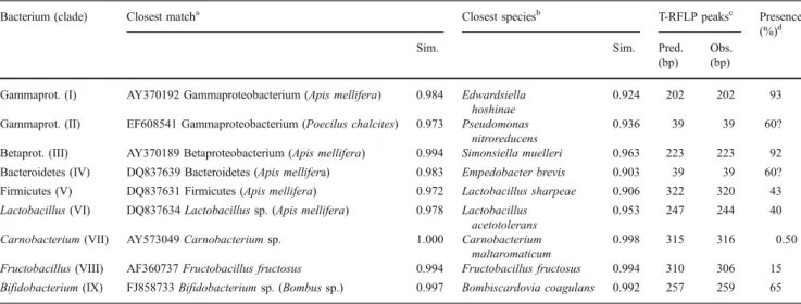
Table 1 SeqMatch results (RDP) of all groups of 16S sequences obtained in this study, similarity scores to closest match among all RDP sequences and type strains only, and corresponding T-RFLP peaks
Bacterium (clade) Closest matcha Closest speciesb T-RFLP peaksc Presence
(%)d
Sim. Sim. Pred.
(bp) Obs. (bp) Gammaprot. (I) AY370192 Gammaproteobacterium (Apis mellifera) 0.984 Edwardsiella
hoshinae
0.924 202 202 93
Gammaprot. (II) EF608541 Gammaproteobacterium (Poecilus chalcites) 0.973 Pseudomonas nitroreducens
0.936 39 39 60?
Betaprot. (III) AY370189 Betaproteobacterium (Apis mellifera) 0.994 Simonsiella muelleri 0.963 223 223 92 Bacteroidetes (IV) DQ837639 Bacteroidetes (Apis mellifera) 0.983 Empedobacter brevis 0.903 39 39 60? Firmicutes (V) DQ837631 Firmicutes (Apis mellifera) 0.972 Lactobacillus sharpeae 0.906 322 320 43 Lactobacillus (VI) DQ837634 Lactobacillus sp. (Apis mellifera) 0.978 Lactobacillus
acetotolerans
0.953 247 244 40
Carnobacterium (VII) AY573049 Carnobacterium sp. 1.000 Carnobacterium maltaromaticum
0.998 315 316 0.50
Fructobacillus (VIII) AF360737 Fructobacillus fructosus 0.994 Fructobacillus fructosus 0.994 310 306 15 Bifidobacterium (IX) FJ858733 Bifidobacterium sp. (Bombus sp.) 0.997 Bombiscardovia coagulans 0.992 257 259 65
aClosest hit with SeqMatch (RDP) for all good quality sequences >1,200 bp: accession number, taxonomic identity (host species) and similarity score
(Sim.) = percent sequence identity over all pairwise comparable positions
bClosest hit with SeqMatch (RDP) for all types of strain sequences: taxonomic identity and similarity score (Sim.)
cPosition of predicted (Pred.) and observed (Obs.) T-RFLP peaks in base pairs, prediction from virtual digest of 16S sequence with HaeIII d
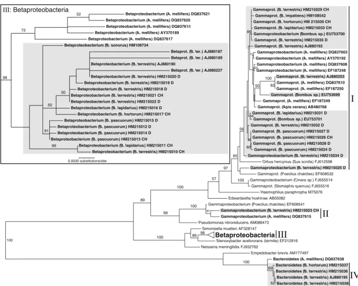
presented in Figs.2and3and Table1. The bacteria can be grouped into nine major clades (I–IX). Figure2reveals four major clades of Gram-negative bacteria. In this tree, clade I and clade II are undescribed Gammaproteobacteria, clade III comprised Betaproteobacteria, and clade IV of Bacter-oidetes. According to the entries in GenBank, the Gam-maproteobacteria in clade I are most closely related to bacteria found in honeybees (Apis mellifera, Apis cerana), but similar bacteria have also been found in aphids (e.g. Cinara sp., Stomaphis quercus) and a ground beetle (Poecilus chalcites). Their closest matches amongst 16S sequences of the type strains in the RDP were Orbus hercynius and Edwardsiella hoshinae. The single sequence obtained for bacteria from clade II shows closest similarity to a bacterium from the honeybee gut and is also close to a bacterium found in the ground beetle P. chalcites (Fig.2). Their closest described relative was found to be Pseudo-monas nitroreducens (Table1). The Betaproteobacteria (III) fall within the Neisseriaceae and are closest to Stenoxy-bacter acetivorans from the termite gut and Simonsiella
muelleri, a human commensal (Fig. 2). They too have
closely related representatives in the honeybee gut. The Bacteroidetes (IV) were found to be closest to Empedo-bacter brevis, but show a low similarity score (0.903, Table1). Again, a closely related bacterium has previously been found in the honeybee gut (Fig.2) [4].
The Gram-positive bacteria (Fig.3) are represented by a novel group of Firmicutes (V), Lactobacillus (VI),
Carnobacterium (VII), Fructobacillus (VIII) and Bifido-bacterium (IX). Clade V is composed of Firmicutes highly divergent from any described bacterial species, but similar bacteria have also been reported from the
honeybee gut before (Fig. 3). Furthermore, a species of
Lactobacillus was found (VI), showing close affinities with lactobacilli previously described from the crop of honeybees [53]. A single sequence from the gut of B. hortorum was identical to Carnobacterium maltaromaticum (VII) and another one from B. terrestris almost identical to Fructobacillus fructosus (VIII). A group of Bifidobac-teria (IX) from different Bombus species was also found in this study, similar to Bifidiobacteria previously isolated
from bumblebees (Fig.3) [43].
Grouping according to sampling location in the phylogenetic tree was not observed in those cases where 16S rRNA gene sequences for one clade of bacteria were obtained from several bumblebee individuals. However, the Betaproteobacteria (clade III) are separated into different well-supported clades according to their host species (Fig. 2).
Comparative Analysis of Gut Bacterial Communities of Different Bombus Species
The two-dimensional NMDS analysis (STRESS1=0.199)
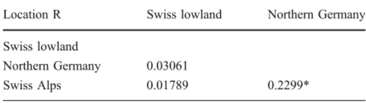
of the T-RFLP profiles from six species and three localities reveals considerable overlap of the structure of the communities, both between and sampling locations and species (Figs.4and 5). Whilst this indicates a high degree of similarity of microbial communities in the sampled bumblebee individuals, significant differences between host species as well as localities were also observed. An ANOSIM revealed a pairwise significant difference between samples from the Swiss Alps to Northern Germany (Table2
and Fig. 4), but not for the other pairwise comparisons
between sampling locations. Most of the pairwise compar-isons between species, however, indicated highly significant differences, but with mostly moderate to low R values (R<0.5, Table 3). The highest degree of separation was observed between B. pascuorum and B. soroeensis (R=0.64), and B. pascuorum and B. terrestris (R=0.53). B. hortorum and B. pascuorum had the most distinct microbiota, being significantly different from most other host species. B. terrestris in contrast showed less separation from the other species, with low R values especially for the German samples. Accordingly, the NMDS plot shows a high amount of scatter for B. terrestris individuals, whereas individuals of the other species tend to be more clustered in certain areas of the plot, especially B. pascuorum and B. soroeensis (Fig.5).
No difference was observed within one host species when comparing the microbiota between sampling locations for B. pascuorum and B. lapidaries, with R values around 0 Number of clones 50 45 40 35 30 25 20 15 10 5 0 Number of clades 4 3 2 1
Figure 1 Individual-based rarefaction curves for the 16S clone libraries from guts of 14 different bumblebee individuals. Individual lines represent the diversity in the different host individuals
(Table 3). A slight difference was, however, detected between B. terrestris from Switzerland and Germany.
Of the 49 T-RFLP peaks (the taxonomic units) recorded in total from all 199 bumblebee individuals, most were rare; only 11 peaks were detected in more than 20% of the sampled bumblebees. The rare peaks remained mostly unidentified because no corresponding 16S sequence could be obtained from the clone libraries, with the exception of a Carnobacterium (VII) found only in a single profile and a Fructobacillus found in only 15% of all individuals.
Assuming that the peaks at a certain position observed in the different profiles always correspond to the predicted peaks of the bacterial taxa identified in the clone libraries
(Table 1 and Figs. 2 and 3), distributions of the most
common bacteria in the bumblebee gut were as follows
(Fig.6). The most dominant bacteria in the bumblebee gut are one species from each of the following groups: Gammaproteobacteria (clade I, present in 92.5% of all individuals), Betaproteobacteria (III, 91.5%), and Firmi-cutes (V, 42.7%) and one species each of the genera Lactobacillus (VI, 40.2%) and Bifidobacterium (IX, 64.8%). In 60.3% of all samples, a peak, possibly corresponding to clade IV (Bacteroidetes), was observed. A single clone from one of the clone libraries also yielded a 16S sequence of an unknown Gammaproteobacterium (clade II) that produces a T-RFLP peak at the same position (39 bp, Table1). An unequivocal identification of this peak is therefore not possible; however, the presence of the Bacteroidetes bacterium in several of the clone libraries might indicate this bacterium to be more widespread than
0.2 substitutions/site
Gammaprot. (B. pascuorum) HM215028 D Gammaprot. (A. mellifera) EF187250
Gammaprot. (Apis cerana) AB480768
Gammaprot. (B. pascuorum) HM215027 D
Gammaprot. (B. pascuorum) HM215024 D Gammaprot. (B. terrestris) HM215029 CH
Gammaproteobacterium (Poecilus chalcites) EF608541
Gammaproteobacterium (B. terrestris) HM215023 CH
Gammaproteobacterium (A. mellifera) DQ837603
Gammaproteobacterium (A. mellifera) DQ837615
Gammaproteobacterium (A. mellifera) EF187248
Stenoxybacter acetivorans (termite) EF212916
Gammaprot. (A. mellifera) DQ837610 Gammaprot. (B. impatiens) HM108542
Gammaprot. (B. pascuorum) HM215026 CH Gammaproteobacterium (A. mellifera) AY370192
Gammaprot. (A. mellifera) EF187249
Haemophilus paraphropha M75076
Neisseria meningitidis FJ932762
Bacteroidetes (B. terrestris) HM215038 Bacteroidetes (B. terrestris) HM215036 Edwardsiella hoshinae AB05082
Pseudomonas nitroreducens AM088473
Bacteroidetes (A. mellifera) DQ837638 Gammaproteobacterium (Bombus sp.) EU753700
Gammaprot. (B. terrestris) HM215032 D Gammaprot. (B. terrestris) HM215035 D
Gammaprot. (Bombus sp.) EU753699
Gammaproteobacterium (B. terrestris) HM215025 D Gammaprot. (B. terrestris) AJ880193
Empedobacter brevis AM177497 Simonsiella muelleri AF328147
Gammaproteobacterium (B. terrestris) HM215034 D Gammaproteobacterium (A. mellifera) DQ837608
Gammaprot. (B. terrestris) AJ880203
57 87 69 72 100 100 98 55 86 60 100 99 100 100 86 62 100 56 98 89 97 82 98 65 100 50 Gammaprot. (B. hortorum) HM 215030 CH Gammaprot. (B. lapidarius) HM215033 CH Gammaprot. (B. lapidarius) HM215031 D Gammaprot. (Bombus sp.) EU753701
Gammaprot. (Poecilus chalcites) EF608532 Gammaproteobacterium (Cinara sp.) FJ655514 Gammaprot. (Stomaphis quercus) FJ655516
Orbus hercynius (Sus scrofa) FJ612598
Bacteroidetes (B. hortorum) HM215037
Bacteroidetes (B. terrestris) AJ880195 0.0030 substitutions/site
Betaproteobacterium (B. terrestris) HM215020 D Betaproteobacterium (A. mellifera) AY370189
Betaprot. (B. ter.) AJ880227 Betaproteobacterium (A. mellifera) DQ837621
Betaproteobacterium (B. pascuorum) HM215014 D Betaproteobacterium (B. terrestris) HM215022 D
Betaproteobacterium (B. lapidarius) HM215011 CH Betaproteobacterium (B. terrestris) HM215010 CH Betaproteobacterium (B. sonorus) HM108734
Betaproteobacterium (A. mellifera) DQ837620
Betaproteobacterium (A. mellifera) DQ837617
Betaproteobacterium (B. terrestris) HM215021 CH
Betaproteobacterium (A. mellifera) DQ837618
Betaprot. (B. ter.) AJ880189
Betaproteobacterium (B. terrestris) HM215019 D Betaproteobacterium (B. pascuorum) HM215015 D Betaproteobacterium (B. terrestris) HM215018 D 52 91 99 72 50 50 98
Betaprot. (B. ter.) AJ880187
Betaproteobacterium (B. terrestris) AJ880190
Betaproteobacterium (B. lapidarius) HM215016 D Betaproteobacterium (B. pascuorum) HM215012 D Betaproteobacterium (B. pascuorum) HM215013 CH Betaproteobacterium (B. hortorum) HM215017 CH III: Betaproteobacteria Betaproteobacteria
I
II
III
IV
Figure 2 Maximum likelihood tree for 16S sequences of Proteobac-teria and Bacteroidetes from this study (clades I to IV) and related sequences from GenBank. Clade III (Betaproteobacteria) is drawn to different scales in the upper left corner. Figures on branches are bootstrap support values (500 replicates); taxon labels denote
bacterium (host species), GenBank accession numbers and sampling location (CH—Swiss lowlands, Jura of Basel; D—Northern Germany, Celle). Bacteria from honeybee and bumblebee guts in bold font, bacteria from the bumblebee gut additionally marked in grey boxes
the Gammaproteobacterium (clade II). The average stan-dardized peak signal intensity as a proxy for the relative abundance of the different bacteria within a host generally corresponds to the percentage of infected individuals
(Fig. 6). For example, the two most common bacteria
(clades I and III) are also the most abundant bacteria within infected hosts, whilst less common bacteria tend to be less abundant in infected hosts (e.g. clade VIII in B. lucorum/ cryptarum and B. soroeensis; Fig.6).
The aforementioned groups can generally be detected in all of the observed Bombus species at all localities (Fig.6), with the exception of the Firmicutes bacterium (clade V)
which was not found in B. soroeensis (Fig. 6). Marked
differences in the frequency of some of the bacteria in
different host species can be observed (Fig. 6). For
example, B. hortorum relatively rarely harboured the otherwise much more common Gammaproteobacterium (I) and Betaproteobacterium (III), but was colonized more than expected by Lactobacillus (VI) and Fructobacillus (VIII). Certain patterns relating to the sampling location can also be found. For example, firmicute (V) is almost absent in the
Swiss Alps, and Fructobacillus (VIII) seems to mostly occur in the Swiss lowlands (Fig.6).
Discussion
The gut bacterial community of central European bumble-bees appears to contain relatively few abundant species, which consistently appear in all examined Bombus species and localities. This low diversity is most likely not an artefact of limited sampling effort, as indicated by the rarefaction analyses of the clone libraries (Fig.1). Bacteria in this abundant group belong to different phyla including the Gamma- and Betaproteobacteria, Bacteroidetes, Firmi-cutes and Actinobacteria. They generally are quite distant to any of the already described bacterial species. For most of the taxa and groups identified here, similar bacteria have been found in the honeybee gut [4,12,39,44,51]. The 16S sequences of gut bacteria from different bumblebee individuals and species generally form distinct clades with respect to similar bacteria from the honeybee gut (Figs. 2
0.2 substitutions/site
Lactobacillus sp. (B. terrestris) AJ880205
Lactobacillus sp. (A. mellifera) EF187240
Carnobacterium maltaromaticum M58825
Bifidobacterium sp. (A. mellifera) EF187237
Firmicute (B. pascuorum) HM215041 D
Lactobacillus sp. (A. mellifera) EF187244
Lactobacillus sp. (Bombus sp.) EU753702
Firmicute (B. terrestris) HM215045 D Firmicute (B. terrestris) HM215047CH
Lactobacillus sp. (A. mellifera) EF187243
Firmicute (B. terrestris) HM215042 CH
Lactobacillus sakei AB362607
Bifidobacterium asteroides EF187235
Lactobacillus sp. (A. mellifera) EF187242
Lactobacillus sp. (B. terrestris) AJ971929
Bifidobacterium sp. (A. mellifera) EF187231
Bifidobacterium coryneforme EF187238
Fructobacillus sp. (B. terrestris) HM215039 CH
Lactobacillus sharpeae M58831
Bifidobacterium sp. (A. mellifera) EF187236 Lactobacillus sp. (A. mellifera) EF187245
Bifidobacterium sp. (B. pascuorum) HM215049 D Bombiscardovia coagulans (B. lapidarius) EU127550
Lactobacillus sp. (B. terrestris) AJ880228 Lactobacillus sp. (A. mellifera) AY370183
Lactobacillus acetotolerans M58801
Firmicute (B. terrestris) AJ880198
Lactobacillus sp. (B. ter.) AJ880197
Firmicute (B. lapidarius) HM215044 CH
Fructobacillus fructosus AF360737
Bifidobacterium sp. (A. mellifera) EF187232 Carnobacterium sp. (B. hortorum) HM215040 CH
Lactobacillus sp. (Bombus sp. body surface) EU753703
Bifidobacterium sp. (B. pascuorum) HM215050 CH
Firmicute (B. terrestris) HM215046 D
Firmicute (B. pascuorum) HM215043 CH
Bifidobacterium sp. (A. mellifera) EF187234 Lactobacillus kunkeei (A. mellifera) EF187239
Lactobacillus sp. (A. mellifera) EF187241
Bifidobacterium sp. (Bombus sp.) FJ858733 Lactobacillus sp. (B. terrestris) AJ880194
Firmicute (A. mellifera) DQ837631
Lactobacillus sp. (B. terrestris) HM215048 CH
Bifidobacterium sp. (B. lucorum) EU127549 Lactobacillus sp. (A. mellifera) DQ837634
Bifidobacterium sp. (B. terrestris) HM215051 D 97 100 94 89 85 62 72 100 52 99 100 95 79 62 63 79 54 57 100 78 85 79 98 100 98 75
VI
V
VII
VIII
IX
Figure 3 Maximum likelihood tree for 16S sequences of Firmicutes and Actinobacteria from this study (clades V IX) and related sequences from GenBank; bootstrap support values (500 replicates) on branches. For further explanations, see Fig.2
-1 -0.5 0 0.5 1 1.5 2 Dimension 2 -1 -0.5 0 0.5 1 1.5 2 Dimension 1
Swiss lowlands (Jura of Basel) Northern Germany (Celle) Swiss alps (Engadin) Location
Figure 4 NMDS plot of 16S T-RFLP profiles for 199 bumble-bee individuals. Different symbols indicate different sampling localities -1 -0.5 0 0.5 1 1.5 2 Dimension 2 -1 -0.5 0 0.5 1 1.5 2 Dimension 1 B. hortorum B. lapidarius B. lucorum/cryptarum B. pascuorum B. soroeensis B. terrestris Species Figure 5 NMDS plot of 16S T-RFLP profiles for 199 bumblebee individuals. Different symbols indicate different
and3). These bacteria may thus be specialized inhabitants of the bumblebee gut and similar but distinct from those observed in honeybees. This is in agreement with the findings of Martinson et al. [44] who recorded similar bacteria in two species of Northern American bumblebees and several species of honeybees, but not in a variety of solitary bees. This similarity between bacterial communities in honeybees and bumblebees is most likely not caused by the sampling method as a variety of extraction and PCR protocols have previously been used in the study of bacterial communities in bees [4,12,39,44,51] with similar results,
The observed sequence divergence of 2–5% between related bacteria in honeybees and bumblebees (Table1) has to be judged against the background of a sequence divergence rate at the 16S rRNA locus of 1–2% per 50 million years [52] and an estimated split of the honeybee and bumblebee linage around 90 million years ago [25]. Hence, this fits well with a scenario where these bacteria became separated as these two host groups diverged. Additional sequence data for bacteria from the other tribes of corbiculate bees (Meliponini and Euglossini) could help expand on this hypothesis. Bumblebees originated only 25– 40 million years ago, and most speciation events have occurred within the last 10 million years [31]. Therefore, as a note of caution, the 16S rRNA gene might be too highly
conserved to yield a fine resolution of these bacteria in the recently diverged Bombus species. Using T-RFLP profiles furthermore reduces the resolution as several bacteria need to have different terminal restriction sites to be effectively differentiated. Hence, our results reflect robust differences, but will have to be refined by further studies.
The group of Betaproteobacteria found in this study not only appears to be differentiated between honeybees and bumblebees but also shows different clades perhaps specialized to different bumblebee host species regardless of their sampling location (Fig.2). This result will have to be confirmed by examining a wider range of bumblebee species, but indications for co-divergence of the closely related genus Simonsiella with their mammalian host have been found previously [29]. Whilst co-speciation of bacterial symbionts with their hosts has mostly been observed in intracellular bacteria [38], extracellular insect gut bacteria have also been reported to have co-speciated with their plataspid bug hosts [34,41].
With exception of sequences from related bacteria in the honeybee gut, for the majority of 16S sequences obtained in this study, no similar sequences were found among the more than two million 16S sequences stored in GenBank at the time of this study, many of which originate from environmental samples [5]. Therefore, these bacteria seem to constitute a specialized endogenous community in the bumblebee alimentary tract rather than bacteria accidentally taken up from the environment passing the gut. An exception to this may be the two strains with a high similarity to C. maltaromaticum and F. fructosus, which have been described from outside the bumblebee gut on
decaying plant or animal matter [17, 45] and might
therefore be unspecific bacteria taken up from the environ-ment by the bees. Bacteria in bees could also come from another source—nectar and pollen. Yet, there is very
Table 3 Results of ANOSIM comparing T-RFLP profiles of all species pairs
B. hortorum CHL B. lapidarius CHL B. lapidarius D B. luc./cryp. CHA B. pascuorum CHL B. pascuorum D B. soroeensis CHA B. terrestris CHL B. hortorum CHL B. lapidarius CHL 0.23*** B. lapidarius D 0.12 −0.03 B. luc./cryp. CHA 0.31*** 0.14*** 0.27 B. pascuorum CHL 0.47*** 0.26*** 0.38** 0.43*** B. pascuorum D 0.46*** 0.20*** 0.27 0.39*** 0.06 B. soroeensis CHA 0.29*** 0.23*** 0.30 0.23** 0.64*** 0.57*** B. terrestris CHL 0.23** 0.30*** 0.27 0.36*** 0.53*** 0.43*** 0.33*** B. terrestris D 0.42*** 0.09 0.04 0.12 0.09 0.00 0.31*** 0.29*
Species with samples from more than one location were split by sampling locations (CHL Swiss lowlands, CHA Swiss Laps, D Northern Germany)
*p<0.05; **p<0.01; ***p<0.0001 (R values, Bonferroni-corrected) Table 2 Results of ANOSIM comparing T-RFLP profiles of all sampling locations
Location R Swiss lowland Northern Germany
Swiss lowland
Northern Germany 0.03061
Swiss Alps 0.01789 0.2299*
limited knowledge about bacteria living inside the pollen and nectar of flowers. Culture-independent studies of this potential microbial habitat are curiously absent, and one of the few culture-dependent studies found bacteria to be virtually absent in floral nectar [21]. This absence of bacteria has been attributed to plant-produced antimicrobial secondary compounds found in floral nectar [21]. However, recent studies have shown a surprising abundance of yeasts in nectar [30], transmitted by pollinating insects like bumblebees [8], pointing at the possibility of other microbial organisms inhabiting flowers. Flowers would represent a likely site of horizontal transmission for bacteria in bees. Such a transmission route has previously been demonstrated for the trypanosomatid C. bombi, an intestinal parasite of bumblebees [15], and RNA viruses in hyme-nopterans including bees [61]. This horizontal transmission route might result in a stable and consistent mutualistic association of the bee hosts and their bacteria, as has been found in symbionts of stinkbugs [40]. The apparent absence of these bacteria in solitary bees [44] points, however, towards a role of sociality in transmission. The life history of bumblebees with gynes staying in their mother colony several days after emergence and founding new colonies after hibernation in the next season [23] would facilitate vertical transmission. The higher probability of transmis-sion of beneficial microbes within a colony and to the daughter colonies might thus represent an additional benefit
of sociality in bumblebees and honeybees [44, 49]. This
mechanism has also been suggested in termites [32].
Significant differences between the gut floras of different
bumblebee species were found in this study (Fig. 5 and
Table 3). Additionally, a comparison of the microbiota
between sampling localities within one host species showed no difference for B. pascuorum and B. lapidaries, in contrast to a comparison with different host species at the same site (Table3). This further strengthens the argument for the existence of species-specific bacterial gut commu-nities in these hosts across geographical distances. These might relate to differences in the host ecology and physiology, thereby selecting different communities of bacteria in the gut. The sampled Bombus species have, for example, different preferred flower types, with the long-tongued B. hortorum visiting flowers with long corolla tubes, whilst the short-tongued B. terrestris is a generalist visiting a broad spectrum of flowers with short corolla tubes, but also robbing nectar from flowers with long corolla tubes [23]. Accordingly, the more specialized B. hortorum has very distinct bacterial communities (Table3), whereas the generalist B. terrestris shows more variation and little distinction than the other examined Bombus species. The communities in B. terrestris from Northern Germany were also found to be significantly different from those in Switzerland, perhaps indicating a higher plasticity of the microbiota in this species. More extensive sampling of different bumblebee species is of course needed to substantiate these points. As discussed above, the signifi-cant differences among host species might also be driven by a predominantly vertical transmission of bacteria from a Host species/Locality CHA CHL D B.ter B.sor B.pas B.luc B.lap B.hor Phylotype (Clade) I III IV/II V VI VIII IX 0.0 0.1 0.2 0.3 0.4 Mean standardized signal intensity 65 88 100 100 86 98 96 89 94 60 100 100 100 86 90 99 85 94 80 81 88 2 100 60 45 55 94 15 22 8 73 0 72 70 42 4 65 41 58 46 27 25 25 51 44 45 9 4 16 5 16 4 30 4 40 44 92 86 59 60 65 58 76
Figure 6 Heatmap of relative abundances and frequencies of different T-RFLP peaks in different bumblebee species and sampling localities. Different shades indicate average standardized signal intensity of individual T-RFLP peaks for the infected individuals, representing relative abundances of the different bacterial phylotypes. Numbers in squares indicate the percentage of individual hosts of each group in which the respective peak was detected. For the Roman numerals
coding for the different bacterial phylotypes, see Table1and Figs.2
and 3. Host species/locality: B.hor B. hortorum (n=20), B.lap B. lapidarius (n=32), B.luc B. lucorum/cryptarum (n=24), B.pas B. pascuorum (n=44), B.sor B. soroeensis (n=22), B.ter B. terrestris (n= 57), D Northern Germany (n=69), CHL Swiss lowland (n=84), CHA Swiss Alps (n=46)
mother colony to a daughter colony via the young queen whilst horizontal transmission would remain rare.
Our analysis could not test the functional role of the bacteria, nor is much known about these roles. However, the bacteria identified here are most likely non-pathogenic because, on one hand, they belong to the generally non-pathogenic lactic acid bacteria, Bifidobacteria or Bacter-oidetes that are commonly found as part of the healthy gut flora of other organisms, including vertebrates and honey-bees [22,46,53]. On the other hand, some of the identified bacteria occur in almost every bumblebee individual checked here (clades I and III, the Gamma- and Betapro-teobacteria), which is indicative of their possible role as mutualists or commensals, but less likely so as pathogens.
Furthermore, the presence of an apparently highly conserved and specialized community in the bumblebee gut across different host species and geographic distances makes a functional relevance of these bacteria for their hosts seem likely. As in other insects [13], they could play a role in host immune defence. The main route of infection of insect pathogens is through ingestion and invasion of the gut, followed by the colonization of the haemocoel through the midgut wall [64]. Exclusion of potential pathogens from the gut is therefore an essential part of the insect immune system. In addition to direct control of pathogens by the insect immune system through, for example, the production of antimicrobial peptides and reactive oxygen species [64], the resident gut flora might play an important protective
role as well [3, 13], either by producing antimicrobial
substances themselves [19] or through competitive exclusion of newly invading bacteria [14]. Gilliam [22] speculated about a similar role of the gut microbiota of honeybees, and recently, the lactic acid bacteria of honeybees have been shown to produce antimicrobial substances and efficiently protect larvae against the foulbrood-causing bacterial patho-gen P. larvae [19]. As the bacteria found in the bumblebee gut are highly similar to those in the honeybee gut, they may possess a protective role for their hosts as well.
Bacteria have furthermore been found to play a key role in the adaptation of herbivorous insects to novel food resources and subsequently in the diversification and ecological success of this group [38]. Even though the diet of bees consisting of pollen and nectar is highly nutritious [59], the highly resistant pollen wall has to be degraded first to make these nutrients available. Bee-specific symbiotic bacteria could aid in this process, either in the host gut or in
stored bee bread [22, 65]. They could also help in
preserving the stored provisions of honeybees and bum-blebees by the production of antimicrobial substances [65]. In conclusion, we have provided the first detailed survey of bacteria in the bumblebee gut for different species from Central Europe. The resident gut bacteria are surprisingly well conserved and species poor, but apparently highly
specialized to this group of organisms. Whilst this indicates a possibly strong interaction with their hosts as well as a functional role, further studies are needed to elucidate this. Acknowledgements We are thankful to Roland Bürki for assistance in the lab during his undergraduate project. Regula Schmid-Hempel, Anna Lazzaro and Nikki E. Freed gave helpful advice on the lab work and data analysis. Martina Tognazzo collected and dissected the bumblebees from Switzerland. Parts of the data analysed in this paper were generated in the Genetic Diversity Centre of ETH Zurich. Bumblebee samples from Germany were collected with permission from the Untere Naturschutzbehörde Landkreis Celle (Lothar Sander); bumblebee samples from the Swiss National Park were collected with permission of the Forschungskommission des Schweizerischen Natio-nalparks (Dr. Thomas Scheurer). Financial supported by the Swiss National Science Foundation (grant no. 31003A-116057 to PSH).
References
1. Abdo Z, Schüette UME, Bent SJ, Williams CJ, Forney LJ, Joyce P (2006) Statistical methods for characterizing diversity of microbial communities by analysis of terminal restriction frag-ment length polymorphisms of 16S rRNA genes. Environ Micro-biol 8(5):929–938
2. Altschul SF, Madden TL, Schaffer AA, Zhang J, Zhang Z, Miller W, Lipman DJ (1997) Gapped BLAST and PSI-BLAST: a new generation of protein database search programs. Nucleic Acids Res 25:3389–3402
3. Azambuja P, Garcia ES, Ratcliffe NA (2005) Gut microbiota and parasite transmission by insect vectors. Trends Parasitol 21 (12):568–572
4. Babendreier D, Joller D, Romeis J, Bigler F, Widmer F (2007) Bacterial community structures in honeybee intestines and their responses to two insecticidal proteins. FEMS Microbiol Ecol 59:600–610
5. Benson DA, Karsch-Mizrachi I, Lipman DJ, Ostell J, Sayers EW (2010) GenBank. Nucleic Acids Res 38(suppl 1):D46–D51 6. Bray JR, Curtis JT (1957) An ordination of the upland forest
communities of southern Wisconsin. Ecol Monogr 27:325–349 7. Breznak JA, Brune A (1994) Role of microorganisms in the
digestion of lignocellulose by termites. Annu Rev Entomol 39:453–487
8. Brysch-Herzberg M (2004) Ecology of yeasts in plant–bumblebee mutualism in Central Europe. FEMS Microbiol Ecol 50:87–100 9. Clarke KR (1993) Non-parametric multivariate analyses of
changes in community structure. Aust J Ecol 18(1):117–143 10. Cole JR, Wang Q, Cardenas E, Fish J, Chai B, Farris RJ,
Kulam-Syed-Mohideen AS, McGarrell DM, Marsh T, Garrity GM, Tiedje JM (2009) The Ribosomal Database Project: improved alignments and new tools for rRNA analysis. Nucleic Acids Res 37:D141– D145
11. Colla SR, Otterstatter MC, Gegear RJ, Thomson JD (2006) Plight of the bumble bee: pathogen spillover from commercial to wild populations. Biol Conserv 129(4):461–467
12. Cox-Foster D, Conlan S, Holmes EC, Palacios G, Evans JD, Moran NA, Quan P, Briese T, Hornig M, Geiser DM, Martinson V, vanEngelsdorp D, Kalkstein AL, Drysdale A, Hui J, Zhai J, Cui L, Hutchinson SK, Simons JF, Egholm M, Pettis JS, Lipkin WI (2007) A metagenomic survey of microbes in honey bee colony collapse disorder. Science 318(5848):283–287
13. Dillon RJ, Dillon VM (2004) The gut bacteria of insects: nonpathogenic interactions. Annu Rev Entomol 49:71–92
14. Dillon RJ, Vennard CT, Buckling A, Charnley AK (2005) Diversity of locust gut bacteria protects against pathogen invasion. Ecol Lett 8(12):1291–1298
15. Durrer S, Schmid-Hempel P (1994) Shared use of flowers leads to horizontal pathogen transmission. Proc R Soc Lond B Bio 258 (1353):299–302
16. Durrer S, Schmid-Hempel P (1995) Parasites and the regional distribution of bumblebee species. Ecography 18:114–122 17. Endo A, Okada S (2008) Reclassification of the genus
Leuconos-toc and proposals of Fructobacillus fructosus gen. nov., comb. nov., Fructobacillus durionis comb. nov., Fructobacillus ficulneus comb. nov. and Fructobacillus pseudoficulneus comb. nov. Int J Syst Evol Microbiol 58:2195–2205
18. Field JG, Clarke KR, Warwick RM (1982) A practical strategy for analysing multispecies distribution patterns. Mar Ecol Prog Ser 8:37–52
19. Forsgren E, Olofsson TC, Vásquez A, Fries I (2010) Novel lactic acid bacteria inhibiting Paenibacillus larvae in honey bee larvae. Apidologie 41:99–108
20. Fraune S, Bosch TCG (2010) Why bacteria matter in animal development and evolution. BioEssays 32:571–580
21. Gilliam M, Moffett JO, Kauffeld NM (1983) Examination of floral nectar of citrus, cotton, and Arizona desert plants for microbes. Apidologie 14(4):299–302
22. Gilliam M (1997) Identification and roles of non-pathogenic microflora associated with honey bees. FEMS Microbiol Lett 155:1–10
23. Goulson D (2003) Bumblebees: their behaviour and ecology. Oxford University Press, Oxford
24. Goulson D, Lye GC, Darvill B (2008) Decline and conservation of bumble bees. Annu Rev Entomol 53:191–208
25. Grimaldi D, Engel MS (2005) Evolution of the insects. Cambridge University Press, New York
26. Guarner F, Malagelada JR (2003) Gut flora in health and disease. Lancet 361(9356):512–519
27. Guindon S, Gascuel O (2003) A simple, fast, and accurate algorithm to estimate large phylogenies by maximum likelihood. Syst Biol 52(5):696–704
28. Hammer Ø, Harper DAT, Ryan PD (2001) PAST: Paleontological Statistics Software Package for Education and Data Analysis. Palaeontol Electronica 4(1):9
29. Hedlund BP, Kuhn DA (2006) The genera Simonsiella and Alysiella. In: Dworkin M, Falkow S, Rosenberg E, Schleifer K, Stackebrandt E (eds) The Prokaryotes: a handbook on the biology of bacteria, vol 5. Springer, New York, pp 828–839
30. Herrera CM, Vega C, Canto A, Pozo M (2009) Yeasts in floral nectar: a quantitative survey. Ann Bot-London 103:1415–1423 31. Hines H (2008) Historical biogeography, divergence times, and
diversification patterns of bumble bees (Hymenoptera: Apidae: Bombus). Syst Biol 57(1):58–75
32. Hongoh Y, Deevong P, Inoue T, Moriya S, Trakulnaleamsai S, Ohkuma M, Vongkaluang C, Noparatnaraporn N, Kudo T (2005) Intra- and interspecific comparisons of bacterial diver-sity and community structure support coevolution of gut microbiota and termite host. Appl Environ Microbiol 71 (11):6590–6599
33. Hooper LV, Midtvedt T, Gordon JI (2002) How host–microbial interactions shape the nutrient environment of the mammalian intestine. Annu Rev Nutr 22:283–307
34. Hosokawa T, Kikuchi Y, Nikoh N, Shimada M, Fukatsu T (2006) Strict host–symbiont cospeciation and reductive genome evolution in insect gut bacteria. PLoS Biol 4(10):1841–1851
35. Huber T, Faulkner G, Hugenholtz P (2004) Bellerophon: a program to detect chimeric sequences in multiple sequence alignments. Bioinformatics 20(14):2317–2319
36. Hugenholtz P, Goebel BM, Pace NR (1998) Impact of culture-independent studies on the emerging phylogenetic view of bacterial diversity. J Bacteriol 180(18):4765–4774
37. Hutchison DW, Strasburg JL, Shaffer C (2005) Cleaning micro-satellite PCR products with Sephadex™in 96-well filtration plates enhances genotyping quality. Biotechniques 38(1):56–58 38. Janson EM, Stiremann JO, Singer MS, Abbot P (2008)
Phytoph-agous insect–microbe mutualisms and adaptive evolutionary diversification. Evolution 62(5):997–1012
39. Jeyaprakash A, Hoy MA, Allsopp MH (2003) Bacterial diversity in worker adults of Apis mellifera capensis and Apis mellifera scutellata (Insecta: Hymenoptera) assessed using 16S rRNA sequences. J Invertebr Pathol 84:96–103
40. Kikuchi Y, Hosokawa T, Fukatsu T (2007) Insect–microbe mutualism without vertical transmission: a stinkbug acquires a beneficial gut symbiont from the environment every generation. Appl Environ Microbiol 73(13):4308–4316
41. Kikuchi Y, Hosokawa T, Nikoh N, Meng X, Kamagata Y, Fukatsu T (2009) Host–symbiont co-speciation and reductive genome evolution in gut symbiotic bacteria of acanthosomatid stinkbugs. BMC Biol 7:2
42. Killer J, Kopečný J, Mrázek J, Rada V, Benada O, Koppová I, Havlík J, Straka J (2009) Bifidobacterium bombi sp. nov., from the bumblebee digestive tract. Int J Syst Evol Microbiol 59:2020– 2024
43. Killer J, Kopečný J, Mrázek J, Rada V, Dubná S, Marounek M (2010) Bifidobacteria in the digestive tract of bumblebees. Anaerobe 16(2):165–170
44. Martinson VG, Danforth BN, Minckley RL, Rueppell O, Tingek S, Moran N (2011) A simple and distinctive microbiota associated with honey bees and bumble bees. Mol Ecol 20(3):619–628 45. Laursen BG, Lene B, Cleenwerck U, Vancanneyt M, Swings J,
Dalgaard P, Leisner JJ (2005) Carnobacterium divergens and Carnobacterium maltaromaticum as spoilers or protective cultures in meat and seafood: phenotypic and genotypic characterization. Syst Appl Microbiol 28(2):151–164
46. Ley RE, Lozupone CA, Hamady M, Knight R, Gordon JI (2008) Worlds within worlds: evolution of the vertebrate gut microbiota. Nat Rev Microbiol 6:776–788
47. Li F, Hullar MAJ, Lampe JW (2007) Optimization of terminal restriction fragment polymorphism (TRFLP) analysis of human gut microbiota. J Microbiol Meth 68(2):303–311
48. Liu WT, Marsh TL, Cheng H, Forney LJ (1997) Characterization of microbial diversity by determining terminal restriction fragment length polymorphisms of genes encoding 16S rRNA. Appl Environ Microbiol 63(11):4516–4522
49. Lombardo MP (2008) Access to mutualistic endosymbiotic microbes: an underappreciated benefit of group living. Behav Ecol Sociobiol 62(4):479–497
50. Moeseneder MM, Arrieta JM, Muyzer G, Winter C, Herndl GJ (1999) Optimization of terminal-restriction fragment length polymorphism analysis for complex marine bacterioplankton communities and comparison with denaturing gradient gel electrophoresis. Appl Environ Microbiol 65(8):3518–3525 51. Mohr KI, Tebbe CC (2006) Diversity and phylotype consistency
of bacteria in the guts of three bee species (Apoidea) at an oilseed rape field. Environ Microbiol 8(2):258–272
52. Ochman H, Elwyn S, Moran NA (1999) Calibrating bacterial evolution. Proc Natl Acad Sci 96(22):12638–12643
53. Olofsson TC, Vásquez A (2008) Detection and identification of a novel lactic acid bacterial flora within the honey stomach of the honeybee Apis mellifera. Curr Microbiol 57(4):356–363 54. Olofsson TC, Vásquez A (2009) Phylogenetic comparison of
bacteria isolated from the honey stomach of honey bees Apis mellifera and bumble bees Bombus spp. J Apic Res 48(4):233–237
55. Otti O, Schmid-Hempel P (2007) Nosema bombi: a pollinator parasite with detrimental fitness effects. J Invertebr Pathol 96:118–124
56. Posada D (2008) jModelTest: phylogenetic model averaging. Mol Biol Evol 25:1253–1256
57. Ramette A (2007) Multivariate analyses in microbial ecology. FEMS Microbiol Ecol 62:142–160
58. Rees GN, Baldwin DS, Watson GO, Perryman S, Nielsen DL (2004) Ordination and significance testing of microbial commu-nity composition derived from terminal restriction fragment length polymorphisms: application of multivariate statistics. A van Leeuw J Microb 86:339–347
59. Roulston TH, Cane JH (2000) Pollen nutritional content and digestibility for animals. Plant Syst Evol 222:187–209
60. Schmid-Hempel P, Reber Funk C (2004) The distribution of genotypes of the trypanosome parasite, Crithidia bombi, in populations of its host, Bombus terrestris. Parasitology 129 (2):147–158
61. Singh R, Levitt AL, Rajotte EG, Holmes EC, Ostiguy N, vanEngels-dorp D, Lipkin WI, de Pamphilis CW, Toth AL, Cox-Foster DL (2010) RNA viruses in hymenopteran pollinators: evidence of inter-taxa virus transmission via pollen and potential impact on non-Apis hymenopteran species. PLoS ONE 5(12):e14357
62. Thompson JD, Higgins DG, Gibson TJ (1994) CLUSTAL W: improving the sensitivity of progressive multiple sequence
alignment through sequence weighting, position-specific gap penalties and weight matrix choice. Nucleic Acids Res 22:4673– 4680
63. Tringe SG, Hugenholtz P (2008) A renaissance for the pioneering 16S rRNA gene. Curr Opin Microbiol 11(5):442–446
64. Vallet-Gely I, Lemaitre B, Boccard F (2008) Bacterial strategies to overcome insect defences. Nat Rev Microbiol 6:302–313 65. Vásquez A, Olofsson TC (2009) The lactic acid bacteria involved
in the production of bee pollen and bee bread. J Apic Res 48 (3):189–195
66. Velthuis HHW, van Doorn A (2006) A century of advances in bumblebee domestication and the economic and environmental aspects of its commercialization for pollination. Apidologie 37:421–451
67. Vorburger C, Gehrer L, Rodriguez P (2010) A strain of the bacterial symbiont Regiella insecticola protects aphids against parasitoids. Biol Lett 6(1):109–111
68. Weisburg WG, Barns SM, Pelletier DA, Lane DJ (1991) 16S ribosomal DNA amplification for phylogenetic study. J Bacteriol 173(2):697–703
69. Williams PH (1998) An annotated checklist of bumble bees with an analysis of patterns of description (Hymenoptera: Apidae, Bombini). Bull Nat Hist Mus Lond Entomol 67:79–152 70. Williams PH, Osborne JL (2009) Bumblebee vulnerability and