Abstract. We report that caveolin-1, one of the major
structural protein of caveolae, interacts with TCP-1, a hetero-oligomeric chaperone complex present in all eu-karyotic cells that contributes mainly to the folding of actin and tubulin. The caveolin-TCP-1 interaction entails the first 32 amino acids of the N-terminal segment of caveolin. Our data show that caveolin-1 expression is needed for the induction of TCP-1 actin folding function in response to insulin stimulation. Caveolin-1 phosphory-lation at tyrosine residue 14 induces the dissociation of
DOI 10.1007/s00018-005-5551-z © Birkhäuser Verlag, Basel, 2006
caveolin-1 from TCP-1 and activates actin folding. We show that the mechanism by which caveolin-1 modulates TCP-1 activity is indirect and involves the cytoskeleton linker filamin. Filamin is known to bind caveolin-1 and to function as a negative regulator of insulin-mediated sig-naling. Our data support the notion that the caveolin-fil-amin interaction contributes to restore insulin-mediated phosphorylation of caveolin, thus allowing the release of active TCP-1.
Keywords. Caveolin, TCP-1, folding, insulin, filamin, chaperone
Introduction
Caveolae are 50–100-nm invaginated plasma membrane domains present in non-lymphoid and neuronal cells. They participate in four main processes, namely, inter-nalization of pathogens and endocytosis of nutriments [1–4], cholesterol transport, signal transduction and cell transformation [1]. Numerous signaling molecules [src kinases, growth factors, G proteins, phospholipase C-g,
protein kinase-C (PKC) and Ras] have been reported to interact with caveolin-1, a major protein component of
caveolae and membrane lipid rafts [5], thus suggesting that these membrane domains play a crucial role in as-sembling signaling complexes. Caveolin-1 has been shown to function as a tumor suppressor, and phosphorylation of caveolin-1 or reduction of its expression level have been correlated with cell transformation [1, 6]. Caveolin-1 is an integral membrane protein that forms homo-oligomers and shows an unusual hairpin-like structure, exposing both the N and C termini to the cytosol [7]. Functionally, the N-terminal segment of caveolin-1 is of great impor-tance and contains an oligomerization site (residues 61– 101) overlapping with a scaffolding domain (residues 82– 101), which binds to diverse signaling molecules [5, 8, 9]. Moreover, caveolin-1 residue Y14 is phosphorylated by the insulin receptor (IR) in response to insulin stimulation [10, 11], or by c-Src or c-Abl in response to cell transfor-mation and during oxidative or hyperosmotic stresses [11, 12]. Finally, caveolin-1 is expressed as two isoforms
a and b, the later lacking the first 32 N-terminal residues
Research Article
Caveolin-1 interacts with the chaperone complex TCP-1 and
modulates its protein folding activity
M.-A. Douceya, +,*, F. C. Bendera, ++, D. Hessb, J. Hofsteengeband C. Brona
aDepartment of Biochemistry, University of Lausanne, chemin des Boveresses 155, 1066 Epalinges (Switzerland) b Friedrich Miescher-Institute, P.O. Box 2543, 4002 Basel (Switzerland)
Received 17 November 2005; received after revision 1 December 2005; accepted 17 February 2006 Online First 29 March 2006
*Corresponding author.
+ Present address: Laboratory of AIDS immunopathogenesis, Cen-tre Hospitalier Universitaire Vaudois, Rue du Bugnon BT 02-252, 1011 Lausanne (Switzerland), Fax:+41-213141070, e-mail: ma_ [email protected].
++Present address: Department of Microbiology, School of Dental Medicine, University of Pennsylvania, Philadelphia, PA 19104 (USA).
due to alternate initiation of the translation of a single mRNA transcript [13]. The functional difference between the two isoforms remains, however, unclear [7, 13, 14]. TCP-1 is a hetero-oligomeric, double-torus-shaped mol-ecular complex composed of eight different highly con-served polypeptides. It appears as 12–16-nm particles [15, 16], and is abundant in the eukaryotic cytosol. TCP-1 is ubiquitously expressed in all eukaryotic organisms and all mammalian cell types thus far investigated, and is essential for cell survival [17]. TCP-1 assists in protein folding in the cytosol in a process that is Mg dependent and requires ATP hydrolysis [18–20]. In contrast to GroEL, which assists in the folding of a large variety of proteins, TCP-1 folds mainly actin and tubulin and only a limited number of other cytosolic proteins [16, 19, 20]. Although GroEL also binds actin and tubulin, these two proteins undergo their final stage of folding only when associated to TCP-1. TCP-1 identifies actin and tubulin
via specific subunits of its apical region around the
cen-tral channel domain and assists actin in reaching its ma-ture conformation by folding its nucleotide binding site [21–23].
In this report, we provide evidence for a specific interac-tion of TCP-1 and the N-terminal region of caveolin-1 a,
which hampers the actin folding function of the se-questered chaperone molecules. The phosphorylation of caveolin residue Y14, which has been reported to be in-volved in several caveolin-mediated signaling pathways including insulin cell activation [10–12], blocks that in-teraction and restores the function of TCP-1. The fact that the folding activity of TCP-1 is also impaired in cells ex-pressing caveolin Y14F mutant is suggestive for an indi-rect effect of caveolin on the activity of TCP-1. Using in-sulin as a cell activation effector, we show that the spe-cific interaction of TCP-1 with caveolin-1 prevents the caveolin-1 from binding and inactivating the cytoskeleton linker filamin known to inhibit insulin-mediated signals transduced by the IR [26, 27].
Experimental procedures Materials
Chemicals were purchased from Sigma (Buchs, Switzer-land), except for protein A-Sepharose beads, CNBr-ac-tivated Sepharose 4B, and dry polyacrylamide gel strips (pH 3–10; ImmobilineTMDryStrip) from Amersham Bio-sciences (Uppsala, Sweden), fugene 6 from Roche Applied Science (Rotenkreuz, Switzerland), lipofectAMINE2000 from Invitrogen (Frederick, MA), and porcine sequenc-ing grade trypsin from Promega (Madison, WI).
Polyclonal rabbit anti-caveolin antibody (Transduction Laboratories, Lexington, KY), polyclonal rabbit anti-TCP1b and monoclonal rat TCP1a (CTA-191)
anti-bodies (StressGen Biotechnologies Corp., Victoria, BC,
Canada) were used for immunodetection and immuno-precipitation, respectively. Monoclonal mouse anti-phos-photyrosine (4G10) and polyclonal anti-insulin receptor
b subunit antibodies were from Upstate Biotechnology
(Lake Placid, NY). Monoclonal antibody (clone 83–14) to the anti-IR (Biosource International, Nivelles, Belgium) was used for immunoprecipitation. Horseradish peroxi-dase-conjugated goat anti-rabbit IgG and anti-mouse IgG were from Santa Cruz Biotechnology (Santa Cruz, CA). Mouse anti-human filamin antibody (MAB 1680) was from Chemicon International (Temecula, CA).
Methods
Plasmid, cell culture, cell treatment and transient transfections. HT29 cells were cultured in RPMI 1640
medium supplemented with 10% fetal calf serum, 5mg/
ml penicillin and 5mg/ml streptomycin (GIBCO-BRL
Life Technologies Inc., Paisley, UK). HuH7, M2 and A7 cell lines were grown as described previously [24, 25]. The plasmids placIOP-cav-1 and placIOP-mock that allow, respectively, isopropyl-b-D-thiogalactopyranoside
(IPTG)-inducible expression of caveolin-1 or the corresponding mock construct were used for cell transfection as previ-ously described [6]. The Y14 to F mutation in placIOP-cav-1 was introduced by PCR using the QuikChange site di-rected mutagenesis kit from Stratagene (Gebouw, CA). Dog caveolin a and b were inserted after PCR and all
in-serts checked by sequencing.
HuH7 cells were transiently transfected with caveolin
a-or b-encoding plasmids using fugene 6. M2 and A7 cells
were transfected by calcium phosphate coprecipitation as described previously [26]. Caveolin expression levels in transfected HuH, M2 and A7 cells were comparable to the physiological expression of caveolin in Swiss mouse 3T3 fibroblasts. However, overexpression of caveolin was ob-tained by IPTG induction at high concentration. ATP/ MgCl2were both added to the cells at 4 mM for 15 min at 37 °C. M2 and A7 cells were stimulated with insulin as previously described [25], and HuH7 cells were treated with deoxyglucose or insulin at 1 U/ml 2 days after trans-fection.
Synthetic peptides and affinity chromatography.
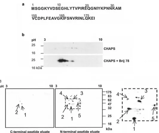
Cave-olin-derived peptides (Fig. 1a) were synthesized with an automated solid-phase synthesizer with Fmoc chemistry, purified by reverse-phase HPLC and characterized by mass spectrometry (MS) and amino acid analysis. Pep-tides were coupled to CNBr-activated Sepharose 4B fol-lowing the instructions of the supplier.
Fifteen dishes (15 cm in diameter) of HT29 cells were lysed for 5 h on ice in 30 ml 20 mM Tris pH 8.0 contain-ing 1% Brij 78, and the lysate was clarified by centrifu-gation at 10 000 g for 10 min. The supernatant was
ap-plied overnight at 4 °C to columns containing peptide-de-rivatized Sepharose. Following extensive washing with lysis buffer supplemented with 0.4 M NaCl, the columns were eluted with 0.1 M glycine, pH 2.4. and the proteins precipitated with chloroform/methanol.
2D gel electrophoresis and MS identification of pro-teins. 2D gel electrophoresis was performed as described
[27], with the following modification in the first dimen-sion of the gel: the precipitated proteins were solubilized in 40 mM Tris base containing 7 M urea and 2 M thio-urea, 4% CHAPS, 1% Brij 78, 0.001% bromophenol blue and 0.8% resolyte 4–8, 18 mM DTT and 2 mM tributyl-phosphine. The 2D gels were stained with colloidal silver [28]. The protein spots were excised from the gel, cleaved with trypsin [29] and the generated peptides were ana-lyzed by capillary liquid chromatography tandem mass spectrometry (LC-MS/MS) using a Magic C18 100mm ¥10 cm HPLC column (Spectronex, Switzerland)
con-nected on line to an ion trap Finnigan DecaXP (Thermo-Finnigan, CA). The eluting peptides were ionized by
elec-trospray ionization, and the peptide ions detected were automatically selected and fragmented in the ion trap. In-dividual MS/MS spectra, containing sequence informa-tion for a single peptide, were compared with the program TurboSequest [30] against the human subset of the pro-tein sequence database Swiss-Prot 40.36. This resulted in the identification of the peptide and, by association, the protein in the spot.
Immunoprecipitation, immunoblotting and sucrose density gradient. Cells were lysed for 4 h on ice in
20 mM Tris pH 8.0 containing 1% Brij 78 and protease inhibitors (9.6mg/ml benzamidine hydrochloride, 0.9 mg/
ml leupeptin and 1.9mg/ml antipain). An equal amount of
protein lysate was immunoprecipitated and analyzed by immunoblotting as described previously [31]. Quantita-tion of the chemiluminescent signals were performed with ImageJ 1.34s quantification software (NIH, USA). Caveolin-1-enriched fractions were obtained by isolation on sucrose density gradient following solubilization in Triton X-100 as described [7].
Figure 1. Proteins interacting with the N- and C-terminal peptides of caveolin-1. (a) N- and C-terminal peptides derived from caveolin-1. (b) Low-density detergent insoluble microdomains from caveolin positive Swiss mouse NIH 3T3 cells were isolated by sucrose density gra-dient. Proteins were resolved by 2D SDS-PAGE using a pH gradient from 3 to 10 in denaturing buffer supplemented either CHAPS or CHAPS/Brij 78 mixed micelles for protein solubilization in the first dimension. Recovery and focalization of caveolin were assessed by Western blotting using caveolin-specific antibodies. (c) HT29 cell lysates were passed through N- and C-terminal peptide affinity columns and the eluates were subjected to 2D SDS-PAGE using a pH gradient from 3 to 10 in the first dimension and 12% polyacrylamide gel in the second dimension. The proteins were revealed by silver staining and the right panel shows a blow-up of the N-terminal peptide eluates. Comparable results were obtained in two independent experiments.
DNase I-inhibition assay. Following two washes with
PBS, the cells were lysed for 4 h on ice in 20 mM Tris pH 8.0 containing 0.3% Triton X-100 and protease inhibitors (45mg/ml benzamidine hydrochloride, 5 mg/ml leupeptin
and 10mg/ml antipain). The lysate was clarified by
cen-trifugation for 5 min at 10 000 g. The DNase I-inhibition assay was performed as described previously [32, 33] us-ing 2 ml substrate solution containus-ing 0.04 mg/ml DNA and 10 U DNase I. The inhibition was measured by adding 25mg protein of cell lysate to the substrate
solu-tion for 2 min at 26 °C and changes in OD at 260 nm were measured. Dissociation of pre-existing F actin to G actin
in vitro requires thermal or chemical unfolding of actin
[32, 33]. Therefore, in a mixture of filamentous and mon-omeric actin present in a cell extract, only the monmon-omeric form will inhibit DNase I [32, 33].
Results
The N-terminal segment of caveolin-1 aa interacts with TCP-1
Peptide derived from the N terminus of caveol1 in-teracts with TCP-1. To examine the structural and
func-tional binding properties of caveolin-1 a, we used a
pep-tide corresponding to the first 32 N-terminal residues of caveolin-1 a and, as a control, a peptide comprising the
last 23 C-terminal residues of caveolin-1 (Fig. 1a). N-and C-terminal synthetic peptides were covalently at-tached to Sepharose beads and used for affinity chro-matography with HT29 cell extracts. Eluted proteins were separated by 2D SDS-PAGE. Due to their resistance to non-ionic detergents, solubilization and recovery of membrane proteins from lipid rafts or caveolae after iso-electric focusing is often of very low efficiency. Good re-covery and solubilization of caveolin (Fig. 1b) and of other raft resident proteins such as Lck, Fyn and Thy-1 (data not shown) were only obtained when CHAPS was combined with 1% Brij 78 (see Methods section). The HT29 cell line was chosen because, as most human colon carcinoma cell lines, it expresses caveolin-1 at very low levels, thus avoiding competition with the
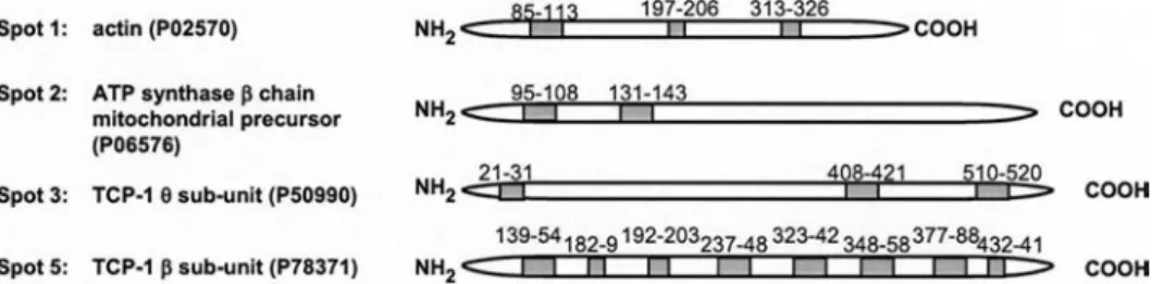
immobi-lized caveolin peptide. A limited number of proteins were specifically eluted from both N- and C-terminal peptide affinity chromatography columns (Fig. 1c). Following trypsin digestion, the resulting peptide fragments were separated and analyzed by LC-MS/MS, and proteins from spots 1, 2, 3 and 5 were identified using the sequence in-formation of the peptide fragments obtained by MS/MS (Fig. 2). Spots 1 and 2 were found in both preparations, although higher amounts were eluted from the N-terminal peptide column. They were shown to contain actin and the mitochondrial ATP synthase beta chain, respectively (Figs 1c and 2). By contrast, spots 3, 4, and 5 were exclu-sively present in the eluate of the N-terminal peptide affinity column. No protein could be identified in spot 4 and the other detectable spots due to an insufficient amount of material. Unexpected was the identification of spots 3 and 5 as TCP-1 subunits q and b, respectively
(Figs 1c and 2). Indeed caveolin has not been reported as being a substrate of the chaperone complex TCP-1. More-over, using a protein blast search, we did not find any se-quence homology between the N-terminal segment of caveolin-1 a, or truncated portions of this segment, and
any other protein of the uniprot database, suggesting that this interaction might be unique. Finally, among the di-versity of proteins interacting with caveolin [5, 9], a com-parable association with a molecular chaperone complex has been previously demonstrated with HSP56, cyclo-philin 40 and cyclocyclo-philin A. Although the complex also involves annexin II, the precise site of interaction remains unknown. The function of this caveolin-chaperone com-plex is the transport of cholesteryl ester from caveolae through the cytosol to an internal membrane [34, 35].
Caveolin-1 interacts with TCP-1 in cells. To determine
whether caveolin interacts with TCP-1 in cells, we trans-fected caveolin-1 a or b in human hepatoma HuH or
hu-man melanocyte A7 cells that express very low levels of caveolin-1 and 2 [24]. The amount of caveolin expressed in transfected cells was physiological and comparable to endogenous expression of caveolin in Swiss mouse 3T3 cells (data not shown). The level of association of cave-olin-1 a to TCP-1 was determined as the ratio between
Figure 2. The N-terminal segment of caveolin-1 interacts specifically with TCP-1. The protein spots were excised from the 2D gels, di-gested with trypsin and extracted peptides were separated by capillary LC and sequenced by MS/MS, gray boxes show the peptides for which the sequences were obtained by MS/MS. The proteins were identified with the program TurboSquest searching the Swiss-Prot 40.36 database; their primary accession number is indicated in parentheses.
the amount of caveolin-1 a co-precipitated with
anti-TCP-1 antibodies and the amount of caveolin-1 a present
in the lysate. Comparison of such quantitations obtained for caveolin-1 a and b indicated that the latter isoform is
only marginally associated with TCP-1 since its co-pre-cipitation was always 10 fold lower than the one observed with the a isoform (Fig. 3a). These data confirm the
pre-dominant role of the 32 N-terminal residues of caveolin-1 in the interaction with TCP-caveolin-1, and yet suggests an ad-ditional minor site of interaction in caveolin-1 b. In
ad-dition, the interaction was stabilized by Mg and Mn cations and disrupted by chelation of divalent cations with EDTA (Fig. 3b). Finally, TCP-1 immunoprecipitates did not contain caspase 3 nor transferrin receptor, al-though these proteins were both present in large amount in the cell lysates (data not shown).
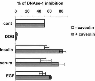
To confirm that caveolin interacts with TCP-1 in intact cells, we examined the effect of this interaction on TCP-1 actin folding function. Since caveolin-TCP-1 is enriched in caveolae which contain tyrosine kinases and various growth factor receptors (including IR), we stimulated HUH cells with serum, insulin or epidermal growth fac-tor (EGF) that induce caveolin phosphorylation to a vari-able extent [10, 11]. We measured the impact of these ef-fectors on TCP-1 actin folding activity in the presence or absence of caveolin expression. TCP-1 assists actin in reaching its mature conformation by folding the nu-cleotide binding site of monomeric G actin, which in turn can bind DNase-1 with affinities in the nanomolar range and thus inhibits its endonuclease activity in vitro [22, 32, 33]. Determination of DNase-1 inhibition in cell extracts therefore enables the measurement of monomeric G actin concentration and allows the actin folding activity of TCP-1 in cells to be assessed [33]. Increase in DNase-1 inhibition was observed in caveolin transfected HuH cells, but not in mock transfected cells, upon insulin, EGF or serum stimulation. Statistical evaluation of these re-sults by paired two-tail t-tests, however, revealed that only insulin can be considered as inducing a significant in-crease of DNase-1 inhibition (p < 0.05), whereas the ef-fect of EGF remains at the lower limit of statistical sig-nificance, and the data obtained with the serum cannot at
that point be considered as significant. In addition, the differences between untreated or treated non-transfected cells were not significant (0.2 < p < 0.5). Deoxyglucose was used as a control to block TCP-1 activity (Fig. 4). These data show that upon insulin stimulation caveolin affects TCP-1 actin folding function in intact cells. Fur-thermore, the absence of a significant increase in DNase-1 inhibition in mock-transfected cells upon insulin stim-ulation (p < 0.5) suggests that the mechanism by which caveolin modulates TCP-1 activity is indirect.
Insulin-mediated phosphorylation of caveolin on Y14 induces the dissociation of the caveolin-TCP-1 com-plex and promotes TCP-1 folding activity. To
under-stand the mechanism by which caveolin modulates TCP-1 actin folding activity, we investigated how insulin-me-diated cell activation increases TCP-1 actin folding in caveolin-expressing cells (Fig. 4). Insulin has been re-Figure 3. TCP-1 associates with caveolin-1 in cells. (a) HuH cells untransfected (-) or transfected with caveolin-1 aor bwere lysed, and the total lysates or immunoprecipitates with anti-TCP-1aantibodies were resolved on SDS-PAGE (12% reducing) and analyzed by West-ern immunoblotting (ib) with antibodies specific for TCP-1 and caveolin. Caveolin aor blysates and immunoprecipitates shown corre-spond to the same amount of protein. (b) TCP-1 was immunoprecipitated from HuH cells transfected with caveolin-1 ain the presence of EDTA or divalent cations and the immunoprecipitates analyzed as described in (a). Shown is one out of three independent experiments.
Figure 4. Caveolin expression modulates TCP-1 actin folding ac-tivity upon cell stimulation. HuH cells untransfected or transfected with caveolin were stimulated with insulin, EGF or serum for 5 min or pretreated with deoxyglucose for 1 h at 37 °C, lysed and the con-tent of monomeric G actin in the whole cell lysate determined by DNase-1 inhibition assay (see Methods section). Shown are the mean and standard deviation of three independent experiments.
ported to induce caveolin phosphorylation [12] primarily
at residue Y14, a reaction which is likely to result directly from the engagement of IR [10]. To determine whether
the insulin-mediated phosphorylation of the N-terminal region of caveolin affects its association with TCP-1, HuH cells transfected with caveolin a were stimulated
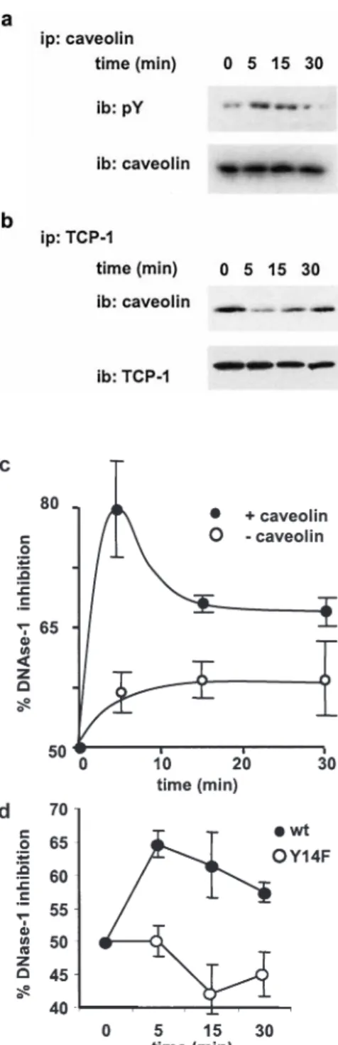
with insulin. After various periods of time up to 30 min, the interaction between caveolin and TCP-1 was analyzed by immunoprecipitation. Insulin treatment caused transient phosphorylation of caveolin, with an optimum reached after 5 min of stimulation (Fig. 5a). The maximal phos-phorylation of caveolin correlated with a minimal bind-ing to TCP-1 (Fig. 5b), suggestbind-ing that the phosphoryl-ation of caveolin inhibits TCP-1 binding (Fig. 5c). We then examined the role of caveolin phosphorylation on the actin folding activity of TCP-1. To this end, we measured the DNase-1 inhibition effect in HuH caveolin transfectants upon insulin stimulation. A maximal inhibi-tion was detected in cell extracts after 5 min of stimula-tion (Fig. 5c), which corresponds to the maximum of caveolin phosphorylation (Fig. 5a). A control experiment performed with mock-transfected cells showed no signif-icant effect of insulin treatment.
In addition, we generated a caveolin mutant in which the phosphorylation site at Y14 was replaced by a phenylala-nine. Wild-type and mutant caveolin expression were com-parable in both HuH and A7 caveolin-transfected cells (data not shown). Measurement of DNase-1 inhibition showed that wild-type caveolin but not the Y14F mutant induced TCP-1 folding activity in response to insulin stimulation (Fig. 5d). These results demonstrate that caveolin phosphorylation on residue Y14 is required for insulin-induced activation of TCP-1 and that caveolin is necessary to promote the TCP-1 folding activity.
Caveolin-TCP-1 interaction acts as a competitor for caveolin-filamin interaction and thus restores insulin signaling. To further understand the complex role of
cave-olin in the control of TCP-1 biological activity, we explored the possibility that caveolin might interfere with mecha-nisms regulating insulin-mediated cell activation. This was based on the observation that the actin-cross-linking pro-tein filamin interacts with the IR, thereby inducing a deac-tivation of insulin-mediated signaling events [25]
Since caveolin-1 also binds filamin [36], we hypothe-sized that this interaction could interfere with the IR-fil-amin association, and thus restore insulin-mediated sig-naling and activation of TCP-1.
To test this hypothesis, A7 filamin-expressing cells were transfected with caveolin-1 a and the interaction between
IR and filamin analyzed by co-immunoprecipitation. In-deed, expression of caveolin impaired IR-filamin interac-tion (Fig. 6a) by 45–50% as determined by the quantita-tion of the relative intensity of ECL signals (see Methods section). The impact of the competition between filamin Figure 5. Insulin-mediated phosphorylation of caveolin induces
caveolin-TCP-1 complex dissociation and activation of actin fold-ing. Kinetic of caveolin phosphorylation (a) and caveolin associ-ation with TCP-1 (b) were examined upon addition of insulin to caveolin-transfected HuH cells. Cell lysates were immunoprecip-itated with anti-caveolin- (a) or anti-TCP-1- (b) specific antibod-ies and the immunoprecipitates resolved by SDS-PAGE and ana-lyzed by Western blotting using phosphotyrosine (pY), anti-caveolin or anti-TCP-1bantibodies. (c) Aliquots of the same HuH cell lysates were used to determine their content of monomeric G actin in by DNase-1 inhibition assay. (d) HuH cells transfected with either wild-type or Y14F mutant caveolin were stimulated with insulin for 5 min, lysed and the content of monomeric G actin determined by DNase-1 inhibition assay. In (c) and (d) the mean and standard deviation of three independent experiments are shown.
binding to caveolin or the IR on TCP-1 activation was then assessed using A7 and the derived filamin-deficient M2 cell lines. Both cell types were transfected with a plasmid encoding caveolin-1 a under the control of an
IPTG-in-ducible promoter, and the activity of TCP-1 was deter-mined by the measure of DNase-1 inhibition following insulin stimulation.
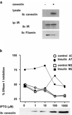
In A7 cells, in the absence of caveolin expression, no change in DNase-1 inhibition was detected upon 10 min of insulin treatment (Fig. 6b). By contrast, following induction of caveolin expression with graduated doses of IPTG, a significant increase in DNase-1 inhibition was observed at 100mM IPTG, corresponding probably to
the optimal level of caveolin-1 expression inducing the strongest effect. Lower or higher expression levels of caveolin failed to induce any detectable change in DNase-1 inhibition upon insulin stimulation (Fig. 6b). Thus in A7 cells, like in HuH cells, caveolin regulates its folding activity via its association with TCP-1 (Fig. 5c). By contrast to A7 cells, when M2 filamin-deficient cells were used, insulin induced a stronger DNase-1 inhibition in the absence of caveolin expression (Fig. 6b). This con-firms the selective impairment in insulin signaling by fil-amin [25]. Importantly, in both A7 and M2 cells express-ing levels of caveolin higher than that obtained with 100mM and 1000 mM IPTG, respectively, insulin
stimu-lation failed to induce DNase-1 inhibition, indicating that caveolin can inhibit TCP-1 activation (Fig. 6b). Further evidence for the direct inhibitory effect of caveolin on TCP-1 actin folding function was provided by the exper-iment using the caveolin mutant Y14F, which showed im-paired activation of TCP-1 upon insulin stimulation (Fig. 5d). This effect was independent of the level of cav-eolin expression (data not shown). In addition, at low ex-pression levels (up to 10mM IPTG), caveolin can restore
insulin-mediated activation of TCP-1 in A7 cells (Fig. 6a) Figure 6. Role of caveolin in insulin-induced activation of TCP-1.
(a) Interaction of the IR with filamin in A7 cells transfected or not with caveolin. The IR was immunoprecipitated with specific anti-bodies and the immunoprecipitates resolved by SDS-PAGE fol-lowed by Western blotting using anti-filamin- or anti-IR-specific antibodies. Equal amounts of protein cell lysates were examined by immunoblotting using anti-caveolin-specific antibodies (top pan-els). (b) Insulin-mediated folding of G actin in filamin-positive (A7) and filamin-negative (M2) cells expressing increasing con-centrations of caveolin. Shown is one representative out of three in-dependent experiments (a and b) at optimal time of DNase-1 inhi-bition, i.e., 10 min after insulin stimulation (b).
Figure 7. Mechanism of caveolin regulation of TCP-1 activity. Filamin, by interacting with the IR, inhibits insulin-mediated signaling. At low expression level, caveolin does not compete efficiently for this interaction but, by associating with TCP-1, inhibits its actin folding function (left panel). At higher expression level, caveolin sequesters filamin from the IR and allows insulin-mediated phosphorylation of caveolin at Y14 and the release of the functionally immobilized TCP-1 (right panel).
by competing for the IR-filamin interaction (Fig. 6b). Taken together these results indicate that caveolin-1 can func-tion as a direct inhibitor of TCP-1 folding activity and as an indirect activator of TCP-1 by competing for the fil-amin-IR interaction.
Discussion
In the present study, we have shown that caveolin binds the chaperone complex TCP-1 and thus contributes to the regulation of its protein folding function. The N-terminal region of caveolin-1 a is crucial for that interaction as
ev-idenced by the specific binding of TCP-1 to a 32-amino acid synthetic peptide corresponding to the N terminus of the protein. Moreover, TCP-1 can be co-immunoprecipi-tated with native caveolin-1 a, whereas only a weak
in-teraction was observed between the chaperone complex and caveolin-1 b, an isoform of caveolin-1 that lacks the
32 N-terminal residues.
Several other proteins were consistently found in associ-ation with the caveolin-1 N-terminal peptide, among which ATP synthase and actin could be identified. The presence of actin is consistent with its specific binding to TCP-1 under the mild dispersing conditions used in our experi-ments. Indeed, the actin-TCP-1 interaction is highly re-sistant to the disruption in mixed micelles of ionic and non-ionic detergents that cause complete disruption of the TCP-1 hexadecamer into its constituent subunits [37]. 2D electrophoresis revealed a number of spots corre-sponding to the various subunits of TCP-1 [38, 39], among which the subunits b and q were in a sufficient
amount to allow their identification by MS. Our data show that TCP-1 interacts with the N-terminal segment of caveolin, but due to the fact that under the experimental conditions used, TCP-1 dissociates partially into its con-stituent subunits, it remains difficult to evaluate the pro-portion of TCP-1 complexes associated with caveolin. Our data are consistent with a quantitative proteomic analysis demonstrating that both caveolin and TCP-1 par-titioned in glycolipid-enriched membrane microdomains [40]. Considering that the cytosolic chaperone complex TCP-1 is expected to partition in the detergent soluble fractions, its localization in rafts might result from its as-sociation with caveolae cytosolic organelles.
Using a DNase I-inhibition assay, which measures the amount of folded actin [32, 33], we provide evidence that caveolin exerts two competing effects on TCP-1 actin folding function. Caveolin, by binding directly to TCP-1, inhibits the chaperone actin folding function as observed in filamin-deficient cells expressing large amounts of caveolin (Fig. 6b). Further evidence for a direct inhibition of TCP-1 by caveolin were obtained by the observation that the Y14F caveolin mutant does not induce TCP-1 ac-tivation nor dissociation from TCP-1 upon insulin
stimu-lation (Fig. 5). These data demonstrate that the specific phosphorylation of this residue controls both the cave-olin-TCP-1 interaction and the chaperone folding func-tion (Fig. 7).
By preventing the actin cross-linking protein filamin to bind to the IR, caveolin restores insulin-mediated sig-naling and activates TCP-1 actin folding function (Fig. 6b). The fragment of filamin spanning repeats 22, 23 and the hinge region are involved in the interaction with both caveolin and the IR [25, 36]. Caveolin interacts with TCP-1 mainly through its first 32 N-terminal residues and with filamin via residues 32–101 (Figs 1 and 2). In addition, a possible negative influence of the first 32 N-terminal residues on the strength of this later inter-action has been described [25]. The filamin-caveolin complex sequesters filamin from the IR, and thus pre-vents the phosphorylation of caveolin associated with fil-amin (Fig. 6, right panel). This complex, by sequestering filamin from the IR, restores insulin signaling through the IR-caveolin-TCP-1 complex. Consequently, insulin mediates phosphorylation of caveolin at Y14, which in turn induces the dissociation of caveolin from TCP-1 and the activation of the chaperone (Fig. 6, right panel). Thus, low expression of caveolin in filamin-expressing cells may not be sufficient to compete for filamin-IR interac-tion (Fig. 6. from left to right panel). In contrast, in fil-amin-deficient cells, such a low level of caveolin is suffi-cient to inhibit TCP-1 activity, and to override insulin-mediated activation of TCP-1. Consequently, at a high level of expression of caveolin, the formation of the com-plex with TCP-1 prevails over its insulin-mediated disso-ciation. This is most likely due to the fact that insulin-me-diated phosphorylation of caveolin is transient (Fig. 3a). Moreover, by forming such a complex, the chaperone TCP-1 could modulate the conformation and the func-tions of caveolin. In fact, a key role of caveolin is to act as a negative regulator of signaling molecules, a function that was initially attributed to the scaffolding domain [5, 9] and more recently to a specific active conformation of the protein [41].
Our data show that a functional consequence of caveolin-TCP-1 interaction is the regulation of the insulin-medi-ated mitogenic pathway. Similar effects of TCP-1 actin folding function was obtained upon EGF or serum treat-ment (Fig. 4), indicating that mediators of caveolin phos-phorylation release the functionally sequestered chaper-one. However, it remains to be determined whether the mechanism identified for insulin stimulation is similar for EGF and serum treatment, and represents a more gen-eral mechanism of caveolin regulatory function.
An additional important function of caveolTCP-1 in-teraction may be to regulate the inin-teraction of caveolae with the cortical actin cytoskeleton. Caveolae tend to col-lect in the actin-rich region of the cell membrane and con-tain the actin and caveolin binding protein filamin.
There-fore, by regulating actin folding function of TCP-1, cave-olin may control the vesicular traffic of caveolae between the cell surface and intracellular organelles.
Acknowledgement. We are grateful to Drs T. Stossel and Y. Ohta for
providing the M2 and A7 cell lines (Brigham and Women’s Hospi-tal, Harvard Medical School, Boston, USA). We thank Léonard Bagnoud for technical assistance, Dr. Slavica Masina (Institute of Biochemistry, Epalinges, Switzerland), Dr. Olivier Michielin (IS-REC, Epalinges, Switzerland) and Prof. Andreas Conzelman (Uni-versity of Fribourg, Switzerland) for careful reading of the manu-script. This study was supported by the Swiss National Science Foundation (SNF 31-61960) and the Giorgi-Cavalieri Foundation.
1 Anderson R. G. (1998) The caveolae membrane system. Annu. Rev. Biochem. 67: 199–225
2 Pelkmans L., Kartenbeck J. and Helenius A. (2001) Caveolar endocytosis of simian virus 40 reveals a new two-step vesicu-lar-transport pathway to the ER. Nat. Cell Biol. 3: 473–483 3 Pelkmans L., Puntener D. and Helenius A. (2002) Local actin
polymerization and dynamin recruitment in SV40-induced in-ternalization of caveolae. Science 296: 535–539
4 Shin J. S., Gao Z. and Abraham S. N. (2000) Involvement of cellular caveolae in bacterial entry into mast cells. Science 289: 785–788
5 Okamoto T., Schlegel A., Scherer P. E. and Lisanti M. P (1998) Caveolins, a family of scaffolding proteins for organizing ‘pre-assembled signaling complexes’ at the plasma membrane. J. Biol. Chem. 273: 5419–5422
6 Bender F. C., Reymond M. A., Bron C. and Quest A. F. (2000) Caveolin-1 levels are down-regulated in human colon tumors and ectopic expression of caveolin-1 in colon carcinoma cell lines reduces cell tumorigenicity. Cancer Res. 60: 5870–5878 7 Schlegel A. and Lisanti M. P. (2000) A molecular dissection of
caveolin-1 membrane attachment and oligomerization. Two separate regions of the caveolin-1 C-terminal domain mediate membrane binding and oligomer/oligomer interactions in vivo. J. Biol. Chem. 275: 21605–21617
8 Li S., Couet J. and Lisanti M. P. (1996) Src tyrosine kinases, Galpha subunits and H-Ras share a common membrane-an-chored scaffolding protein, caveolin. Caveolin binding nega-tively regulates the auto-activation of Src tyrosine kinases. J. Biol. Chem. 271: 29182–29190
9 Couet J., Li S., Okamoto T., Ikezu T. and Lisanti M. P. (1997) Identification of peptide and protein ligands for the caveolin-scaffolding domain. Implications for the interaction of caveolin with caveolae-associated proteins. J. Biol. Chem. 272: 6525– 6533
10 Kimura A., Mora S., Shigematsu S., Pessin J. E. and Saltiel A. R. (2002) The insulin receptor catalyzes the tyrosine phos-phorylation of caveolin-1. J. Biol. Chem. 277: 30153–30158 11 Sanguinetti A. R. and Masticke C. C (2003) c-Abl is required
for oxidative stress-induced phosphorylation of caveolin-1 on tyrosine 14. Cell Signal. 15: 289–298
12 Lee H., Volonte D., Galbiati F., Iyengar P., Lublin D. M., Breg-man D. B. et al. (2000) Constitutive and growth factor-regu-lated phosphorylation of caveolin-1 occurs at the same site (Tyr-14) in vivo: identification of a c-Src/Cav-1/Grb7 signaling cassette. Mol. Endocrinol. 14: 1750–1775
13 Scherer P. E., Tang Z., Chun M., Sargiacomo M., Lodish H. F. and Lisanti M. P. (1995) Caveolin isoforms differ in their N-ter-minal protein sequence and subcellular distribution. Identifica-tion and epitope mapping of an isoform-specific monoclonal antibody probe. J. Biol. Chem. 270: 16395–16401
14 Thomsen P., Roepstorff K., Stahlhut M. and van Deurs B. (2002) Caveolae are highly immobile plasma membrane mi-crodomains, which are not involved in constitutive endocytic trafficking. Mol. Biol. Cell 13: 238–250
15 Lewis V. A., Hynes G. M., Zheng D., Saibil H. and Willison K. (1992) T-complex polypeptide-1 is a subunit of a heteromeric particle in the eukaryotic cytosol. Nature 358: 249–252 16 Frydman J. and Hartl F. U. (1996) Principles of
chaperone-as-sisted protein folding: differences between in vitro and in vivo mechanisms. Science 272: 1497–1502
17 Kubota H. (2002) Function and regulation of cytosolic molec-ular chaperone CCT. Vitam. Horm. 65: 313–331
18 Willison K. R., Dudley K. and Potter J. (1986) Molecular cloning and sequence analysis of a haploid expressed gene en-coding t complex polypeptide 1. Cell 44: 727–738
19 Gao Y., Thomas J. O., Chow R. L., Lee G. H. and Cowan N. J. (1992) A cytoplasmic chaperonin that catalyzes beta-actin fold-ing. Cell 69: 1043–1050
20 Yaffe M. B., Farr G. W., Miklos D., Horwich A. L., Sternlicht M. L. and Sternlicht H. (1992) TCP1 complex is a molecular chaperone in tubulin biogenesis. Nature 358: 245–248 21 Llorca O., McCormack E. A., Hynes G., Grantham J., Cordell
J., Carrascosa J. L. et al. (1999) Eukaryotic type II chaperonin CCT interacts with actin through specific subunits. Nature 402: 693–696
22 Llorca O., Martin-Benito J., Ritco-Vonsovici M., Grantham J., Hynes G. M., Willison K. R. et al. (2000) Eukaryotic chaper-onin CCT stabilizes actin and tubulin folding intermediates in open quasi-native conformations. EMBO J. 19: 5971–5979 23 Pappenberger G., Wilsher J. A., Roe S. M., Counsell D. J.,
Willi-son K. R. and Pearl L. H. (2002) Crystal structure of the CCT-gamma apical domain: implications for substrate binding to the eukaryotic cytosolic chaperonin. J. Mol. Biol. 318: 1367–1379 24 Vainio S., Heino S., Mansson J. E., Fredman P., Kuismanen E.,
Vaarala O. et al. (2002) Dynamic association of human insulin receptor with lipid rafts in cells lacking caveolae. EMBO J. 3: 95–100
25 He H. J., Kole S., Kwon Y. K., Crow M. T. and Bernier M. (2003) Interaction of filamin A with the insulin receptor alters insulin-dependent activation of the mitogen-activated protein kinase pathway. J. Biol. Chem. 278: 27096–27104
26 Cunningham C. C., Gorlin J. B., Kwiatkowski D. J., Hartwig J. H., Janmey P. A., Byers H. R. et al. (1992) Actin-binding pro-tein requirement for cortical stability and efficient locomotion. Science 255: 325–327
27 Rabilloud T. (1992) Use of thiourea to increase the solubility of membrane proteins in two-dimensional electrophoresis. Elec-trophoresis 19: 758–760
28 van Oostveen I., Ducret A. and Aebersold R. (1997) Colloidal silver staining of electroblotted proteins for high sensitivity pep-tide mapping by liquid chromatography-electrospray ionization tandem mass spectrometry. Anal. Biochem. 247: 310–318 29 Shevchenko A., Wilm M., Vorm O. and Mann M. (1996) Mass
spectrometric sequencing of proteins silver-stained polyacry-lamide gels. Anal. Chem. 68: 850–858
30 Eng J. K., McCormack A. L. and Yates J. R. III (1994) An ap-proach to correlate tandem mass spectral data of peptides with amino acid sequences in a protein database. J. Am. Soc. Mass Spectrom. 1994. 5: 976–989
31 Doucey M. A., Legler D. F., Faroudi M., Boucheron N., Baum-gaertner P., Naeher D. et al. (2003) The beta1 and beta3 inte-grins promote T cell receptor-mediated cytotoxic T lymphocyte activation. J. Biol. Chem. 278: 26983–26991
32 Schuler H., Lindberg U., Schutt C. E. and Karlsson R. (2000) Thermal unfolding of G-actin monitored with the DNase I-in-hibition assay stabilities of actin isoforms. Eur. J. Biochem. 267: 476–486
33 Blikstad I., Markey F., Carlsson L., Persson T. and Lindberg U. (1978) Selective assay of monomeric and filamentous actin in cell extracts, using inhibition of deoxyribonuclease I. Cell 15: 935–943
34 Uittenbogaard A. and Smart E. J. (2000) Palmitoylation of caveolin-1 is required for cholesterol binding, chaperone
com-plex formation and rapid transport of cholesterol to caveolae. J. Biol. Chem. 275: 25595–25599
35 Uittenbogaard A., Ying Y. and Smart E. J. (1998) Characteriza-tion of a cytosolic heat-shock protein-caveolin chaperone com-plex. Involvement in cholesterol trafficking. J. Biol. Chem. 273: 6525–6532
36 Stahlhut M. and van Deurs B. (2000) Identification of filamin as a novel ligand for caveolin-1: evidence for the organization of caveolin-1-associated membrane domains by the actin cy-toskeleton. Mol. Biol. Cell 11: 325–337
37 Hynes G. M. and Willison K. R. (2000) Individual subunits of the eukaryotic cytosolic chaperonin mediate interactions with binding sites located on subdomains of beta-actin. J. Biol. Chem. 275: 18985–18994
38 Kubota H., Hynes G., Carne A., Ashworth A. and Willison K. (1994) Identification of six Tcp-1-related genes encoding di-vergent subunits of the TCP-1-containing chaperonin. Curr. Biol. 4: 89–99
39 Hynes G., Kubota H. and Willison K. R. (1995) Antibody characterisation of two distinct conformations of the chaper-onin-containing TCP-1 from mouse testis. FEBS Lett. 358: 129–132
40 Foster L. J., De Hoog C. L. and Mann M. (2003) Unbiased quantitative proteomics of lipid rafts reveals high specificity for signaling factors. Proc. Natl. Acad. Sci. USA 100: 5813– 5818
41 Liu P., Rudick M. and Anderson R. G. (2002) Multiple func-tions of caveolin-1. J. Biol. Chem. 277: 41295–41298