Paul Stolzmann Joseph Knight Lotus Desbiolles Willibald Maier Hans Scheffel André Plass Vartan Kurtcuoglu Sebastian Leschka Dimos Poulikakos Borut Marincek Hatem Alkadhi Received: 20 October 2008 Revised: 8 December 2008 Accepted: 21 December 2008 Published online: 4 February 2009
# European Society of Radiology 2009
Remodelling of the aortic root in severe
tricuspid aortic stenosis: implications
for transcatheter aortic valve implantation
Abstract Detailed knowledge of aortic root geometry is a prerequisite to anticipate complications of trans-catheter aortic valve (TAV) implanta-tion. We determined coronary ostial locations and aortic root dimensions in patients with aortic stenosis (AS) and compared these values with normal subjects using computed tomography (CT). One hundred consecutive pa-tients with severe tricuspid AS and 100 consecutive patients without val-vular pathology (referred to as the controls) undergoing cardiac dual-source CT were included. Distances from the aortic annulus (AA) to the left coronary ostium (LCO), right coronary ostium (RCO), the height of the left coronary sinus (HLS), right coronary sinus (HRS), and aortic root dimensions [diameters of AA, sinus of Valsalva (SV), and sino-tubular junc-tion(STJ)] were measured. LCO and RCO were 14.9±3.2 mm (8.2–25.9) and 16.8±3.6 mm (12.0–25.7) in the controls, 15.5±2.9 mm (8.8–24.3) and 17.3±3.6 mm (7.3–26.0) in patients
with AS. Controls and patients with AS had similar values for LCO (P= 0.18), RCO (P=0.33) and HLS (P= 0.88), whereas HRS (P<0.05) was significantly larger in patients with AS. AA (r=0.55,P<0.001), SV (r= 0.54,P<0.001), and STJ (r=0.52,P< 0.001) significantly correlated with the body surface area in the controls; whereas no correlation was found in patients with AS. Patients with AS had significantly larger AA (P<0.01) and STJ (P<0.01) diameters when com-pared with the controls. In patients with severe tricuspid AS, coronary ostial locations were similar to the controls, but a transverse remodelling of the aortic root was recognized. Owing to the large distribution of ostial locations and the dilatation of the aortic root, CT is recommended before TAV implantation in each patient.
Keywords Aortic stenosis . CT coronary angiography . Transcatheter aortic valves . Aortic root geometry
Introduction
Aortic stenosis (AS) represents the most common valvular heart disease in adults [1]. Surgical valve repair or replacement is the treatment of choice for symptomatic patients with severe AS, offering symptomatic relief and reducing mortality [2, 3]. However, more than 30% of patients are not candidates for an operative treatment due to comorbidities [1]. In those patients suffering from severe tricuspid AS, transcatheter aortic valve (TAV) implantation
that may be performed either percutaneously or transapi-cally provides an alternative treatment option to open surgery [4–9].
Potential difficulties of TAV implantation include the optimal design of the stent, the preservation of valve function after the delivering procedure, and the avoidance of coronary obstruction and paravalvular regurgitation [4, 10–14]. Coronary flow restriction occurs either by direct blocking of the implanted stent or from the aortic cusps that are immobilized against the coronary ostia [12–14]. P. Stolzmann . L. Desbiolles .
H. Scheffel . S. Leschka . B. Marincek . H. Alkadhi (*)
Institute of Diagnostic Radiology, University Hospital Zurich, Raemistrasse 100, 8091 Zurich, Switzerland e-mail: [email protected] Tel.: +41-44-2553662 Fax: +41-44-2554443 J. Knight . V. Kurtcuoglu . D. Poulikakos Laboratory of Thermodynamics in Emerging Technologies, Department of Mechanical and Process Engineering, ETH Zurich, Switzerland
W. Maier
Cardiovascular Center, University Hospital Zurich, Zurich, Switzerland A. Plass
Clinic for Cardiovascular Surgery, University Hospital Zurich, Zurich, Switzerland
Therefore, the precise locations of the coronary ostia should be determined prior to the procedure to ensure an unobstructed coronary blood flow [4,12,14]. Paravalvular regurgitation can be avoided by correct TAV positioning and sizing in respect to aortic root dimensions [12].
Echocardiography [3] may be used for determining the aortic root anatomy; however, it has the inherent limitations of being dependent on the individual patient’s constitution, on instrumental settings and transducer position, and on operator skills. On the other hand, cross-sectional imaging modalities, such as magnetic resonance imaging (MRI) and computed tomography (CT), could be used. MRI, however, lacks the superior resolution of CT with regard to valve and coronary artery morphology.
Current multidetector-row spiral CT technology acquires volumetric data with isotropic voxel resolution enables the reconstruction of three-dimensional structures with a high spatial and temporal resolution. Thus, CT allows for an accurate assessment of coronary arteries [15–17] and aortic root morphology [18,19].
In this study, we determined coronary ostial locations and aortic root dimensions in patients with severe tricuspid AS in comparison with subjects without valvular heart disease using CT.
Material and methods
Patients
From August 2006 to April 2008, 100 consecutive patients with severe tricuspid AS and 100 consecutive patients without valvular pathology (the controls) were included. All patients with AS underwent CT for preoperative planning (100%) before aortic valve repair (18%) or replacement (82%). Controls were referred to CT because of atypical chest pain (100%). Demographic data are listed in Table1.
General exclusion criteria for contrast-enhanced CT included nephropathy (serum creatinine level >150 µmol/l) and hypersensitivity to iodine-containing contrast media. Patients were excluded who had aneurysms of the thoracic aorta or had undergone previous surgery on the heart, thoracic aorta, mediastinum, thoracic cage, or lung. In addition, patients with bicuspid aortic valves were excluded from the study.
Transthoracic echocardiography (TTE) was used to demonstrate normal valvular function in the 100 controls and severe tricuspid AS in the 100 patients, according to international guidelines [3]. Combined aortic disease, such as regurgitation, was additionally assessed [3]. TTE was performed within 21 days of CT (time interval 16±7 days, range 0–21 days).
For all patients, clinical data was collected including age, sex, body weight, and body height. The body mass index (BMI) was calculated from the body weight and body height, the body surface area (BSA) was calculated according to Mosteller [20].
The local ethical committee approved this retro-spective study and waived the written informed consent requirement.
CT data acquisition
All patients were imaged using a dual-source CT system (Somatom Definition, Siemens, Forchheim, Germany). No beta-receptor antagonists for heart-rate control were administered before CT. Eighty milliliters of contrast medium (iopromidum, Ultravist 370; Bayer Schering Pharma, Berlin, Germany) were administered at a flow rate of 5 ml/s and followed by 50 ml of a 20% contrast agent/80% saline solution mixture. Contrast agent applica-tion was controlled by bolus-tracking in the ascending aorta (threshold 120HU). CT data acquisition was performed in the cranio-caudal direction during
mid-Table 1 Patient demographics (n=200)
Controls (n=100) Patients with severe tricuspid AS (n=100) P value
Age, years ± SD (range) 61±9 (46–84) 68±10 (47–85) <0.001
Women 44 45 0.50 BMI [kg/m2] 24.6±4.0 (17.6–32.6) 25.4±3.8 (18.1–31.7) 0.16 BSA [m2] 1.84±0.23 (1.53–2.24) 1.83±0.18 (1.62–2.23) 0.96 Risk factors Hypertension 48 70 <0.001 Hypercholesterolemia 36 53 <0.01 Smoker 35 31 0.33 Diabetes mellitus 7 14 0.08
Family history of coronary artery disease 23 16 0.14
inspiration covering the entire heart. CT parameters were: detector collimation 2×32×0.6 mm, slice acquisition 2× 64×0.6 mm by means of a z-flying focal spot, gantry rotation time 330 ms, pitch of 0.2–0.5 depending on the heart rate, tube current time product 330 mAs/rotation, and tube potential 120 V. ECG-pulsing was adapted to the heart rate for radiation dose reduction in all patients as previously recommended [21], resulting in an effective dose of approximately 7–9 mSv [22]. Images were reconstructed in mid-diastole at 70% of the RR-interval with a slice thickness of 0.75 mm (increment 0.5 mm) using soft (B26f) and sharp convolution kernels (B46f).
CT data analysis
Post-processing software (Circulation, Siemens, Forchheim, Germany) was used to obtain the following measurements by electronic calipers.
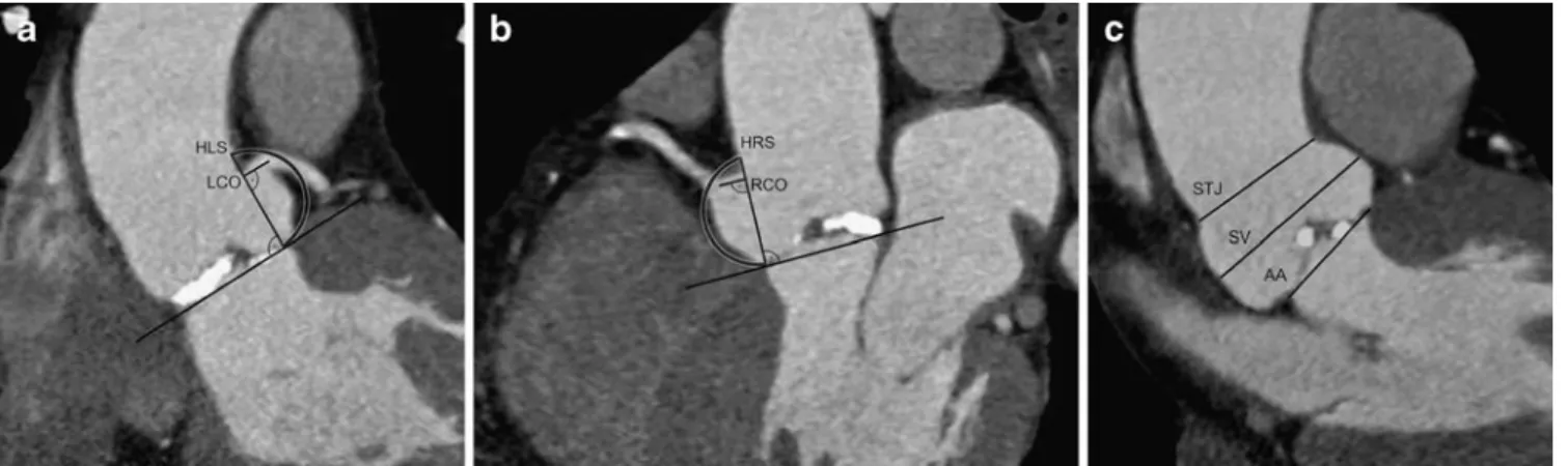
Coronary ostial locations The left coronary ostium (LCO) was located on an oblique coronal reformation orientated orthogonally to the plane of the aortic annulus (AA) (Fig.1a). The right coronary ostium (RCO) was located on an oblique sagittal reformation. Measurements were taken perpendicular from the base of the AA to the center of each coronary ostium (Fig. 1b).
Sinus heights The height of the left coronary sinus (HLS) was determined by moving through reformations parallel to the previously used coronal oblique view (Fig. 1a). Similarly, the height of the right coronary sinus (HRS) was visualized on parallel sagittal oblique reformations (Fig. 1b). All height measurements of the sinuses were taken perpendicular to the plane of the AA.
Aortic root dimensions Aortic root dimensions included the diameter of the AA, the sinus of Valsalva (SV), and sino-tubular junction (STJ). All measurements were taken on an oblique coronal reformation parallel to the previously used coronal oblique views (Fig.1c).
Aortic valve calcification Aortic valve calcification was semi-quantitatively graded by using a previously pub-lished scale [23]: grade 1, mild with small isolated spots of calcification; grade 2, moderate with multiple larger spots of calcification; and grade 3, heavy with extensive calcification of the aortic cusps.
All analyses were performed by a radiologist with 3 years of experience in cardiovascular imaging.
Inter-observer and intra-observer agreement
To test for inter-observer reliability of CT measurements, data from 50 patients (25 random patients of each group) were analyzed by a second radiologist (7 years of experience in cardiovascular imaging). Planes for measure-ments were reformatted by each reader separately. To test for intra-observer variability, the same reader re-analyzed the same 50 datasets after 1month.
Statistical analysis
Numerical values were expressed as frequencies or percen-tages. Age, heart rates, BMI, BSA, ostial locations (i.e., LCO and RCO), sinus heights (i.e., HLS and HRS), dimensions of the aortic root (i.e., AA, SV, and STJ) were normally distributed (as evidenced by the Kolmogorov-Smirnov test) and thus were presented as means ± standard deviations (SDs).
Fig. 1 Measurements of coronary ostial locations and sinus heights with CT. Coronary ostial locations of the LCO were located on an oblique coronal reformation (a) orientated orthogonally to the plane of AA. LCO measurements were made from the base of the AA perpendicularly up to the center of the coronary ostium. Measure-ments for the left sinus height (HLS) were obtained by moving
through parallel planes to visualize its distal attachment (semicircle). The location of the RCO and HRS were measured on an oblique sagittal reformation (b) in a similar fashion. Measurements of the aortic root [i.e., AA, sinus of Valsalva (SV) width, and sino-tubular junction (STJ)] were made on a parallel oblique coronal reformation (c)
Differences in frequencies (i.e., gender, cardiovascular risk factors, LCO or RCO above the STJ) between controls and patients with severe tricuspid AS were assessed using the chi-square test or Fisher’s exact test. Heart rates, BMI, BSA, ostial locations, sinus heights, as well as aortic root dimensions (i.e., AA, SV, and STJ) of controls and patients with severe tricuspid AS were compared using t-tests for unpaired samples. Mean differences between male and female patients were assessed using unpaired t-tests for ostial locations, sinus heights, ostial locations as percen-tage of the sinus height, and aortic root dimensions. Intra-individual comparisons of the LCO, RCO, HLS, and HRS were performed using a paired t-test.
Bland-Altman analysis for intra- and inter-observer agreement was used to assess differences in observations with the mean of observations. Pearsons correlation analysis was used to compare measurements of the different readers and reading sessions as well as to assess the influence of age, BMI and BSA on sinus heights, ostial locations, and aortic root dimensions (i.e., AA, SV, and STJ).
A P value of <0.05 was considered significant. Statistical analysis was performed using commercially available soft-ware (SPSS, release 15.0, Chicago, IL, USA).
Results
All CT measurements could successfully be made in each patient. All patients were in a sinus rhythm with a heart rate during CT of 69±15 bpm (range 47–95 bpm), with no significant difference present between controls and patients with AS (P=0.67).
Of the 100 patients with AS, 42 patients (42%) suffered from additional aortic regurgitation (mild, n=34; moderate, n=6; severe, n=2).
Inter-observer and intra-observer agreement
Bland-Altman analysis revealed minimal differences between the measurement of both observers (i.e., LCO= 0.6±1.7 mm, HLS=0.3±1.6 mm, RCO=0.5±1.8 mm, HRS=0.08±1.6 mm, AA=1.4±1.8 mm, SV=0.8±1.5 mm,
and STJ=1.1±1.4 mm). Minimal differences were also observed when testing intra-observer agreement (i.e., LCO=0.06±1.4 mm, HLS=0.5±1.2 mm, RCO=0.3± 1.5 mm, HRS=0.3±1.3 mm, AA=1.3±0.9 mm, SV=0.9± 1.1 mm, and STJ=1.5±1.1 mm). Inter-observer correlation coefficients ranged from r=0.80–0.93 (P<0.01); and intra-observer correlation coefficients ranged from r=0.79–.95 (P<0.001). Because both the inter-observer and the intra-observer agreements were high, the remaining 150 datasets were analyzed by only one reader.
CT data analysis
Controls
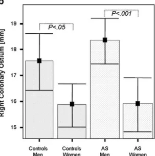
Absolute values of the LCO, RCO, HLS, and HRS as well as the LCO and RCO as percentages of the sinus heights for controls are demonstrated in Table2and Fig.3. There were significant differences between the left and right coronary ostial locations (P<0.001). No differences were found between male and female patients for the LCO (P=0.34), but the RCO showed a significantly (P<0.05) higher location in male patients (Fig.2).
The HLS and the HRS were similar, with no significant difference between the two sides (P=0.08). Significant differences were found between men and women in regard to the HLS (P<0.001) and the HRS (P<0.05), with a higher location in men for both sides.
The LCO was located above the STJ in 4/100 patients (4%), the RCO in 9/100 patients (9%), respectively. A significant difference between the LCO and RCO as percentages of sinus heights (P<0.0001) was found; no differences were found between men and women for the LCO (P=0.28) and the RCO (P=0.57) as percentages of the corresponding sinus heights.
Significant dependencies on BSA were found for the LCO (r=0.21, P<0.05), RCO (r=0.31, P<0.01), HLS (r= 0.50, P<0.001), and HRS (r=0.87, P<0.001). No correla-tions were found between the BSA and both the LCO and RCO as a percentage of sinus height (P=0.22 and P=0.72). No significant dependencies (P>0.05) on age or BMI were found for any of these parameters.
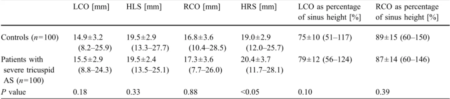
Table 2 Coronary ostial locations and sinus heights in controls and patients with severe tricuspid AS. Data are means ± SDs. Numbers in parentheses are ranges
LCO [mm] HLS [mm] RCO [mm] HRS [mm] LCO as percentage of sinus height [%] RCO as percentage of sinus height [%] Controls (n=100) 14.9±3.2 (8.2–25.9) 19.5±2.9 (13.3–27.7) 16.8±3.6 (10.4–28.5) 19.0±2.9 (12.0–25.7) 75±10 (51–117) 89±15 (60–150) Patients with severe tricuspid AS (n=100) 15.5±2.9 (8.8–24.3) 19.5±2.4 (13.5–25.1) 17.3±3.6 (7.7–26.0) 20.4±3.7 (11.7–28.1) 79±12 (56–124) 87±14 (60–146) P value 0.18 0.33 0.88 <0.05 0.10 0.39
Aortic root dimensions of controls are demonstrated in Table 3. Women showed significant smaller values for all parameters (P<0.05) except for the AA/BSA (P=0.11) when compared with men. Significant dependencies on BSA were found for the AA (r=0.55, P<0.001), SV (r= 0.54, P<0.001), and STJ (r=0.52, P<0.001). No correla-tions were found between any of these parameters and BMI or age (P>0.05).
Patients with severe tricuspid AS
All 100 patients with AS (100%) had calcifications of the aortic cusps. Six patients (6%) were classified to have calcifications of grade 1, 23 to have grade 2 (23%), 52 to have grade 3 (52%), and 19 patients to have grade 4 (19%). Coronary ostia locations for patients with AS are demonstrated in Table2and Fig.3. The RCO was significantly (P<0.001)
higher than the LCO. Significant differences were also found between men and women in regard to the LCO (P<0.01) and RCO (P<0.001), with a higher location for men (Fig.2).
HLS and HRS were similar, with no significant difference between the two sides (P=0.13). Significant differences were found between men and women for HLS (P<0.001) and HRS (P<0.001).
The LCO was located above the STJ in 6/100 patients (6%), the RCO in 8/100 patients (8%), respectively. In one patient (1%), both the LCO and RCO were located above the STJ. The LCO as a percentage of HLS was significantly different from the RCO as a percentage of HRS (P< 0.0001). No differences were found between men and women for the LCO (P=0.51) and RCO (P=0.28) as percentages of the sinus heights.
Weak but significant correlations were found between the BMI and both the LCO (r=−0.20, P<0.05) and the RCO as a percentage of the HRS (r=0.20, P<0.001). In Fig. 2 Gender-specific plot of left (LCO, a) and right ostial
locations (RCO, b) in controls and patients with severe tricuspid AS. Regarding the LCO of controls, men and women had similar measures (P=0.34), whereas male patients with severe tricuspid AS showed significant higher locations (P<0.01) when compared with
female patients. With respect to the RCO, male controls (P<0.05) and male patients with AS (P<0.001) showed significantly higher locations of the RCA than women. Locations of the LCO (P=0.18) and RCO (P=0.33) were similar in controls and in patients with severe tricuspid AS
Table 3 Aortic root dimensions in controls and in patients with severe tricuspid AS. Data are means ± SDs. Numbers in parentheses are ranges
AA [mm] SV [mm] Sino tubular junction [mm]
AA/BSA [mm/m2] SV/BSA [mm/m2] Sino tubular
junction/BSA [mm/m2] Controls (n=100) 23.0±3.1 (16.2–29.6) 33.5±4.2 (26.2–43.8) 25.9±3.3 (18.2–32.5) 12.6±1.6 (8.6–18.3) 18.4±2.2 (13.8–25.6) 14.2±1.9 (10.4–19.8) Patients with severe tricuspid AS (n=100) 24.5±3.2 (16.4–31.7) 34.0±3.6 (25.2–42.5) 27.2±3.5 (21.0–36.8) 13.4±2.0 (8.2–18.6) 18.7±2.6 (13.4–24.5) 15.0±2.4 (10.2–25.8) P value <0.01 0.31 <0.01 <0.01 0.31 <0.05
contrast to controls, no correlations were observed for any parameter with BMI, BSA, or age (P>0.05).
Absolute values of the AA, SV, and STJ as well as those indexed to the BSA are listed in Table3. Female patients showed significantly (P<0.05) smaller values for all parameters except for the STJ/BSA (P=0.16) when compared with male patients. No significant dependencies (P>0.05) on BSA, BMI, or age were revealed for any of the aortic root dimensions in patients with AS.
Comparison between controls and patients with severe tricuspid AS
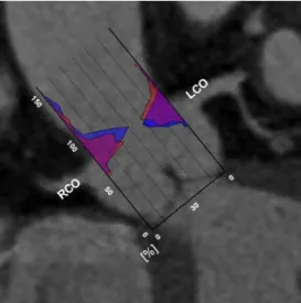
Comparisons of controls and patients with AS in regard to the LCO, RCO, HLS, and HRS are shown in Table2. The rates of patients with the LCO or RCO above the STJ in both groups of controls and patients with AS were similar (P=0.06 and P=0.37). LCO (P=0.10) and RCO as percentages of the sinus heights were similar in controls and patients with AS, but showed a large distribution in both groups (Fig.3).
Patients with AS had significantly larger values of the AA (P<0.01) and STJ (P<0.01) when compared with the controls (Table3, Fig.4). Significant differences for the AA (P<0.05), STJ (P<0.05), AA/BSA (P<0.05), and STJ/ BSA (P<0.05) also were present when comparing controls with patients with AS but no aortic regurgitation. Levels of significances were maintained after normalization to the BSA (Table3).
Discussion
TAV implantation provides the potential for minimally invasive valve replacement in patients with severe tricuspid AS who are not candidates for open surgery because of serious comorbidities [4, 5, 7, 8, 24]. The first human implantation of TAV was performed transapically in a 57-year-old man with calcific AS in 2002 by Cribier et al. [10].
Boudjemline and Bonhoeffer [11] pointed out that the precise placement of TAV is crucial: locations too high above the annulus result in coronary ostial obstruction, whereas locations too low can negatively impact the left ventricular and/or mitral valve function [11]. Potential obstruction of the coronary orifices arises from the aortic cusps being pushed up against the coronary orifices or by the TAV itself [14]. Because obstruction of the coronary arteries represents a risk with potentially catastrophic consequences, the locations of the coronary ostia should be determined to maintain a secure distance of the stent [4,12,14].
Another potential complication of TAV may be para-valvular regurgitation [4,10,11]. Regurgitant blood flow may occur after valve implantation in patients who receive a TAV that is too small for the individual’s AA [6]. Minimizing paravalvular insufficiency by correct position-ing and sizposition-ing is therefore important to improve the patient outcome [6].
In order to anticipate these complications, imaging is performed in patients who are under consideration for valve surgery. CT may be performed and is useful to confirm AS severity and to define valve anatomy [25–27]. This study provides data of coronary ostia locations and aortic root dimensions in patients with severe tricuspid AS and demonstrates differences in these patients when compared with the controls.
Our in-vivo measurements of the HLS and right sinus heights in controls have general agreement with previous post-mortem studies [28, 29]. In controls, the LCO and
Fig. 4 Scheme visualizing the differences of coronary ostial locations as percentages of sinus heights and absolute aortic root dimensions of patients with severe tricuspid AS (red) in proportion to controls (blue). Significant larger measures were revealed for the AA and the STJ in patients with severe tricuspid AS
Fig. 3 Distribution of coronary ostial locations as a percentage of sinus heights overlaid on multiplanar CT reconstruction visualizing the anatomy of the aortic root in a symptomatic patient with severe tricuspid AS. No differences were found in regard to the ostial locations when patients with severe tricuspid AS (red area) were compared with patients without valvular disease (blue area). Note the lower location of the LCO when compared with the ostium of the right coronary artery (RCO)
RCO were located on average at 14.9 mm and 16.8 mm, respectively, above the AA. These values are in concor-dance with those as obtained by Jatene et al. [30] in patients being over 40-years old (LCO: 14.9 mm, RCO: 16.1 mm). The lower location for the LCO in our study is also in accordance with former studies [30,31].
When comparing controls and patients with AS, we found similar sinus heights with the LCO located closer to the AA when compared with the right side. Thus, it appears that the longitudinal anatomy of the coronary ostia and aortic root remains preserved in severe tricuspid AS. Pertaining to TAV, however, the lower location of the LCO per se could be a limiting factor for the design of TAV. The large distribution of coronary ostial heights of 7.7– 28.5 mm (representing 56–150% of sinus heights) suggests preprocedural CT to be important for determining the precise position of the coronary ostia.
The AA and STJ were significantly larger in patients with AS when compared with the controls. This indicates a transverse remodelling of the aortic root, which is still present in patients with AS but without additional aortic regurgitation. This is in line with Crawford et al. [32], who showed that aortic root dilation may regularly be recognized in AS. A possible explanation for the larger aortic root sizes could be given by the higher age of patients with AS when compared with the controls. This is also suggested by the Framingham heart study that recognized age-related differences of aortic root sizes [33]. Additionally, and similar to our study, women had smaller aortic root dimensions [33]. Furthermore, all measurements of the aortic root in controls were sig-nificantly correlated to the BSA, whereas no such corre-lation was found in patients with AS.
A major advantage of CT for the preprocedural planning of TAV lies in its ability to provide—within the same examination—accurate information on concomitant coro-nary artery disease [15–17]. In addition, quantification of aortic valve calcification with CT represents a
comple-mentary information to the severity of AS and provides independent outcome information for these patients [34].
Limitations
First, values are derived from a patient group consisting of Caucasian patients, and hence do not account for possible differences in other ethnic groups. Second, patients with AS were significantly older than controls. This selection bias, however, is generated by the fact that AS generally occurs in the elderly population. Third, results from TTE in the assessment of AS were not compared with results from CT, but has been amply documented in the literature [25, 26, 35, 36]. Finally, the impact of the preprocedural anatomical assessment on patient outcome was not assessed in the present study.
Conclusion
This study provides data on coronary ostial locations and aortic root geometry in patients with severe tricuspid AS in comparison with controls having a normal valve function. In patients with severe tricuspid AS, the longitudinal dimensions including the coronary ostia and sinus heights are maintained, whereas a transverse remodelling of the aortic root including the diameter of the AA and STJ occurs. Considering the large distribution of coronary ostial locations both in controls and in patients with AS, as well as the dilated aortic root in patients with AS, preprocedural cardiac CT is recommended prior to TAV implantation in each individual patient.
Acknowledgements This study was supported by the National Center of Competence in Research, Computer Aided and Image Guided Medical Interventions of the Swiss National Science Foundation.
References
1. Iung B, Baron G, Butchart EG et al (2003) A prospective survey of patients with valvular heart disease in Europe: The Euro Heart Survey on Valvular Heart Disease. Eur Heart J 24:1231– 1243
2. Kvidal P, Bergstrom R, Horte LG et al (2000) Observed and relative survival after aortic valve replacement. J Am Coll Cardiol 35:747–756
3. Bonow RO, Carabello BA, Chatterjee K et al (2006) ACC/AHA 2006 guide-lines for the management of patients with valvular heart disease: a report of the American College of Cardiology/ American Heart Association Task Force on Practice Guidelines (writing Committee to Revise the 1998 guide-lines for the management of patients with valvular heart disease) developed in collaboration with the Society of Cardiovascular Anesthesiologists en-dorsed by the Society for Cardiovas-cular Angiography and Interventions and the Society of Thoracic Surgeons. J Am Coll Cardiol 48:e1–e148
4. Lutter G, Ardehali R, Cremer J et al (2004) Percutaneous valve replace-ment: current state and future prospects. Ann Thorac Surg 78:2199–2206 5. Cribier A, Eltchaninoff H, Tron C et al
(2006) Treatment of calcific aortic ste-nosis with the percutaneous heart valve: mid-term follow-up from the initial feasibility studies: the French experience. J Am Coll Cardiol 47:1214–1223 6. Webb JG, Chandavimol M, Thompson
CR et al (2006) Percutaneous aortic valve implantation retrograde from the femoral artery. Circulation 113:842–850
7. Walther T, Simon P, Dewey T et al (2007) Transapical minimally invasive aortic valve implantation: multicenter experience. Circulation 116:I240–I245 8. Ye J, Cheung A, Lichtenstein SV et al (2007) Six-month outcome of transa-pical transcatheter aortic valve implan-tation in the initial seven patients. Eur J Cardiothorac Surg 31:16–21
9. Eltchaninoff H, Zajarias A, Tron C et al (2008) [Transcatheter aortic valve im-plantation: technical aspects, results and indications]. Arch Cardiovasc Dis 101:126–132
10. Cribier A, Eltchaninoff H, Bash A et al (2002) Percutaneous transcatheter im-plantation of an aortic valve prosthesis for calcific aortic stenosis: first human case description. Circulation 106:3006– 3008
11. Boudjemline Y, Bonhoeffer P (2002) Steps toward percutaneous aortic valve replacement. Circulation 105:775–778 12. Lutter G, Kuklinski D, Berg G et al
(2002) Percutaneous aortic valve re-placement: an experimental study. I. Studies on implantation. J Thorac Cardiovasc Surg 123:768–776 13. Boudjemline Y, Bonhoeffer P (2003)
Percutaneous valve insertion: a new approach? J Thorac Cardiovasc Surg 125:741–742 author reply 742–743 14. Huber CH, Tozzi P, Corno AF et al (2004) Do valved stents compromise coronary flow? Eur J Cardiothorac Surg 25:754–759
15. Gilard M, Cornily JC, Pennec PY et al (2006) Accuracy of multislice com-puted tomography in the preoperative assessment of coronary disease in pa-tients with aortic valve stenosis. J Am Coll Cardiol 47:2020–2024
16. Meijboom WB, Mollet NR, Van Mieghem CA et al (2006) Pre-operative computed tomography coro-nary angiography to detect significant coronary artery disease in patients referred for cardiac valve surgery. J Am Coll Cardiol 48:1658–1665
17. Scheffel H, Leschka S, Plass A et al (2007) Accuracy of 64-slice computed tomography for the preoperative detec-tion of coronary artery disease in patients with chronic aortic regurgita-tion. Am J Cardiol 100:701–706 18. Alkadhi H, Desbiolles L, Husmann L et
al (2007) Aortic regurgitation: assess-ment with 64-section CT. Radiology 245:111–121
19. Lu TL, Huber CH, Rizzo E et al (2008) Ascending aorta measurements as as-sessed by ECG-gated multi-detector computed tomography: a pilot study to establish normative values for trans-catheter therapies. Eur Radiol. doi:10.1007/s00330-008-1182-8
20. Mosteller RD (1987) Simplified calcu-lation of body-surface area. N Engl J Med 317:1098
21. Leschka S, Scheffel H, Desbiolles L et al (2007) Image quality and recon-struction intervals of dual-source CT coronary angiography: recommenda-tions for ECG-pulsing windowing. In-vest Radiol 42:543–549
22. Stolzmann P, Scheffel H, Schertler T et al (2008) Radiation dose estimates in dual-source computed tomography coronary angiography. Eur Radiol 18:592–599
23. Rosenhek R, Binder T, Porenta G et al (2000) Predictors of outcome in severe, asymptomatic aortic stenosis. N Engl J Med 343:611–617
24. Cribier A, Eltchaninoff H, Tron C et al (2004) Early experience with percuta-neous transcatheter implantation of heart valve prosthesis for the treatment of end-stage inoperable patients with calcific aortic stenosis. J Am Coll Cardiol 43:698–703
25. Alkadhi H, Wildermuth S, Plass A et al (2006) Aortic stenosis: comparative evaluation of 16-detector row CT and echocardiography. Radiology 240:47– 55
26. LaBounty TM, Sundaram B, Agarwal P et al (2008) Aortic valve area on 64-MDCT correlates with transesophageal echocardiography in aortic stenosis. AJR Am J Roentgenol 191:1652–1658
27. Feuchtner GM, Muller S, Bonatti J et al (2007) Sixty-four slice CT evaluation of aortic stenosis using planimetry of the aortic valve area. AJR Am J Roentgenol 189:197–203
28. Berdajs D, Lajos P, Turina M (2002) The anatomy of the aortic root. Cardi-ovasc Surg 10:320–327
29. Swanson M, Clark RE (1974) Dimen-sions and geometric relationships of the human aortic valve as a function of pressure. Circ Res 35:871–882 30. Jatene MB, Monteiro R, Guimaraes
MH et al (1999) Aortic valve assess-ment. Anatomical study of 100 healthy human hearts. Arq Bras Cardiol 73:75– 86
31. Cavalcanti JS, de Melo NC, de Vasconcelos RS (2003) Morpho-metric and topographic study of coronary ostia. Arq Bras Cardiol 81:359–362 355–358
32. Crawford MH, Roldan CA (2001) Prevalence of aortic root dilatation and small aortic roots in valvular aortic stenosis. Am J Cardiol 87:1311–1313 33. Vasan RS, Larson MG, Benjamin EJ et
al (1995) Echocardiographic reference values for aortic root size: the Fra-mingham Heart Study. J Am Soc Echocardiogr 8:793–800 34. Messika-Zeitoun D, Aubry MC,
Detaint D et al (2004) Evaluation and clinical implications of aortic valve calcification measured by electron-beam computed tomography. Circulation 110:356–362
35. Pouleur AC, le Polain de Waroux JB, Pasquet A et al (2007) Aortic valve area assessment: multidetector CT compared with cine MR imaging and transtho-racic and transesophageal echocardiog-raphy. Radiology 244:745–754 36. Saam T, Oberhoffer M, Rist C et al
(2008) [Assessment of aortic stenosis after aortic valve replacement: com-parative evaluation of dual-source CT and echocardiography]. Rofo 180:553– 560