HAL Id: in2p3-00025918
http://hal.in2p3.fr/in2p3-00025918
Submitted on 21 Dec 2006
HAL is a multi-disciplinary open access
archive for the deposit and dissemination of
sci-entific research documents, whether they are
pub-lished or not. The documents may come from
teaching and research institutions in France or
abroad, or from public or private research centers.
L’archive ouverte pluridisciplinaire HAL, est
destinée au dépôt et à la diffusion de documents
scientifiques de niveau recherche, publiés ou non,
émanant des établissements d’enseignement et de
recherche français ou étrangers, des laboratoires
publics ou privés.
Experimental evidence for subshell closure in
8
He and
indication of a resonant state in
7
He below 1 MeV
F. Skaza, V. Lapoux, N. Keeley, N. Alamanos, E. C. Pollacco, F. Auger, A.
Drouart, A. Gillibert, D. Beaumel, E. Becheva, et al.
To cite this version:
F. Skaza, V. Lapoux, N. Keeley, N. Alamanos, E. C. Pollacco, et al.. Experimental evidence for
subshell closure in
8He and indication of a resonant state in
7He below 1 MeV. Physical Review C,
American Physical Society, 2006, 73, pp.044301. �10.1103/PhysRevC.73.044301�. �in2p3-00025918�
in
7He below 1 MeV
F. Skaza1 , V. Lapoux1 ,∗ N. Keeley1 , N. Alamanos1 , E.C. Pollacco1 , F. Auger1 , A. Drouart1 , A. Gillibert1 , D. Beaumel2, E. Becheva2, Y. Blumenfeld2, F. Delaunay2, L. Giot3, K.W. Kemper4, L. Nalpas1, A. Obertelli1,A. Pakou5, R. Raabe1,† P. Roussel-Chomaz3, J-L. Sida1, J.-A. Scarpaci2, S. Stepantsov6, and R. Wolski7
1
CEA-SACLAY DSM/DAPNIA/SPhN F-91191 Gif-sur-Yvette, France
2
Institut de Physique Nucl´eaire, IN2P3-CNRS, F-91406 Orsay, France 3
GANIL, Bld Henri Becquerel, BP 5027, F-14021 Caen Cedex, France
4
Department of Physics, Florida State University, Tallahassee, Florida 32306-4350, USA
5
Department of Physics, University of Ioannina, 45110 Ioannina, Greece
6
JINR, FLNR 141980 Dubna, Moscow region, Russia and
7
The Henryk Niewodniczanski Institute of Nuclear Physics, Krakow, Poland (Dated: March 21, 2006)
The spectroscopy of the unstable 8
He and unbound7
He nuclei is investigated via the p(8
He,d) transfer reaction with a 15.7A.MeV 8
He beam from the SPIRAL facility. The emitted deuterons were detected by the telescope array MUST. The results are analyzed within the coupled-channels Born approximation framework, and a spectroscopic factor C2
S = 4.4±1.3 for neutron pickup to the7
Heg.s is deduced. This value is consistent with a full p3/2 subshell for 8
He. Tentative evidence for the first excited state of7
He is found at E* = 0.9±0.5 MeV (width Γ = 1.0 ± 0.9 MeV). The second one is observed at a position compatible with previous measurements, E*=2.9±0.1 MeV. Both are in agreement with previous separate measurements. The reproduction of the first excited state below 1 MeV would be a challenge for the most sophisticated nuclear theories.
PACS numbers: 25.60.-t,21.10.-k,21.10.Jx, 24.10.Eq
I. INTRODUCTION
The binding and excitation energies of the light weakly-bound neutron-rich nuclei are crucial benchmarks for microscopic models. In particular, since the drip-line nucleus8
He has the highest N/Z amongst the bound nuclei, its spectroscopy can help to clarify the isospin-dependent term in the most recent microscopic calcula-tions. The theoretical description of 8He seems
possi-ble either within the framework of microscopic cluster models (resonating group model RGM [1], cluster shell model [2]) or a large-basis no-core shell model (NCSM) space [3]. In the cluster models, the structure of 8He
is described as an alpha core surrounded by 4 neutrons, which constitute a skin or halo. Alternatively, the ground state (gs) of8He may be described as a mixing of the
fol-lowing configurations : 6
He(2+
)+2n and6
He(0+
)+2n [4]. In the simple shell model (SM) picture, the gs of 8He
corresponds to a closed 0p3/2 shell, giving a sum rule estimate of the spectroscopic factor (SF) of C2S = 4.0
for pickup of a neutron to the gs of 7He. In order to
test whether these pictures hold for8
He, we have chosen the (p,d) reaction as a natural spectroscopic tool, also allowing the spectroscopy of7He to be investigated.
7
He is a particle unstable nucleus previously observed at Riken using the p(8He,d) reaction at 50A.MeV [5].
∗E-mail address: vlapoux@cea.fr
†permanent address: IKS, University of Leuven, B-3001 Leuven, Belgium
In this pioneering work, the excitation spectrum for7He
was deduced, with a low-lying resonant excited state at 3.3(3) MeV above the6He+n 0.44 MeV threshold being
observed (E*=2.9 MeV, width Γ = 2.2(3) MeV). This excited state, which mainly decays into α+3n, was in-terpreted as a p1/2 neutron coupled to the 6He core in
its unbound 2+ excited state, and a tentative spin
as-signment of 5/2− was made [5]. This resonance was
also observed at E*=2.95(10) MeV (Γ = 1.9(3) MeV), in the 9
Be(15
N,17
F)7
He reaction [6]. In the break-up experiment [7] with a 8
He beam on a Carbon target, the relative energy of the 6He and neutron fragments
was reconstructed and the fitted spectrum supported the assumption of a resonant state at E* = 0.6(1) MeV (Γ = 0.75(8) MeV). Based on the observation of the6He
fragment, thus excluding the 5/2− configuration
associ-ated to the unbound6He(2+) core, it was discussed as a
possible 1/2− state [7]. Recently however, the low-lying
excited states of7He were studied via the isobaric analog
states (IAS) of7Li [8]. The authors do not confirm the
above result, while they report that the analog of a very broad resonance (1/2−,T=3/2) is located at an
excita-tion energy above 2.2 MeV in7He. From the theoretical
point of view, recent calculations [1, 3, 9] agree in pre-dicting at least 2 resonances, 1/2− and 5/2−, above the
3/2− gs. Microscopic models do not predict any positive
parity states at low energy. In the RGM [1], the 1/2−
and 5/2− states are given by the coupling of a 0p1/2
neutron to the 6He core, in its gs or 2+ excited state,
respectively. We present here the8He(p,d) reaction
per-formed at 15.7A.MeV with better energy resolution and larger angular coverage than previously. The deduced
ex-2 citation spectrum for7He is discussed together with the
analysis of the differential distributions p(8He,d)7He g.s
and p(8He,d)7He∗. It should be noted that the 1/2− and
5/2−excited states were not observed simultaneously in
previous experiments due to the experimental resolution or the selectivity of the observation process.
II. EXPERIMENTAL SET-UP AND ANALYSIS
OF THE EXCITATION SPECTRA
The8He beam impinged on a proton target and the
di-rect reactions were studied by detecting the light recoil. We adopted the same techniques as for the6He(p,p’)
ex-periment presented in [10]. The8He beam was produced
by the ISOL technique and accelerated to 15.7A.MeV by the CIME cyclotron at the SPIRAL facility [11], with no contaminants. The maximum (average) intensity in the experiment was 14000 (5000) p/s. The proton tar-get was a 8.25 mg/cm2 thick polypropylene (CH
2)n foil.
The beam profile and incident angle on the target were monitored event by event by two low-pressure multi-wire beam tracking detectors, CATS [12], located upstream of the target. The whole set-up is shown in Fig.1. The
Plastic Plastic Wall p Polypropylene target CsI SiLi (x,y) Si-strips CATS Faraday beam He 8 MUST He 8 He* 8 or He, 6 He 4 300 µm 3 mm 15 mm
FIG. 1: Experimental set-up. The MUST array was assem-bled in a wall configuration, located 15 cm from the target. It was placed in 2 positions with the vertical axis rotated by an angle of 50◦and 65◦ with respect to the beam axis in the
laboratory frame.
MUST detector [13], an array of 8 three-stage telescopes, detected the recoil deuteron in coincidence with a wall of plastic scintillators measuring the projectile remnant. One module consisted of a 6x6 cm2x-y position sensitive
Si-strip detector (300µm) backed with a Si(Li) (3 mm) and a CsI scintillator. Each strip detector had a mini-mum energy threshold of 0.5 MeV with angular and en-ergy resolutions of 0.4◦ and 50 keV. Proton, deuteron
and triton particles were identified at energies below 6, 8 and 9 MeV, respectively via the correlation between the time-of-flight (TOF) and the energy deposited in the Si-strip stage. For higher energies, the standard ∆E-E technique was applied. The beam and reaction fragments were detected in the forward direction either in a small plastic scintillator at zero degrees or for larger center of mass (c.m.) angles in the plastic wall covering an area
of 50x48 cm2at 75.5 cm from the target. The wall gave
time and energy measurements resulting in mass reso-lution δM/M ' 19%, sufficient to identify roughly 8He
nuclei and substract background in the plastic signals, but not to discriminate6
He from8
He or from4
He. The kinematics of the total energy of the light particle with respect to the scattering angle in the laboratory frame are plotted in Fig. 2a) and b), for events including a pro-ton or a deuteron detected in MUST in coincidence with He isotopes in the plastic wall, respectively. From the proton or deuteron identification and with the kinemati-cal plots of the reaction of interest elastic, inelastic (p,p’) or (p,d) were selected. The elastic data extend from 20 to 110◦
c.m., the transfer data from 27 to 85◦c.m.. The total
number of incident 8
He particles on the (CH2)n target
was 8.17 108
. The excitation spectrum for8
He and the differential cross sections for8He(p,p’) will be presented
in a forthcoming article. The excitation energy spectrum for7He was calculated by the missing mass method from
the measured energy and angle of the scattered deuteron. First, the excitation spectrum was constructed by consid-ering the whole statistics for the p(8He,d) reaction,
re-taining only few events associated to small gates. These gates were chosen to exclude 4He or 6He thus allowing
a strict identification of the 6He (gate denoted ”a”) or 4
He (gate ”b”) in the plastic wall as shown by Fig. 3. The whole statistics, for all c.m. angles, were considered to discuss qualitatively the 7He spectrum respected to
the6He and 4He fragments. Afterwards, for the
quan-titative discussion of the7He spectrum, we will consider
thin c.m. slices in the (p,d) kinematics. We will need to consider a larger gate (noted ”c”) in order to have more statistics in the 7He spectrum. Even if the
discrimina-tion between the4He and6He fragments is not achieved,
having the deuteron in coincidence with either an6He or 4
He fragment will be enough in the second step.
(deg) lab θ 30 40 50 60 70 80 E (MeV) 0 5 10 15 20 25 30 35 40 0 5 10 15 20 25 30 35 40 He,p) 8 p( 3.6 MeV * He 8 He,p’) 8 p( a) (deg) lab θ 30 40 50 60 70 80 0 5 10 15 20 25 30 35 40 0 1 2 3 4 5 6 7 He 7 He,d) 8 p( b)
FIG. 2: Kinematical plot of events for : a) the8
He elastic, inelastic reactions on protons and b) (p,d) transfer. The cal-culated lines of the reactions are drawn to guide the eye, the dashed line in a) represents the (p,p’) to an excited state at 3.6 MeV.
First, the resulting excitation spectra for7
He, associ-ated to gates a and b are displayed in Fig. 4a. and 4b, respectively. The energies are referred to the gs of 7He
the6He(2+)+n threshold (at E*=1.36 MeV compared to 7He gs), a peak corresponding mainly to the7He gs is
ob-served. Above the4He + 3n threshold (at 0.535 MeV), a
broad resonance is observed (Fig. 4b) at E*= 2.9(1) MeV with a width Γ = 2.1(8) MeV. These parameters are in agreement with the values found in [5, 6]. The ratio, 0.6±0.3, between4He and total yields is also compatible
with the previously measured branching ratio Γα+3n/Γtot
=0.7±0.2 [5]. Following the theoretical indication of [1], the resonances observed mainly in coincidence with 6
He or4He would be attributed to 3/2−gs and 5/2−excited
state, respectively. G Temps Cats2 Pl3 8000 8500 9000 9500 10000 10500 11000 11500 12000 (u.a.) D *Pl3 G Pl3 0 2000 4000 6000 8000 10000 12000
(a)
8HeTime of flight Cats-Plastics(arb. units)
C h a rg e P la st ic s (a rb . u n it s) 4He b. 4He+6He c. 6He a.
FIG. 3: Correlation spectrum of the charge in the plastic wall versus TOF. The data correspond to the events of 8
He on proton, for which a light charged particle (p,d, or triton) was
detected in MUST. TOF is between the 2nd beam detector
CATS and the plastics. The contours (gates) indicated in the plot are discussed in the text.
(MeV) He 7 * E c) -1 0 1 2 3 4 5 6 0 50 100 150 200 250 -1 0 1 2 3 4 5 6 0 5 10 15 20 25 30 35 40 45 50 C 1 2 3 B Counts / 200 keV 0 5 10 15 20 25 a) d+6He (MeV) He 7 * E -2 -1 0 1 2 3 4 5 6 0 2 4 6 8 10 12 d+4He b)
FIG. 4: Excitation spectra for7
He with following conditions (see also the text) : a) coincidence with the deuteron and the
6
He fragment in the plastic wall ; b) coincidence with the4
He ; c) the area 1 and 2, 3, B and C correspond to the resonances for the gs and 2 excited states, to the physical and Carbon backgrounds, respectively. The thick solid curve is the total fit including all contributions. The error bars on the points are statistical errors. The insert shows a zoom.
As a second step, to extract the characteristics of the possible resonances, we selected a thin angular slice where the energy straggling can be controlled. The (p,d) reaction was only selected by requiring the deuteron in coincidence with a He fragment (either 4
He or 6
He, as shown in gate ”c” of Fig 3).
We constructed the 7He excitation energy spectrum
for angles between 50 and 60◦
c.m., where the FWHM
resolution in excitation energy is δE∗ = 590 (14) keV.
The corresponding spectrum is presented as points in Fig. 4.c. The shape of the continuum background con-tribution to this spectrum is given by the area B. It was determined by a Monte-Carlo simulation of the physical background produced by few-body kinematics with sev-eral decay channels, and filtered by the experimental re-sponse. The ingredients of the simulation were the phase space calculations of the reaction channels, the detection efficiency and angular acceptance of the telescopes and plastic wall, and the experimental angular and energy resolutions.
The various reaction channels included in the simu-lation of the physical background are enumerated below, and their associated excitation energy spectra are plotted in Fig. 5. The few-body components are coming from : • 3-body phase space, with channels :
8
He+p → d+n+6He(0+) (curve 1); 8
He+p → d+6He(2+)+n (curve 2); 8
He+p → d+5
He+2
n, with interaction in the final state between4
He and one neutron and between the two re-maining neutrons (curve 3);
• 4-body phase space,
8
He+p → d+4He+2n+n interaction in the final state
be-tween 2 amongst the 3 neutrons (curve 4);
8
He+p → d+5
He+n+n interaction in the final state be-tween4
He and one neutron (curve 5); • 5-body phase space,
8
He+p → d+4He+n+n+n (curve 6);
• 3-body phase space, with interaction in the final state between the 3 neutrons,8
He+p → d+4
He+3
n (curve 7). The notation in means that in the calculation the
i neutrons were considered as correlated in the space framework. The channels producing the same kind of background were fitted by a common distribution. The relative contribution of each channel was arbitrary, and in test calculations each of them was considered alter-natively as the main one to check the region in excita-tion energy in which they might contribute mostly. Our knowledge of the microscopic structure of8He did not
al-low to fix unambiguously the components of the physical background produced by the phase space. But several constraints could be applied to the resulting background curve. The overall normalization was left free and deter-mined in order to superimpose the simulated excitation energy distribution with the measured one. The simu-lated shape (decrease and cuts) of the excitation energy spectrum in the energy region above 5 MeV was in agree-ment with the data, showing that all the experiagree-mental effects (angular acceptance, cuts) were well taken into
4 (MeV) He 7 * E -2 -1 0 1 2 3 4 5 6 7
Counts (arb. units)
0 10 20 30 40 50 60 70 80 90
1
2
2
3
4
5
6
7
FIG. 5: Physical background contributions expressed as a function of the excitation energy for 7
He. Each curve is ob-tained by simulation of the various reaction channels detailed in the text.
account.
The background from the Carbon in the target (area C), was estimated by measuring the reactions of
8He (number of incident particles being 1.8 108) on a
Carbon target. The data were fitted with the continuum background contribution (area B), the Carbon compo-nent, and either one (the gs), 2 (gs and resonance at 2.9 MeV) or 3 resonances. The purpose was to check if our data could support the 7He resonant state obtained
by Meister et al. [7] below 1 MeV.
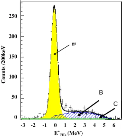
C o u n ts /2 0 0 k eV gs B C E* 7He (MeV) -3 -2 -1 0 1 2 3 4 5 6 250 200 150 100 50 0
FIG. 6: Excitation spectrum for 7
He. Area B and C corre-spond to the physical and Carbon backgrounds, respectively. The thick solid curve is the total fit including gs resonance and all contributions. The error bars on the points are statistical errors.
In the angular range between 50c.m. and 60c.m, the
χ2/N value for the 2-peak fit was 1.5, for the 3-peak
fit it was 0.96. The total curve for the 2-peak fit (no 1 MeV resonance included) is shown for comparison in
Fig. 6. For each angular slice taken into account the same features for the resonances were found and a bet-ter χ2 was obtained when including the 3 resonances
rather than with the 2-peak fit. The result of the best fit obtained with 5 components including 3 resonances is shown in Fig. 4.c. During the fitting procedure, Breit-Wigner (BW) functions folded with the experimental res-olution were adopted to describe the gs (area 1) and the resonances (area 2,3). The 2nd excited state, being em-bedded in the background, was described with position and width of the BW function fixed to previous values : 2.9 MeV and Γ = 2.1 MeV respectively. The param-eters of the gs and the 1st excited state were left free
together with the normalization of the continuum back-ground. The components corresponding to the best fit and total curve are presented in Fig. 4.c. The position of the resonances was found to be independent of angle for various angular slices, which confirms the existence of nuclear states. The resonance curves were unfolded with a Gaussian function to subtract the experimental resolu-tion and are given as BW funcresolu-tions. The gs is located at 0.36(5) MeV above the6He + n threshold, with width
Γ = 0.17(5) MeV.
Even if the resonance cannot be extracted with enough statistics, it is not excluded by our data, and we can indi-cate this 1st excited state at E* = 0.9(5) MeV (1.3 MeV
above threshold) with width Γ = 1.0(9) MeV, which is consistent with the results obtained in [7]. It is in con-trast with the conclusions from [8] based on the observa-tion of the IAS of7
He in 7
Li. Although the background under the 1st excited state is large, it is important to
stress that no physical decay mode was found to be able to produce directly a significant contribution in the re-gion of 1 MeV. It was checked that, when the contribution of a specific reaction channel was enhanced to produce an amount of counts around 1 MeV, then the agreement of the total curve with the data points at higher energies was less good, showing that the excess of counts observed around 1 MeV is rather due to a resonance than to a background effect.
III. ANALYSIS OF THE TRANSFER CROSS
SECTIONS
We now discuss the differential cross sections extracted from our data. Including statistical and systematic er-rors, the normalization of the data for elastic scatter-ing and transfer to the7
He gs has a total uncertainty of ' 15 %, mainly from the subtraction of the background, the acceptance of the detection system (±5%), the target thickness (±5%) and the efficiency in the detection of the incident8
He (±2%).
In order to obtain a SF for neutron pickup to the7
Hegs
from the p(8
He,d) data, a series of coupled-channels Born approximation (CCBA) calculations was carried out using the code Fresco [14]. This analysis requires a p+nucleus potential in the entrance and a d+nucleus
po-tential in the exit channel. The bare d+nucleus popo-tential was the Watanabe type [15], generated by single-folding of proton, neutron + nucleus potentials. Couplings to the deuteron breakup were included in the exit channel us-ing the continuum-discretized coupled-channels (CDCC) formalism, as described in [16] and the transfer step was treated within the usual prior-form distorted-wave Born approximation (DWBA). The calculated p(8He,d)
distri-butions for angles smaller than '30◦
c.m., used to define
the SF, were found to be essentially independent of the choice of entrance potential. This potential was taken from the CH89 parameterization [17] modified to fit to the measured elastic scattering data. In order to test the
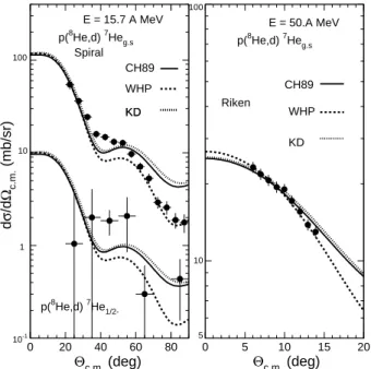
10-1 1 10 102 0 20 40 60 80 10-1 1 10 100 d σ /d Ωc.m. (mb/sr) Θc.m. (deg) E = 15.7 A MeV p(8He,d) 7Heg.s p(8He,d) 7He 1/2-Spiral CH89 WHP KD KD 6 7 8 9 10 20 30 40 50 60 70 80 90 100 0 5 10 15 20 5 10 100 Θc.m. (deg) E = 50.A MeV Riken p(8He,d) 7Heg.s CH89 WHP KD
FIG. 7: Analysis of the (p,d) cross sections to 7
Heg.s and 7
He1/2−obtained at 15.7 and (p,d) 7
Heg.s at 50A.MeV [5]. sensitivity of our results to the choice of n,p+7He
poten-tials, a series of calculations using various global nucleon optical potential systematics was performed. We present results for 3 sets : CH89 for both n and p, Koning and De-laroche (KD) [18] for both neutron and proton, Wilmore and Hodgson [19] for neutron and Perey [20] for pro-ton (WHP). The neutron binding potential for the p/d overlap was given by the Reid soft-core interaction [21], including the small D-state component of the deuteron gs. For the 8
He/7
He overlap we used standard values of R0 = ro × A1/3 fm with ro = 1.25 fm and a = 0.65 fm.
The8He(p,d) transfer data at 15.7A.MeV are presented
in Fig.7 together with the data obtained at 50A.MeV at Riken [5]. The dashed, dotted and solid curves show the cross sections obtained with the WHP, KD, CH89 potentials respectively. At 15.7A.MeV, the best fit C2S
value obtained for each set of potentials corresponds to 4.4 ; at 50A.MeV the C2S values range from 4.0 (KD),
to 4.4 (CH89) and 4.6 (WHP). For a given choice of exit potentials, varying the n+7He binding potential radius
between ro = 1.0 and 1.5 fm was found to lead to vari-ations of up to 20 % in the extracted SF. We therefore obtain a value of C2
S = 4.4 ± 1.3, taking into account all sources of uncertainty. The cross section for transfer to the 0.9 MeV resonance is compatible with an L=1 calcu-lation (spin 1/2− or 3/2−) and upper limit deduced for
the SF is 0.2. Combined with the observation of the6
He fragment, the spin assignment of the resonance found at low energy 0.9(5) MeV is consistent with a 1/2−. The
characteristics of the 7He resonances obtained in
previ-ous experiments are summarized and compared to micro-scopic calculations in Fig. 8.
-1 0 1 2 3 4 5 6 0 2 4 6 8 10 12 14 Eexc ( 7 He) (MeV) experiment Ref. (5) 3/2-thr Γ: 0.16(2) 0.44(2) 2.9(3) Γ=2.2(3) (6) 3/2-0.14(2) 0.44(2) 5/2-2.95(10) Γ=1.9(3) 5.8(3) Γ=4(1) (7) 0.43(2) 0.15(8) 1/2-0.6(1) Γ :0.75(8) this work 3/2-0.36(5) 0.17(5) 1/2-0.9(5) Γ=1.0(9) 5/2-2.9(1) Γ=2.1(8) theory (1) 3/2-0.45 0.12 1/2- 5/2-(3) 3/2-2.33 2.3 3.6 4.4 1/2- 5/2- 3/2-(9) 3/2-0.1(2) 1/2-
5/2-FIG. 8: Experimental and theoretical spectra of 7
He. The threshold energy indicated for the models is deduced from the predicted binding energy for7
He and the energies of the excited states are given with respect to the calculated gs.
Recent predictions of the 1/2− energy given by large
basis NCSM [3] and Quantum Monte-Carlo (QMC) cal-culations [9] are 2.3 and 2.9 MeV, respectively. Note that in Ref. [3] the7He gs is found 2 MeV higher than the
ex-perimental value. In the QMC the agreement is better but the 5/2−is predicted 1.3 MeV higher. In the RGM [1]
the predicted resonant energies are between 2.3-3.8 MeV but could be even lower depending on the assumptions made on the 1/2− resonance.
6
IV. CONCLUSIONS
In the search for the predicted 1/2− 1st excited state,
Golovkov et al. [22] studied the d(6
He,p)7
He reaction; no resonance was found above the gs. But it should be noted that a better microscopic description of the nuclear structure and reactions embedded in the continuum is re-quired in order to understand the measured positions and widths of the excited resonances given by separate exper-iments. Recently, within the recoil corrected continuum SM calculations [23], the conclusion in [8] was found pre-mature. From our work, combined with the conclusions underlined in [23] about the structure of7He(1/2−), this
state could be conceived as more complicated than a sim-ple mixing of6
He(0+
)+n and6
He(2+
)+n configurations. Therefore, these features might not be incompatible : be-ing not simply built on 6He(0+), 7He(1/2−) is not seen
in 6He(d,p) but seen in break-up experiment of 8He ; it
is indicated here in 8He(p,d), and weakly populated due
to its small SF to8
He(0+
). If the observations for the 3 resonant states are confirmed, and compatible with the following quantum numbers : 3/2−, 1/2−, 5/2−, this
se-quence would then be in agreement with a simple SM picture, and well understood in most of the microscopic models. However, the excitation energies are predicted higher than found experimentally. From our results, we can indicate that the 8
He(p,d) reaction is the best one for a tentative measurement of the first excited state in
7He. Combining good 4,6,8He separation as in [5] and
energy resolution with our present technique would help in clarifying the characteristics of this state.
In conclusion, from the8He(p,d)7He reaction, we have
observed the7He gs, the excited state around 3 MeV, and
have indication for the 1stexcited state below 1 MeV. We
have obtained a value for C2S supporting a relatively
pure (p3/2)4
ν configuration for the8He gs.
Acknowledgments
The help of P. Gangnant, J.-F. Libin (GANIL), L. Peti-zon and M. Vilmay (IPN-Orsay), during the preparation of the experiment, is gratefully acknowledged.
[1] J. Wurzer and H.M. Hofmann, Phys. Rev. C 55, 688 (1997).
[2] M.V. Zhukov, A.A. Korsheninnikov, and M.H. Smelberg, Phys. Rev. C 50 R1 (1994).
[3] P. Navr`atil and B.R. Barrett, Phys. Rev. C 57, 3119 (1998).
[4] A.A. Korsheninnikov et al., Phys. Rev. Lett. 90, 082501 (2003).
[5] A.A. Korsheninnikov et al., Phys. Rev. Lett. 82, 3581 (1999).
[6] H.G. Bohlen et al., Phys. Rev. C 64, 024312 (2001). [7] M. Meister et al., Phys. Rev. Lett. 88, 102501 (2002). [8] G. V. Rogachev et al., Phys. Rev. Lett. 92, 232502
(2004).
[9] S.C. Pieper, V.R. Pandharipande, R.B. Wiringa, and J. Carlson, Phys. Rev. C 64, 014001 (2001).
[10] A. Lagoyannis et al., Phys. Lett. B 518, 27 (2001). [11] A. C. Villari et al., Nucl. Phys. A693, 465 (2001). [12] S. Ottini et al., Nucl. Instrum. Methods Phys. Res. A
431, 476 (1999).
[13] Y. Blumenfeld et al., Nucl. Instrum. Methods Phys. Res. A 421, 471 (1999).
[14] I. J. Thompson, Comput. Phys. Rep. 7, 167 (1988). [15] S. Watanabe, Nucl. Phys. 8 (1958) 484.
[16] N. Keeley, N. Alamanos, and V. Lapoux, Phys. Rev. C 69, 064604 (2004).
[17] R.L. Varner, W.J. Thompson, T.L. McAbee, E.J. Lud-wig, and T.B. Clegg, Phys. Rep. 201, 57 (1991). [18] A.J. Koning and J.P. Delaroche, Nucl. Phys. A713, 231
(2003).
[19] D.Wilmore and P.E.Hodgson, Nucl. Phys. 55, 673 (1964). [20] F.G. Perey, Phys. Rev. 131, 745 (1963).
[21] R.V. Reid, Jr., Ann. Phys. (N.Y.) 50, 441 (1968). [22] M.S. Golovkov et al., Physics of Atomic Nuclei 64, 1244
(2001).