HAL Id: inserm-00381948
https://www.hal.inserm.fr/inserm-00381948
Submitted on 8 Jul 2009
HAL is a multi-disciplinary open access archive for the deposit and dissemination of sci-entific research documents, whether they are pub-lished or not. The documents may come from teaching and research institutions in France or abroad, or from public or private research centers.
L’archive ouverte pluridisciplinaire HAL, est destinée au dépôt et à la diffusion de documents scientifiques de niveau recherche, publiés ou non, émanant des établissements d’enseignement et de recherche français ou étrangers, des laboratoires publics ou privés.
Triadin binding to the C-terminal luminal loop of the
ryanodine receptor is important for skeletal muscle
excitation contraction coupling.
Sanjeewa Goonasekera, Nicole Beard, Linda Groom, Takashi Kimura, Alla
Lyfenko, Andrew Rosenfeld, Isabelle Marty, Angela Dulhunty, Robert Dirksen
To cite this version:
Sanjeewa Goonasekera, Nicole Beard, Linda Groom, Takashi Kimura, Alla Lyfenko, et al.. Triadin binding to the C-terminal luminal loop of the ryanodine receptor is important for skeletal muscle excitation contraction coupling.. Journal of General Physiology, Rockefeller University Press, 2007, 130 (4), pp.365-78. �10.1085/jgp.200709790�. �inserm-00381948�
The Journal of General Physiology
J. Gen. Physiol. © The Rockefeller University Press $30.00
Cite by DOI:10.1085/jgp.200709790
1 of 14
A R T I C L E
Triadin Binding to the C-Terminal Luminal Loop of the Ryanodine
Receptor is Important for Skeletal Muscle Excitation–Contraction
Coupling
Sanjeewa A. Goonasekera,1 Nicole A. Beard,2 Linda Groom,1 Takashi Kimura,2 Alla D. Lyfenko,1
Andrew Rosenfeld,1 Isabelle Marty,3 Angela F. Dulhunty,2 and Robert T. Dirksen1
1Department of Pharmacology and Physiology, University of Rochester, Rochester, NY 14642
2Division of Molecular Bioscience, John Curtin School of Medical Research, Australian National University, P.O. Box 334, Canberra,
ACT, 2601, Australia
3INSERM U607; CEA Grenoble, DRDC, F38054 Grenoble cedex, France
Ca2+ release from intracellular stores is controlled by complex interactions between multiple proteins. Triadin is a transmembrane glycoprotein of the junctional sarcoplasmic reticulum of striated muscle that interacts with both calsequestrin and the type 1 ryanodine receptor (RyR1) to communicate changes in luminal Ca2+ to the release machinery. However, the potential impact of the triadin association with RyR1 in skeletal muscle excitation–con-traction coupling remains elusive. Here we show that triadin binding to RyR1 is critically important for rapid Ca2+ release during excitation–contraction coupling. To assess the functional impact of the triadin-RyR1 interaction, we expressed RyR1 mutants in which one or more of three negatively charged residues (D4878, D4907, and E4908) in the terminal RyR1 intraluminal loop were mutated to alanines in RyR1-null (dyspedic) myotubes. Coimmuno-precipitation revealed that triadin, but not junctin, binding to RyR1 was abolished in the triple (D4878A/D4907A/ E4908A) mutant and one of the double (D4907A/E4908A) mutants, partially reduced in the D4878A/D4907A double mutant, but not affected by either individual (D4878A, D4907A, E4908A) mutations or the D4878A/E4908A double mutation. Functional studies revealed that the rate of voltage- and ligand-gated SR Ca2+ release were reduced in proportion to the degree of interruption in triadin binding. Ryanodine binding, single channel recording, and calcium release experiments conducted on WT and triple mutant channels in the absence of triadin demonstrated that the luminal loop mutations do not directly alter RyR1 function. These fi ndings demonstrate that junctin and triadin bind to different sites on RyR1 and that triadin plays an important role in ensuring rapid Ca2+ release during excitation–contraction coupling in skeletal muscle.
I N T R O D U C T I O N
Ca2+ signaling in most cells depends on Ca2+ release
from intracellular stores. The effi ciency of Ca2+ release
is determined by the Ca2+ binding capacity of the store
proteins and the activity of Ca2+ release channels in the
store membrane. The Ca2+ store in striated muscle, the
SR, plays a central role in the vital functions of move-ment, respiration, and heart beat (Rossi and Dirksen, 2006). The key Ca2+ binding protein in the SR is
calse-questrin (CSQ) and the Ca2+ release channel is the
ry-anodine receptor (RyR) (Zhang et al., 1997; Beard et al., 2004). CSQ not only binds Ca2+ but also regulates
Ca2+ release by communicating with the RyR via two
intermediary proteins, triadin and junctin (Beard et al., 2002; Gyorke et al., 2004; Wei et al., 2006), which are found in many tissues and play a ubiquitous role in Ca2+
signaling. Both are transmembrane proteins that bind to CSQ and the RyR (Jones et al., 1995) to form a CSQ/ triadin/junctin/RyR “luminal Ca2+ transduction machine”
that is central to Ca2+ release unit function (Beard et al.,
2002, 2004; Gyorke et al., 2004; Wei et al., 2006) and assembly (Tijskens et al., 2003).
Triadin is a junctional SR protein discovered in 1990 by Brandt et al. (1990) that was originally proposed to play a critical role in excitation–contraction (EC) cou-pling (Kim et al., 1990). However, since the bulk of the protein is located within the SR lumen where it binds to CSQ and the RyR (Knudson et al., 1993b; Guo and Campbell, 1995), it is currently believed to facilitate
cross-talk between CSQ and the RyR (Beard et al., 2002), rather than directly infl uence EC coupling (Gyorke et al., 2004). Junctin was later discovered and thought to have a comparable function to triadin, due to its similar structure and ability to also bind CSQ and the RyR (Jones et al., 1995; Zhang et al., 1997; Tijskens et al., 2003). The specifi c residues in RyR1 that bind triadin and junctin are functionally relevant and, therefore, of
S.A. Goonasekera and N.A. Beard contributed equally to this work. Correspondence to Robert T. Dirksen: Robert_Dirksen@URMC. Rochester.edu
Abbreviations used in this paper: 4-cmc, 4-chloro-m-cresol; CSQ, calse-questrin; DHPR, dihydropyridine receptor; EC, excitation-contraction; RyR1, ryanodine receptor type-1; WT, wild type.
on September 12, 2007
www.jgp.org
great interest. A putative triadin binding site was re-cently identifi ed in the terminal intraluminal loop of RyR1 between residues 4860 and 4917 (Lee et al., 2004). More recently, alanine substitution of three specifi c neg-atively charged residues within this region (D4878, D4907, and E4908) was found to disrupt triadin binding to full-length RyR1 and also alter the magnitude and kinetics of caffeine-induced Ca2+ release (Lee et al., 2006).
How-ever, the relative impact of disrupting the triadin–RyR1 interaction on Ca2+ release during EC coupling, potential
direct effects of the mutations on RyR1 function that are unrelated to triadin binding, or effects of the mutations on junctin binding to RyR1 have not been investigated.
Here we demonstrate that the intraluminal RyR1 resi-dues D4878, D4907, and E4908 contribute unequally (D4907 > E4908 > D4878) to triadin binding to RyR1 and that this interaction is an important regulator of both voltage- and ligand-induced SR Ca2+ release in
skeletal muscle. Moreover, we found that disruption of triadin binding to RyR1 did not affect junctional target-ing of RyR1, RyR1 enhancement of dihydropyridine receptor (DHPR) calcium channel activity (retrograde coupling; see Dirksen, 2002 for review), or the ability of junctin to bind to RyR1. We propose a model whereby triadin binding to RyR1 enhances release channel open-ing in response to both voltage- and ligand-induced ac-tivation and that this activity is important for ensuring robust and rapid calcium release during EC coupling.
M A T E R I A L S A N D M E T H O D S
Preparation of Mutants and Nuclear Microinjection of Dyspedic Myotubes
The D4878A (∆M1), D4907A (∆M2), E4908A (∆M3), D4878A/
D4907A (∆M1,2), D4878A/E4908A (∆M1,3), D4907A/E4908A
(∆M2,3), and D4878A/D4907A/E4908A (∆M1,2,3) mutations were
introduced into a full-length rabbit RyR1 cDNA using standard two-step site-directed mutagenesis. All sequences generated and modifi ed by PCR were checked for integrity by sequence analysis. Primary cultures of dyspedic myotubes were cultured from skele-tal myoblasts isolated from newborn dyspedic mice (Nakai et al., 1996; Avila et al., 2001). After allowing myoblasts to differentiate into multinucleated myotubes for 4–7 d, nuclei of individual
myo-tubes were microinjected with cDNAs encoding CD8 (0.1μg/μl)
and one of the RyR1 constructs at a concentration of 0.5μg/μl.
Expressing myotubes were subsequently identifi ed 48–72 h after injection by incubation with CD8 antibody-coated beads (Dyna-beads, Dynal ASA). All animals were anaesthetized and humanely killed following procedures that were reviewed and approved by the University Committee on Animals Resources at the University of Rochester School of Medicine and Dentistry.
Measurements of Electrically Evoked and Agonist-induced
Ca2+ Transients in Myotubes
Intracellular Ca2+ measurements in intact myotubes were
ob-tained from Indo-1 AM (TefLabs Inc.) loaded myotubes (Avila et al., 2001). Cytosolic dye within a rectangular region of the cell was excited at 350 nm and fl uorescence emission at 405 and 485 nm
was measured at 100 Hz sampling frequency using a 40× oil
objective, a photomultiplier detection system (Photon Technology
International), and results are presented as the ratio of 405 and
485 nm (F405/F485). These Ca2+ measurements were conducted in
a normal rodent Ringer’s solution consisting of (in mM) 145
NaCl, 5 KCl, 2 CaCl2, 1 MgCl2, 10 HEPES, pH 7.4. Electrically
evoked Ca2+ transients were elicited using fi eld stimulation (8 V,
20 ms) applied every 10 s. A caffeine (30 mM) or
4-chloro-m-cresol (4-cmc, 500 μM) bolus was delivered at the end of the
pro-tocol to assess release channel expression and SR Ca2+ content.
Data are presented as mean ± SEM with signifi cance accepted at
P < 0.01 (unpaired Student’s t test).
Simultaneous Measurements of Macroscopic Ca2+ Currents
and Transients in Myotubes
Whole-cell patch clamp experiments were used to simultaneously
measure voltage-gated L-type Ca2+ currents and Ca2+ transients
(Avila et al., 2001). Peak L-current magnitude was normalized to cell capacitance (pA/pF) and plotted as a function of membrane
potential (Vm) and fi tted according to
= max m− rev + G1/2− m G
I G (V V )/(1 exp[(V V )/k ]), (1)
where Gmax is the maximal L-channel conductance, Vm is test
po-tential, Vrev is extrapolated reversal potential, VG1/2 is the voltage
for half-maximal activation of Gmax, and kG is a slope factor.
Rela-tive changes in intracellular Ca2+ in these experiments were mea-sured after dialysis with K5-Fluo-4 salt. Relative changes in Fluo-4
fl uorescence (∆F/F) were measured either 30 or 200 ms after de-polarization, plotted as a function of Vm, and fi tted according to
ΔF/F ( F/F= Δ max)/{1 exp[(V+ F1/2−V )/k ]},m F (2) where (∆F/F)max is the calculated maximal change in fl
uores-cence, VF1/2 is the voltage for half-maximal activation of (∆F/F)max,
and kF is a slope factor. The bell-shaped voltage dependence of
∆F/F measurements obtained in ∆M1,2,3- and ∆M2,3-expressing
myotubes was fi tted according to the following equation:
( 3 )
ΔF/F (( F/F)= Δ max((Vm−V )/k'))/(1 exp((Vrev + F1/2−V )/k )),m F where (∆F/F)max, Vm, Vrev, VF1/2, and kF have their usual meanings.
The additional variable k’ is a scaling factor that varies with (∆F/F)max (Sheridan et al., 2003; Goonasekera et al., 2005). Pooled
current–voltage (I-V) and fl uorescence–voltage (∆F/F-V) data in Table I are expressed as mean ± SEM. All patch clamp experiments were conducted in an external solution consisting of (in mM) 145 TEA-Cl, 10 CaCl2, and 10 HEPES (pH 7.4). The
internal patch pipette solution consisted of (in mM) 145 Cs-Aspartate, 10 CsCl, 0.1 Cs2-EGTA, 1.2 MgCl2, 5 Mg-ATP, 0.2
K5-Fluo-4, and 10 HEPES (pH 7.4).
Measurements of 4-cmc-induced Ca2+ Transients
in HEK293 Cells
HEK293 cells plated on coverslips were transfected with either WT RyR1 or ∆M1,2,3 cDNA (Jiang et al., 2002). 2 d after
transfec-tion, cells were loaded with 5 μM Fura-2 AM for 30 min at 37°C in Ringer’s solution. Coverslips of Fura-2–loaded cells were then mounted in a tissue chamber on the stage of an epifl uorescence-equipped inverted microscope. Cells were sequentially excited at 340- and 380-nm wavelength and fl uorescence emission at 510 nM was collected using a high-speed IMAGO-QE CCD camera (TILL Photonics). The results are presented as the ratio of F340/F380 (collected at 6.25Hz). Maximal increase in intracellular
Ca2+ induced by addition of 4-cmc (500 μM) was defi ned as the
difference between peak and baseline fl uorescence ratios dur-ing agonist exposure.
on September 12, 2007
www.jgp.org
Purifi cation of Triadin and Junctin
Triadin and junctin were purifi ed from rabbit skeletal muscle as previously described (Mulvey and Ohlendieck, 2003) except that after separation by electrophoresis, proteins were eluted by
gentle agitation at 37°C in a buffer containing 0.5% CHAPS, 20
mM MOPS, and 150 mM NaCl (pH 7.4). Proteins were then dialyzed twice against 20 mM MOPS, 150 mM NaCl, pH 7.4. Purifi -cation was enhanced by immunoselection with anti-junctin or anti-triadin.
[3H]Ryanodine Binding and Single Channel Recording
of Purifi ed RyRs
WT and ∆M1,2,3 RyR1 constructs were expressed and purifi ed
from HEK293 cells as previously described (Kimura et al., 2005;
Kimura et al., 2007). [3H]Ryanodine binding was performed as
previously described (Kimura et al., 2005), except that 5mg of purifi ed RyR1 protein was used in all experiments and in some
experiments preincubation with triadin (5μg/ml) was done for
30min on ice, before initiating [3H]Ryanodine binding. Single
channel experiments were conducted as described in (Kimura et al., 2005), using the following conditions. To incorporate purifi ed
RyRs (WT or ∆M1,2,3), the standard incorporation solution
con-sisted of the following: cis: 230 mM Cs-methanesulfonate, 20 mM
CsCl, 5 mM CaCl2, and 10 mM TES (pH 7.4); and trans: 30 mM
Cs-methanesulfonate, 20 mM CsCl, 1 mM CaCl2, and 10 mM TES
(pH 7.4). Immediately after channel incorporation, 200 mM Cs-methanesulfonate was added to the trans chamber (to ensure
recording solution symmetry) and cis Ca2+ was adjusted to 1 mM,
10 μM, or 100 nM by the addition of Ca2+ chelator (BAPTA).
Chan-nel activity was analyzed over 10–30-s periods of continuous activity
at +40 and −40 mV. Slow fluctuations in the baseline were
cor-rected using an in-house program written by Dr. D.R. Laver. Chan-nel activity was measured either as “mean current” (average of all
data points) or as open probability (Po), using threshold analysis
with the program Channel 2, (developed by Drs. P.W. Gage and M. Smith, JCSMR). Measurements of mean current, performed on 30-s records from bilayers containing one to four channels, in-cluded all channel activity from small subconductance openings to maximum levels. Mean current divided by the maximum
cur-rent provides an approximate measure of Po. Po, mean open times
(To), and mean closed times (Tc) were measured directly during
periods of recording in which the opening of only a single chan-nel was detected. Threshold levels for chanchan-nel opening and
clos-ing were set at 20% of the maximum single channel conductance
in order to exclude baseline noise. Immunoprecipitations
Precleared purifi ed skeletal muscle triadin or junctin were incu-bated with either anti-triadin or anti-junctin antibody coupled to protein A/G sepharose and incubated with purifi ed recombinant
RyR1s (expressed in HEK293 cells) for 2 h at 4°C in a buffer
con-taining 20 mM MOPS, 150 mM NaCl, pH 7.4. Immunoprecipi-tates were washed three times in 20 mM MOPS, 150 mM NaCl (pH 7.4), proteins eluted from the beads by boiling for 2–3 min in Laemmli sample buffer, and subjected to SDS-PAGE and im-munoblot. Identical results were obtained using similar buffers
containing 1 mM Ca2+. Reverse immunoprecipitation was
per-formed, using a sepharose/anti-RyR/RyR complex to assess binding of purifi ed triadin and junctin to immobilized RyRs. Anti-RyR was obtained from Sigma-Aldrich. Each immunoprecipitation was
re-peated at least three times (n ≥ 3). Where appropriate, percent
binding was calculated from three separate immunoprecipitates via immunoblot image analysis using the Genetools analysis software. Immunofl uorescence Labeling
Expressing dyspedic myotubes plated on glass coverslips were fi xed and immunostained with a mouse monoclonal anti-RyR
antibody (34C, 1:10; Developmental Studies Hybridoma Bank) and a sheep polyclonal anti-DHPR antibody (1:200; Upstate
Biotechnology) overnight at 4°C as previously described (Graves
and Hinkle, 2003). On the following day, coverslips were washed with PBS three times each for 5 min and then incubated for 1 h at room temperature in blocking buffer containing a 1:500 dilution of Alexa Fluor 488–labeled donkey anti-mouse IgG (Molecular Probes) and 1:500 dilution of rhodamine-labeled donkey anti-sheep IgG (Jackson ImmunoResearch Laboratories Inc.) and washed with PBS (three times for 5 min each). Cover-slips were mounted on glass slides and images obtained using a Nikon Eclipse-C1 confocal microscope (Nikon Instruments
Inc.) and a 40× oil objective. All confocal images were sampled
at a spatial resolution (pixel diameter) of 100 nm. A similar protocol was used to immunostain and obtain images from
WT RyR1- and ∆M1,2,3-expressing HEK293 cells using antibody
34C (1:20) and a rhodamine-conjugated goat anti-mouse IgG (1:2,000).
Figure 1. Triadin and junctin binding to RyR1 luminal loop
mutants. (A) Proposed interaction between triadin and the ter-minal RyR1 luter-minal loop. (Top) Amino acid sequence for a por-tion of the luminal loop between the fi nal two transmembrane domains of the rabbit RyR1 protein. Negatively charged amino acids mutated in this study (D4878, D4807, and E4809 on the
left and M1, M2, and M3 on the right in the schematic) are shown
in italics. (Bottom) Representation of the triadin interaction with the terminal luminal loop of RyR1 is modifi ed from Fig. 5 of Lee et al. (2004). (B and C) Western blot analysis of proteins immunoprecipitated with anti-triadin (B) or anti-junctin (C). After incubation of the WT or mutant RyR1 (listed at the bottom of the blot) with either triadin (B) or junctin (C). Immunopre-cipitations were performed and the immunoprecipitated pro-tein analyzed by Western Blot using anti-RyR and anti-triadin (B) or anti-junctin (C).
on September 12, 2007
www.jgp.org
R E S U L T S
Three Negatively Charged Residues in the C-terminal Intraluminal Loop of RyR1 Unequally Coordinate Triadin Binding
To determine the relative importance of the three nega-tively charged RyR1 residues identifi ed by Lee et al. (2004) (Fig. 1 A) for triadin and junctin binding to full-length RyR1, we assessed the effects of a comprehensive series of mutations to these residues in triadin and junc-tin coimmunoprecipitation. Purifi ed skeletal triadin or junctin were coupled to protein A/G sepharose via anti-triadin or anti-junctin antibodies (respectively), and as-sociation with WT and mutated RyR1 constructs was determined by incubation with purifi ed recombinant RyR1 constructs. In contrast to Lee et al. (2004), triadin binding to RyR1 was not altered by any of the single mu-tants (D4878A, ∆M1; D4907A, ∆M2; E4908A, ∆M3) or for
D4878A/E4908A (∆M1,3) (Fig. 1 B), with similar amounts
of each mutant binding to triadin as that observed for WT RyR1. However, a 50% ± 12% (n = 3) reduction in triadin binding was observed for D4878A/D4907A (∆M1,2)
while triadin binding to D4907A/E4908A (∆M2,3) and
D4878A/D4907A/E4908A (∆M1,2,3) was undetectable
(Fig. 1 B). Identical results were obtained in the absence of calcium (as in Lee et al., 2004) and in the presence of 1 mM Ca2+. In addition, identical results were also
ob-served in reverse coimmunoprecipitation experiments in which purifi ed WT RyR1 or ∆M1,2,3 were coupled to
protein A/G sepharose via anti-RyR and their ability to pull down purifi ed triadin and junctin was detected (un-published data). In marked contrast to triadin, none of the luminal loop mutations signifi cantly altered the abil-ity of RyR1 to interact with junctin (Fig. 1 C) since the
amount of each RyR1 mutant construct associated with junctin was similar to that of WT RyR1.
Our results in Fig. 1 were not infl uenced by the use of purifi ed proteins. In initial experiments, solubilized SR was used as a source of triadin to pull down WT or ∆M1,2,3 RyR1. The SR was exposed to 10 mM Ca2+ to
in-hibit the interaction between CSQ and either triadin or junctin (Beard et al., 2005) and to minimize interactions between triadin and junctin (Zhang et al., 1997), before exposure to anti-triadin protein A/G sepharose. We found that exposure to 10 mM Ca2+ also dissociated triadin from
the RyR. When the Ca2+ concentration was lowered
ap-propriately, the anti-triadin/native triadin complex pulled down WT RyR1, but not ∆M1,2,3 RyR1, as was
ob-served using purifi ed triadin in Fig. 1. Interestingly, in the light of experiments reported by Lee et al. (2006), we also performed experiments using triadin anti-body to pull down triadin from solubilized SR vesicles isolated from adult rabbit SR with either 0 or 1 mM Ca2+ and found that the anti-triadin antibody pulled
down a suite of proteins that included the RyR, tria-din, CSQ, junctin, histidine-rich protein, and several other unidentifi ed proteins.
The Triadin Binding-defi cient RyR1 Mutant (∆M1,2,3) Lacks
Electrically Evoked Ca2+ Release and Exhibits Slowed
Kinetics of Agonist-induced Ca2+ Release
To determine the consequences of disrupting triadin binding to RyR1 on skeletal muscle EC coupling, we ex-pressed either WT RyR1 or ∆M1,2,3 in myotubes derived
from RyR1-null (dyspedic) mice. Extracellular electrical stimulation (Fig. 2 A, closed triangles) evoked robust and rapid global Ca2+ transients in intact Indo-1 AM–
loaded WT RyR1-expressing myotubes (Fig. 2 A, left),
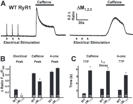
Figure 2. Effects of ∆M1,2,3 on ligand-induced
Ca2+ release. (A) Representative indo-1 ratio traces
obtained from intact dyspedic myotubes
express-ing either WT RyR1 (left) or ∆M1,2,3 (right)
fol-lowing electrical stimulation (fi lled triangles) and caffeine application (horizontal bars). (B) Average maximal magnitude of electrically evoked (left), caffeine-induced (middle), and 4-cmc–induced
(right) Ca2+ release. (C) Average time to peak
(TTP, left) and t1/2 of decay (middle) for
caffeine-induced Ca2+ release and time to peak 4-cmc Ca2+
release (TTP, right). *, P < 0.01.
on September 12, 2007
www.jgp.org
but not in uninjected (naive) dyspedic myotubes (un-published data). However, while average resting Indo-1 ratios were not signifi cantly different (resting F405/F485
was 0.60 ± 0.01, n = 28, and 0.56 ± 0.01, n = 27, for WT RyR1- and ∆M1,2,3-expressing myotubes, respectively),
∆M1,2,3-expressing myotubes lacked electrically evoked
Ca2+ transients (Fig. 2 A, right, and Fig. 2 B). To confi rm
functional ∆M1,2,3 expression, a maximal concentration
(30 mM) of caffeine or 4-cmc (500μM) was applied to both WT- and ∆M1,2,3-expressing myotubes (Fig. 2, A
and B). Ca2+ release in response to caffeine was reduced
in ∆M1,2,3-expressing myotubes (Fig. 2 B) and the kinetics
of caffeine-induced and 4-cmc–induced Ca2+ release
(time to peak and t1/2 of decay) were signifi cantly slowed
(Fig. 2, A and C).
The Triadin Binding-defi cient RyR1 Mutant (∆M1,2,3)
Supports Retrograde, but not Orthograde, DHPR–RyR1 Coupling
Simultaneous whole-cell patch clamp measurements of voltage-gated L-type Ca2+ currents and intracellular
Ca2+ transients were used to determine the ability of the
∆M1,2,3 triadin binding-defi cient mutant to support the
retrograde and orthograde signals of skeletal muscle EC coupling (Fig. 3 A). Naive dyspedic myotubes exhib-ited small (<1 pA/pF) L-type Ca2+ currents (Fig. 3 C,
open squares) and lacked voltage-gated Ca2+ release
(Fig. 3 D, open squares) (Nakai et al., 1996). Expression of WT RyR1 (Fig. 3 A) restored robust ( 10 pA/pF)
voltage-gated L-type Ca2+ currents (bottom traces) and
Ca2+ transients (top traces). Interestingly, while
expres-sion of ∆M1,2,3 (Fig. 3 B) similarly enhanced L-type Ca2+
current density, Ca2+ release was markedly slower and
reduced in magnitude. Voltage-gated Ca2+ transients in
WT RyR1-expressing myotubes exhibited the character-istic sigmoidal voltage dependence of skeletal muscle EC coupling (Fig. 3 D, fi lled circles), while Ca2+ transients
in ∆M1,2,3-expressing myotubes exhibited a bell-shaped
voltage dependence (Fig. 3 D, fi lled triangles) that mir-rored that of the L-type Ca2+ current (Fig. 3 C, fi lled
triangles). The bell-shaped Ca2+ transients in ∆M 1,2,3
-ex-pressing myotubes largely arose from Ca2+ infl ux–induced
Ca2+ release through ∆M
1,2,3 channels as they were
markedly reduced by blockers of both the L-type Ca2+
channel (0.5 mM Cd2++ 0.2 mM La3+, open symbols in
Fig. 3 E) and the RyR (100μM ryanodine, open symbols in Fig. 3 F).
RyR1 Mutations that Disrupt Triadin Binding Do Not Directly Alter Release Channel Function
The observed alterations in ligand and voltage-gated Ca2+
release in ∆M1,2,3-expressing dyspedic myotubes
docu-mented in Figs. 2 and 3 could either be due to effects of the mutations on triadin regulation of RyR1 or to direct effects of the mutations on channel function that are independent of triadin binding. To test for potential direct effects of the mutations, we compared the subcel-lular localization and function of WT and ∆M1,2,3 RyR1
Figure 3. Effects of ∆M1,2,3 on orthograde and retro-grade DHPR-RyR1 coupling. (A) Representative L-type
Ca2+ currents (bottom traces) and intracellular Ca2+
transients (top traces) resulting from 200-ms
depolariza-tions to −50, −10, +30, and +70 mV in a WT
RyR1-expressing myotube. (B) Representative L-type Ca2+
currents (bottom traces) and intracellular Ca2+ transients
(top traces) resulting from 200-ms depolarizations to
−50, −10, +30, and +70 mV in a ∆M1,2,3-expressing
myo-tube. (C and D) Average voltage dependence of peak
L-type Ca2+ current density (C) and intracellular Ca2+
transients (D) in naive dyspedic myotubes (open squares),
WT RyR1-expressing (closed circles), and ∆M1,2,3
-express-ing (closed triangles) myotubes. (E) Inhibition of L-type
Ca2+ currents (with 0.5 mM Cd2+/0.2 mM La3+, open
symbols) markedly reduced (86 ± 7%, n = 5 at +30 mV)
Ca2+ transients in ∆M1,2,3-expressing myotubes (triangles)
but only minimally reduced (16 ± 9%, n = 5 at +30 mV)
Ca2+ transients in WT RyR1-expressing myotubes (circles).
(F) Blockade of Ca2+ release with 100 μM ryanodine
(open symbols) markedly reduced depolarization-induced
Ca2+ transients in both WT RyR1- (circles) and ∆M1,2,3
-expressing (triangles) myotubes.
on September 12, 2007
www.jgp.org
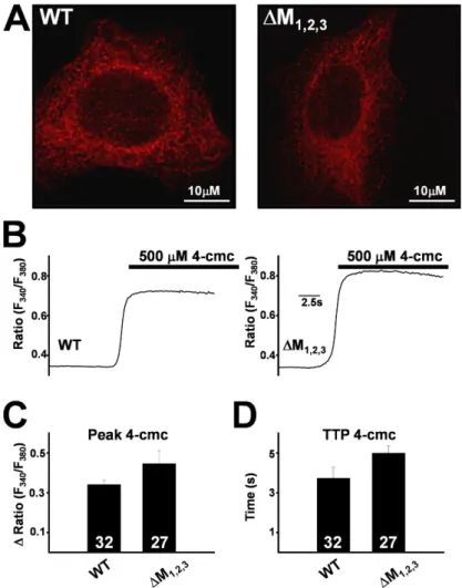
channels following expression in HEK293 cells (Figs. 4 and 5). WT and ∆M1,2,3 channels exhibited identical
re-ticulated ER localization (Fig. 4 A) following expression in HEK293 cells. Since HEK293 cells do not express tri-adin and 4-cmc activates Ca2+ release through expressed
RyR1 channels (Fessenden et al., 2000), we compared 4-cmc–induced Ca2+ release responses in WT- and ∆M
1,2,3
-expressing HEK293 cells. The magnitude and kinetics of 4-cmc–induced Ca2+ release was not signifi cantly
dif-ferent (P > 0.05) between WT- and ∆M1,2,3-expressing
HEK293 cells (Fig. 4, B–D).
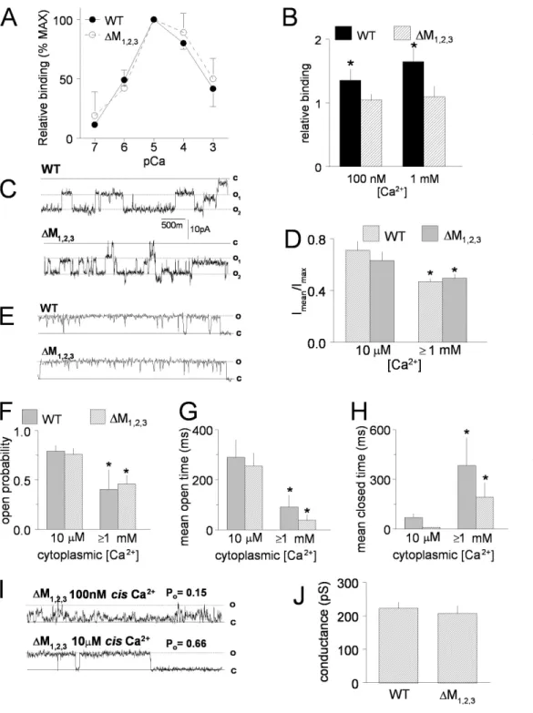
We next compared the function of purifi ed WT and ∆M1,2,3 channels following expression in HEK293 cells
(Fig. 5). WT and ∆M1,2,3 channels exhibited identical
Ca2+ dependence of [3H]ryanodine binding, with
bind-ing (which indirectly refl ects Po) increasing with channel
activation between 100 nM and 10 μM cis Ca2+ (EC 50 was
4.14 ± 0.34 and 3.99 ± 0.17 μM for WT and ∆M1,2,3
chan-nels, respectively) and then similarly declining as cis Ca2+
rises above 10 μM (Fig. 5 A). The data in Fig. 5 A are expressed relative to maximum binding in 10 μM Ca2+ in
order to reduce variations between ER preparations. Maximal [3H]ryanodine binding was not signifi cantly
different between WT and ∆M1,2,3 channels (Bmax was
4.8 ± 0.6 and 4.5 ± 0.1 pM/mg for WT and ∆M1,2,3
channels, respectively). Preincubation of recombinant WT RyR1 channels with 5 μg/ml triadin increased [3H]ryanodine binding relative to binding in the absence
of triadin. In contrast, incubation with triadin had no effect on maximal [3H]ryanodine binding to ∆M
1,2,3 RyR1
(Fig. 5 B). These results demonstrate a strong functional consequence of triadin binding to WT RyR1 and the lack of any functional change when triadin is added to triadin binding-defi cient ∆M1,2,3 channels. Previous
stud-ies have shown an inhibitory interaction between the cytoplasmic domain of triadin and RyR1 (Ohkura et al., 1998; Groh et al., 1999).
The activity of single ∆M1,2,3 channels in bilayers was
similar to that of WT channels (Fig. 5, C–J). In six of eight experiments using either WT or ∆M1,2,3 channels, current
recordings showed periods where more than one chan-nel opened in the bilayer with similar openings in WT and mutant channels (Fig. 5 C). To include data from these recordings, open probability of 30-s recordings was calculated as the mean current in the recording (average of all data points) normalized to the maximum current (maximum open level), i.e., Imean/Imax (Fig. 5 D) (Beard
et al., 2002). In each experiment, channel activity was initially recorded in 5 mM cis Ca2+ to aid in channel
in-corporation, cis Ca2+ concentration was then decreased
Figure 4. Subcellular localization and Ca2+ release
function of WT RyR1 and ∆M1,2,3 channels expressed in
HEK293 cells. (A) Both WT RyR1 (left) and ∆M1,2,3 (right) channels exhibit similar reticulated ER
expres-sion. (B) Representative fura-2 ratio (F340/F380) traces
obtained from WT RyR1- (left) and ∆M1,2,3-expressing
(right) HEK293 cells following addition of 500 μM 4-cmc (bar). Average peak (C) and time to peak (TTP) 4-cmc responses (D) obtained from WT RyR1- and
∆M1,2,3-expressing HEK293 cells.
on September 12, 2007
www.jgp.org
to 10 μM and subsequently returned to 1 mM. Since there was no signifi cant difference in channel activity between 1 or 5 mM cis Ca2+, data at these two
concentra-tions were combined and reported as ≥1 mM in Fig. 5 (D–H). In agreement with the [3H]ryanodine binding
data (Fig. 5 A) and previous studies (Laver et al., 1997), WT channel activity fell signifi cantly when the cis Ca2+
concentration was increased from 10 μM to ≥1 mM Ca2+.
However, no signifi cant difference was observed between WT and ∆M1,2,3 channels (Fig. 5, D and F). All recordings
showed periods (10–30 s) of single channel opening (Fig. 5 C) that were also analyzed for open probability, mean open time, and mean closed time using a standard 20% threshold-crossing criterion (see Materials and methods).
Figure 5. Properties of
puri-fi ed WT RyR1 and ∆M1,2,3
channels expressed in HEK293
cells. (A) Ca2+ dependence of
[3H]ryanodine binding (%
maximum binding) to WT
(closed circles) and ∆M1,2,3
(closed squares) channels. (B) Effect of preincubation
with 5 μg/ml purifi ed triadin
on [3H]ryanodine binding
in the presence of either
100 nM or 1 mM Ca2+ (n =
6 for each). *, P < 0.05.
[3H]ryanodine binding to
pu-rifi ed RyR1 in the presence of triadin (fi lled bars) is normal-ized to binding to purifi ed RyR1 in absence of triadin (crosshatched bars). (C) Rep-resentative single channel records from artifi cial lipid bi-layers incorporated with two purifi ed WT (top) and two
purifi ed ∆M1,2,3 (bottom)
channels at −40 mV in the
presence of 1 mM trans
(lumi-nal) Ca2+ and 10 μM cis
(cyto-plasmic) Ca2+. The channels
opened from the closed level
(c) to either single open (o1)
and double open (o2) levels.
(D) Average data from four WT RyR1 channels and four
∆M1,2,3 channels showing
open probability measured as
mean current (Imean)
normal-ized to maximum current
(Imax) during periods in which
only one or two channels were open in the bilayer. Open probability decreased when
the cis Ca2+ was increased
from 10 μM to either 1 mM or
5 mM (data for 1 and 5 mM cis
Ca2+ at +40 and −40 mV were
grouped in the average data).
*, P < 0.05. (E) Periods of
sin-gle channel activity in record-ings from bilayers containing
purifi ed WT (top) and ∆M1,2,3
(bottom) channels at +40 mV
with 1 mM trans (luminal) Ca2+ and 10 μM cis (cytoplasmic) Ca2+. (F–H) Average open probability (F), mean open time (G), and mean
closed time (H) for 30-s recordings from four WT and four ∆M1,2,3 channels in the presence of 10 μM cis Ca2+ and combined data for 1
and 5 mM cis Ca2+. *, P < 0.05. (I) Data from a ∆M1,2,3 channel recorded fi rst in 100 nM cis Ca2+ (top) and then in 10 μM cis Ca2+
(bot-tom). The open probability for 30 s of channel activity at each Ca2+ concentration is given above each record. (J) Average single channel
conductance from four WT RyR1 channels and four ∆M1,2,3 RyR1 channels.
on September 12, 2007
www.jgp.org
Open probability values obtained with this method (Fig. 5 F) were very similar to those estimated from Imean/Imax
(Fig. 5 D). The decline in open probability when cis Ca2+ concentration was increased from 10 μM to ≥1 mM was due to an abbreviation of the mean open time (Fig. 5 G) and prolongation of the mean closed time (Fig. 5 H). These changes in mean open and closed times were simi-lar in WT and ∆M1,2,3 channels.
In one experiment with a ∆M1,2,3 channel, the cis Ca2+
concentration was lowered to 100 nM and then increased to 10 μM (Fig. 5 I). As expected from the [3H]ryanodine
binding data (Fig. 5 A), the open probability of this channel was greater with 10 μM cis Ca2+ than with 100 nM cis Ca2+, increasing from 0.15 to 0.66; the mean open time increased from 10.9 to 54.4 ms, and the mean closed time fell from 58.3 to 27.4 ms. The changes in open probability, mean open time, and mean closed time when Ca2+ concentration was increased from 100 nM to 10 μM in this ∆M1,2,3 channel are similar to changes in
the activity of WT channels that occur over the same range of Ca2+ concentrations. Finally, the ∆M1,2,3
muta-tion did not signifi cantly affect unitary conductance of RyR1 channels (Fig. 5 J) and the conductance at +40 mV was similar to that at −40 mV (Fig. 5, compare C with E or I). Average Cs+ conductance was 222 ± 19 pS for WT RyR1 and 207 ± 24 pS for ∆M1,2,3. Taken together,
the data in Figs. 4 and 5 demonstrate that WT and ∆M1,2,3 channels exhibit similar channel function in the
absence of triadin.
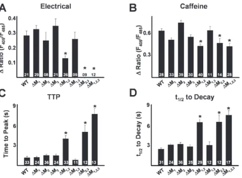
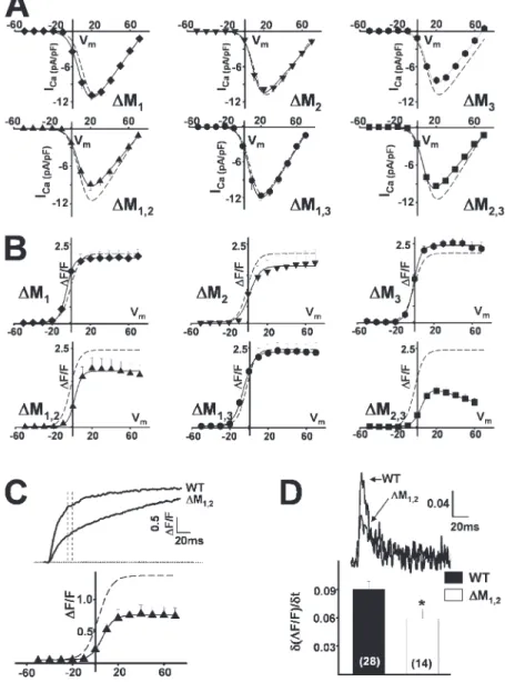
Effects of Other RyR1 Luminal Loop Mutations on Ligand- and Voltage-gated Ca2+ Release
Indo-1 Ca2+ measurements in intact myotubes (Fig. 6) and whole-cell voltage clamp (Fig. 7 and Table I) exper-iments were also conducted in myotubes expressing
each of the single (∆M1, ∆M2, and ∆M3) and double
(∆M1,2, ∆M2,3, and ∆M1,3) RyR1 mutations. Electrically
evoked release (Fig. 6 A), ligand-induced release (Fig. 6, B–D), and both retrograde (Fig. 7 A and Table I) and orthograde coupling (Fig. 7 B and Table I) were not sig-nifi cantly different between WT RyR1-expressing myo-tubes and either ∆M1-, ∆M2-, ∆M3-, or ∆M1,3-expressing
myotubes. On the other hand, effects of the ∆M2,3
muta-tion were similar to those of ∆M1,2,3 in that ∆M2,3
-expressing myotubes also lacked electrically evoked Ca2+
release (Fig. 6 A), exhibited slowed kinetics of caffeine-induced Ca2+ release (Fig. 6, C and D), normal L-type
Ca2+ channel activity (Fig. 7 A and Table I), and lacked
sigmoidal voltage-gated Ca2+ release (Fig. 7 B).
Interest-ingly, reduced triadin binding to the ∆M1,2 mutant (Fig.
1 B) was accompanied by a signifi cant reduction in the magnitude of electrically evoked Ca2+ release (Fig. 6 A)
and a slowing in ligand-induced Ca2+ release (Fig. 6, C
and D). In patch clamp experiments, there was a ten-dency (P = 0.05) for a reduction in voltage-gated Ca2+
release measured at the end of 200-ms depolarizations in ∆M1,2-expressing myotubes (Table I). However, for
release assessed 30 ms after the start of the test pulse (to approximate release during a brief action potential), voltage-gated Ca2+ release (Fig. 7 C and Table I) and the
maximum rate of Ca2+ release (Fig. 7 D), approximated
from the peak of the fi rst derivative of the Ca2+ transient
elicited at +70 mV (δ(∆F/F)/δt), were signifi cantly (P < 0.01) reduced in ∆M1,2-expressing myotubes.
Triadin Binding-defi cient RyR1 Mutants Exhibit Normal Junctional Targeting
The reduction in voltage-gated Ca2+ release documented
in ∆M1,2-, ∆M2,3-, and ∆M1,2,3- expressing myotubes (Fig.
7, B and C) could result from a lack of proper targeting
Figure 6. Effects of the terminal luminal
loop RyR1 mutants on electrically evoked
and caffeine-induced Ca2+ release. (A–D)
Bar graphs summarizing the effects of each of WT RyR1 and the different terminal
RyR1 luminal loop mutants (∆M1, ∆M2,
∆M3, ∆M1,2, ∆M1,3, ∆M2,3, and ∆M1,2,3) on
electrically evoked Ca2+ release (A), peak
caffeine-induced (30 mM) Ca2+ release
(B), and the average time to peak (C) and
t1/2 of decay (D) of caffeine-induced Ca2+ transients.
on September 12, 2007
www.jgp.org
of the mutant release channels to DHPR-containing SR–sarcolemmal junctions. However, our fi nding that all three of these triadin binding-defi cient mutant RyR1 channels restored retrograde DHPR Ca2+ channel
con-ductance to a similar degree as WT RyR1 (Fig. 6 A and Table I) provides strong functional evidence that each mutant was expressed, properly targeted to SR–sarco-lemmal junctions, and interacted with DHPRs present in the junction. Nevertheless, we also compared DHPR and RyR subcellular localization in WT-, ∆M1,2-, ∆M2,3-,
and ∆M1,2,3-expressing myotubes in coimmunofl
uores-cence labeling experiments (Fig. 8). Consistent with the observed restoration of retrograde coupling for each of these constructs, the RyR1 mutants that displayed de-fects in triadin binding and electrically evoked Ca2+
re-lease exhibited a punctate appearance (Fig. 8, left) that overlapped with discrete clusters of DHPR fl uorescence (Fig. 8, middle). This similar punctate appearance indi-cates correct junctional targeting of each construct and is particularly evident in the merged images (Fig. 8, right),
in which green and red indicate RyR1 and DHPR, re-spectively, and yellow represents common regions of punctate appearance.
D I S C U S S I O N
Since its initial discovery in 1990 (Brandt et al., 1990), triadin has been shown to be enriched and embedded in the SR membrane of the triad (Knudson et al., 1993a), to interact directly with both CSQ and RyR1 (Guo and Campbell, 1995), to inhibit RyR channels when applied to the cytoplasmic side (Ohkura et al., 1998), to activate channels when applied to the luminal side (Gyorke et al., 2004), and to contribute to the luminal Ca2+
transduc-tion machinery that regulates Ca2+ release channel
activity in skeletal muscle (Beard et al., 2004). Lee et al. (2004) identifi ed three negatively charged amino acids (D4878, D4907, and E4908) in an RyR1 luminal loop that were each required for the isolated loop to bind a specifi c positively charged triadin KEKE motif (residues
Figure 7. Effects of the terminal luminal loop
RyR1 mutants on orthograde and retrograde cou-pling. (A and B) Average voltage dependence of
L-type Ca2+ current density (A) and
depolariza-tion-induced Ca2+ transients (B) in ∆M1-, ∆M2-,
∆M3-, ∆M1,2-, ∆M1,3-, and ∆M2,3-expressing
myo-tubes. Dashed lines representing the average volt-age dependence obtained from WT-expressing myotubes are shown for comparison. (C)
Repre-sentative depolarization-induced (test potential =
+70 mV) Ca2+ transients from WT RyR1- and
∆M1,2-expressing myotubes (top). Voltage
depen-dence of Ca2+ transients measured 30 ms after
the start of the test pulse (bottom). (D)
Differen-tials of fl uorescence traces (+70 mV) taken
dur-ing the initial phase of depolarization (top). Bar
graph of average peak differential (δ(∆F/F)/δt)
(bottom). The number of experiments is given
within each bar. *, P < 0.01.
on September 12, 2007
www.jgp.org
200–232) (Fig. 1 A). Our results provide the following important and novel contributions with regard to tria-din bintria-ding/regulation of RyR1: (a) a single mutation to any of these three negatively charged residues is not suffi cient to modify RyR1–triadin association and the combination of the different mutations demonstrates that each residue contributes unequally (D4907 > E4908 > D4878) to triadin binding (Fig. 1 B), (b) the RyR1 luminal loop mutations do not directly alter release channel function (Figs. 4 and 5), (c) luminal loop mu-tations that disrupt triadin binding do not alter either junctin binding (Fig. 1 C) or RyR1 junctional targeting (Fig. 8), and (d) triadin binding to RyR1 ensures rapid and robust Ca2+ release during both voltage- and
ligand-induced activation (Figs. 2, 3, 6, and 7; and Table I). Recently, Lee et al. (2006) found that triadin binding was reduced and the kinetics of caffeine-induced cal-cium release slowed following infection of RyR-null 1B5 myotubes with herpes simplex virus-1 virions packaged with either ∆M2 or ∆M1,2,3 compared with that observed
for WT RyR1 (Lee et al., 2006). We found a similar slow-ing in the kinetics of caffeine-induced calcium release in ∆M1,2,3-expressing primary dyspedic myotubes (Fig.
2 C). However, unlike Lee et al. (2006), we found that triadin binding to RyR1 as well as the kinetics of caffeine-induced calcium release were unaltered by the ∆M2
mu-tation (Fig. 6, C and D). Differences between the two studies observed for the effects of the ∆M2 mutation are
not entirely clear, but may involve differences between the cells, expression methods, and biochemical/analytical approaches used in the two studies.
Importantly, results presented here are the fi rst to characterize the impact of triadin binding to RyR1 on the orthograde and retrograde signals of EC coupling. Specifi cally, we found that triadin binding to RyR1
en-hances release channel activity during both voltage and ligand activation and that this critical regulation of re-lease channel activity ensures robust and rapid calcium release during skeletal muscle EC coupling. This idea is further supported by our observation that [3H]ryanodine
binding to WT RyR1, but not ∆M1,2,3, is signifi cantly
en-hanced by the addition of purifi ed triadin (Fig. 5 B) and that triadin increases the open probability of RyR chan-nels incorporated into planar lipid bilayers (Gyorke et al., 2004). In addition, our study is also the fi rst to demon-strate that the mutations in RyR1 used to disrupt triadin binding do not directly affect release channel activity, retrograde RyR1–DHPR signaling, or the ability of junctin to bind to RyR1.
Defects observed for the RyR1 mutants could result either from effects of the mutations on triadin binding, direct effects on the channel, or both. We show that these mutations do not directly affect RyR1 channel function; WT and ∆M1,2,3 channels expressed in HEK293 cells
exhibited similar Ca2+ dependence of [3H]ryanodine
binding (Fig. 5 A), open probability (Fig. 5, D and F), mean open (Fig. 5 G) and closed (Fig. 5 H) times, con-ductance (Fig. 5 J), and Ca2+ release activity (Fig. 4, B–D).
Moreover, purifi ed triadin increased [3H]ryanodine
binding to WT RyR1, but not to ∆M1,2,3 (Fig. 5 B). In
ad-dition, WT RyR1 and triadin binding-defi cient mutants expressed in dyspedic myotubes were activated by Ca2+
(Fig. 3 D), caffeine (Fig. 2 B), 4-cmc (Fig. 2 B), and fully restored retrograde DHPR coupling (Fig. 7 A and Table I). The slight reduction in maximal caffeine-induced Ca2+
release (Fig. 6 B) observed for the triadin binding-defi cient mutants may result from a combination of slow Ca2+
re-lease inactivating some channels and the fact that triadin binding to RyR1 enhances Ca2+ gating (Fig. 5 B). Finally,
effects of the luminal loop mutants on voltage-gated
T A B L E I
Parameters of Fitted IV and FV Curves
Gmax kG VG1/2 Vrev (∆F/F)max kF VF1/2
nS/nF mV mV mV mV mV RyR1 (n = 27) 246 ± 12 4.8 ± 0.2 9.8 ± 1.0 74.6 ± 1.5 2.5 ± 0.2 4.8 ± 0.3 −2.7 ± 0.9 RyR1 (n = 27) (30 ms) 175 ± 10 5.5 ± 0.2 12.0 ± 1.0 70.9 ± 1.3 1.4 ± 0.1 5.1 ± 0.3 2.7 ± 0.9 ∆M1 (n = 17) 232 ± 9 4.7 ± 0.5 8.7 ± 1.4 74.5 ± 1.7 2.1 ± 0.2 3.6 ± 0.5 −5.1 ± 0.9 ∆M2 (n = 18) 247 ± 14 5.0 ± 0.4 6.5 ± 1.5 75.7 ± 2.1 2.3 ± 0.3 4.2 ± 0.5 −2.0 ± 1.8 ∆M3 (n = 12) 211 ± 17 4.6 ± 0.3 11.1 ± 0.9 68.6 ± 1.8 2.0 ± 0.2 3.6 ± 0.3 −2.4 ± 0.7 ∆M1,2 (n = 12) 180 ± 11 4.5 ± 0.3 9.2 ± 1.4 76.6 ± 2.3 1.8 ± 0.3 2.9 ± 0.3 1.9 ± 1.1 ∆M1,2 (n = 12) (30 ms) 132 ± 10 5.7 ± 0.2 12.3 ± 1.8 70.3 ± 2.0 0.8 ± 0.1b 4.8 ± 0.3 7.0 ± 1.1b ∆M1,3(n = 6) 259 ± 14 4.8 ± 0.3 8.9 ± 1.0 73.6 ± 2.3 2.4 ± 0.3 3.7 ± 0.6 −5.0 ± 2.5 ∆M2,3 (n = 17) 187 ± 8 4.2 ± 0.4 6.9 ± 1.2 75.5 ± 2.1 0.9 ± 0.1a 3.4 ± 0.3 10.5 ± 1.3a ∆M1,2,3 (n = 18) 200 ± 11 3.6 ± 0.2 13.5 ± 1.2 75.0 ± 2.0 0.8 ± 0.1a 4.0 ± 0.3 4.3 ± 1.1a
Values represent mean ± SEM for n experiments. Parameters for the voltage dependence of Ca2+ current (I-V) and Ca2+ release (∆F/F) were obtained
by fi tting myotubes within each group separately to the appropriate equation (I-V, Eq. 1; ∆F/F, Eqs. 2 and 3) as described in Materials and methods. Gmax,
maximal L-channel conductance; (∆F/F)max, maximal change in relative fl uo-4 fl uorescence; Vrev, L-channel reversal potential; VG1/2 and VF1/2, potential at
which G and F are half-maximal, respectively; kG and kF, slope factors for I-V and ∆F/F, respectively. aP < 0.01, compared to WT (200 ms).
bP < 0.01, compared to WT (30 ms).
on September 12, 2007
www.jgp.org
Ca2+ release coincided perfectly with independent
bio-chemical determination of triadin binding; mutations that did not affect binding (∆M1, ∆M2, ∆M3, and ∆M1,3)
did not affect orthograde coupling, mutations that eliminated binding abolished orthograde coupling (∆M1,2,3 and ∆M2,3), and mutations that partially
dis-rupted binding partially reduced orthograde coupling (∆M1,2). Since the binding of purifi ed triadin to
puri-fi ed WT or mutant RyR1 proteins in our experiments is likely to be thermodynamically different from in vivo binding that occurs in the SR, we cannot completely rule out the possibility that the effects of the RyR1 mutations on release and triadin binding are coincidentally related. Nevertheless, our results provide strong evidence that altered voltage- and ligand-induced Ca2+ release of the
∆M1,2,3, ∆M1,2, and ∆M2,3 mutants result from defects in
triadin binding rather than direct effects of the mutations on channel function.
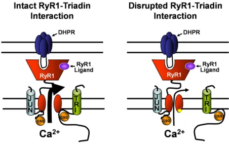
Fig. 9 provides a simple model that ties together previously published results with the fi ndings of this study. Under normal conditions (Fig. 9, left), triadin binds to both CSQ and the terminal luminal loop of RyR1, forming a quaternary complex along with junc-tin that maximizes SR Ca2+ release following activation
of RyR1 by either endogenous (e.g., DHPR) or exoge-nous (e.g., caffeine or 4-cmc) triggers. In this model, mu-tations in RyR1 that interfere with triadin binding are proposed to disrupt this critical RyR1 regulatory mecha-nism and result in the dissociation of the triadin–CSQ complex from the release channel (Fig. 9, right). Since we found that junctin binding to RyR1 is not affected by any of the RyR1 luminal loop mutations, a junctin–CSQ
Figure 8. Triadin binding-defi cient
RyR1 mutants exhibit normal targeting to DHPR-containing junctions. (A) Dou-ble immunofl uorescence labeling in
rep-resentative WT- (fi rst row), ∆M1,2- (second
row), ∆M2,3- (third row), and ∆M1,2,3
-expressing (fourth row) myotubes with antibodies against RyR1 (left) and the DHPR (middle). The “merged images” (right) emphasize common regions of puncta (yellow foci) containing ex-pressed RyR1 proteins and endogenous DHPRs in clusters that represent junc-tions of the SR with the sarcolemma.
on September 12, 2007
www.jgp.org
complex is shown to interact with an alternate RyR1 site in Fig. 9, suggesting that RyR1 activity may be differen-tially regulated by triadin–CSQ and junctin–CSQ. Our results indicate that triadin binding to RyR1 primes the channel for ligand/voltage sensor activation in order to ensure rapid Ca2+ release kinetics following either
volt-age- or ligand-induced activation. In the absence of this critical interaction of triadin–CSQ with RyR1, the kinet-ics of ligand- (Fig. 2 C and Fig. 6 C) and voltage-induced (Fig. 7, C and D) Ca2+ release is markedly slowed. In the
case of release stimulated by a brief action potential fol-lowing neuromuscular transmission, this reduction in the rate of release is suffi cient to essentially abolish Ca2+
release during EC coupling (Fig. 2 B and Fig. 6 A). Overexpression of triadin in rat skeletal myotubes was shown to signifi cantly reduce KCl depolarization-induced Ca2+ release with minimal effects on either
caffeine-/4-cmc–induced Ca2+ release or L-type Ca2+
current density (Rezgui et al., 2005). Triadin overexpres-sion could result in either increased (due to enhanced levels of triadin) or decreased (due to overexpression causing triadin aggregation, Knudson et al., 1993a) tria-din bintria-ding to RyR1. In either case, the results of Rezgui et al. (2005) indicate that the EC coupling machinery is exquisitely sensitive to triadin– RyR1 stoichiometry. Our results are consistent with this notion since disruption of triadin binding to RyR1 also markedly reduced ortho-grade, but not retroortho-grade, DHPR–RyR1 coupling. The similar effects on voltage-gated Ca2+ release observed
following triadin overexpression and triadin dissocia-tion from RyR1 could be explained by effects either of disrupting triadin binding to RyR1 or of free triadin on the EC coupling machinery.
Preliminary reports have produced confl icting results regarding effects of triadin defi ciency on EC coupling. For example, KCl-induced Ca2+ release is similar in
my-otubes derived from WT and pan-triadin knockout mice (Shen, X., J.R. Lopez, P.D. Allen, and C.F. Perez. 2006.
Biophysical Society Meeting. Abstr. 328) but signifi -cantly reduced and slowed following transient siRNA-mediated knockdown of triadin and junctin in C2C12 myotubes (Wang, Y., L. Xinghai, H. Duan, T. Fulton, and G. Meissner. 2007. Biophysical Society Meeting. Abstr. 1237). The second fi nding is consistent with our results. The absence of an effect on depolarization-induced Ca2+ release in pan-triadin knockout mice could
result from a multitude of different compensatory mech-anisms commandeered to correct for the loss of a critical RyR1 regulatory protein. In fact, skeletal muscle function and EC coupling is also not markedly altered following knockout of the skeletal muscle isoform of calsequestrin (CSQ1; Paolini et al., 2007). Similarly, cardiac SR Ca2+
storage and depolarization-induced Ca2+ release are
essentially normal following either global knockout of CSQ2 (Knollmann et al., 2006) or cardiac-specifi c knockout of the Na/Ca exchanger (Henderson et al., 2004), respectively. The surprising lack of an effect of genetic ablation of either CSQ1/2 or the Na/Ca ex-changer on EC coupling certainly does not mean that these proteins are not normally important for calcium storage and removal, respectively. Rather, remarkable compensatory changes in Ca2+ release unit assembly
and effi ciency were found to counteract CSQ and Na/ Ca exchange defi ciency in these animals. Conceivably, similar compensatory changes might also account for the absence of a marked effect of triadin ablation of skeletal muscle EC coupling.
More than 15 different central core disease mutations in RyR1 have been identifi ed in the terminal RyR1 luminal loop shown here to markedly infl uence triadin binding and ligand/voltage-gated Ca2+ release. A number of these
mutations reduce Ca2+ release without signifi cantly
affect-ing either SR Ca2+ content or retrograde coupling to the
DHPR (Avila et al., 2003). Thus, it will be important for fu-ture studies to determine if any of these disease mutations in the RyR1 terminal luminal loop diminishes Ca2+ release
Figure 9. Proposed model for triadin regulation
of DHPR and ligand activation of RyR1. The qua-ternary CSQ–triadin–RyR1–junctin interaction tethers CSQ close to the release channel pore and promotes high probability release channel opening (depicted by a large arrow) following ei-ther DHPR or ligand activation (left). Disruption of triadin binding to RyR1 results in a similar re-duction in SR Ca2+ release (depicted by a small arrow) following either DHPR or ligand activa-tion (right). A junctin–CSQ complex is shown to bind to a separate RyR1 site from that of triadin.
on September 12, 2007
www.jgp.org
during EC coupling by altering the critical regulatory interaction of triadin with the SR Ca2+ release channel.
We would like to thank Dr. Paul D. Allen (Brigham and Women’s Hospital, Harvard Medical School, Boston, MA) for providing ac-cess to the dyspedic mice used in this study and to Linda Groom for excellent technical assistance. The anti-junctin antibody used in this study was generously provided by Dr. Steve Cala (Wayne State University, Detroit, MI).
This work was supported by research grants from the National Institutes of Health (AR44657 to R.T. Dirksen) and the Australian National Health and Medical Research Council (316937 to A.F. Dulhunty).
Olaf S. Andersen served as editor. Submitted: 23 March 2007 Accepted: 22 August 2007
R E F E R E N C E S
Avila, G., J.J. O’Brien, and R.T. Dirksen. 2001. Excitation-contrac-tion uncoupling by a human central core disease mutaExcitation-contrac-tion in the ryanodine receptor. Proc. Natl. Acad. Sci. USA. 98:4215–4220. Avila, G., K.M. O’Connell, and R.T. Dirksen. 2003. The pore region
of the skeletal muscle ryanodine receptor is a primary locus for excitation-contraction uncoupling in central core disease. J. Gen.
Physiol. 121:277–286.
Beard, N.A., M.M. Sakowska, A.F. Dulhunty, and D.R. Laver. 2002. Calsequestrin is an inhibitor of skeletal muscle ryanodine recep-tor calcium release channels. Biophys. J. 82:310–320.
Beard, N.A., D.R. Laver, and A.F. Dulhunty. 2004. Calsequestrin and the calcium release channel of skeletal and cardiac muscle. Prog.
Biophys. Mol. Biol. 85:33–69.
Beard, N.A., M.G. Casarotto, L. Wei, M. Varsanyi, D.R. Laver, and A.F. Dulhunty. 2005. Regulation of ryanodine receptors by
calseques-trin: effect of high luminal Ca2+ and phosphorylation. Biophys. J.
88:3444–3454.
Brandt, N.R., A.H. Caswell, S.R. Wen, and J.A. Talvenheimo. 1990. Molecular interactions of the junctional foot protein and di-hydropyridine receptor in skeletal muscle triads. J. Membr. Biol. 113:237–251.
Dirksen, R.T. 2002. Bi-directional coupling between dihydropyridine receptors and ryanodine receptors. Front. Biosci. 7:d659-d670. Fessenden, J.D., Y. Wang, R.A. Moore, S.R. Chen, P.D. Allen, and
I.N. Pessah. 2000. Divergent functional properties of ryanodine receptor types 1 and 3 expressed in a myogenic cell line. Biophys. J. 79:2509–2525.
Goonasekera, S.A., S.R. Chen, and R.T. Dirksen. 2005. Reconstitution
of local Ca2+ signaling between cardiac L-type Ca2+ channels and
ryanodine receptors: insights into regulation by FKBP12.6. Am. J.
Physiol. Cell Physiol. 289:C1476–C1484.
Graves, T.K., and P.M. Hinkle. 2003. Ca2+-induced Ca2+ release in
the pancreatic β-cell: direct evidence of endoplasmic reticulum
Ca2+ release. Endocrinology. 144:3565–3574.
Groh, S., I. Marty, M. Ottolia, G. Prestipino, A. Chapel, M. Villaz, and M. Ronjat. 1999. Functional interaction of the cytoplasmic domain of triadin with the skeletal ryanodine receptor. J. Biol.
Chem. 274:12278–12283.
Guo, W., and K.P. Campbell. 1995. Association of triadin with the ryanodine receptor and calsequestrin in the lumen of the sarco-plasmic reticulum. J. Biol. Chem. 270:9027–9030.
Gyorke, I., N. Hester, L.R. Jones, and S. Gyorke. 2004. The role of calse-questrin, triadin, and junctin in conferring cardiac ryanodine re-ceptor responsiveness to luminal calcium. Biophys. J. 86:2121–2128.
Henderson, S.A., J.I. Goldhaber, J.M. So, T. Han, C. Motter, A. Ngo, C. Chantawansri, M.R. Ritter, M. Friedlander, D.A. Nicoll, et al.
2004. Functional adult myocardium in the absence of Na+-Ca2+
ex-change: cardiac-specifi c knockout of NCX1. Circ. Res. 95:604–611. Jiang, D., B. Xiao, L. Zhang, and S.R. Chen. 2002. Enhanced basal
activity of a cardiac Ca2+ release channel (ryanodine receptor)
mutant associated with ventricular tachycardia and sudden death.
Circ. Res. 91:218–225.
Jones, L.R., L. Zhang, K. Sanborn, A.O. Jorgensen, and J. Kelley. 1995. Purifi cation, primary structure, and immunological char-acterization of the 26-kDa calsequestrin binding protein (junc-tin) from cardiac junctional sarcoplasmic reticulum. J. Biol. Chem. 270:30787–30796.
Kim, K.C., A.H. Caswell, J.A. Talvenheimo, and N.R. Brandt. 1990. Isolation of a terminal cisterna protein which may link the di-hydropyridine receptor to the junctional foot protein in skeletal muscle. Biochemistry. 29:9281–9289.
Kimura, T., M. Nakamori, J.D. Lueck, P. Pouliquin, F. Aoike, H. Fujimura, R.T. Dirksen, M.P. Takahashi, A.F. Dulhunty, and S. Sakoda. 2005. Altered mRNA splicing of the skeletal muscle ryanodine
receptor and sarcoplasmic/endoplasmic reticulum Ca2+-ATPase in
myotonic dystrophy type 1. Hum. Mol. Genet. 14:2189–2200. Kimura, T., S.M. Pace, L. Wei, N.A. Beard, R.T. Dirksen, and A.F.
Dulhunty. 2007. A variably spliced region in the type 1 ryanodine receptor may participate in an inter-domain interaction. Biochem.
J. 401:317–324.
Knollmann, B.C., N. Chopra, T. Hlaing, B. Akin, T. Yang, K. Ettensohn, B.E. Knollmann, K.D. Horton, N.J. Weissman, I. Holinstat, et al. 2006. Casq2 deletion causes sarcoplasmic reticulum volume
in-crease, premature Ca2+ release, and catecholaminergic
polymor-phic ventricular tachycardia. J. Clin. Invest. 116:2510–2520. Knudson, C.M., K.K. Stang, A.O. Jorgensen, and K.P. Campbell.
1993a. Biochemical characterization of ultrastructural localiza-tion of a major junclocaliza-tional sarcoplasmic reticulum glycoprotein (triadin). J. Biol. Chem. 268:12637–12645.
Knudson, C.M., K.K. Stang, C.R. Moomaw, C.A. Slaughter, and K.P. Campbell. 1993b. Primary structure and topological analysis of a skeletal muscle-specifi c junctional sarcoplasmic reticulum glyco-protein (triadin). J. Biol. Chem. 268:12646–12654.
Laver, D.R., T.M. Baynes, and A.F. Dulhunty. 1997. Magnesium inhi-bition of ryanodine-receptor calcium channels: evidence for two independent mechanisms. J. Membr. Biol. 156:213–229.
Lee, E.H., D.W. Song, J.M. Lee, G. Meissner, P.D. Allen, and D.H. Kim.
2006. Occurrence of atypical Ca2+ transients in triadin-binding
de-fi cient-RYR1 mutants. Biochem. Biophys. Res. Commun. 351:909–914. Lee, J.M., S.H. Rho, D.W. Shin, C. Cho, W.J. Park, S.H. Eom, J. Ma,
and D.H. Kim. 2004. Negatively charged amino acids within the intraluminal loop of ryanodine receptor are involved in the inter-action with triadin. J. Biol. Chem. 279:6994-7000.
Mulvey, C., and K. Ohlendieck. 2003. Use of continuous-elution gel electrophoresis as a preparative tool for blot overlay analysis.
Anal. Biochem. 319:122–130.
Nakai, J., R.T. Dirksen, H.T. Nguyen, I.N. Pessah, K.G. Beam, and P.D. Allen. 1996. Enhanced dihydropyridine receptor channel activity in the presence of ryanodine receptor. Nature. 380:72–75. Ohkura, M., K. Furukawa, H. Fujimori, A. Kuruma, S. Kawano, M.
Hiraoka, A. Kuniyasu, H. Nakayama, and Y. Ohizumi. 1998. Dual regulation of the skeletal muscle ryanodine receptor by triadin and calsequestrin. Biochemistry. 37:12987–12993.
Paolini, C., M. Quarta, A. Nori , S. Boncompagni, M. Canato, P. Volpe, P.D. Allen, C. Reggiani, and F. Protasi. 2007. Re-organized stores and impaired calcium handling in skeletal muscle of mice lacking calsequestrin-1. J. Physiol. 10.1113/jphysiol.2007.138024. Rezgui, S.S., S. Vassilopoulos, J. Brocard, J.C. Platel, A. Bouron, C.
Arnoult, S. Oddoux, L. Garcia, M. De Waard, and I. Marty. 2005.
on September 12, 2007
www.jgp.org
Triadin (Trisk 95) overexpression blocks excitation-contraction coupling in rat skeletal myotubes. J. Biol. Chem. 280:39302–39308. Rossi, A.E., and R.T. Dirksen. 2006. Sarcoplasmic reticulum: the
dynamic calcium governor of muscle. Muscle Nerve. 33:715–731. Sheridan, D.C., L. Carbonneau, C.A. Ahern, P. Nataraj, and R.
Coronado. 2003. Ca2+-dependent excitation-contraction coupling
triggered by the heterologous cardiac/brain DHPR β2a-subunit in skeletal myotubes. Biophys. J. 85:3739–3757.
Tijskens, P., L.R. Jones, and C. Franzini-Armstrong. 2003. Junctin and calsequestrin overexpression in cardiac muscle: the role of
junctin and the synthetic and delivery pathways for the two pro-teins. J. Mol. Cell. Cardiol. 35:961–974.
Wei, L., M. Varsanyi, A.F. Dulhunty, and N.A. Beard. 2006. The con-formation of calsequestrin determines its ability to regulate skel-etal ryanodine receptors. Biophys. J. 91:1288–1301.
Zhang, L., J. Kelley, G. Schmeisser, Y.M. Kobayashi, and L.R. Jones. 1997. Complex formation between junctin, triadin, calse-questrin, and the ryanodine receptor. Proteins of the cardiac junctional sarcoplasmic reticulum membrane. J. Biol. Chem. 272:23389–23397.
on September 12, 2007
www.jgp.org


![[PDF] Materiel bureautique PDF document de formation | Cours Bureautique](data:image/gif;base64,R0lGODlhAQABAIAAAP///wAAACH5BAEAAAAALAAAAAABAAEAAAICRAEAOw==)