The Development of Functional Inputs to a Neural Circuit: Synaptic
Strength Before and After the Activity-Dependent Maturation of the
Retinogeniculate System
byCarsten Dietrich Hohnke
Submitted to the Department of Brain and Cognitive Sciences in partial fulfillment of the requirements for the degree of
SCHERING
PtO"
Doctor of Philosophy MASSACHUSETTS INSTITUTE
MAR-
0
at the
MASSACHUSETTS INSTITUTE OF TECHNOLOGY LIBRARIES
August 1999
@ Massachusetts Institute of Technology 1999. All rights reserved.
/1
Department of Brain and Cognitive Sciences August 4, 1999 Certified by Accepted by . -Mriganka Sur Professor Thesis Supervisor Gerald Schneider Chairman, Departmental Committee on Graduate Students Author
The Development of Functional Inputs to a Neural Circuit:
Synaptic Strength Before and After the Activity-Dependent
Maturation of the Retinogeniculate System
by
Carsten Dietrich Hohnke
Submitted to the Department of Brain and Cognitive Sciences On August 4, 1999, in partial fulfillment of the
requirements for the degree of Doctor of Philosophy
Abstract
The activity-dependent development of appropriate connections in the central nervous system relies on mechanisms that are also used in the modification of the efficacy of synaptic
transmission. Consequently, it has been proposed that long-term changes in the efficacy of synapses mediate the stabilization and withdraw of axon branches. This thesis comprises four sets of experiments designed to explore this hypothesis. That is, does synaptic efficacy, by some measure, change during a period when neural circuitry is being shaped by neural activity? The experiments were conducted using whole-cell patch-clamp recordings of excitatory postsynaptic currents (EPSCs) to probe synaptic efficacy throughout a period of activity-dependent axonal reorganization, ON/OFF sublamination, in the ferret lateral geniculate nucleus of the thalamus
(LGN). Because, in the context of information transfer, synaptic efficacy should be considered
relative to the voltage deflection required to initiate an action potential in the postsynaptic cell, I first extensively explored the development of the electrophysiological properties of relay cells in the LGN. While some of these properties change significantly during ON/OFF, sublamination, the difference between action-potential threshold and resting membrane potential is fixed. Thus, the contribution to information transfer at a single synapse can be compared by examining synaptic currents before and after this period. This was accomplished in the next set of experiments by recording spontaneous EPSCs (sEPSCs) at resting membrane potentials as a measure of charge transfer at a single synapse. These conductances have been shown to increase in many types of long-term enhancement of synaptic efficacy. However, I show that the
properties of sEPSCs recorded at the beginning of ON/OFF sublamination were not different from those recorded after its completion. The third set of experiments, then, investigated the development of charge transfer that is active at depolarized membrane potentials as well as the development of the ratio of the two types of conductances. These two measures likewise are held constant during this period of axon reorganization. The last set of experiments takes a
slightly different view of synaptic efficacy. Namely, because it is the retinogeniculate axon as a
whole, that is undergoing reorganization, I asked whether the contribution to membrane
depolarization of a single retinogeniculate axon, rather than one of its synapses, changes during
ON/OFF sublamination. As an extension of that exploration, I investigated whether the number
period in order to determine whether there is, in fact, a functional reorganization of input taking place. The number of retinogeniculate fibers innervating a cell in the LGN decreases during
ON/OFF sublamination while the strength of the remaining fibers increases. As a whole, the
results from the experiments in this thesis provide evidence for the functional reorganization of retinogeniculate input in parallel with a remarkable stability and normalization in synaptic transmission during a period of intense anatomical and physiological, activity-dependent change.
Thesis Supervisor: Mriganka Sur
Contents
ABSTRACT ... 2
CONTENTS ... 4
ACKNOW LEDGEMENTS ... 6
INTRODUCTION... 8
OUTLINE OF THE DISSERTATION ... 9
PUBLICATIONS ... 12
CHAPTER O NE: LITERATURE REVIEw ... 13
ACTIVITY-INDEPENDENT VS. ACTIVITY-DEPENDENT DEVELOPMENT ... 13
Target Recognition is Activity-Independent ... 13
Precise Addressing is Activity-Dependent... 15
Activity May Play an Instructive Role in the Development of Connections ... 17
M ECHANISM S OF ACTIVITY-DEPENDENT DEVELOPMENT... 19
NM DA Receptors and Nitric Oxide... 19
Neurotrophins ... 23
LONG-TERM POTENTIATION... 23
RESEARCH ON THE M AMMALIAN VISUAL SYSTEM ... 25
The Ferret Lateral Geniculate Nucleus as a M odel System ... 27
SUMMARY ... 29
FIGURE LEGENDS... 32
CHAPTER TW o: G ENERAL M ETHODS... 36
EXPERIMENTAL SETUP ... 36
DATA ACQUISITION... 39
The Effects of Access Resistance ... 40
DATA ANALYSIS... 42
Detection of M iniature Events... 42
Statistics ... ... 44
Interpretation of M inimal Stimulation ... 48
FIGURE LEGENDS... 52
CHAPTER THREE: THE DEVELOPMENT OF RELAY CELL MEMBRANE PROPERTIES DURING THE DEVELOPMENT OF ON/OFF SUBLAMINATION IN THE FERRET LATERAL GENICULATE NUCLEUS ... 58
ABSTRACT... 58 INTRODUCTION... 58 M ETHODS ... 60 RESULTS... 61 DISCUSSION ... 65 FIGURE LEGENDS... 70
CHAPTER FOUR: STABLE PROPERTIES OF SPONTANEOUS EPSCS AND MINIATURE RETINAL EPSCS DURING THE DEVELOPMENT OF ON/OFF SUBLAMINATION IN THE FERRET LATERAL GENICULATE NUCLEUS... 81
ABSTRACT ... 81
INTRODUCTION... 81
M ETHODS ... 85
RESULTS... 85
Spontaneous EPSCs ... 85
D ISCUSSION ... ... 93
Comparison with Other Systems ... 95
Retinogeniculate Transmission during Sublaminar Segregation... 97
FIGURE LEGENDS... 100
CHAPTER FIVE: THE DEVELOPMENT OF THE AMPA/NMDA RATIO OF EPSCS DURING ON/OFF SUBLAMINATION IN THE FERRET LATERAL GENICULATE NUCLEUS ... 116
ABSTRACT... 116
INTRODUCTION... 117
M ETHODS ... 119
RESULTS... 120
The Development of Spontaneous NMDA Receptor-Mediated EPSCs ... 120
The Development of the A/N Ratio... 122
D ISCUSSION... 125
Development of Spontaneous NM DA Receptor-M ediated EPSCs ... 127
The Development of the A/N Ratio... 128
FIGURE LEGENDS ... 132
CHAPTER Six: THE DEVELOPMENT OF RETINAL AXON INPUT ONTO RELAY CELLS IN THE FERRET LATERAL GENICULATE NUCLEUS DURING THE DEVELOPMENT OF ON/OFF SUBLAMINATION ... 141
ABSTRACT ... 141
INTRODUCTION ... 142
M ETHODS ... 144
RESULTS ... 146
Single Fiber Stim ulation... 146
Increasing Stimulation ... 150
D ISCUSSION ... 154
Single Fiber Stim ulation... 154
Increasing Stimulation ... 156
FIGURE LEGENDS... 161
CHAPTER SEVEN: SUMM ARY AND CONCLUSIONS... 175
SUM MARY ... 175
CONCLUSIONS ... 177
Acknowledgements
I wish to thank my thesis advisor, Mriganka Sur. Mriganka has, of course, been deeply involved
in my work and it is immeasurably improved because of it. Mriganka has also been a never-ending source of generosity and encouragement. The freedom and resources that he has given me to steer my own course has been, at times, a burden and, at times, a joy, but always
exceptionally rewarding. I am extremely grateful for the many opportunities, both inside the lab and outside of it, which he has made available to me and for the counsel he has provided on my professional development.
Guosong Liu, Peter Dayan, Murray Sherman, and Matt Wilson, my other committee members, and Sacha Nelson, a sort of honorary committee member, have helped to make my thinking clearer. Our conversations about my work, and science in general, have been helpful.
Diana Smetters has been my guardian angel-a really, really smart one. Diana taught me all she could with the time that she had and then sent me to Woods Hole to learn more. We overlapped at MIT for only six months in 1994 and 1995, but for some reason she is still willing to answer all of my dopey questions. She is in all ways my mentor and friend and I am very luck for it.
The Neural Systems and Behavior summer course at the Marine Biological Laboratory in Woods Hole, MA was a singular experience. Detailing what I've gained scientifically and personally from NS&B would fill a book and perhaps I'll write it someday. For now, let me
simply say that whenever I am discouraged with science, or I hear that others are, I think of Ron Calabrese, Janice Weeks, Mike Nusbaum, Farzan Nadim, Glen Prusky, Dawn Blitz, Ben Philpot, Suzie Renn, Mark Bower and others from NS&B '95, and I am reassured that the endeavor is in good hands.
It is trite, but true that graduate students learn more from their colleagues than from their classes. MIT has done me a great favor by introducing me to those that arrived, at the
Department of Brain and Cognitive Sciences with me in the Fall of 1994: Rose, Loren, Voichita, Heather, James, Edson, and Andreas. And those that have come after who have been good and often helpful company: Dan, Max and Andrea. I'd also like to thank the current and former members of the Sur lab for their help and company. These are Diana, Sacha, Alessandra, Louis, Chenchal, Bhavin, Chris, Karina, Shona, Jitendra, Courtney, James, Serkan, Casto, Al, Valentin, and Jen. All have, to some degree or another, in some way or another, made my work more enjoyable. A special note of thanks to Tara McHugh for many laughs and enjoyable
conversations, but espcially for the numerous times that her professionalism and help has made my work easier and safer.
It has been my great fortune to benefit from Sherri Hitz's companionship during most of my thesis work. She has supported me in so many ways throughout the last four years-I can't imagine having accomplished this without her in my life. My scientific thinking, such as it is, has profited greatly from the conversations that we have had in the library at Woods Hole, those in our small kitchen on Beacon Hill, and many more. Sherri's stunningly broad knowledge of biology and her exceptional experimental hands have eased my labor countless times and have improved the outcome of my work.
My family has sent me care packages and they have come to visit and they have called.
They have sent me jokes by email and listened to me bitch and moan; they have counseled me and wished me well. Their unconditional belief that I would succeed and emerge from the other side of this adventure has been invaluable to me in getting over the rough spots. Thank you Mutti, Cordula, Oma, Dad and Carol.
Finally, I would like to thank Kent Shellenberger, my former boss at Parke-Davis. Over ten years ago when I first expressed my interest in neuroscience, Kent took that interest seriously and went out of his way to nurture it. Since that time he has always responded warmly and helpfully to my requests for advice.
The research presented in this thesis has been supported by NIH Grants 07023 and
Introduction
Introduction
We are shaped, to an extent, by our environment. In early life the environment takes on a
particularly important role. A wealth of experimental data show that the acquisition of language, appropriate social behavior, and other cognitive abilities are exquisitely
dependent on external stimuli during critical or sensitive periods (or
"experience-expectant" periods [Greenough et al., 1987]). For example, while the ability to develop
language may be largely guaranteed by "hard-wired" mechanisms, the specific phonetic make-up of a child's language and the rate at which it is learned depend heavily on the
feedback the child receives from the environment (for review see Gleitman and Newport,
1995). Similarly, the classic experiments of Harlow demonstrate that inadequate social
interaction in early life impairs a rhesus monkey's ability to respond appropriately to
normal social cues (Gleitman, 1995).
Even more striking and well documented is the effect of the environment on
normal sensory development. Children with visual deficits (e.g., strabismus or
amblyopia) who are not treated in early life recover with much greater difficulty (if at all)
than those who are. Similarly, newborn kittens and primates deprived of normal visual
experience subsequently show deficits on a wide range of visual behaviors (Movshon,
1976; Carlson, 1990). These effects are not limited to mammals. Certain species of birds
must receive appropriate auditory signals during early development if they are to learn
species-specific songs (for reviews see Mooney 1995; Bottjer et al., 1997; Ball and
Outline of the Dissertation
almost certainly due to the inappropriate wiring or patterns of connectivity between neurons that also results from visual deprivation (Hubel and Wiesel, 1970; LeVay et al.,
1980; Sur et al., 1982; Antonini and Stryker, 1996).
Consequently, a fundamental question of developmental neurobiology is: How do
the more than 100 billion neurons in the human brain establish the correct connections
amongst themselves and the motor output (i.e., muscles)? Classic experiments have shown that neural activity is crucial for the establishment of the appropriate neural circuitry in many systems. More recent experiments have uncovered important components of the mechanisms involved in this activity-dependent development. Interestingly, these components are also involved in the modification of synaptic
efficacy'. In this thesis I explore the connection between those two phenomena. That is,
I measure the development of synaptic efficacy during a time when neural circuitry is
being shaped by neural activity.
Outline of the Dissertation
Chapter one is a brief review of the body of research on which the work presented here
draws, namely, activity-dependent development of neural connectivity and the possible
1I use the term "efficacy" (and sometimes "strength") throughout this thesis and wish to clarify exactly what I mean by it here. The power of a synapse to produce an effect stems from multiple sources. These include: the probability that a vesicle of transmitter is released from the presynaptic terminal in response to an action potential, the number of molecules of transmitter contained within a vesicle, the degree to which transmitter is neutralized by reuptake and/or enzymatic agents in the synaptic cleft, the affinity of a postsynaptic receptor for a ligand, and the current flux through the receptor-mediated channel. The experiments presented here do not rigorously assay the probability of transmitter release. Consequently, the term "efficacy," when referring to the results, is overly broad, but the alternatives are unwieldy.
Introduction
role therein for activity-dependent modifications of synaptic efficacy. As described above, appropriate stimulation by the environment is crucial for the normal development of animals. Chapter One continues by recalling work characterizing early, independent axon development and contrasting that with investigations of the activity-dependent refinement of the initial neural circuitry. In particular, I review the results of experiments that detail the effects of stimulus deprivation on the anatomical and
physiological properties of the nervous system. Those experiments were followed by attempts to characterize the specific molecular components of activity-dependent development including postsynaptic receptors, diffusible, retrograde messengers, and growth factors. In the following section I go on to describe the regulation of the time periods during which activity-dependent mechanisms are most critical. Thereafter, the review explores the similarities and possible relationship between activity-dependent development and the modification of synaptic efficacy. Finally I introduce the
mammalian visual system as a model for examining activity-dependent development and describe the system that I've used in the experiments presented in this thesis, the ferret
LGN. I explain why the ferret LGN is the right system to use and summarize what is
known about its development during the period when neural activity is shaping its neural
circuitry.
Chapter Two describes the general methods that were used in all the experiments
presented in this thesis. It begiris with a description of the experimental setup: slice
preparation, whole-cell patch-clamp recording from relay cells in the LGN, and
Outline of the Dissertation
data were acquired in general, and the unavoidable corruption of the measured signals by the access resistance of the recording pipette. The last section explains the automated signal detection routines that I coded and used, and an explanation of the types of statistical analyses that I performed.
Chapter Three through Chapter Six present the data collected in four sets of experiments. The first set of experiments presents a large body of data that I've gathered on the development of the electrophysiological properties of relay cells in the LGN. These data are important for determining the relative effect of synaptic input on
information transfer. In the discussion I compare the results to what is known about the development of relay cell anatomy. The next set of experiments investigated the
development of single inputs onto relay cells of the LGN that activate conductances at resting membrane potentials. These experiments were designed as a first-pass at answering the question of whether, on average, the strengths of synapses onto a neuron are increased after the axons converging on that neuron have stabilized. The third set of experiments investigated conductances that are active at depolarized membrane
potentials. These experiments recognized that, at times when the postsynaptic neuron is depolarized, an additional type of neurotransmitter receptor contributes to synaptic efficacy. They also allowed me to examine the development of the ratio of resting and
voltage dependent synaptic currents. The last set of experiments explored the development of the strength of rietinogeniculate axons and the convergence of retinal axons onto relay cells in the LGN. That is, what is the development of the contribution to
Introduction
particular relay cell, and how does this number change over the course of the activity-dependent reorganization of retinal axons in general? Or, just how muchfunctional
reorganization of the neural circuitry is going on?
Because, for the most part, chapters three through six were written as stand-alone articles (indeed, chapter four has been published, more below), there is some repetition of content between them. Overwhelmingly the repetition is found in the introduction
sections of the chapters (the method sections have been collapsed as much as possible
into Chapter Two: General Methods). Hopefully (and I believe this to be true) the
redundancy will be found to be more useful than burdensome in that it is the main themes of the "big picture" which are repeated.
Chapter seven summarizes the findings from the data chapters and presents
overall conclusions.
Publications
Chapter Four has been published (Journal of Neuroscience, 1999, 19:236-247). The
literature review relies heavily on two reviews that I wrote together with Mriganka Sur as
do many of the themes presented in the introduction. One of the reviews is published
(Mental Retardation and Developmental Disabilities Research Reviews, 1999, 5:51-59)
and the other is in press. The former is referred to throughout the thesis as "Hohnke and
Sur, 1999b". Parts of those reviews benefited enormously from discussions with and
Activity-Independent vs. Activity-Dependent Development
Chapter One: Literature Review
Activity-Independent vs. Activity-Dependent Development
At some point during the term in most introductory psychology courses a discussion of the "nature vs. nurture" question arises (or innate vs. acquired, modularity vs. domain-generality, Chomsky vs. Piaget, etc.). The question is to what extent are an organism's traits and abilities inherited vs. learned during its lifetime? Keeping in mind that the
separation of developmental influences into genetic and environmental is probably a false dichotomy (see Barkow et al., 1992 for an interesting discussion), it is useful to restate
the question in neurophysiological terms: To what extent is the wiring of a nervous
system activity-independent vs. activity-dependent? Research on the initial growth,
target recognition, and final connectivity of axons suggests that there are, broadly
speaking, two phases of axon development. Initially, axons from sensory neurons grow
from the periphery into their targets in the central nervous system. Upon arriving at their
destination, axons probe large areas of tissue, but finally are restricted to a smaller area
within which they make the "right" connections. The first of these phases, target
recognition, does not require neural activity while the latter, appropriate point-to-point
connectivity, does.
Target Recognition is Activity-Independent
Experiments in systems in which the growth of individual axons could be manipulated
Chapter One: Literature Review
discriminate amongst and follow to appropriate targets. Growth of these axons along the
correct paths and into the correct targets is not affected by the absence of activity; indeed,
the initial phase of axon growth largely proceeds prior to the formation of synapses and
the onset of neural activity (e.g., Blagburn et al., 1996). How then do the pioneer neurons
find their way? The answer seems to be that the path of the earliest axons is determined
by myriad attractive and repulsive cues in the extracellular matrix. It is likely that
neurons rely on several of the available cues in order to select the correct pathway (for
reviews see Tessier-Lavigne and Goodman, 1996; Kolodkin, 1996).
Once a retinal axon, for example, reaches its target structure, it must still
determine an appropriate place to contact specific neurons within that structure. Early
work on axon guidance in the optic tectum by Roger Sperry and his colleagues in the
1950s led to the chemoaffinity hypothesis. According to this view, axons from the retina
grow to the correct location in the tectum by following specific chemical labels
(Sperry, 1963). Thus, when the optic nerve is crushed, retinal axons regenerate and grow
into the tectum with remarkable specificity, recreating an ordered map of visual space.
Sperry's view led to the search for chemical "labels", many of which have been
discovered to be cell surface molecules on the projecting and target neurons. However,
when only a subset of retinal axons is permitted to regrow into the tectum, they are not
restricted to those areas that they would occupy had all the retinal axons been allowed to
regenerate. Rather, this subset of axons grows to occupy the entirety of the available
tissue, suggesting that chemical labels are only one (early and coarse) mechanism by
Activity-Independent vs. Activity-Dependent Development
Precise Addressing is Activity-Dependent
When axons reach their targets they must finally make connections with the neurons that
reside there. Is it possible that the molecular guides that led the axons this far can finish
the job? Sperry proposed exactly that. In particular Sperry suggested that specific
surface molecules on incoming fibers "recognize" matching molecules on target neurons.
Given a sufficient diversity of these types of molecules, precise point-to-point
connectivity could develop. Yet, there are over a 100 billion neurons in a human brain
each of which usually make hundreds (if not thousands) of contacts (Kandel et al., 1991).
The volume of "data" that would be required to "program" appropriate connectivity is
prohibitively large. That is, genetic coding of specific connectivity is a prohibitively
large task.
Indeed, axons and dendrites do not initially grow into the appropriate wiring
scheme. In the visual system, axons from both retinae grow into areas of the LGN that
later in development are occupied by axons from only one or the other retina (Rakic,
1977; Linden et al., 1981; Shatz, 1983). Likewise, axons from the LGN carrying inputs
from the two eyes are initially intermingled in layer IV of the visual cortex (LeVay et al.,
1978; 1980), but subsequently reorganize such that the input from one eye is segregated
from the other (Hubel and Wiesel 1963; Hubel et al., 1977).
If not molecular cues, then what informs the formation of the correct connectivity
between neurons? Neural activity plays an important role during this stage of
development. While in some cases the appropriate patterning of inputs does not require
Chapter One: Literature Review
and Sur, 1995). For example, in the ferret, retinogeniculate axons segregate in an
activity-dependent manner during the first four postnatal weeks to form laminae and
sublaminae (Linden et al., 1981; Stryker and Zahs, 1983; Hahm et al., 1991; more on the
ferret LGN below).
Activity is also crucial to the development of the appropriate wiring in structures
involved in later stages of visual processing. The segregation of left and right eye inputs
in the visual cortex is disrupted after neonatal lid suture (Hubel and Wiesel, 1970; Hubel
et al., 1977; Sherman and Spear, 1982) and intraocular injections of tetrodotoxin (TTX;
Stryker and Harris, 1986). That is, normally, individual axon arbors projecting from the
LGN to the visual cortex develop distinct clusters of terminations consistent with the
segregation of the inputs from the two eyes. However, when retinal activity is blocked
by TTX, most of the axon arbors fail to develop terminal clusters (Antonini and Stryker, 1993).
Interestingly, many of the activity-dependent events in the visual system occur
prior to any environmentally driven visual experience by the animal. The formation of
eye-specific laminae and ON/OFF sublaminae in the ferret LGN, ocular dominance
columns in monkeys, and orientation tuning properties of visual cortical neurons all occur
prior to eye-opening (and in monkeys, in utero). The stereotyped neural activity required
during this early period of development may derive from endogenously generated
Activity-Independent vs. Activity-Dependent Development
(Galli and Maffei, 1988). In the ferret, these bursts form spontaneous waves of neural
activity that sweep across the retina during the time that retinogeniculate axons are
reorganizing (Wong et al., 1993). Axons from the two eyes that overlap in the LGN rely
on these waves of activity to compete for space (Penn et al., 1998). Similar kinds of
correlated activity during early development have also been described in the cerebral
cortex (Katz and Shatz, 1996).
After precise connectivity has been achieved, it must, in some systems, be
maintained. Experimentally induced eye-specific stripes in the frog optic tectum
desegregate if neural activity is blocked after their formation (Reh and
Constantine-Paton, 1985; Cline and Constantine-Constantine-Paton, 1989). Similarly, while maps of orientation
and ocular dominance in the kitten visual cortex develop in the absence of neural activity,
they deteriorate if activity is not present after three weeks of age (Crair et al., 1998).
Activity May Play an Instructive Role in the Development of Connections
It seems clear that activity is necessary for the development of precise connectivity in the
visual system. However, it does not automatically follow that activity is sufficient. The
results that I have reviewed show only that neural activity is permissive, not that it is
instructive. That is, activity is required for axon growth and connectivity to move
forward, but may not necessarily play a role in determining appropriate connections. The
recognition of correct contacts may be mediated by a mechanism that does not rely on
TTX is a potent toxin found in certain species of puffer fish that prevents action potentials by blocking voltage-gated sodium channels.
Chapter One: Literature Review
activity.
Yet, patterned activity is crucial. When neural activity in the retina is eliminated,
simultaneously stimulating both of the optic nerves with electrodes does not restore the
pattern of ocular dominance columns. Only when the optic nerves are stimulated
asynchronously does the development of the ocular dominance columns proceed
normally (Stryker and Strickland, 1984). Similarly, when all retinal ganglion cells of the
retinae are induced to fire together by stroboscopic illumination, the retinotopic maps
formed by retinal axons in the optic tectum do not fine-tune their connections (Schmidt
and Eisele, 1985; Becker and Cook, 1990; Brickley et al., 1998). In these cases retinal
axons experience a normal amount of activity, but it is not patterned appropriately. These
experiments, then, suggest that patterned activity is instructive as well as permissive.
The term "instructive" is used here only to imply that particular patterns of
activity (and not simply activity itself) are required for the normal development of the
visual system. It is not meant to imply a particular view vis-a-vis the ongoing debate
between contructivism and selectionism (e.g., Jerne 1967; Changeux and Danchin 1976;
Edelman 1987; Rakic et al., 1986; Purves et al., 1996; Quartz and Sejnowski, 1997).
That the environment "instructs" the developing brain is the central tenet of contructivism
which contrasts with the selectionist claim that the environment merely 'selects'
appropriate neural circuits from a preexisting repertoire. Much of the activity-dependent
literature reviewed here is often'taken as evidence for selectionism (and the data
presented in Chapter Six is also consistent with that view), though there is equally
Mechanisms of Activity-Dependent Development
dendrites. As with many such debates, the final resolution is likely to be a subtle and
complex mixture of the two; perhaps along the lines suggested by Greenough and
colleagues (e.g., Black and Greenough, 1986). Indeed, both elaboration in correct target
areas (i.e., instructed growth) and pruning of incorrect connectivity (i.e., selection) are
known to occur.
Mechanisms of Activity-Dependent Development
We know that blocking neural activity disrupts the normal development of precise
connectivity, but why? What are the subsequent links in the chain that translate neural
activity into morphological changes (i.e., changes in the structure of the nervous system)?
It appears that N-methyl-D-aspartate (NMDA) receptors, nitric oxide (NO), and
neurotrophins are involved. These molecules are also involved in most types of
long-term potentiation (LTP) of synaptic transmission, a mechanism proposed for altering
synaptic strength in the adult hippocampus and cerebral cortex. Thus, activity-dependent
development and LTP appear to be closely related phenomena.
NMDA Receptors and Nitric Oxide
NMDA receptors are one of two types of neurotransmitter receptors that respond to the
amino acid glutamate, a major excitatory neurotransmitter. Other receptors do not
respond to NMDA, but rather to a-amino-3 hydroxy-5 methyl-4 isoxazole proprionic
acid (AMPA) and are grouped together as the AMPA receptors. The role of the NMDA
Chapter One: Literature Review
terminal are required simultaneously for calcium flux through the associated ion channel. The existence of coincidence detection at synapses was implied by Hebb (1949) who proposed that a synapse is strengthened in proportion to the product of the activity of its pre- and postsynaptic sides. In the early 1970s Bliss and Lomo (1973) discovered that synaptic strengthening could, indeed, be induced in the nervous system. They showed that brief, high frequency stimulation of pathways in the hippocampus, a region critical for learning and memory, produced an increase in synaptic strength subsequently termed LTP. A decade later it was discovered that LTP in the CAl region of the hippocampus could not be induced if the coincidence detecting NMDA receptors were blocked, thus providing evidence of "Hebbian" synapses in the brain. Subsequent experiments have examined whether NMDA receptors are also involved in activity-dependent
development.
For example, the development of the relay cells of the LGN is upset in the ferret when NMDA receptors are blocked (more below). Interestingly, postsynaptic NMDA receptors also play a role in the development of their presynaptic inputs. Early evidence that NMDA receptors might be involved in activity-dependent plasticity of retinal axons came from experiments in which an extra eye was implanted into a frog embryo. The retinal projections from the third eye grow into the optic tectum and must share that space with the projections from one of the normal retinae. Suprisingly, the projections from the two retinae do not intermingle, but rather segregate into rostrocaudally oriented zones that are reminiscent of the ocular dominance columns found in the visual cortex of higher vertebrates (Constantine-Paton and Law, 1978). Chronically blocking the NMDA
Mechanisms of Activity-Dependent Development
receptors in the optic tectum results in the gradual desegregation of these eye-specific
zones (Cline et al., 1987; Cline and Constantine-Paton, 1989) and blockade in the LGN
disrupts ON/OFF sublamination (Hahm et al., 1991; Figure 1). In the frog, treatment
with NMDA reduces the number of retinal axon branches (Yen et al., 1995). These
experiments suggest that NMDA receptor-mediated activity may serve to modulate the
size, complexity and location of retinal axons.
NMDA receptors are also involved in the development of the physiological
properties of neurons in the visual cortex, presumably as a consequence of their role in
anatomical plasticity. In the mature visual cortex, neurons are responsive to stimulation
from either eye. However, when one eye is deprived of visual stimuli during early
development, neurons become responsive only to the non-deprived eye. When NMDA
receptors are chronically blocked during monocular deprivation, more neurons than
normal continue to be responsive to both eyes (Bear et al., 1990). Not only are NMDA
receptors involved in this activity-dependent decoupling of inappropriate inputs, they are
also involved in the strengthening of appropriate ones. When kittens are reared in the
dark, visual cortical neurons remain unselective for orientation. When, subsequently, one
eye receives normal visual experience, neurons develop normal, strong orientation
selectivity. Chronic blockade of NMDA receptors during the late period of visual
experience, however, blocks that development of orientation selectivity (Bear et al.,
1990).
In some cases, however, NMDA receptors do not seem to be involved in
Chapter One: Literature Review
depend on NMDA receptor activation (Smetters et al., 1994), nor do certain types of LTP
(more below). Additionally, there is an important caveat to the interpretation of
NMDA-blockade experiments. NMDA receptors are involved in the normal transmission of
patterned visual information and inhibiting their activation may simply result in a
generalized attenuation of postsynaptic responsiveness to stimuli. That is, it may not be
the special coincidence detection properties of NMDA receptors that are involved in
developmental plasticity, but rather their contribution to normal synaptic transmission
(Fox and Daw, 1993). However, recent experiments that reduced the expression of a
particular subunit of the NMDA receptor have shown that the ocular dominance shift
resulting from monocular deprivation continues to be disrupted, but with minimal effects
on the general responsiveness to and selectivity of visual stimuli (Roberts et al., 1998).
If blocking NMDA receptors (which are found on postsynaptic membranes)
results in the reorganization of presynaptic retinal axons, then there must be a signal that
travels back from the post- to the presynaptic terminal. NO is an attractive candidate for
the required retrograde messenger: its production requires the presence of calcium which
enters through NMDA receptors, and it can diffuse out of the postsynaptic terminal and
signal the presynaptic terminal. There is some evidence that NO plays exactly that role in
LTP (more below). Additionally, NO synthase (NOS), the precursor of NO, is
developmentally regulated in the ferret LGN (more below). Similarly, NOS expression
peaks in the chick tectum coincidentally with the retraction of an aberrant retinal
projection (Williams et al., 1994), and the inhibition of NOS during this period prevents
Long-Term Potentiation
Neurotrophins
Neurotrophins mediate growth and survival in a number of systems and may be the
mechanism whereby activity is translated into structural changes. For example, the levels
of mRNA that encode a receptor for a particular neurotrophin vary with visual experience
in the rat visual cortex (Castren et al., 1992). Additionally, neurotrophins are involved in
the formation of ocular dominance columns in the cat visual cortex. The expression of
truncated trkB, a neurotrophin receptor, is well correlated with the critical periods in
visual development (Allendoerfer et. al., 1994) and blocking the neurotrophins that act on
trkB prevents ocular dominance column formation (Cabelli et al., 1995). Similarly,
neurotrophin expression in the frog retina coincides with the patterning of retinal axons in
the tectum (Cohen-Cory and Fraser, 1994). More specifically, neurotrophins have been
shown to modulate both axonal and dendritic growth in the visual system. When a
particular neurotrophin is injected into the optic tectum of live tadpoles, the branching
and complexity of optic axon terminal arbors is rapidly increased (Cohen-Cory and
Fraser, 1995). Localized delivery of neurotrophins to the visual cortex during monocular
deprivation rescues geniculocortical axons of the deprived eye from the atrophy that
normally results (Riddle et al., 1995). Neurotrophins also modulate the growth of
dendrites in the developing visual cortex (McAllister et al., 1996). Lastly, neurotrophins
can induce increases in long-term synaptic efficacy that may lead to the stabilization of
appropriate inputs (Kang and Schuman, 1995).
Chapter One: Literature Review
The type of activity-dependent development described above has components that seem
to be shared with LTP (Shatz, 1990; Goodman and Shatz, 1993; Cramer and Sur, 1995).
For example, like activity-dependent development, many types of LTP require patterned
electrical activity, NMDA receptor activation and NO production (for review see Bliss
and Collingridge, 1993). Consequently, numerous researchers have suggested that LTP
underlies the stabilization of synapses in developing sensory structures
(Constantine-Paton, et al., 1990; Kandel and O'Dell, 1992; Goodman and Shatz, 1993; Cramer and Sur,
1995; Katz and Shatz, 1996).
LTP can be induced in visual structures as well as in the hippocampus where it
has been most actively studied. In the rat visual cortex, the ability to induce LTP is
tightly correlated with the critical period for developing binocular connections. When the
critical period is delayed by rearing rats in the dark, the period during which LTP can be
easily induced is also shifted (Kirkwood et al., 1995). LTP of inhibitory synapses is also
more easily induced in young visual cortex than in old (Komatsu, 1994). Additionally, in
the ferret LGN, NMDA-dependent long-term enhancement of synaptic efficacy can be
induced during the time that retinogeniculate axons are reorganizing (Mooney et al.,
1993). However, in the mouse visual cortex, the development of ocular dominance
columns can proceed normally despite genetically induced defects in several forms of
LTP (Hensch et al., 1998).
That LTP can be induced in particular regions of the nervous system and that the
ease of induction is correlated with periods of exceptional plasticity does not imply that
Research on the Mammalian Visual System
is, in fact, occurring during development comes from investigations of so-called "silent
synapses." Silent synapses are synapses at which no functional AMPA receptors are
present (Liao et al., 1995; Isaac et al., 1995). Shortly after birth a significant proportion
of silent synapses are found in the rat and mouse thalamocortical system and rat visual
cortex, but the proportion declines during early development (Isaac et al., 1997; Rumpel
et al., 1998; Golshani and Jones, 1999). The early, silent synapses are converted to
functional synapses using LTP induction protocols, require NMDA receptor activation to
do so, and the ease of LTP induction declines in parallel with the reduction in the
proportion of silent synapses (Isaac et al., 1997; Rumpel et al., 1998). Morphological
evidence suggests that AMPA receptors are entirely absent from silent synapses. That is,
it is not the case that they are present, but non-functional or "turned-off," rather, at least
in a significant proportion of hippocampal synapses, AMPA receptors do not appear until
later in development although NMDA receptors are present early on (Petralia et al., 1999;
Liao et al., 1999).
Research on the Mammalian Visual System
Research on the role of neural activity in the development of the nervous system has
taken place in many systems. For example, the development of vocalization in songbirds
has proven to be a valuable model for examining the neural mechanisms underlying
behavior (for reviews see Mooney 1995; Bottjer et al., 1997; Ball and Hulse, 1998).
Studies in the peripheral nervous system were the first to suggest that neural activity
Chapter One: Literature Review
the auditory, somatosensory, and olfactory areas of the central nervous system, many
experiments have demonstrated the crucial role of neural activity in early development
(e.g., Sanes and Constantine-Paton, 1983; Rees et al., 1985; Baker et al., 1993; Sanes and
Takacs, 1993; Pasic et al., 1994; Killackey et al., 1995; Hyson and Rubel, 1995; Zirpel
and Rubel, 1996; Philpot et al., 1997a; Philpot et al., 1997b; Sigg et al., 1997). The
majority of the research on the activity-dependent development of neuronal connectivity,
however, has taken place in the mammalian visual system. Visual experience is easy to
manipulate-incoming light to one or both eyes can be precisely controlled, contact
lenses or goggles can provide calculated distortions, images can be presented on
computer screens, and so on. In many mammals, the visual system, being the primary
sensory system, occupies the largest portion of sensory processing tissue in the brain.
Both subcortically and in the cortex, dramatic anatomical patterns of connectivity (Figure
2), such as ocular dominance columns in layer IV of the cortex and eye-specific laminae
and sublaminae in the thalamus, provide convenient assays of manipulations of visual
experience. The precise "tuning" of neurons in the visual system to features in the
environment (e.g., the orientation and direction of contours or the binocular disparity of
an object) also provides a useful physiological measure of appropriate connectivity.
These anatomical and physiological properties of the mammalian visual system are very
similar from species to species.
Lastly, vision can be use'd to probe general mechanisms of development. For
example, in animals in which retinal projections are induced to provide input to the
Research on the Mammalian Visual System
cortex (Sur et al., 1988; Roe et al., 1990; Roe et al., 1992). Additionally, many areas of
the developing neocortex have the ability to take on the characteristics of other,
functionally different areas when transplanted at an early age (for review see O'Leary et
al., 1995). Consequently, whatever developmental mechanisms are at play in the visual
system are likely to be involved in other sensory systems.
The Ferret Lateral Geniculate Nucleus as a Model System
The ferret LGN, in particular, is an excellent system in which to explore the relationship
between dependent development and changes in synaptic efficacy. The
activity-dependence of ON/OFF sublamination is well-characterized and requires many of the
components also required for the modification of synaptic efficacy. Additionally, it has
been demonstrated that retinogeniculate synaptic efficacy can be modulated and
developmental measurement of synaptic efficacy is aided by the characteristics of the
neural circuitry of the LGN.
As noted briefly above, retinogeniculate axons in the ferret segregato in an
activity-dependent manner during early postnatal life. More specifically, they segregate
to form eye-specific laminae during the first two postnatal weeks (Linden et al., 1981)
and subsequently, within each of the eye-specific laminae, inputs from ON-center and
OFF-center retinal ganglion cells segregate to form sublaminae by then of the fourth
postnatal week (Stryker and Zahs, 1983; Hahm et al., 1991). Segregation of retinal inputs
into eye-specific laminae is modulated by retinal activity (Shatz and Stryker, 1988; Penn
Chapter One: Literature Review
Development of the LGN during ON/OFF segregation is also disrupted by the
blockade of NMDA receptors or NO (Hahm et al., 1991; Cramer et al., 1996; Figure 1).
For example, blocking NMDA receptors during the third postnatal week upsets ON/OFF
sublamination. Retinogeniculate axon arbors terminate in inappropriate areas of the LGN
and/or have arbors that are too large (Hahm et al., 1991; Figure 1). Finally, NOS is
developmentally regulated in the ferret LGN. Between one and five weeks after birth,
NADPH-diaphorase (which is colocalized with NOS) is expressed in LGN cells, but not
before or after. The peak of expression at four weeks is coincident with the segregation
of retinogeniculate axons into ON/OFF sublaminae (Cramer et al., 1995). Indeed,
inhibiting NOS during the third and fourth postnatal weeks significantly reduces normal
sublamination (Cramer et al, 1996; Figure 1).
Not only is normal neural activity critical for appropriate retinogeniculate axon
development, it is also involved in the development of its major postsynaptic target, the
relay cells of the LGN. Normally, relay cell dendrites increase in complexity in the
weeks after birth, adding branches and small appendages along the branches (Sutton and
Brunso-Bechtold, 1991). The addition of branches and appendages ordinarily pauses
during the third postnatal week. However, when d-APV, an NMDA receptor antagonist,
is infused into the thalamus during this time, relay cells show an increase in branching
and appendage addition (Rocha and Sur, 1995). Intracranial infusions of TTX in fetal
cats cause an increase in the derisity of dendritic spines in the LGN (Dalva et al., 1994).
Similarly, relay cells show an increase in dendritic branching and in the number of
Summary
Sur, 1995). Later in development, after eye-opening, the normal elimination of transient dendritic spines is delayed following early eye enucleation (Sutton and Brunso-Bechtold,
1993).
In addition to being a good model of activity-dependent development, the LGN is a useful system for studying the development of synaptic efficacy. First, there are only two major classes of neurons in the LGN, relay cells and interneurons, allowing for sampling from a relatively homogeneous population of cell types (Sherman and Koch,
1990). The relay cells can be distinguished from interneurons by both anatomical and
physiological characterisitcs (Pape and McCormick, 1995). Second, because the optic tract forms an easily identifiable bundle along the lateral edge of the LGN, access to the input fibers in question, the retinogeniculate fibers, is unencumbered. Third, Mooney et
al. (1993) have shown that the efficacy of retinogeniculate synapses can be modified in response to high-frequency stimulation. Fourth, the largely feed-forward connectivity of retinogeniculate inputs eliminates any contamination (i.e., disynaptic responses) from lateral inputs, and more generally, an enormous amount of information is known about the circuitry of the LGN (for reviews see Sherman and Koch, 1990; Sherman and
Guillery, 1996).
Summary
Both activity-dependent and activity-independent mechanisms are at play during the early development of the visual system. Activity-independent mechanisms are involved in coarse target recognition, but activity is required for establishing precise connectivity.
Chapter One: Literature Review
One major question is whether, during the refinement of connections, activity is
instructive or permissive. In the adult, neural activity involved in learning and memory
must, at some level, be instructive (e.g., the neural activity associated with learning a new
concept from reading this chapter must instruct synaptic change rather than simply permit
unguided synaptic change to occur) and experiments suggest that the quality as well as
the quantity of visual input is important. One model that emerges from these
considerations is that neural activity is exploited by the nervous system at different stages
of development for progressively more precise manipulations. At the earliest stages of
development axons find their targets without the aid of activity. Subsequently, patterned
spontaneous activity is generated by the nervous system itself to guide precise
connectivity within the target. Later, stimulus driven activity is required to dynamically
maintain the complex synaptic relationships that mediate routine, but sophisticated, perceptual and cognitive processing (retinotopy in the three-eyed frog and the orientation
maps in the visual cortex require ongoing activity after relatively precise connections
have already been laid down). Lastly, after sufficient wiring has been established, activity instructs specific changes in synaptic connectivity that result in learning and
memory.
Activity-dependent development employs mechanisms that are similar to those
used for long-term increases in synaptic strength. NMDA receptors, nitric oxide and
neurotrophins play critical roles-in both phenomena. However, there are more than just a
few cases of both types of events that do not rely on these three components. It is likely
Summary
system.
The ferret retinogeniculate system is a good model for investigating the link
between activity-dependent development and changes in synaptic efficacy.
Retinogeniculate axon segregation into laminae and sublaminae requires neural activity
and, more specifically, NMDA receptor activation and NO production. Additionally,
long-term changes in synaptic efficacy can be induced in the LGN during the period that
segregation is occurring. Throughout this period the electrophysiological and anatomical
properties of the relay cells in the LGN are undergoing numerous changes that are
Chapter One: Literature Review
Figure Legends
Figure 1. The segregation of ON- and OFF-center retinal axons within each of the
eye-specific layers in the ferret LGN depends on neural activity during the third and fourth
postnatal weeks. (A) At birth, denoted by postnatal day 0 (PO), retinal axons from both
eyes have invaded the LGN and arborize throughout its extent. (B) At the beginning of
the second postnatal week (P7), axon arbors from the left and right eyes have largely
segregated from one another forming an ipsilateral eye lamina (Al) and a contralateral
eye lamina (A). (C) Within the Al and A laminae, axons from ON- and OFF-center
retinal ganglion cells are intermingled during the first two to three postnatal weeks. (D)
Finally, by the fourth postnatal week, ON- and OFF-center axons have segregated from
one another to form sublaminae (denoted by dashed lines) in both of the eye-specific
laminae. (E) If NMDA receptors in the LGN are blocked during the period that ON/OFF
sublaminae segregate, or if the production of NO is blocked, normal sublamination is
disrupted and axons can be too large or not restricted to their appropriate layer.
Sublamination is also prevented if retinal activity is blocked by intraocular TTX
injections, presumably because of similar abnormalities in axon arbor size and placement.
Figure 2. The primary visual pathway and examples of the activity-dependent
development of brain circuits. (A) Retinal ganglion cells mediating perception of a visual
hemifield project their axons to the contralateral LGN. Relay cells of the LGN innervate
layer IV of the visual cortex. (B) Early in development axons from the ipsilateral and
Figure iFigure LegendsFigure 1
to from eye-specific laminae and, shortly thereafter, sublaminae that receive inputs from
either ON- or OFF-center retinal ganglion cells. (C) Similarly, axons from both LGN
laminae initially overlap at their target, layer IV of the visual cortex, and are subsequently
refined such that they occupy distinct ocular dominance columns. (D) Maps of the
orientation preference of neurons (depicted as shades of gray) in visual cortex show
relatively little clustering of neurons with like orientation preferences in early
development. As the circuitry matures, maps reveal extensive clustering of similar
orientation preferences in a pinwheel-like organization. (E) Developing pyramidal
neurons in the superficial layers of visual cortex extend their axons horizontally in a
relatively crude manner showing little preference for any particular area within their
reach. As development proceeds, the axonal arborization is elaborated in areas of similar
'41 Chapter One
Figure 1
A. PO
B. P7
C. PI
D.
P28
E. P28 TTX, NMDA
or NOS Blockade
A Al4
A AOk Figure 1Figure 2 Chapter One
Figure 2
Retinae
Early
Lateral Geniculate Nucleus
Late A. B. D. Early Late C. Early Visual Cortex Late
Ipsi Contra Ipsi Contra Eye Eye Eye Eye
E. Early Late
7~Z~
psi ontra Ipsi OFF ON
Eye Eye Eye Contra Eye
Chapter Two: General Methods
Chapter Two: General Methods
Experimental Setup
Whole-cell patch-clamp recordings (Blanton et al., 1989) were made during current- and
voltage-clamp of LGN relay cells in thalamic slices (400 gM thick) from young
(postnatal day 12 [P12] to P31) ferrets (Figure 1). The animals were deeply anesthetized
with sodium pentobarbital (35 mg/kg, i.p.) and decapitated. A block of tissue including
the thalamus was rapidly removed and placed in a cold solution (4*C) containing (in
mM): NaCl (126), KCl (3), MgSO4 (2), NaHCO3 (25), NaHPO4 (1), CaCl2 (2.5), and
dextrose (10), saturated with 95% 02, 5%CO2 (pH 7.4). In some experiments NaCl was
replaced with equiosmolar sucrose (252 mM; Aghajanian and Rasmussen, 1989), and
kynurenic acid (0.5 mM) was added to minimize excitotoxicity during slicing. The
cortex was dissected away and the remaining thalamus was sliced in the horizontal or
I
coronal plane with a Vibratome (Ted Pella, Model 1000).
Slices were maintained at room temperature and continuously superfused with the
NaCl based solution described above. Before "blind" recording, a slice was transferred
to an interface-type chamber and given 45 minutes to equilibrate to 320C. In later
experiments slices were transferred to a submersion chamber and recordings were
performed at room temperature inder visual control with DIC-enhanced optics.
In both cases, the boundaries of the LGN and the position of the recording
electrode were visible and recordings were made from relay cells in the A or Al lamina.
Experimental Setup I identified, and limited recordings to, relay cells based on a number of criteria. Class 3
cells (e.g., Guillery, 1966), presumptive interneurons (McCormick and Pape, 1988),
generally have somata smaller than relay cells. In experiments using DIC-enhanced
optics, it was possible to target recordings to cells with medium and large somata.
During blind recordings, the smaller size of Class 3 cells, their oval shape' (Sutton and
Brunso-Bechtold, 1991), and their low proportion of total LGN cells (Sherman and Koch,
1990; Sutton and Brunso-Bechtold, 1991), minimized the probability of successfully
patching onto such a cell. These facts were confirmed during the experiments using
DIC-enhanced optics. More importantly however, are the electrophysiological differences
between interneurons and relay cells. Most (White and Sur, 1992) relay cells, but not
interneurons, have low-threshold calcium currents that are activated after
hyperpolarization (McCormick and Pape, 1988; Pape and McCormick, 1995). The vast
majority of the cells that I recorded from had hyperpolarization deinactivated
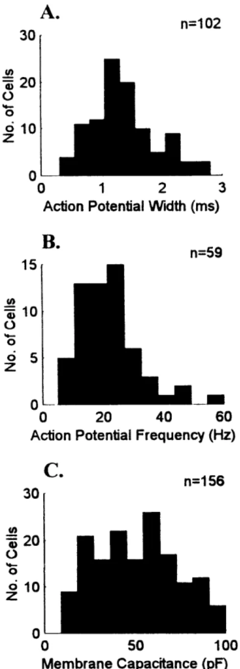
low-threshold calcium currents (Figure IA). Addtionally, interneurons have significantly
narrower action potential widths and higher frequency action potential generation than
relay cells (McCormick and Pape, 1988; Pape and McCormick, 1995). Histograms of
these parameters for the cells that I recorded from do not suggest more than one
population of cell type (Figure 2A,B). Finally, the smaller soma sizes of interneurons
should translate into smaller membrane capacitance. However, as with action potential
width and frequency, membrane capacitance is not obviously distributed into two
Chapter Two: General Methods
populations (Figure 2C), though it is more broadly distributed and may represent
multiple, overlapping distributions; perhaps due to the different somata sizes of
interneurons and relay cells, but more likely due to the different somata sizes of X and Y
type relay cells.
Patch pipettes were pulled from borosilicate glass (World Precision Instruments)
on a horizontal pipette puller (Sutter Instruments) to tip resistances of 3-8 Mo. The
pipettes were filled with (in mM): potassium gluconate (125), KCl (10), HEPES (10),
sodium EGTA (1), CaCl2 (0.1), MgCl2 (2), Na-ATP (2), Na-GTP (0.2), or cesium
gluconate (120), HEPES (10), sodium EGTA (1), CaCl2 (0.1), MgCl2 (2), Na-ATP (2),
Na-GTP (0.1); the pH was adjusted to 7.3.
Retinal afferents were stimulated by delivering constant current through a bipolar
stimulating electrode positioned in the optic tract at the lateral edge of the slice. Cortical
afferents were stimulated in the perigeniculate nucleus just medial to the LGN. The
particular stimulation protocols used are detailed in the method sections of each of the
data chapters.
Likewise, the particular use of pharmacological agents is described in the method
sections of each of the data chapters. In summary, the agents used were: lidocaine
N-ethyl bromide quaternary salt (QX-314, Research Biochemicals International [RBI]) for
the intracellular blockade, and tetrodotoxin (TTX, Sigma) for the extracellular blockade,
of sodium channels; bicuculline'methiodide (BMI, Sigma),
Data Acquisition
2,3-dione (CNQX disodium, RBI), D-2-amino-5-phosphonopentanoic acid (D-AP5,
RBI), and x-methyl-4-carboxyphenylglycine (MCPG, RBI) for the extracellular blockade
of GABAA, AMPA, NMDA, and metabotropic glutamate receptors, respectively. BMI
(50 pM) was used in all recordings in Chapters Five and Six. The data in Chapter Three
is not affected by synaptic currents so recordings both with and without BMI present are
pooled. The effect of the presence or absence of BMI is examined explicitly in Chapter
Four.
Data Acquisition
Recordings were obtained with an Axopatch-200 amplifier (Axon Instruments). Data
were acquired off-line with the pClamp data acquisition software (Axon Instruments), digitized using a Neurocorder encoding unit (Neurodata), and stored on video tape and
computer disk for off-line analysis. Recordings were low-pass filtered at 1 to 5 kHz and
sampled at 10 kHz.
Cells that had resting membrane potentials more hyperpolarized than -40 mV
upon patch rupture and generated overshooting action potentials were considered for
analysis. For examination of resting potential properties, all cells were voltage-clamped
at -60 mV.
Cell membrane capacitance was charged using the independent circuitry in the
Axopatch-200 designed for that purpose. Briefly, values for access resistance and
membrane capacitance can be accurately determined through canceling whole-cell