HAL Id: inserm-00143572
https://www.hal.inserm.fr/inserm-00143572
Submitted on 20 Dec 2017
HAL is a multi-disciplinary open access
archive for the deposit and dissemination of
sci-entific research documents, whether they are
pub-lished or not. The documents may come from
teaching and research institutions in France or
abroad, or from public or private research centers.
L’archive ouverte pluridisciplinaire HAL, est
destinée au dépôt et à la diffusion de documents
scientifiques de niveau recherche, publiés ou non,
émanant des établissements d’enseignement et de
recherche français ou étrangers, des laboratoires
publics ou privés.
Peter Robinson, Emilio Arteaga-Solis, Clair Baldock, Gwenaëlle
Collod-Béroud, Patrick Booms, Anne de Paepe, Harry Dietz, Gao Guo, Penny
Handford, Daniel Judge, et al.
To cite this version:
Peter Robinson, Emilio Arteaga-Solis, Clair Baldock, Gwenaëlle Collod-Béroud, Patrick Booms, et
al.. The molecular genetics of Marfan syndrome and related disorders.. Journal of Medical Genetics,
BMJ Publishing Group, 2006, 43 (10), pp.769-87. �10.1136/jmg.2005.039669�. �inserm-00143572�
REVIEW
The molecular genetics of Marfan syndrome and related
disorders
P Robinson, E Arteaga-Solis, C Baldock, G Collod-Be´roud, P Booms, A De Paepe, H C Dietz,
G Guo, P A Handford, D P Judge, C M Kielty, B Loeys, D M Milewicz, A Ney, F Ramirez,
D P Reinhardt, K Tiedemann, P Whiteman, M Godfrey
. . . . J Med Genet 2006;000:1–23. doi: 10.1136/jmg.2005.039669
Marfan syndrome (MFS), a relatively common autosomal
dominant hereditary disorder of connective tissue with
prominent manifestations in the skeletal, ocular, and
cardiovascular systems, is caused by mutations in the gene
for fibrillin-1 (FBN1). The leading cause of premature
death in untreated individuals with MFS is acute aortic
dissection, which often follows a period of progressive
dilatation of the ascending aorta. Recent research on the
molecular physiology of fibrillin and the pathophysiology
of MFS and related disorders has changed our
understanding of this disorder by demonstrating changes
in growth factor signalling and in matrix-cell interactions.
The purpose of this review is to provide a comprehensive
overview of recent advances in the molecular biology of
fibrillin and fibrillin-rich microfibrils. Mutations in FBN1
and other genes found in MFS and related disorders will be
discussed, and novel concepts concerning the complex and
multiple mechanisms of the pathogenesis of MFS will be
explained.
. . . .
See end of article for authors’ affiliations . . . . Correspondence to: Dr Peter N Robinson, Institute of Medical Genetics, Charite´ University Hospital, Humboldt University, Augustenburger Platz 1, 13353 Berlin, Germany; peter.robinson@charite.de Revised version received 7 March 2006 Accepted for publication 9 March 2006
. . . .
M
arfan syndrome (MFS; MIM 154700) i;
s arelatively common autosomal dominant hereditary disorder of connective tissue with prominent manifestations in the skeletal, ocular, and cardiovascular systems. MFS is caused by mutations in the gene for fibrillin-1 (FBN1). Many affected individuals have a char-acteristic habitus with tall stature, long slender limbs (dolichostenomelia), arachnodactyly, sco-liosis, and pectus excavatum or carinatum. Ectopia lentis affects up to 80% of individuals with MFS and is almost always bilateral. The leading cause of premature death in untreated individuals with MFS is acute aortic dissection, which follows a period of progressive dilatation of the ascending aorta. Recent comprehensive treatments of the clinical aspects of MFS have been published.1 2
A review of the molecular genetics of MFS appeared in these pages in the year 2000.3In the
half decade since then, remarkable progress has been made in elucidating the molecular anatomy of both fibrillin and the fibrillin-rich microfibrils as well as in understanding the molecular pathogenesis of MFS. Initial ideas about the pathogenesis of MFS concentrated on a static dominant negative model based on the concept
of fibrillin-rich microfibrils as purely architec-tural elements in the extracellular matrix. Recent findings greatly enhanced our understanding of the pathogenesis of MFS by demonstrating changes in growth factor signalling and other changes in matrix-cell interactions, which has set the stage for attempts to develop novel forms of treatment.
The current review will focus on the fibrillin-LTBP gene family and tissue organisation of microfibrils, mutations in genes associated with MFS and related phenotypes, the structure of fibrillin-1 domains, FBN1 mutations and proteo-lysis, and the genetics of MFS in mouse models. The current review will highlight advances published after the first review; interested read-ers are referred to the previous article for more information on other topics.3
THE FIBRILLIN-LTBP GENE FAMILY
The fibrillins and the latent-TGFb-binding pro-teins (LTBPs) form two closely related protein families with structural and non-structural func-tions in the extracellular matrix. Both families are characterised by a modular domain structure with repeated cysteine-rich modules.
Members of the fibrillin-LTBP family have both structural and non-structural functions. Three closely related fibrillins have been described. Fibrillin-24and the recently discovered
fibrillin-35have a domain organisation identical
to that of fibrillin-1 and an overall level of amino acid identity of between 61% and 69%. Several lines of evidence suggest that the fibrillins have both overlapping and unique functions.5 6 All
three fibrillins are structural components of
Abbreviations: BMP, bone morphogenetic protein; cbEGF, calcium binding epidermal growth factor; CCA, congenital contractural arachnodactyly; CMN, cystic medial necrosis; CSGE, conformation sensitive gel electrophoresis; DHPLC, denaturing high performance liquid chromatography; EBP, elastin-binding protein; ECM, extracellular matrix; HNPCC, hereditary non-polyposis colorectal cancer; LAP, latency-associated peptide; LDS, Loeys-Dietz aortic aneurysm syndrome; LLC, large latent complexes; LTBPs, latent-TGFb-binding proteins; MAGP-1, microfibril-associated glycoprotein-1; MFS, Marfan syndrome; MFS2, type 2 Marfan syndrome; MMP, matrix metalloproteinase; MMR, mismatch repair; MSI, microsatellite instability; NMR, nuclear magnetic resonance; PTC, premature termination codon; SSCP, single stranded conformation polymorphism; TAAD, thoracic ascending aortic aneurysms and dissections; TbRII, type II TGFb receptor; TGFb, transforming growth factor-b; UMD, Universal Mutation Database; WMS, Weill-Marchesani syndrome
microfibrils4 5 7; however, fibrillin-2 and fibrillin-3 are
pre-ferentially expressed in embryonic developmental stages,4 5 8
whereas fibrillin-1 is expressed from the gastrula to throughout adult life.9 In addition, distinct phenotypes are
observed in fbn1 and fbn2 gene targeting experiments in mice; since microfibrils are assembled in both mice, some overlap of architectural functions appears likely.5 10–12
The LTBPs associate with transforming growth factor-b (TGFb), thereby regulating its secretion and spatial and temporal activation; in humans, four members of the LTBP family are known, three of which undergo alternative splicing.13TGFb is synthesised as a homodimeric proprotein,
whereby the dimeric propeptide is cleaved intracellularly from the growth factor; the propeptide is called the latency-associated peptide (LAP) because TGFb cannot bind to its surface receptors when it is bound to the LAP. The LAP in turn is usually disulfide-bonded to an LTBP; this aggregate is referred to as the large latent complexes (LLC). The LTBPs thus have a dual function: as structural components of the extracellular matrix and as modulators of TGFb availability (the reader is referred to Rifkin14 and Todorovic et al15 for
recent reviews on this topic).
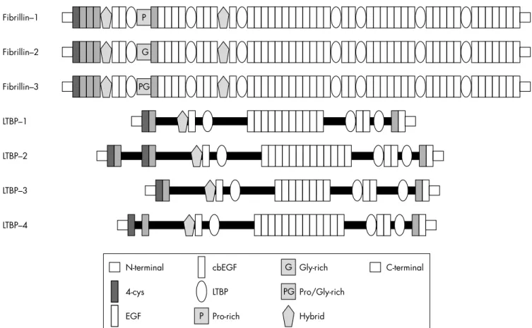
The fibrillins and LTBPs display a remarkably similar domain structure made up of repeated cysteine-rich struc-tural modules including multiple copies of an epidermal growth factor-like module (EGF) and an 8-cysteine (8-cys) module found only in the fibrillins and LTBPs (fig 1). EGF modules are approximately 45 residues in length and are characterised by six conserved cysteine residues that form three intramodule disulfide bonds. Forty three of the 47 EGF modules in fibrillin-1 and many of those in the various LTBPs additionally conform to a consensus sequence, (D/N)-X-(D/ N)(E/Q)Xm(D/N)*Xn(Y/F), that mediates calcium binding in
the N-terminal region of the module (m and n are variable, and an asterisk denotes b-hydroxylation).16
The 8-cys module (also variously referred to in the literature as the 8-cysteine module, the TB module, or the LTBP module) occurs only in the fibrillins and LTBPs. An experiment using a recombinant construct of one of the seven 8-cys modules of fibrillin-1 showed that the structure is stabilised by four intradomain disulfide bonds.17Some 8-cys
domains can mediate binding to the LAP-TGFb complex.18 19
Not all domains have this function; a recent study showed that LTBP1 and LTBP3 bound efficiently to TGFb, LTBP4 bound weakly, and LTBP2 and fibrillin-1 and fibrillin-2 did not bind.20 An additional cysteine-rich module with simila-
=
rities to both the cbEGF module and the 8-cys module, termed the 8-cys hybrid module,21 can mediate
intermole-cular disulfide bonding between fibrillin-1 monomers, which may be an important step in the assembly of microfibrils.22
The fibrillins share a globular C-terminal domain of about 120 residues with the fibulins23; in fibrillin-1, this module
may be involved in homotypic interactions.24
TISSUE ORGANISATION OF MICROFIBRILS
Fibrillin microfibrils are widely distributed extracellular matrix multimolecular assemblies comprised of fibrillin and other proteins. The microfibrils endow elastic and non-elastic connective tissues with long range elasticity. They direct tropoelastin deposition during elastic fibrillogenesis and form an outer mantle for mature elastic fibres. Microfibril arrays are also abundant in dynamic tissues that do not express elastin, such as the ciliary zonules of the eye. Analysis of fibrillin-rich microfibrils by transmission electron microscopy has revealed them to have a diameter of 8–12 nm, a tubular appearance, and beaded periodicity, and to contain glyco-proteins. Isolated fibrillin-rich microfibrils have a complex ‘‘beads on a string’’ appearance, and are extensible.
Fibrillin–1 P Fibrillin–2 G Fibrillin–3 PG LTBP–1 LTBP–2 LTBP–3 LTBP–4 G PG P N-terminal 4-cys P EGF cbEGF LTBP Pro-rich Gly-rich Pro/Gly-rich Hybrid C-terminal
Figure 1 The fibrillin-1 gene FBN1 spans about 235 kb of genomic DNA on chromosome 15q21.1, and has a transcript size of 9749 nucleotides; the
<
coding sequence of FBN1 is spread over 65 exons,21and three alternatively spliced non-coding 59 exons have been described.249With severalMicrofibrils form loosely packed bundles in roughly parallel alignment. These bundles adopt tissue-specific architectures that are dictated by cells, and by the strength and direction of forces put upon the tissue. They are found in locations that are subject to repeated mechanical stresses, and in the proximity of basement membranes, and they serve a critical biomechanical anchoring role in dynamic connec-tive tissues.25
Elastic fibre formation is a developmentally regulated process in which tropoelastin (the soluble precursor of mature elastin) is deposited on a preformed template of fibrillin microfibrils.26 Mature elastic fibres are a composite
biomaterial with an outer microfibrillar mantle and an inner core of amorphous cross-linked elastin with some embedded microfibrils. The proportion of microfibrils to elastin appears to decline with age, with adult elastic fibres often having only sparse peripheral mantles of microfibrils.
A recent investigation by mass spectrometry of the composition of purified fibrillin-rich microfibrils from non-elastic and non-elastic tissues showed that in all microfibril preparations, fibrillin-1 was abundant and the only fibrillin isoform detected. Isolated microfibrils, extracted from tissues by enzyme digestions or homogenisation, have a ‘‘beads on a string’’ appearance with untensioned periodicity of ,56 nm.27 28 Unextracted hydrated zonular microfibrils
appeared, by quick freeze deep etch microscopy, to be more tubular, suggesting that molecular components are lost or that there is a major molecular rearrangement on extrac-tion.29 The molecular basis of the ‘‘beads’’, and fibrillin-1
alignment in microfibrils remain unclear. Models of fibrillin-1 alignment
The details of the precise molecular architecture of fibrillin-1 alignment within microfibrils are not entirely clear, and several models have been proposed. An intermolecular fibrillin-1 transglutaminase cross-link30 provides molecular
constraints, although mass spectrometry has shown that not all fibrillin-1 molecules within tissue microfibrils are cross-linked.31
The ‘‘hinge’’ model, based on detailed scanning transmis-sion electron microscopy, mass mapping, automated electron tomography, and atomic force microscopy data,28 32–34predicts
maturation from an initial parallel head to tail alignment to an approximately one third stagger (,100 nm) that would allow transglutaminase cross-link formation, and further packing i
>
nto a more energetically favourable ,56 nm untensioned form.The one third staggered model was suggested on the basis of extrapolation of molecular dimensions,35 the crystal
structure of fibrillin-1 cbEGF/TB/cbEGF domain arrays,36
and calcium binding studies of TB/cbEGF flexibility.37
Interested readers can find further discussion of issues surrounding fibrillin-1 alignment and microfibrillar architec-ture in Baldock et al,28Davis et al,29, Kielty et al,34and Lee et
al.36
Early studies of isolated microfibrils revealed a number of highly stretched microfibrils with periodicities up to ,150 nm, which suggested that microfibrils may have elastic properties.27Stretching of isolated intact microfibrils has been
achieved using surface tension forces and molecular comb-ing.33 These studies showed that microfibrils behave as
relatively stiff elastic filaments which can perform anchoring roles in ciliary zonules and other basement membrane interfaces. It was confirmed by x ray diffraction that hydrated microfibril bundles are elastic, and suggested that elasticity in microfibril-rich tissues may arise, in part, from reversible alterations in supra-microfibrillar arrangements. Proteolytic damage to microfibrils that may occur in MFS or ageing, may generate stretched microfibrils that may have lost their elastic properties.25 38
ASSEMBLY OF MICROFIBRILS
Today, a fragmented picture has emerged of the events and molecules involved in the assembly process from profibrillins to mature tissue microfibrils. The mechanisms include profibrillin processing, self-assembly, regulatory events, cross-link formation, and maturation of microfibrils. Microfibril assembly in individuals with MFS
Early immunofluorescence studies with dermal fibroblasts or skin biopsies obtained from individuals with MFS showed reduced or qualitatively altered fibrillin networks as com-pared to controls.39–41However, such altered patterns were not
observed in all samples.41 42 One possible interpretation of
these data is that a subset of mutations in fibrillin-1 compromises the assembly process and thus the formation of microfibril networks. Further evidence for this interpreta-tion comes from pulse chase experiments using dermal fibroblasts from individuals with MFS. These analyses showed deficiencies at different levels such as fibrillin synthesis and secretion, as well as deposition into the extracellular matrix.43 44 Despite the differences in the
secreted amount of fibrillin, a large portion of the fibroblasts studied showed impaired incorporation of the mutant fibrillin into the extracellular matrix, suggesting functional disturbances in early stages of the assembly mechanism.45 46
In cases where higher order assembly into beaded micro-fibrils was observed, the ultrastructural appearance showed
R522N
Cancer
LDAS
Extracellular Protein kinase
del32–37 A(10): frameshift L308P T315M Y336N A355P G357W V387M V447A N435S L452M R460 skipEx6 S449F E526G R528H R528C R528H R537C
Figure 2 Mutations in TGFBR2 found in Loeys-Dietz aortic aneurysm syndrome (LDS)112and related hereditary disorders111 113and representative
missense mutations found in colon carcinoma166 250and breast carcinoma.251The TGFBR2 protein is drawn according to UniProt entry P37173 and
comprises an N-terminal signal sequence, an extracellular domain, a transmembrane domain (stippled rectangle), a cytoplasmatic region of unknown significance (the white box), the serine-threonine protein kinase domain, and a C-terminal domain (black rectangle).
several types of abnormalities including diffuse, frayed, or poorly defined interbead domains, or variable interbead periodicities.47–49 These consequences potentially could also
emerge from functional problems in the assembly mechan-ism. In summary, although molecular evidence is still lacking, it seems clear that a certain subset of mutations in fibrillin-1 leading to MFS and other microfibrillopathies directly affects microfibril assembly mechanisms.
Role of propeptide processing in fibrillin assembly Fibrillins are phylogenetically old proteins occurring in species from jellyfish to human. All known fibrillins possess highly conserved basic recognition sites (RX(K/R)R) for processing by endoproteinases of the furin/PACE type within the unique N- and C-terminal domains. It has been shown by several groups and methodologies that fibrillin-1 is indeed processed at these recognition sites.22 50–54 Processing
pro-duces a 17 or 20 residue N-terminal propeptide depending on the actual cleavage site for the signal peptide, and a 140 residue C-terminal propeptide. Due to the size of the propeptide, C-terminal processi
?
ng is accessed much better experimentally and thus has received more attention. For fibrillin-1, it has been demonstrated that processing of the C-terminal propeptide is required for deposition into the extracellular matrix, suggesting that profibrillin-1 conversion to mature fibrillin-1 plays a regulatory role in assembly into higher order aggregates.43 50 53On the molecular level, it is notclear how the presence of a C-terminal propeptide prevents matrix deposition and assembly. The propeptide may interact and mask self-assembly sites in fibrillin-1.24 A mutation in
fibrillin-1 (R2726W) associated with isolated skeletal fea-tures of MFS was shown to interfere with normal processing at the C-terminal end and thus disturbed the incorporation of the mutated protein into the extracellular matrix.50 Other
mutations close to processing sites may have similar consequences. Virtually no information is available as regards the functional role of the N-terminal processing. However, by analogy, it is predicted that the N-terminal propeptide also regulates assembly of fibrillins.
Self-assembly of fibrillins
As described in the ‘‘Tissue organisation of microfibrils’’ section, the molecular organisation of fibrillin-1 in micro-fibrils has been analysed by various groups resulting in a number of different models for the alignment of fibrillin in microfibrils.7 28 30 35 36 51 Despite the differences in these
models in terms of stagger and molecular condensation of individual molecules, common to all models is a head to tail orientation of fibrillin-1 molecules as originally proposed by Sakai and coworkers in 1991.7 Another commonly accepted
property of microfibrils is the involvement of six to eight fibrillin molecules per cross-section of the interbead region.28 55 56
Correlation of antibody epitopes in the fibrillin-1 molecule with the location of the corresponding epitopes in micro-fibrils clearly revealed that the terminal ends of the fibrillin molecules are situated in or close to the beads.7 28 51 Data
obtained with recombinant fibrillin-1 fragments have estab-lished direct interaction in a homotypic N- to C-terminal fashion.57These results established a linear head to tail
self-assembly mechanism for fibrillin-1. Heterotypic interactions between fibrillin-1 and fibrillin-2 in an N- to C-terminal fashion suggested that both fibrillin isoforms can be organised within the same microfibril,57 and colocalisation
of both isoforms has indeed been demonstrated by double immunogold labelling in tissue microfibrils.8Electron
micro-scopy after rotary shadowing of full length recombinant fibrillin-1 suggested that the interaction epitopes are rela-tively close to the terminal ends.57These results were further
substantiated by analyses of smaller overlapping fibrillin-1
fragments in various ligand interaction assays, positioning the interaction sites in the N-terminal region encoded by
@
exons 1–8 and the C-terminal region encoded by exons 57– 65.24In addition to a linear head to tail self-interaction, there is evidence that lateral homotypic interactions also play a role in fibrillin assembly. Reducible homodimer formation early during biosynthesis was observed for recombinant fragments of fibrillin-1 and -2 spanning from the proline and glycine-rich domains, respectively, to the second 8-cys/TB domain,58
A
as well as for smaller recombinant fragments of the proline-rich region of fibrillin-1 and the glycine-proline-rich region of fibrillin-2 including flanking domains.59Additionally,
homo-typic lateral interactions have been observed with an N-terminal recombinant fibrillin-1 fragment encoded by exons 1–8, as well as with an C-terminal fragment encoded by exons 57–65.24The lateral homotypic interactions between
N-terminally and C-N-terminally located fragments as well as the head to tail linear interactions between the N- and C-terminal ends are of high affinity with dissociation constants in the low nanomolar range, indicating that both types of mechanisms are highly relevant for initial assembly stages.24 57
Intermolecular cross-link formation in fibrillin assembly
Two types of intermolecular cross-links important for the stability of microfibrils have been identified: reducible disulfide bonds and non-reducible e(c-glutamyl)lysine cross-links. Intermolecular disulfide bond formation is apparent early in the assembly of microfibrils since higher molecular weight disulfide-bonded aggregates containing fibrillin can be observed after a few hours in cell or organ cultures.22 60Most of the highly conserved cysteine residues in
fibrillins are predicted to stabilise individual domains through intramolecular disulfide bonds.17 35 36 One cysteine
residue in the first hybrid domain of human fibrillin-1 and -2 has been shown to be available for intermolecular cross-links on the surface of the molecule.22
Additional data suggest that other cysteine residues, which are normally involved in intramolecular domain stabilisation, may be reshuffled to participate in intermolecular cross-links. Cysteine residues in the first and second 8-cys/TB domain
B
have been suggested for such a role based on the propensity of various recombinant fibrillin-1 and -2 fragments to form reducible homodimers.58 59 Such a mechanism would likelyrequire the enzymatic activity of one or more protein disulfide isomerases on the cell surface or in the extracellular matrix.
Non-reduciblee(c-glutamyl)lysine cross-links, catalysed by transglutaminases, have been identified in microfibrils extracted from various tissues.30 61 62 Detailed analyses of
microfibrils from human tissues have identified transgluta-minase cross-links in the N- and C-terminal regions of fibrillin-1, as well as a high overall content of these cross-links.30In addition to fibrillin-1, another prominent protein
in microfibrils, microfibril-associated glycoprotein-1 (MAGP-1), was also characterised as a substrate for transglutami-nase.63 It is possible that besides homotypic fibrillin-1
transglutaminase cross-links, heterotypic fibrillin-1-MAGP-1 cross-links may be present in microfibrils. Zonular fibres in the eye have been demonstrated to be a target for transglutaminase 2.64Biomechanical analyses of microfibrils
suggested that transglutaminase cross-links play an impor-tant role in strengthening the microfibrils.65Other potential
roles of the transglutaminase cross-links may include correct lateral alignment of fibrillin or other molecules as a prerequisite for downstream assembly events. Mutations disrupting transglutaminase cross-link sites likely result in
serious consequences for microfibril assembly and stability. To gain insight into such potential mechanisms in MFS and other microfibrillopathies, it will be important to identify the exact amino acid residues involved and the time course of transglutaminase cross-link formation.
Accessory molecules important for fibrillin assembly In addition to self-assembly and cross-linking mechanisms, other molecules may be involved in the assembly process of microfibrils. Several regions in fibrillin-1 have been identified as interacting with heparin/heparan sulfate with high affinity.66–68In cell culture assembly assays, these
glycosami-noglycans inhibit the formation of microfibrillar net-works,66 67leading to the hypothesis that heparan sulfate or
heparan sulfate containing proteoglycans may have regula-tory functions in the assembly of microfibrils. Recently, the heparan sulfate containing proteoglycan perlecan was iden-tified as a molecule which interacts with fibrillin-1 and with microfibrils close to basement membrane zones.69 Reduced
amounts of microfibrils in basement membrane zones of perlecan null mice may reflect a potential role for perlecan in microfibril assembly.69
MICROFIBRIL-ASSOCIATED PROTEINS
In addition to fibrillin, several other proteins are integral components of the microfibrils or associated with them. These can be grouped into small non-fibrillin proteins that are integral parts of fibrillin-rich microfibrils (table 1) and proteins that can associate with fibrillin-rich microfibrils but do not serve an integral structural function (table 2). We will not attempt a comprehensive review of the non-fibrillin proteins but rather will summarise the most important functions that have been attributed to these proteins and their interaction with fibrillin. Readers are referred to Gibson70and the references in the tables 1 and 2 for further
information. It appears likely that more interacting proteins remain to be discovered in light of a recent proteomics study of fibrillin-rich microfibrils showing copurification of a number of proteins in addition to fibrillin-1. For instance, c-crystallin copurified with zonular microfibrils, suggesting an interaction that could contribute to zonule anchorage to the lens.31
Structure and assembly of microfibrils
One role of non-fibrillin microfibrillar proteins is as structural constituents of the microfibrils. For instance, microfibril-associated glycoprotein-1 (MAGP-1) is a small glycoprotein with an apparent molecular weight of 31 kDa that is covalently bound to microfibrils by disulfide linkages71and
is specifically located on the beads of the beaded-filament structure of the microfibrils.72MAGP-2 has a more restricted
tissue distribution than MAGP-1,73 suggesting it may have
tissue- or developmental stage-specific functions. MAGP-1 and MAGP-2 bind to distinct regions of fibrillin-1 and it has been suggested that this may help to regulate microfibrillar assembly.74
Interaction with other matrix components
Interactions between fibrillin-rich microfibrils and tropoelas-tin are important for the formation of elastic fibres. In addition to interactions between fibrillin-1 and tropoelastin,75
MAGP-1 interacts with tropoelastin76 77 in a way that is
important for tropoelastin deposition.78Microfibrils interact
with a variety of other extracellular matrix structures and some of these interactions are mediated by non-fibrillin proteins. For instance, MAGP-1 binds to the a3chain of type
VI collagen. Since type VI collagen microfibrils and fibrillin-rich microfibrils are often found near to one another in some extracellular matrices, it is plausible that MAGP-1 may mediate a molecular interaction between type VI collagen microfibrils and fibrillin-containing microfibrils.79
Table 1 Small non-fibrillin proteins that are integral parts of fibrillin-rich microfibrils in at least some tissues and developmental stages
Protein Chromosome Potential functions MAGP-1 1p36.1–p35 Tropoelastin deposition78
Tropoelastin binding76 89
Binding to fibrillin-176 90
Binding to fibrillin-291
Ternary complex with tropoelastin and biglycan77
Posttranslational modifications92
Ternary complex with tropoelastin and biglycan93
Binding to type VI collagen79
Substrate for transglutaminase63
MAGP-2 12p12.3–p13.1 Binding to fibrillin-174 90
Binding to fibrillin-274
RGD-mediated cell attachment94
Interaction with Jagged180
AAAP-40 5q32–q33.2 40 kDa protein95 96
(MAGP-3)
MFAP1 (AMP) 15q15–q21 54 kDa protein that is processed to a 36 kDa protein97 98
MFAP3 5q32–q33.1 41 kDa serine-rich protein99
MFAP4 17p11.2 Colocalisation to elastic fibres100
(MAGP-36) Role in elastogenesis101
Table 2 Proteins that can associate with fibrillin-rich microfibrils but do not serve an integral structural function in them
C
Protein Class of protein Potential biological role of interaction Elastin – Tropoelastin deposition78
Regulation of microfibril formation75
LTBP-1 Fibrillin-LTBP Sequestering of latent TGFb102
LTBP-2 Fibrillin-LTBP Structural role?103
LTBP-4 Fibrillin-LTBP Sequestering of latent TGFb102
Versican Proteoglycan (hyalectin) Link fibrillin-microfibrils to versican/hyaluronan network fibrillin-LTBP104
Perlecan Proteoglycan (hyalectin) Anchoring microfibrils to basement membranes and in the biogenesis of microfibrils69
Heparin/heparan sulfate Proteoglycan Binding of related heparan sulfate chains may regulate cell-surface assembly of fibrillin66
Decorin Small dermatan sulfate proteoglycan Induction of fibrillin-1 expression in renal fibroblasts and mesangial cells105
Biglycan Small dermatan sulfate proteoglycan Role in elastogenesis77
Fibulin-2 Fibulin Mediate/modulate attachment of fibrillin to tropoelastin106
Fibulin-5 Fibulin Regulation of the initial deposition of tropoelastin on to microfibrils107
BMP-7 Bone morphogenetic protein Regulation of limb patterning12
Interaction with cells
It has become increasingly clear that fibrillin-rich microfibrils have functions that are not directly related to structural integrity but rather have to do with growth factor metabolism and triggering cellular signals. As will be discussed in more detail below, the latent transforming growth factor-b binding proteins (LTBP) are a family of secreted glycoproteins, three of which play an important role in the regulation of TGFb regulation.15 In addition to TGFb, microfibrils could
con-ceivably be involved in the regulation of other growth factors, although little experimental evidence is available at this point.14 MAGP-2 can interact wi
EX
th Jagged1 and induce its shedding; it is thus conceivable that MAGP-2 may be able to modulate the Notch signalling pathway.80There is evidence ofinteraction between fibrillin-1 and at least one bone morphogenetic protein (BMP),81 82and fibrillin-2-rich
micro-fibrils and BMP-7 have been shown to functionally interact in the regulation of limb patterning in a mouse model.12
Finally, an interesting area of research is the question whether novel cell signalling pathways are triggered in MFS or other diseases of the microfibrils, and whether fragments of the proteins of the microfibrils can acquire novel signalling properties through exposure of otherwise cryptic binding sites, a phenomenon that has been well described in several other disorders such as osteoarthritis.83
As will be discussed below, there is evidence that fibrillin fragments can induce matrix metalloproteinase (MMP) expression, and it is well known that elastin fragments can induce MMP expression.84 85
Enzymatic activity
The enzyme lysyl oxidase, which is involved in the cross-linking of tropoelastin monomers, has been localised to the interface between extracellular bundles of amorphous elastin and the microfibrils.86 Although there is no evidence of a
direct interaction between fibrillin and lysyl oxidase to date, it is plausible that interactions with fibrillin or other microfibrillar components might be important for elastogen-esis.70 Interestingly, ADAMTS10,87 an extracellular matrix
protease, is mutated in the autosomal recessive form of Weill-Marchesani syndrome (WMS).88The dominant form of WMS
is caused by mutations in fibrillin-1, suggesting, perhaps, a potential interaction between ADAMTS10 and fibrillin-1. However, the substrates of ADAMTS10 or indeed other potential functions of this molecule remain to be elucidated. Mutations in FBN1, FBN2, and other genes associated with MFS and related phenotypes
Mutations in FBN1 were discovered in individuals with MFS in 1991,109 and subsequently mutations in FBN2 were
discovered in individuals with a phenotypically related disorder, congenital contractural arachnodactyly (CCA).110
More recently, mutations in the genes for TGFBR1 and TGFBR2 were found in several disorders with varying degrees of overlap with classic MFS.111–113Genetic loci for other forms
of isolated aortic dilatation and dissection have been identified. The identification of mutations in these genes has provided significant insight into the pathogenetic path-ways involved in MFS and related disorders, and further insight is to be expected from characterising the full spectrum of mutations associated with these disorders and from identifying the full set of genes in which mutations cause related disorders of connective tissue. The following sections present the current state of knowledge on FBN1, FBN2, and TGFBR2 mutations and on loci involved in isolated aortic aneurysm and dissection.
MUTATION ANALYSIS OF THE FBN1 GENE IN INDIVIDUALS WITH MFS: SENSITIVITY, METHODS, AND CLINICAL INDICATIONS
In most instances, the diagnosis of MFS can be made on clinical grounds. A set of clinical diagnostic criteria, as summarised in the Ghent nosology,114define major criteria
with high diagnostic specificity and minor criteria with less specificity. In order to make a diagnosis of MFS, the Ghent nosology requires a combination of major criteria in at least two organ systems and involvement of a third organ system. In the majority of cases, these criteria allow the establish-ment or exclusion of the diagnosis. However, the interpreta-tion of these criteria is not always obvious for a number of reasons. Firstly, MFS is known for its extensive phenotypic variability both within and between families, which may cause underdiagnosis of the condition.115Secondly,
establish-ing a diagnosis of MFS in children can be difficult because several manifestations of MFS are age-dependent and may not yet be present in childhood. Thirdly, clinical overlap exists between MFS and other, so called Marfan-like conditions which share some of the features of MFS but do not necessarily have the same outcome.
Therefore, if the Ghent criteria are fulfilled, then the diagnosis of MFS is certain. In adult patients presenting with a small number of non-specific skeletal manifestations often seen in MFS, the diagnosis is unlikely, but a full clinical evaluation including echocardiography and ophthalmologic examination is indicated.
In situations of clinical uncertainty, molecular analysis of the FBN1 gene therefore seems a logical aid to the clinical diagnosis of MFS. In practice, however, the large and complex structure of the FBN1 gene and the wide scope of FBN1 mutations have hampered clinical implementation of FBN1 testing. Moreover, literature data show great variation in detection rates of FBN1 mutations and methodologies used. This can be accounted for by a variety of factors such as the type of mutational analysis method, the substrate (genomic versus cDNA) and, most importantly, the accuracy of the clinical diagnosis. The studies that have looked at the sensitivity and specificity of FBN1 mutation analysis show substantial differences in one or several of these factors.
The first mutational studies, performed on cDNA, showed low efficiency of FBN1 mutation detection using SSCP (single stranded conformation polymorphism) and yielded mutation rates of 9% to 23%.116 117Subsequent studies, using genomic
DNA as template for analysis of the 65 individual exons of the FBN1 gene, obtained higher detection rates but varied according to the type of screening method used.118–120
Initially the best results were obtained with CSGE (con-formation sensitive gel electrophoresis), with detection rates ranging from 57% to 90%.42 121 122 Subsequent studies have
EO
shown that mutation detection by DHPLC (denaturing high performance liquid chromatography) is highly efficiency,123
although a relatively high false-positive rate may be an issue.124 It now appears that DHPLC is the most efficient
approach for mutation detection also because of its potential for automation when combined with robotic PCR.
The most important factor influencing the mutation detection rate appears to be the clinical homogeneity or heterogeneity of the patient population. Indeed, several studies have shown that the incidence of FBN1 mutations is significantly higher in patients who fulfil the MFS diagnostic criteria than in patients who do not. In a large study including a cohort of 94 MFS patients and 77 patients with MFS related phenotypes, Loeys et al125 found FBN1
detection rates of 66% versus 5%, respectively. They demon-strated that fulfilling the clinical diagnosis of MFS in itself is a good predictor of the outcome of FBN1 mutation analysis. Katzke et al120supported these findings by their study which
showed a much higher incidence of mutations in a group of MFS patients versus those with an MFS related condition, and concluded that clinical overdiagnosis is the most important explanation for low FBN1 mutation detection rates. Two other studies123 126also showed that the majority of
patients in whom an FBN1 mutation was found, met the clinical diagnosis of MFS, supporting the opinion that the robustness of selection criteria is the most important determinant of the outcome of mutational studies.
Several clinical situations can occur in which molecular studies of the FBN1 gene may be helpful. In patients who present with skeletal, cardiovascular, and/or possibly other manifestations of MFS but have no involvement of the ocular system, it can be difficult to establish the diagnosis strictly on clinical grounds, particularly in the absence of a positive family history. Here, however, MRI studies can reveal the presence of dural ectasia in which case the diagnostic criteria may still be met. Several mutational studies which report FBN1 mutations in Marfan-like patients have not verified the presence or absence of dural ectasia, so that the possibility remains that the diagnostic criteria are in fact met. In cases where no definitive conclusion can be reached with the clinical data, molecular analysis of the FBN1 gene is an alternative option.
Because of the evolving nature of the phenotype, particu-larly so for the cardiovascular and skeletal manifestations, children with suspected MFS may not yet fulfil the diagnostic criteria. In those instances, it is better to postpone a final diagnosis until later. The identification of an FBN1 mutation in children or young adults not (yet) fulfilling the diagnostic criteria can help to identify those who need to be clinically followed with particular attention.
FBN1 mutations have been identified in a range of phenotypes, the type 1 fibrillinopathies, with greater or lesser degrees of clinical overlap with MFS (table 3). A decision as to whether mutation analysis is indicated when such phenotypes are suspected needs to be made on an individual basis.
In addition to the more or less well delineated disorders listed in table 3, FBN1 mutations can be found in individuals with Marfan-like disorders who do not fulfil the criteria of the Ghent nosology.127It is recommend that accepted clinical
guidelines for the care and management of MFS are applied in these cases even if the criteria of the Ghent nosology are not fulfilled, because it is possible that complications such as aortic dilatation can emerge at any age.
Finally, the availability of a molecular test also allows prenatal or preimplantation diagnosis for prospective parents. This is one option for which requests appear to be steadily increasing.
In summary, after stringent clinical selection, a detection rate of up to 90% is currently achievable in FBN1 molecular testing of patients with classic MFS.128 This allows the
implementation of mutational studies in clinical practice.
THE UMD FBN1 DATABASE: A DATABASE FOR FBN1 MUTATIONS IMPLICATED IN MFS AND RELATED PHENOTYPES
The UMD FBN1 database (http://www.umd.be) was created in 1995 in an effort to standardise the information regarding FBN1 mutations using UMD (Universal Mutation Database) software.143–149 The database follows the guidelines on
mutation databases of the Hugo Mutation Database Initiative including nomenclature of mutations.150
The mutation records of the database include point mutations, large and small deletions, insertions, and splice mutations in the FBN1 gene. Each record contains the molecular and clinical data for a given mutation in a standardised, easily accessible, and summary form; if available, data on fibrillin protein biosynthesis classification groups45are included.
To date, 601 FBN1 mutations are available online. The mutations are spread throughout almost the entire gene without obvious predilection for any given region. Approximately 12% of mutations are recurrent.149
The mutation studies performed to date generally have concentrated on screening the 65 coding exons of FBN1. For the most part, methods capable of detecting larger deletions were not applied and it is not clear how much flanking sequence or other regions of the gene were investigated. It is also unclear whether, once a mutation was identified, the remaining gene regions were regularly and fully evaluated.
Many different kinds of mutation have been identified in FBN1. Point mutations are the most common mutational event, with nonsense and missense mutations comprising about 10% and 60% of all reported mutations. The most common missense mutations substitute cysteine residues that form disulfide bonds within one of the cbEGF or 8-cys domains, but missense mutations creating novel cysteine residues in these modules are also common. The majority of the remaining mutations in these modules affect residues of the calcium consensus sequence. About a quarter of missense mutations affect modules other than cbEGF, and for the most Table 3 Type 1 fibrillinopathies
Syndrome Clinical features Reference MFS See text See text
Neonatal MFS Severe end of clinical spectrum Kainulainen et al,117Booms et al129
Atypically severe MFS Severe and early onset cardiovascular complications Putnam et al,130
Tiecke et al,131
Ectopia lentis Mainly ocular findings Lo¨nnqvist et al,132
Ades et al,133
Kyphoscoliosis Progressive kyphoscoliosis of variable severity Ades et al134
Familial arachnodactyly Dolichostenomelia and arachnodactyly Hayward et al135
Familial thoracic ascending aortic See text aneurysms and dissections
MASS phenotype Mitral valve prolapse, aortic dilatation without dissection, Dietz et al136
skeletal and skin abnormalities
Shprintzen-Goldberg syndrome Craniosynostosis, a marfanoid habitus, and skeletal, Sood et al,137
Kosaki et al138
Robinson et al139
neurological, cardiovascular, and connective tissue anomalies
Isolated skeletal features Tall stature, scoliosis, pectus excavatum, arachnodactyly Milewicz et al50
New variant of MFS Skeletal features of MFS, joint contractures, ectopia lentis, Sta˚hl-Hallengren et al,140
Black et al,141
no cardiovascular manifestations
Weill-Marchesani syndrome Short stature, brachydactyly, joint stiffness, and Faivre et al142
(autosomal dominant) characteristic eye abnormalities
Although classic MFS is by far the most common disorder associated with FBN1 mutations, several other disorders with overlapping clinical findings have been described due to mutations in FBN1.
part, the pathophysiological mechanisms of these mutations remain unclear.
Small insertions, deletions, or duplications represent about 13% of all reported mutations. The majority of these mutations create a premature termination codon (PTC). Another 13% of reported mutations consist of various classes of splicing errors, most commonly affecting canonical splice sequences at exon/intron boundaries. Many splice site mutations in FBN1 result in in-frame exon skipping, such that the mutant fibrillin-1 lacks an entire cbEGF domain. Such mutations can be associated with a particularly severe phenotype.151Some exon-skipping mutations in FBN1 result
in a frameshift152with reduced mutant RNA levels through
nonsense-mediated decay of the mutant transcript.153 A
nonsense mutation136and a silent exonic mutation154in exon
51 have been reported as inducing in-frame skipping of the entire exon 51 and demonstrate the existence of an exonic splicing enhancer.155 156
Global analysis of FBN1 mutations reveals two classes of mutations. The first type, which represents more than one third of the mutations, contains mutations predicted to result in shortened fibrillin-1 molecules, including nonsense muta-tions, splicing errors, insermuta-tions, and duplicamuta-tions, as well as in-frame or out-of-frame deletions. These mutations are likely to result in nonsense-mediated decay resulting in reduction in the level of the mutant allele. The second type represents slightly less than two thirds of the mutations and contains missense mutations, mostly located in cbEGF-like modules. They can be subclassified into: (a) mutations creating or substituting cysteine residues potentially impli-cated in disulfide bonding and consequently in the correct folding of the monomer; (b) amino acids implicated in calcium binding and subsequently in interdomain linkage, structural integrity of affected domains, and increased protease susceptibility; and (c) other mutations that might affect the conformation of affected modules, interdomain packing, or other functions such as protein-protein interac-tions.
Elucidating the molecular basis of MFS and related fibrillinopathies is the major goal of the teams working on this subject.149 157 The extreme clinical variability, the
diffi-culties associated with clinical diagnosis, and the low detection rate of mutations in this large gene all conspire to negatively impact on progress. At present it is not possible to predict the phenotype for a given FBN1 mutation. On the one hand, mutations affecting different positions within a given module may be associated with quite different phenotypes. On the other hand, mutations affecting an analogous residue within two different modules may also be associated with differing phenotypes. Therefore, it is apparent that neither the location of the affected structural module in the protein nor the position of the altered residue is, in itself, sufficient to predict potential genotype-phenotype correlations.158 The
high degree of intrafamilial variability suggests that environ-mental and perhaps stochastic factors or modifying genes are important for the phenotypic expression of disease. The level of the expression of the normal fibrillin-1 allele159 and
hyperhomocysteinaemia related to the C677T methylenete-trahydrofolate reductase polymorphism160 have been
pro-posed as factors that modify the clinical severity of MFS. The elucidation of the full range of modifying factors in MFS represents an interesting area for further research.
MUTATIONS IN TGFBR1 AND TGFBR2
Signalling by TGFb family cytokines controls a variety of cellular processes including proliferation, differentiation, and apoptosis; propagation of signalling into the cell is mediated by a family of type 1 and type 2 receptors including the type 1 and type 2 TGFb receptor. TGFb binds first to type II
receptors, allowing subsequent incorporation of type I receptors into a ligand-receptor complex involving a TGFb dimer and four receptor molecules. The signal is then propagated into the cell by means of phosphorylation of the Smad proteins.161
Linkage to chromosome 3p24.2–p25 was demonstrated for a large family with a Marfan-like phenotype for whom linkage to FBN1 and FBN2 had previously been excluded.162 163
This disorder has been termed MFS type II (MIM 154705) and shares some of the cardiovascular and skeletal features of classic MFS.
Identification of a chromosomal breakpoint disrupting the gene encoding the TGFb receptor 2 (TGFBR2) in a boy with short stature, dural ectasia, and several skeletal and cardiovascular manifestations of MFS led to the identifica-tion of three further missense mutaidentifica-tions in four families or individuals with manifestations of MFS in whom FBN1 mutations had been ruled out (one mutation was found in two unrelated families).111All the mutations were found in
the serine-threonine kinase domain of the TGFb receptor 2. More recently, a new aortic aneurysm syndrome with hypertelorism, bifid uvula or cleft palate, and generalised arterial tortuosity with ascending aortic aneurysm together with other findings such as craniosynostosis, mental retarda-tion, and congenital heart disease was described; this disorder, Loeys-Dietz aortic aneurysm syndrome (LDS; MIM 609192), was shown to be associated with mutations in the genes for either TGFb receptor type 1 or TGFb receptor type 2 resulting in perturbations of TGFb signalling.112As will
be noted below, mutations of the arginine at position 460 of TGFBR2 have been identified in individuals with thoracic
EP
ascending aortic aneurysms and dissections (TAAD), although there appears to be some degree of phenotypic overlap with LDS in many affected individuals.Microsatellite instability (MSI) is a prominent feature in hereditary non-polyposis colorectal cancer (HNPCC) and some forms of acquired colon cancer. Defects in mismatch repair (MMR) genes and associated coding region MSI can cause frameshift mutations with functional inactivation of affected genes, thereby providing a growth advantage to MMR deficient cells. TGFBR2 is one of the most commonly affected genes with mutations in a polyadenine tract in exon 3 being found in up to 90% of cases of microsatellite-instable sporadic and HNPCC associated colon cancer.164 Germline
mutations in TGFBR2, however, are a rare cause of HNPCC.165
Interestingly, one mutation (R528H) was found both as a somatic mutation in colon cancer166 167 and as a germline
mutation in LDS.112 At present, there is no evidence that
individuals with LDS are at increased risk for colon cancer. It should be noted that there has been some controversy as to whether certain TGFBR2 mutations lead to a phenotype that is identical or at least very similar to that of classic MFS, thus justifying the diagnosis of type 2 MFS (MFS2). The question about locus heterogeneity for MFS has been addressed by several studies. Historical linkage data in MFS families showed a cumulative LOD score for the FBN1 locus in excess of 100, which provides evidence for a single predominant locus for MFS. On the other hand, none of the individuals with LDS112fulfilled the clinical diagnostic criteria
for MFS. In a previous study, Loeys et al128identified 86 FBN1
mutations in a cohort of 93 patients with classic MFS. None of the remaining patients had a TGFBR2 or TGFBR1 mutation. Overall, this suggests strongly that FBN1 is the predominant if not sole locus for MFS.128However, there have been reports
of individuals with TGFBR2 mutations diagnosed with MFS2 without features characterising LDS.168 Given that the
aneurysms in LDS appear to be more aggressive than those in MFS and the fact that the cardiovascular involvement is also characterised by arterial aneurysms throughout the
arterial tree and marked arterial tortuosity, it is clinically very important to be aware of the potential differences between these two syndromes. Detailed clinical characterisation of individuals with TGFBR2 mutations will be required to determine if a subset of these mutations is associated with MFS2 or whether the diagnosis of LDS is more appropriate.
MUTATIONS IN FBN2 AND CONGENITAL CONTRACTURAL ARACHNODACTYLY
The discovery of a second fibrillin gene led to the genetic association of fibrillin-2 encoded by FBN2 with congenital contractural arachnodactyly (CCA). CCA or Beals syndrome is characterised by a marfanoid habitus. In addition to the tall, slender asthenic appearance, most individuals with CCA have crumpled ears, flexion contractures, severe kyphosco-liosis, and muscular hypoplasia.169–172 The ear abnormalities
are characterised as a folded upper helix of the external ear. In most patients, contractions of major joints (knees, elbows, ankles) are present at birth. The proximal interphalangeal joints display flexion contractures (that is, camptodactyly). Contractures of the hip, adducted thumbs, and clubfoot may also occur. Bowed long bones and muscular hypoplasia are additional musculoskeletal findings in CCA. Contractures usually resolve with time. Arachnodactyly (long slender fingers and toes) is present in most individuals with CCA. The greatest morbidity in CCA is caused by progressive kypho/scoliosis that can begin in early infancy. It is present in about half of all affected individuals. The spinal abnormal-ities are progressive. Severe thoracic cage abnormalabnormal-ities with associated scoliosis may cause restrictive pulmonary dis-ease.173
While CCA shares some clinical characteristics with MFS (table 4), it does not share the usually shortened life expectancy.
Putnam et al110were the first to identify mutations in two
unrelated individuals with CCA; both were cysteine substitu-tions. It is important t
EQ
o note that in contrast to the distribution of FBN1 mutations causing MFS throughout the coding region, the FBN2 mutations so far identified in CCA appear to cluster between exons 23 and 34. The homologous region of FBN1, the so called neonatal region, contains the greatest percentage of mutations from MFS patients at the most severe end of that disorder’s clinical spectrum.117Virtually all of the known FBN2 mutations are ofthe calcium binding epidermal growth factor-like (cbEGF) domains.174–178
Molecular studies of only one individual with severe/lethal CCA have been performed.179 This individual had an exon
splicing mutation that caused the skipping of exon 34, a cbEGF-like domain. Significantly, this individual’s mother was a somatic mosaic with one third of her fibroblasts also harbouring the same exon 34 mis-splicing mutation. Therefore, one can speculate that there is a threshold for certain mutations causing skeletal perturbations versus severe developmental abnormalities in the cardiovascular and gastrointestinal systems.
Although individuals with CCA usually do not have aortic involvement, aortic root dilatation does occur in some cases
and screening for aortic involvement should be performed in individuals with this disorder.180
Fibrillin-2 in development and animal models
The temporal and spatial expression of fibrillin-2 has been examined in several species. In human fetal aorta, antibodies to fibrillin-2 were found to stain most intensely in the media, where elastic fibres are most abundant. In human elastic cartilage, fibrillin-2 localised to the cartilaginous core while fibrillin-1 localised primarily to the surrounding connective tissue.4 Fibrillin-1 and fibrillin-2 demonstrate a similar
spatial and temporal distribution in most tissues during
ER
early human embryonic development. Exceptions included the kidney, liver, rib anlagen, and notochord.181 Similarstudies in the developing mouse showed that in most tissues fibrillin-2 was expressed earlier than fibrillin-1.182
Studies in the chick have shown that fibrillin-2 (called JB-3 in the early literature) is expressed very early in develop-ment and is found in the regions of heart developdevelop-ment.183The
early expression of the fibrillins has led to speculation that they may mediate the tensile forces that shape the early embryo.184A possible role for fibrillin-2 in lung development
has been shown in a rat model. Studies of fetal lung explants demonstrated abnormal branch morphogenesis when the explants were incubated with antisense oligonucleotides to fibrillin-2.185
Browning et al186described a mouse with syndactyly (sne)
that was derived from chemical mutagenesis of murine embryonic stem cells. They showed that sne (now renamed syfp-3J) was an allele of the sy locus. sy is the shaker-with-syndactylism mouse, a radiation mutant with a chromosome 18 (syntenic to human chromosome 5) contiguous gene deletion syndrome.187 The deleted region contains the gene
encoding fibrillin-2. Some spontaneously occurring mouse models with syndactyly also mapped to the sy locus (syfpand syfp-2J). All three syfp mutations are FBN2 mutations.186 188
Additional evidence that absence of FBN2 leads to syndactyly came from gene targeting studies.12The FBN22/2knockout
mouse displayed the same type of syndactyly observed in the sy mice. Interestingly, two of the fibrillin-2 mutations in the syfpmice were outside the neonatal region, that is, the area in which all of the human CCA mutations have been found.188
These findings have led to the obvious speculation that fibrillin-2 mutations outside the neonatal region, for exam-ple, may lead to other human phenotypes.188
FAMILIAL THORACIC ASCENDING AORTIC ANEURYSMS AND DISSECTIONS
Cystic medial necrosis (CMN) is known to be associated with syndromes such as MFS, but is more frequently found in the absence of an associated phenotypic syndrome. Reports of families with autosomal dominant inheritance of thoracic aortic aneurysms leading to type A dissections (TAAD) with medial necrosis on pathologic examination indicate that single gene mutations can cause medial necrosis in the absence of an associated syndrome.189 190Additionally, medial
necrosis of the proximal aorta with aneurysms/dissections is associated with other heritable diseases such as Turner syndrome,191 Noonan syndrome,192 Ehlers-Danlos
syn-drome,193patent ductus arteriosus,194 195 and bicuspid aortic
valve.196 197
Initial studies showed that first degree relatives of probands with non-syndromic TAAD have a higher risk of thoracic aortic aneurysms and sudden death compared with a control group.198 199 In addition, these studies support the
ES
hypothesis that genetic factors play a role in the aetiology of TAADs in patients who do not have an identified syndrome causing aortic disease. Milewicz and colleagues described six families with aortic aneurysms and dissections, all of whom Table 4 Clinical features of congenital contractural
arachnodactyly
Marfanoid habitus
Flexion contractures of multiple joints including elbows, knees, hips, fingers
Kyphoscoliosis (sometimes severe) Muscular hypoplasia
demonstrated autosomal dominant inheritance associated with decreased penetrance and variable age of onset of the aortic disease.189 Various studies indicate that the aortic
disease in the majority of these families is not due to a mutation in the FBN1 gene or other genes encoding vascular proteins, such as COL3A1.189 200 201
More recently, several loci for non-syndromic TAAD have been mapped. The first locus, termed TAAD1, was mapped to chromosome 5q13–14 with a maximum LOD score of 4.74 with the marker D5S2029.202This locus was confirmed by an
independent study in a Finnish population where approxi-mately one half of the families studied show evidence of linkage to TAAD1.203 The critical interval containing the
defective gene maps to a 7.8 cM region. Another locus for familial aortic aneurysms and dissections has been mapped to the long arm of chromosome 11 (11q23–24) using a single large family.204In contrast to the families linked to TAAD1,
the clinical phenotype of the family linked to the FAA1 locus indicated a diffuse vascular aetiology. Apart from dilatation in the sinuses of Valsalva, involvement of other aortic segments and arteries was also observed, such as dilatation in the abdominal aorta and left subclavian artery. In addition, the disease was fully penetrant with aortic imaging in the family described. The FAA1 locus is a rare cause of the vascular condition as indicated by the fact that no other families demonstrate linkage of the phenotype to markers at this locus.
Another locus for TAAD was mapped to a 25 cM region on chromosome 3p24–25 using another large family with multiple members with aneurysms and dissections of the thoracic aorta. The disease in the family was characterised as autosomal dominant with decreased penetrance and variable age of onset.205Eighteen TAAD families described previously
failed to show linkage to 3p24–25, indicating that TAAD2 is a minor locus for TAAD.202 204It was recently determined that
mutations in the transforming growth factor beta receptor type II gene (TGFBR2) is the cause of disease at the TAAD2 locus.113 The TGFBR2 gene was screened for missense,
nonsense, and exon splicing errors and mutations were found in four out of 80 unrelated families with familial TAAD, indicating that TGFBR2 mutations are a relatively rare cause of familial TAAD. Although most vascular disease in these families involved ascending aortic aneurysms leading to type A dissections, affected family members also had descending aortic disease and aneurysms of other arteries, including cerebral, carotid, and popliteal aneurysms. Strikingly, all four families carried mutations that affected arginine at amino acid 460 in the intracellular domain, suggesting a mutation hot spot for familial TAAD and establishing a strong genotype-phenotype correlation between familial TAAD and mutations at this location. Structural analysis of the TGFBR2 serine/threonine kinase domain reveals that R460 is strategically located within a highly conserved region of this domain and that the amino acid substitutions resulting from these mutations will interfere with the receptor’s ability to transduce signals. A surprising observation in these families was that there is no evidence of an increased susceptibility to cancer in families with germline TGFBR2 mutations, despite evidence in the literature that somatic TGFBR2 mutations occur in a variety of cancers.
In summary, TAAD is a genetically heterogeneous disease that may be inherited in conjunction with a syndrome or as a non-syndromic predisposition for TAAD. Studies of familial TAAD due to TGFBR2 mutations have highlighted the dysregulation of the TGFb pathway as a mechanism leading to aneurysm formation. Clinically, it is important to perform cardiovascular evaluation on first degree relatives of indivi-duals with suspected non-syndromic familial aortic
dissec-tion, because of the possibility of potentially life-threatening vascular disease in an individual with obvious phenotypic signs of the disorder.
THE EFFECTS OF FIBRILLIN MUTATIONS ON THE STRUCTURE OF FIBRILLIN-1 DOMAINS
In order to understand the effects of fibrillin mutations on the structure and function of the tissues affected by MFS, it will ultimately be necessary to understand the effects of mutations on protein structure. It has not been possible to determine the structure of the entire fibrillin-1 protein or even of larger fragments thereof. However, studies on recombinant polypeptides comprising up to several cbEGF or 8-cys modules have provided significant insight.
The two predominant structural modules in fibrillin-1 are the calcium-binding epidermal growth factor-like domain (cbEGF) and the transforming growth factor b binding protein-like (TB or 8-cysteine) domain. High resolution structures of the fibrillin-1 domain fragments cbEGF32–33, TB6, and cbEGF12–13 have been solved previously using nuclear magnetic resonance (NMR) which identifies the solution structure of the molecule.17 35 206More recently, x ray
crystallography has been used to solve the structure of the triple domain fragment, cbEGF22-TB4-cbEGF2336 and
demonstrated a calcium-stabilised tetragonal pyramidal conformation. An RGD integrin binding site localises to the tip of a b-sheet within TB4 and is thus accessible to cell-surface integrins. Comparative sequence alignments of the linker regions from cbEGF-TB domains within fibrillin-1 suggest that the relative orientation of cbEGF22-TB4 is likely to be preserved at homologous sites within fibrillin-1. In contrast, the variation in amino acid number and composi-tion of TB-cbEGF linker sequences suggests that these pairs will adopt different orientations with respect to one another within fibrillin-1, and may contribute to the biomechanical properties of microfibrils. This is supported by the variability of Kdvalues of TB-cbEGF pairs from fibrillin-1 (see below). A
model of a large region of fibrillin-1 (cbEGF11-TB5) has been generated from this combined structural information, which suggests that although the protein is in an extended conformation, it is not simply linear. A significant bend is introduced by the packing of cbEGF22 against TB4. Based on this structure, a staggered model for assembly of fibrillin-1 into the microfibril has been suggested as an alternative to the proposed organisation based on extracellular matrix
ET
studies.36Single cbEGF domains expressed from fibrillin-1 and other proteins usually display low affinity binding in the mM range. However, in fibrillin-1, and in many other proteins, the cbEGF domains are often arranged as repeating tandem arrays. On covalent linkage of an N-terminal cbEGF, the affinity of the C-terminal cbEGF increases.207 The bound
calcium, together with the hydrophobic packing interaction, performs a key structural role in restricting interdomain flexibility35 208 and therefore protects the modules against
proteolytic cleavage.209 210 Dynamics studies show that the
most stable region of a cbEGF pair is in the vicinity of the interdomain calcium-binding site.208 Analysis of different
cbEGF domain pairs has, however, identified a range of affinities from 350 mM to 300 nM, suggesting that primary sequence variation, in addition to the pairwise domain interaction, must also influence affinity.211 212 A study of
heterologous TB-cbEGF domain pairs37has shown that most
of these domain pairs bind Ca2+ considerably more tightly than previously observed, with Kd values as low as 9 nM.
These data suggest that under physiological conditions, many fibrillin-1 cbEGF domains will be fully saturated and may impart rigidity to the native protein. However, the TB6-cbEGF32 domain pair, with a Kdof 1.6 mM,213 appears the
most likely of the TB-cbEGF domain pairs to be flexible and may contribute to the extensibility and elasticity of the microfibrils.
Insights into the role of a number of disease-causing FBN1 mutations in the pathogenesis of MFS have been gained from NMR, calcium chelation, and limited proteolysis studies of recombinantly expressed fragments of fibrillin-1. Reduction of calcium binding caused by substitution of a calcium ligand or destabilisation of the interdomain interface would be predicted to produce a less extended, more flexible structure within a region of fibrillin-1. This may result in increased proteolytic susceptibility due to exposure of enzyme-specific cryptic cleavage sites. The effect of a missense mutation on protease susceptibility of a cbEGF domain can, however, be influenced by a number of factors such as the particular residue mutated and the position of the mutant domain within the fibrillin-1 peptide.
The structural effects of the pathogenic mutations C1977Y and C1977R which disrupt the 1–3 disulphide bond of cbEGF30 and are therefore predicted to cause misfolding, have been studied in a cbEGF29–31 triple construct using the combined methods of NMR, chelation, and limited proteo-lysis.212 The substitutions caused loss of Ca2+ binding to
cbEGF30, consistent with intradomain misfolding and disrupted cbEGF29–30 domain-domain packing. Surprisingly, the calcium binding properties of cbEGF29 and cbEGF31 were unaffected, suggesting these cysteine substitutions have relatively localised effects confined to the N-terminal end of the mutant domain (fig 3A). However, a disruption of the 5–6 disulphide bond by a C750G substitu-tion which affects the C-5 residue of cbEGF7 in an EGF4-TB3 fragment caused increased proteolytic susceptibility of cbEGF8.214This is presumably due to disruption of domain
packing between cbEGF7 and 8 and hence reduction of the calcium binding affinity of cbEGF8. Cysteine substitutions are therefore likely to have different structural effects, which depend on the particular disulphide bond affected, and hence result in a variety of pathogenic mechanisms.
These studies, together with earlier reports, emphasise the structural heterogeneity that can be introduced into fibrillin-1 by different FBNfibrillin-1 mutations. In the case of the folding substitution, G1127S, in cbEGF13, it was shown215 216that the
mutant domain retained the ability to bind calcium (fig 3B). Studies210 213of the effects of the calcium binding
substitu-tion, N2144S, in domain pairs demonstrated that, while the structure of the mutant domain was unaffected, its ability to bind Ca2+ was reduced (fig 3C). Calcium binding substitu-tions which occur in the context of a cbEGF domain pair can, however, result in more significant structural changes.210 217
For example, the protein engineered N2183S substitution in cbEGF33 (fig 3D) resulted in an increased proteolytic susceptibility of the cbEGF32–33 domain pair and the lack of calcium dependent protection indicated the absence of Ca2+binding to the mutant domain.210Thus, as with cysteine
substitutions, calcium-binding substitutions may cause vari-able intramolecular effects dependent upon domain context. It is evident that the structural effects of different FBN1 missense mutations are complex. In the neonatal region of fibrillin-1, for example, missense mutations which affect structurally analogous calcium ligands in different cbEGF domains or cause substitution of different ligands coordinat-ing the same Ca2+ (D1113G and N1131Y) produce varying phenotypes. Three missense mutations, K1043R, I1048T, and V1128I, which have no clear structural effect (although the I1048T substitution does introduce a glycosylation consensus sequence) are found to cluster on one face of a model constructed for the cbEGF11–15 region of fibrillin-1. An unstructured, extended loop, present in cbEGF12 between cysteines 5 and 6, may also localise to this face of the model
and be involved in intra- or intermolecular contacts.206
Analysis of the model shows that substitutions that may affect the calcium-binding properties of cbEGF12 give rise to severe phenotypes. An increase in the intrinsic flexibility of this region resulting from defective calcium binding could distort a potential binding interface, which may be important for the microfibril assembly process and/or interactions with other microfibril components.
A correlation between the in vitro structural effects of amino acid substitutions with their cellular behaviour and consequences for intracellular trafficking and secretion is important for understanding the pathogenesis of MFS. Fibrillin-1 biosynthesis, processing, and matrix deposition have been studied by pulse-chase analyses of patient fibroblast cell cultures.44 45 218 The interpretation of such
pulse-chase studies, however, is complicated by the presence of normal fibrillin-1 produced from the wild type allele, which cannot be distinguished from the mutant product. In order to study the fate of mutant fibrillin-1, a recombinant system has been developed using a fibroblast host cell.219
In this system, fibrillin-1 fragments containing two cysteine substitutions associated with classic MFS, C1117Y and C1129Y in cbEGF13, were retained intracellularly in the
EU
cbEGF31 cbEGF30 C1977R/Y cbEGF29 A cbEGF14 cbEGF13 G1127S cbEGF12 B cbEGF32 N2144S TB6 C cbEGF33 cbEGF32 N2144S cbEGF33 cbEGF32 N2183S D
Figure 3 Schematic illustration of the variable effects of missense mutations on calcium binding in multi-domain fragments from fibrillin-1. Calcium binding properties were assessed by NMR, limited proteolysis, and (for A) calcium chelation. Bound calcium is depicted as a grey circle and the absence of detectable binding as an open circle. The grey diagonal striped circle indicates reduced binding.