HAL Id: inserm-00270200
https://www.hal.inserm.fr/inserm-00270200
Submitted on 4 Apr 2008HAL is a multi-disciplinary open access archive for the deposit and dissemination of sci-entific research documents, whether they are pub-lished or not. The documents may come from teaching and research institutions in France or abroad, or from public or private research centers.
L’archive ouverte pluridisciplinaire HAL, est destinée au dépôt et à la diffusion de documents scientifiques de niveau recherche, publiés ou non, émanant des établissements d’enseignement et de recherche français ou étrangers, des laboratoires publics ou privés.
response to first-line antiretroviral therapy.
Olivia Peuchant, Rodolphe Thiébaut, Sophie Capdepont, Valerie
Lavignolle-Aurillac, Didier Neau, Philippe Morlat, François Dabis, Hervé
Fleury, Bernard Masquelier
To cite this version:
Olivia Peuchant, Rodolphe Thiébaut, Sophie Capdepont, Valerie Lavignolle-Aurillac, Didier Neau, et al.. Transmission of HIV-1 minority-resistant variants and response to first-line antiretroviral therapy.: Transmission of minority resistant HIV-1. AIDS, Lippincott, Williams & Wilkins, 2008, 22 (12), pp.1417-23. �10.1097/QAD.0b013e3283034953�. �inserm-00270200�
Transmission of HIV-1 minority resistant variants and response to first-line antiretroviral therapy
O Peuchant 1, R Thiébaut 2, S Capdepont 1, V Lavignolle-Aurillac 2, D Neau 2,3, P Morlat 3,
F Dabis 2, H Fleury 1, B Masquelier 1 and the ANRS CO3 Aquitaine Cohort *
1 CHU de Bordeaux, Laboratoire de Virologie, and Université Victor Segalen Bordeaux 2,
EA 2968, F-33076, Bordeaux, France
2 INSERM U593 and U875, ISPED, Université Victor Segalen, F-33076, Bordeaux, France
3 CHU de Bordeaux, Département de Maladies infectieuses, Bordeaux, France
* Participants listed in appendix
Short title: Transmission of minority resistant HIV-1
1 Table, 2 Figures
Text : 3483 words
Abstract : 250 words
Presented in part at the 14th Conference on Retroviruses and Opportunistic infections, Los
Abstract
Background: The transmission of drug-resistant HIV-1 can impair the virological response to
antiretroviral therapy (ART). Minority resistant variants (RV) have been detected in acute
seroconverters. We investigated the clinical relevance of the detection of majority and of
minority RV in an observational study in ART- naïve, recently infected patients.
Methods: We included patients infected between 1996 to 2005, with a plasma sample
obtained < 18 months after seroconversion and prior to ART initiation. Majority RV were
determined by direct population sequencing. Minority RV were searched by allele-specific
PCR for the mutations K103N and M184V in RT and L90M in protease. The association
between resistance and viro-immunological response to ART was estimated by using a
piecewise linear mixed model.
Results: Majority RV were detected in 23/172 (13.4%) patients. Patients with majority RV
had a lower mean plasma viral load (VL) and higher mean CD4 cell count at baseline
compared to those without resistance. The decrease in VL between one and 6 months on ART
was significantly steeper in patients with sensitive viruses compared to patients with majority
RV (p=0.029). Minority RV were detected in 21/73 (29%) patients with wild-type viruses at
sequencing analysis. The presence of minority RV did not modify baseline VL and CD4 cell
count, and did not affect the changes in VL and CD4 cell count.
Conclusion: The transmission of majority RV, but not of minority RV, influenced the
response to ART in this prospective study. The detection of the transmission of minority RV
warrants further clinical validation.
Introduction
The selection of HIV-1 variants with resistance to antiretroviral drugs (ARV) is one important
factor limiting the effect of antiretroviral therapy (ART). In ART-treated patients with
detectable plasma HIV-1 RNA, the prevalence of resistance to at least one drug can reach up
to 80% [1, 2]. The transmission of drug resistant HIV-1 has been widely documented and the
prevalence of resistance in drug -naïve patients has been shown to reach 10% or more in
Western Europe and in North America [3-5]. Phylogenetic analyses have shown that
transmitted resistant variants could originate not only from treated patients but also from
acutely-infected, drug-naïve patients [6]. Some studies have shown the impact of the
transmission of resistant variants leading to worse virological and immunological responses to
first-line ART [7-9], but other studies failed to show such an influence [10].
The detection of transmitted resistant variants has been processed in the vast majority of
studies by using conventional nucleotide sequencing methods after PCR amplification of the
viral genome from plasma RNA. However, these bulk sequencing procedures cannot detect
minority variants below a detection threshold of 20% of the global population. The detection
of minor populations of drug-resistant HIV-1 has been recently reported in acute
seroconverters [11]. Thus even in the absence of resistance in the current dominant genotype,
the possibility remains that minority species may exist against which the treatment will prove
less effective.
In this study we report the detection of majority resistant variants and of minority resistant
variants in a cohort of recently HIV-1 infected patients, and we study the influence of
Patients and Methods Study population
The patients included in this retrospective study were HIV-1 infected patients followed-up at
the Bordeaux University Hospital within the ANRS CO 3 Aquitaine Cohort. The Aquitaine
Cohort is a prospective hospital-based cohort of HIV-1-infected patients under routine clinical
management [12], initiated in 1987 in the Bordeaux University Hospital and four other public
hospitals in Aquitaine by the Groupe d'Epidémiologie Clinique du Sida en Aquitaine
(GECSA). Inclusion criteria were: all adults who were in- or out-patients of the participating
hospital wards with HIV-1 infection confirmed by Western blot testing, regardless of clinical
stage, either having at least one follow-up after the first report or with a known date of death,
and having given informed consent. Patients were included in the present study if they had a
reliably estimated date of HIV-1 seroconversion between 1996 and 2005, either with a
laboratory evidence of acute infection (antibody negative with plasma HIV-1 RNA and/or p24
antigen positivity, or evolving antibody response), or with previously seronegative test within
3 years of the first seropositive test. In this latter case, the estimated date of seroconversion
was the mid point between the last negative and the first positive serology. Included patients
had plasma HIV-1 RNA and CD4 cell count measurements available before therapy and at
least at one time point during the first six months on ART.
Among the patients with an accurate estimation of their date of HIV infection between 1996
and 2005 and starting ART, conventional genotypic resistance analysis could be processed in
a subgroup of patients with available sample before ART and viral load and CD4 cell counts
measurements on ART. The study of minority resistant variants could then be processed on a
subgroup of patients originating from this initial study and with available sample and no
genotypic resistance detected by sequence analysis.
Genotypic resistance analysis was processed on the 1st available plasma sample within 18
months of the estimated date of seroconversion and before initiation of ART. HIV-1 reverse
transcriptase (RT) and protease genes were sequenced according to the Agence Nationale de
Recherche sur le SIDA (ANRS) consensus protocol; the details of the methods appear at
www.hivfrenchresistance.org. Resistance mutations were reported as listed by the
International AIDS Society-USA Panel (www.iasusa.org, update October 2005), including
revertant mutations at RT codon 215, and/or the ANRS algorithm version 2006
(www.hivfrenchresistance.org), excluding minor protease mutations and the V118I RT
mutation. Genotypic resistance analysis was processed retrospectively for most patients,
because these tests are recommended in France for all antiretroviral –naïve patients only
since 2006[13] (ref Yeni). Genotypic resistance analysis results were available before the
choice of regimen only for patients with diagnosis of HIV-1 primary infection since 2000.
Quantification of minority resistant variants by allele-specific PCR
Minority populations of drug-resistant variants were detected from plasma samples by
real-time, allele-specific PCR (ASPCR) for the RT mutations K103N and M184V and the protease
mutation L90M. ASPCR was processed on a subgroup of patients originating from the initial
group with conventional genotypic resistance analysis, with available plasma samples and/or
RT-PCR products and no resistance to the subsequent ART regimen as detected by direct
sequencing. We used a method adapted from Metzner et al [11]. RT and protease were first
reverse transcribed and PCR–amplified from plasma RNA, as described above for the
genotypic resistance analysis. The PCR products were then submitted to nested real-time PCR
enabling the specific amplification of wild type or mutated sequences, or whole population for
each considered mutation. For the detection of the K103N mutation, the primers were IN
K103N CCGCAGGGTTAAAAAAGAIC-3’ (nt 2839-2858) and Pol 3002
and Pol 2801 TCAAGACTTCTGGGAAGTTCA-3’ (nt 2801-2821) and Pol 3122
5’-TGCTGCCCTATTTCTAAGTCA-3’ (nt 3122-3134) for the amplification of a 334 bp
fragment corresponding to the whole viral population and comprising RT codons 103 and
184. For the detection of the mutation M184V, the primers IN M184V
CCAGACATAGTTATCTATCAATAIG-3’ (nt 3075-3099) and N35
5’-CCTACTAACTTCTGTATGTCATTGACAGTCCAGCT-3’ (nt 3300-3333) enabled the
detection of the mutated population, and Pol 2801 and Pol 3122 the detection of the whole
population. For the detection of the L90M protease mutation, the primers were Pol 2316
GCTCTATTAGATACAGGAGCAG-3’ (nt 2316-2337) and either INL90M
5’-TGCAACCAATCTGAGTCIT-3’ (nt 2520-2538) for the detection of the mutated population,
or INL90 5’-TGCAACCAATCTGAGTCIA-3’for the detection of the wild type population.
In order to overcome the possibility of nonamplification due to subtypespecific or
-unspecific polymorphic variations in the HIV genome, when we designed primers screened by
ASPCR we took into account the polymorphic variations observed in the bulk sequencing
results. The real-time PCR reactions were performed on the Light Cycler 1.5 with the
LightCycler FastStart DNA Master SYBR Green I® kit (Roche Diagnostics, Meylan, France).
For the quantitation of the different viral populations, DNA standards were prepared by PCR
amplification of the RT or protease gene of different pNL4-3 HIV-1 plasmids carrying or not
the mutations L90M, K103N and M184V (gift from Dr François Clavel, INSERM U552,
Paris, France). The presence or absence of each mutation in the corresponding PCR products
was confirmed by sequencing. The standards were tested in duplicate for each experiment,
and the standards were prepared by serial dilution from 106 to 102 copies per reaction. The
viral PCR products were tested in triplicate at three different dilutions (10-1, 10-2 and 10-3).
The specificity of the detection of the amplified product was checked using the analysis of the
mutated sequences reported to the whole (mutated + wild type) sequences ± standard
deviation. The discriminatory ability of each assay was tested as previously described [14], in
reciprocal experiments by adding 106 copies of non complementary DNA (106 – 10 copies) of
either wild type or mutant DNA with the oligonucleotides specific to the wild type or mutated
(for L90M) or to the whole population and mutated (for M184V and K103N) population,
taken into account the standard deviation.
Samples presenting mutations detected by bulk sequencing analysis and samples scored as
wild type by cloning analysis were used as positive and negative controls.
The minority resistant study was done all retrospectively, and could thus not influence the
choice of treatment regimens.
Evaluation of the ASPCR assay
The detection of minority resistant variants within the global plasma viral population was
assessed by using the ASPCR. This method uses allele-specific oligonucleotides with
discriminatory abilities for the detection of either wild-type or mutant sequences. In order to
overcome non-specific amplification with wild-type primers at RT codons 103 and 184V, we
modified the technique published by Metzner et al [11] by quantifying the global viral
population instead of the wild type population. The sensitivities of the assay for detecting a
mutated sequence out of the global population were shown to be 0.08%, 0.4% and 0.3% for
the mutations L90M, K103N and M184V, respectively. The specificity and sensitivity of the
method were always successfully checked on positive and negative controls.
Quantification of plasma HIV-RNA
Plasma HIV-1 RNA was measured using bDNA (Quantiplex® , Bayer Diagnostics, Eragny,
France, threshold 500 or 50 copies/ml) or RT-PCR (Cobas TaqMan® , Roche Diagnostics,
Statistical methods
The association between genotypic resistance and plasma HIV-1 RNA or CD4 levels was
estimated at treatment initiation and during the first six months on ART. Repeated
measurements of HIV RNA and CD4+ cell count were analysed using a piecewise linear
mixed model accounting for the intra-patient correlation. Resistance was defined as genotypic
resistance to at least one drug of the ART regimen. The effect of resistance was tested on the
baseline value at ART initiation, on the short-term slope (until one month) and on the
long-term slope (after one month). Furthermore, the estimation of model parameters took into
account the left-censoring of HIV RNA due to undetectable measures [15]. Models were
adjusted for the type of assay used to quantify plasma HIV RNA. All analyses were
performed with SAS Software (SAS v9.0, SAS Institute, Cary, NC, USA). We studied
separately the influence of majority resistant variants and of minority resistant variants.
Results
Patients’ characteristics and drug resistance in the bulk viral population
Among 295 patients with an accurate estimation of their date of HIV infection between 1996
and 2005 and starting ART, 172 patients had an available genotype before ART and viral load
and CD4 cell counts measurements on ART and were therefore included in the study. The
characteristics of the included patients are shown in table 1 and were not different from those
of the initial population. The median time from estimated seroconversion to the date of
sampling for resistance genotypic analysis was 5.2 months (IQR: 1.7-10.4). Genotypic
resistance was detected by sequence analysis in 23/172 (13.4%) HIV-1 strains. Resistance to
NRTIs was detected in 20 patients, resistance to NNRTIs in 11 patients and resistance to PIs
in seven patients. Fifty patients were treated by 2-3 NRTIs, 83 patients received PI-containing
ART, and 39 received NNRTI-based ART. Twenty-two (12.8%) patients received less than
according to the presence or absence of genotypic resistance. The median delay between the
date of seroconversion and the date of beginning ART was 9.7 months (IQR 2-16) and 11.9
months (IQR: 5-15) in patients with sensitive and in patients with resistant viruses,
respectively (p = 0.41). Finally, 16 (9.3%) patients were infected by an HIV-1 strain
presenting genotypic resistance to at least one drug comprised in the subsequent ART
regimen.
Transmission of minority resistant variants
The research of minority resistant variants could be processed for 73 patients originating from
the 172 patients with conventional genotypic resistance analysis, who presented similar
characteristics to the whole population and were treated by: 2-3 NRTIs (n=21); two NRTIs +
one NNRTI (n=14), or two NRTIs + one PI (n=38). Ten of 73 (13.7%) patients were infected
by HIV-1 non-B subtypes. Mutations were searched by ASPCR when they were not detected
by direct sequencing and had a putative impact on response to ART. Minority variants were
detected in 21/73 (29%) patients: K103N was detected in 3/14 (21%) patients, M184V in
18/72 (25%) and L90M in 0/40 (0%) patients. Minority variants represented 2.5% to 7.1% of
the total viral population for the mutation K103N and 1.3% to 23.5% for M184V. In patients
for whom the M184V was detected as minority population, the subsequent ART regimens
were non boosted-PI-based (n=5), boosted-PI-based (n= 5), NRTI-based (n= 5), and
efavirenz-based (n= 3).
Association between transmitted resistance and response to ART
Majority variants and response to ART
The association between the transmission of majority resistant variants detected by direct
sequencing and the virological and immunological outcome on ART was studied on 172
patients with a 6-month follow-up on ART. The median number of viral load and CD4
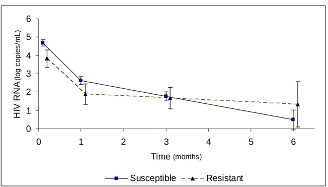
virological response is shown in Figure 1A. At baseline before ART, the baseline plasma
HIV-1 RNA was significantly lower in the resistant group compared to the susceptible group
(3.81 log10 copies/ml vs 4.68 log log10 copies/ml, p=0.002). There was then no difference in
the decay of plasma HIV-1 RNA between baseline and month 1 (-1.93 log10 copies/ml for
resistant group vs -2.04 log10 copies/ml for susceptible group, p= 0.48). However, the
continuing decrease in viral load was significantly steeper in the susceptible group compared
to the mutated group between month 1 and 6 (-0.43 log10 copies/ml/month vs -0.11 log10
copies/ml/month, p= 0.029).
The baseline CD4 (+) cell count was significantly higher in the resistant group compared to
the susceptible group (560 cells/µl vs 438 cells/µl, p= 0.04)(Figure 1B). The two groups had
then parallel increases in CD4 (+) cell counts on ART leading to the same difference in CD4+
level at 6 months (682 cells/µl vs 533 cells/µl, p= 0.04).
Seven patients presented at least one of three mutations 184V, 103N or 90M as a majority
mutation population and tended to have a lower baseline HIV RNA load (difference -0.67,
p=0.08) and the change in HIV RNA at months 1 and 6 was similar (p=0.65 and p=0.79)
compared to the patients without mutations.
Minority resistant variants and response to ART
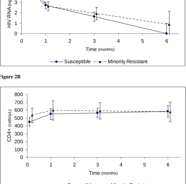
The influence of the minority resistant variants was studied in a subgroup of 73 patients. The
resistant group (with minority resistant variants) and the susceptible group (with no resistance
to the ART regimen) had comparable plasma HIV-1 RNA levels at baseline (4.71 log10
copies/ml vs 4.81 log10 copies/ml, p= 0.68) (Figure 2A). There was then no difference in the
evolution of the plasma HIV-1 RNA in the two groups between baseline and month 1
(-1.99 log10 copies/ml in susceptible group vs –2.17 log10 copies/ml in resistant group,
p=0.49) and between month 1 and month 6 (-0.54 log10 copies/ml in susceptible group vs
The comparison of the baseline CD4 (+) cell counts in the two groups showed a non
significant trend towards higher CD4 (+) cell counts in the resistant group compared to the
susceptible group (451 cells/µl vs 534 cells/µl, p= 0.14) (Figure 2B). No significant
difference in the evolution of the CD4 (+) cell counts could was then shown between the two
groups (p=0.50 during the first month and p=0.52 later on).
Discussion
In our cohort of patients with recent HIV-1 infection and subsequent ART, enrolled between
1996 to 2005, we could detect the transmission of resistant HIV-1 in 13.4% of patients by
using a conventional sequencing method. This prevalence is comparable to other studies in
France or in Europe in the same period [3, 4, 7]. We then investigated the transmission of
minority resistant variants in a subgroup of patients with no resistance detected by bulk
sequencing. We used a modified version of the ASPCR method in order to detect accurately
both the minority mutated genomes, with a limit of detection at < 0.5% of the total
population, and the total number of amplified genomes. We detected minority mutated
variants associated with resistance to the subsequent ART in 29% of patients. This finding is
in accordance with previous data obtained in acute seroconverters [11] and suggests that
minority resistant variants can be detected not only at the onset of the primary infection but
also later on. Similar data were recently reported from an American cohort of
antiretroviral-naïve patients [16] showing that low frequency mutations were equally found in
recently-infected persons and infections of longer duration. This result is also in agreement with model
predictions according to the rapid turnover, the high mutation rate and large viral population
size [17]. We cannot rule out the possibility that the minority variants could have been
generated de novo after transmission, even if, considering the loss of viral fitness conferred by
M184V there is no reason that this mutation could have been selected in absence of drug,
It is important to note that our results were obtained by investigating only three mutations
(K103N and M184V in the RT and L90M in the protease), thus the estimation of the
frequency of minority resistant variants would have been certainly higher by searching for
more mutations. When we studied the virological response in the group of patients having
K103N, M184V or L90M as majority population, we could not show any difference in
comparison with the group of patients without mutations. However, according to the restricted
number of patients with the mutations of interest, the statistical power for finding a significant
difference was limited (about 60% for finding a difference of 0.5 log10 copies/mL at a given
time).
The mutation more often detected by ASPCR in our study was the M184V mutation. We
have two explanations for this finding: first, we showed previously that M184V was the most
frequent mutation in patients on ART with detectable viral load [1], thus this mutation is
likely to be frequently transmitted; second, the M184V mutation confers an impaired
replicative capacity to the virus [18], and in absence of 3TC or FTC, the M184V variants
rapidly disappear in the majority population.
We could investigate the virological and immunological responses to ART in our patients
according to the presence or absence of resistant variants in the majority or only in the
minority population. The detection of a baseline resistance in the majority population was
shown to be associated with a less pronounced decrease in plasma HIV-1 RNA between one
and six months on therapy. This finding is in accordance with previous studies, suggesting
that transmitted resistance could lead to sub-optimal response to first-line therapy, supporting
the guidelines which recommend performing genotypic resistance analysis prospectively in all
patients at the time of diagnosis of the HIV infection [13, 19]. Interestingly, the detection of
transmitted majority resistant variants was associated with a lower baseline plasma viral load
variants. Of note, this difference in the CD4 cell count was not determined by the clinician
decision of when to start ART. The higher CD4 cell counts in the resistant group were
conserved over six months on therapy, suggesting an immunological benefit in the short term.
The detection of minority resistant variants by ASPCR was not associated to a worse
virological response to ART in our study, contrary to the association found with the majority
resistant variants. This finding could be explained by several hypotheses; the most commonly
detected mutation by ASPCR was M184V. This mutation codes the resistance to 3TC and
FTC but can reverse the resistance to AZT and tenofovir; Moreover, the decreased replicative
capacity conferred by M184V is likely to result in lower viral loads and the continuation of
3TC-containining regimens despite the presence of M184V is not necessarily associated with
a worse virological outcome [20]. It is also possible that the putative negative effect of the
M184V on the virological response – if it exits- could have been masked by the antiviral
effect of the other drugs, in majority protease inhibitors. Finally, we cannot exclude that the
limited statistical power could have lead to the non-detection of the influence of minority
variants in our study, since we could only perform ASPCR in a subgroup of patients.
However, we maximised the statistical analyses by estimating slopes using repeated
measurements of markers and taking into account undetectable viral load [14]. Furthermore,
our data are very concordant with those recently reported from the Zurich-PHI Study [21] that
found minority M184V variants in 14.9% of patients and minority K103N variants in 5.4% of
patients without association to virological failure on early therapy. By contrast, Johnson et al
[22] found an association between the detection of baseline minority resistant variants and
virological response in patients enrolled in an abacavir/lamivudine/efavirenz trial. Hence, the
heterogeneity due to the various treatment regimens used in our study population may explain
In conclusion, our study confirms the need for the detection of transmitted resistant variants
by conventional sequencing methods in routine practice. The detection of minority resistant
variants in nearly one-third of the investigated patients was not associated with the virological
outcome on therapy but warrants further clinical validation in order to precise it’s potential
interest for the management of ART.
Acknowledgements
The authors thank all patients included in the study. We are indebted to Pascal Bonot and
Marie-Hélène Shrive for excellent technical assistance. Authors’ contributions: O Peuchant
developed and processed the allele-specific PCR; R Thiébaut developed the statistical model
and participated in the writing of the manuscript; S Capdepont made the sequence analysis
and phylogeny; V Lavignolle-Aurillac made the statistical analysis; D Neau and P Morlat
were responsible for the clinical diagnosis and follow-up of the patients; F Dabis coordinated
the Aquitaine Cohort; H Fleury directed the virology laboratory; and B Masquelier
coordinated the study and wrote the manuscript.
References
1. Costagliola D, Descamps D, Assoumou L, et al. Prevalence of HIV-1 Drug
Resistance in Treated Patients: A French Nationwide Study. J Acquir Immune
Defic Syndr 2007.
2. Richman DD, Morton SC, Wrin T, et al. The prevalence of antiretroviral drug
resistance in the United States. Aids 2004,18:1393-1401.
3. Masquelier B, Bhaskaran K, Pillay D, et al. Prevalence of transmitted HIV-1 drug
resistance and the role of resistance algorithms: data from seroconverters in the CASCADE collaboration from 1987 to 2003. J Acquir Immune Defic Syndr
2005,40:505-511.
4. Wensing AM, van de Vijver DA, Angarano G, et al. Prevalence of drug-resistant
HIV-1 variants in untreated individuals in Europe: implications for clinical management. J Infect Dis 2005,192:958-966.
5. Weinstock HS, Zaidi I, Heneine W, et al. The epidemiology of antiretroviral drug
resistance among drug-naive HIV-1-infected persons in 10 US cities. J Infect Dis
2004,189:2174-2180.
6. Brenner BG, Roger M, Routy JP, et al. High rates of forward transmission events
7. Chaix ML, Desquilbet L, Cottalorda J, et al. Sub-Optimal response to HAART in
patients treated at time of primary HIV-1 infection and infrected with HIV resistant strains. Antivir Ther 2005,10:S127.
8. Little SJ, Holte S, Routy JP, et al. Antiretroviral-drug resistance among patients
recently infected with HIV. N Engl J Med 2002,347:385-394.
9. Grant RM, Hecht FM, Warmerdam M, et al. Time trends in primary HIV-1 drug
resistance among recently infected persons. Jama 2002,288:181-188.
10. Pillay D, Bhaskaran K, Jurriaans S, et al. The impact of transmitted drug resistance
on the natural history of HIV infection and response to first-line therapy. Aids
2006,20:21-28.
11. Metzner KJ, Rauch P, Walter H, et al. Detection of minor populations of
drug-resistant HIV-1 in acute seroconverters. Aids 2005,19:1819-1825.
12. Lazaro E, Coureau G, Guedj J, et al. Change in T-lymphocyte count after initiation
of highly active antiretroviral therapy in HIV-infected patients with history of Mycobacterium avium complex infection. Antivir Ther 2006,11:343-350.
13. Yeni P. Prise en charge thérapeutique des personnes infectées par le VIH. Paris:
Médecine-Sciences Flammarion; 2006.
14. Metzner KJ, Bonhoeffer S, Fischer M, et al. Emergence of minor populations of
human immunodeficiency virus type 1 carrying the M184V and L90M mutations in subjects undergoing structured treatment interruptions. J Infect Dis
2003,188:1433-1443.
15. Thiébaut R, Jacmin-Gadda H. Mixed models for longitudinal left-censored
repeated measures. Computer Methods and Programs in Biomedicine
2004,74:255-260.
16. Johnson J, Li J-F, Wei X, Lipscomb J, Smith A, Heneine W. Sensitive testing
demonstrates a high prevalence of transmitted drug resistance among
conventionally genotyped wildtype HIV-1 infections. Antivir Ther 2007,12:S46.
17. Coffin JM. HIV population dynamics in vivo: implications for genetic variation, pathogenesis, and therapy. Science 1995,267:483-489.
18. Wainberg MA. The impact of the M184V substitution on drug resistance and viral fitness. Expert Rev Anti Infect Ther 2004,2:147-151.
19. Hirsch MS, Brun-Vezinet F, Clotet B, et al. Antiretroviral drug resistance testing in
adults infected with human immunodeficiency virus type 1: 2003
recommendations of an International AIDS Society-USA Panel. Clin Infect Dis
2003,37:113-128.
20. Castagna A, Danise A, Menzo S, et al. Lamivudine monotherapy in HIV-1-infected
patients harbouring a lamivudine-resistant virus: a randomized pilot study (E-184V study). Aids 2006,20:795-803.
21. Metzner KJ, Rauch P, Von Wyl V, et al. Prevalence of minority quasispecies of
drug-resistant HIV-1 in patients with primary HIV-1 infection in Zurich in the years 2002-2006. Antivir Ther 2007,12:S47.
22. Johnson J, Li J-F, Wei X, et al. Low-frequency mutations substantially increase the
prevalence of transmitted drug resistance and greatly strengthen the relationship between resistance mutations and virological failure. 14th conference on
Legends to the figures
Figure 1: Mean levels of plasma HIV RNA (A) and CD4 cell count (B) predicted by a
piecewise linear model according to the presence of majority resistant variants. N= 172
patients, Aquitaine Cohort, 1996-2005.
Susceptible: baseline HIV-1 genotype predicting full sensitivity to the first-line ART.
Resistance: baseline HIV-1 genotype predicting resistance to at least one drug of the first-line
ART.
Figure 2: Mean levels of plasma HIV RNA (A) and CD4 cell count (B) predicted by a
piecewise linear model according to the presence of minority resistant variants. N= 73
patients, Aquitaine Cohort, 1996-2005.
Susceptible: baseline HIV-1 genotype predicting full sensitivity to the first-line ART.
Resistance: baseline HIV-1 genotype predicting resistance to at least one drug of the first-line
Table 1: Characteristics of the 172 patients included in the study N (%) Exposure category
Sex between men 106 (62%) Sex between men
and women
59 (34%)
Injecting drug use 3 (2%) Other/unknown 4 (2% ) Sex Male 140 (81% ) Female 32 (19%) HIV-1 subtype B 142 (83%) Non-B 30 (17%) Year of seroconversion 1996-1999 2000-2003 2004-2005 77 (45%) 72 (42%) 23 (13%) Median Age (years, IQR) 32 (27-40.5) Median CD4+ cell count
(cells/µl)
426 (314-579) Median plasma HIV-1 RNA 4.7 (4.1-5.3) First-line ART
2-3 NRTIs NRTIs+ NNRTI NRTIs+ PIs Resistance to at least one drug of the regimen
49 (28.5%) 37 (21.5%) 86 (50%)
16 (9.3%)
Abbreviations: ART: antiretroviral therapy; NRTI: nucleoside reverse transcriptase inhibitor;
Figure 1 A 0 1 2 3 4 5 6 0 1 2 3 4 5 6 Time (months) HI V RNA ( log c o pi es /m L ) Susceptible Resistant Figure 1 B 0 100 200 300 400 500 600 700 800 900 0 1 2 3 4 5 6 Time (months) CD4 + ( c el ls /µ L) Susceptible Resistant
Figure 2A 0 1 2 3 4 5 6 0 1 2 3 4 5 6 Time (months) HI V RNA ( log c o pi es /m L )
Susceptible Minority Resistant
Figure 2B 0 100 200 300 400 500 600 700 800 0 1 2 3 4 5 6 Time (months) CD4 + ( c e lls /µ L )
Appendix
Composition of the GECSA (Groupe d’Epidémiologie Clinique du SIDA en Aquitaine) –
ANRS CO3 Aquitaine cohort:
Coordinator: F. Dabis. Epidemiology and Methodology: G. Chêne, F. Dabis, S.
Lawson-Ayayi, R. Thiébaut, M. Winnock. Infectious diseases and Internal medicine: M. Bonarek,
F. Bonnal, F. Bonnet, N. Bernard, O. Caubet, L. Caunègre, C. Cazanave, J. Ceccaldi, P.
Couzigou, FA Dauchy, C. De La Taille, S. De Witte, M. Dupon, P. Duffau, H. Dutronc, S.
Farbos, MC. Gemain, C. Greib, K. Lacombe, D. Lacoste, S. Lafarie-Castet, P. Loste, D.
Malvy, P. Mercié, P. Morlat, D. Neau, A. Ochoa, JL. Pellegrin, JM. Ragnaud, S. Tchamgoué,
J.F. Viallard. Immunology: P. Blanco, JF. Moreau. Virology: H. Fleury, ME. Lafon, B.
Masquelier, I. Pellegrin. Pharmacology: D. Breilh. Pharmacovigilance: G.
Miremont-Salamé. Data collection and Data management: MJ. Blaizeau, M. Decoin, S. Delveaux, S.
Geffard, S. Gillet, C. Hannapier, S. Labarrère, V. Lavignolle-Aurillac, B.