Chapter 1 Basic Life Support
BLS; Basic Life Support
■BLS Task Force Chairmen
Koichi Tanigawa, Takashi Nakagawa ■BLS Task Force Members
Masami Ishikawa, Akinori Takeuchi, Choichiro Tase, Eiichiro Noda, Norifumi Mabuchi, Hiroya Wakamatsu
■Editorial Board
Kunio Ohta, Tetsuya Sakamoto, Naoki Shimizu, Hiroshi Nonogi, Tetsuo Hatanaka ■Co-Chairmen
Kazuo Okada, Seishiro Marukawa
■1 Introduction
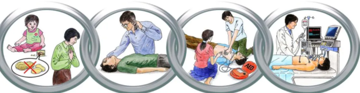
Victims of cardiac arrest or asphyxia are in the dire medical emergency, where the links of four actions called “Chain of Survival” are necessary to save their lives and return them to their previous state of health. These four actions are (Fig 1):
1. Prevention of conditions leading to cardiac arrest
2. Immediate recognition of cardiac arrest and activation of the emergency medical service system
3. Basic life support (CPR and AED)
4. Advanced life support and post-cardiac arrest care
Fig 1 Chain of survival for all ages. The links in the chain are: prevention, immediate
recognition and activation, early CPR and rapid defibrillation, and advanced life support with
integrated post-cardiac arrest care
Prevention of conditions leading to cardiac arrest refers to preventing diseases or events that may lead to cardiac arrest or respiratory arrest. It is important for children to avoid getting involved in accidents, such as traffic accidents, choking and near-drowning. As for adults, to recognize initial symptoms of acute coronary syndromes or stroke is crucial so that patients are able to receive medical treatment before they suffer cardiac arrest.
Immediate recognition begins with the suspicion that a person who has suddenly collapsed or is unresponsive is in cardiac arrest. When recognizing the possibility of cardiac arrest, call out for help, notify the EMS system (by calling 119) and help to quickly get the victim an AED and healthcare professionals or EMS personnel with emergency medical equipments.
Basic life support (BLS) is a series of treatments to maintain respiration and circulation in the victim. BLS includes cardiopulmonary resuscitation (CPR) with chest compressions and rescue breathing, and defibrillation with an AED, which can be performed immediately by any lay person, and is considered to have a major role in neurologically intact survival of the victim.
Advanced life support (ALS) is a treatment provided using drugs and medical instruments to the victim who does not achieve return of spontaneous circulation after BLS. After resuscitation, intensive care at a specialized medical facility as needed can increase the possibility of the victim's return to a normal life.
Prior to the 2005 CoSTR, it had been reported that BLS of early activation of the EMS system, early CPR and early defibrillation had larger impact on neurologically intact survival of the victims of out-of-hospital cardiac arrest, compared to ALS including tracheal intubation and drug administration.1 Subsequent studies pointed out that, during CPR provided by a lay rescuer, attempting rescue breathing caused considerable interruptions of chest compressions, and the significance of chest compressions was emphasized.2 This concept has been incorporated in the 2010 CoSTR. The JRC guidelines recommend that, when cardiac arrest is witnessed, CPR should be started with chest compressions rather than with ventilations to minimize delay in starting compressions.
These BLS guidelines are developed as common approaches by lay rescuers with different backgrounds in the scenes where they deal with victims of all age groups, and therefore the algorithm for victims in a cardiopulmonary emergency adopted here is targeted both at adults and children. Higher effectiveness of CPR performed by any rescuer is expected by standardizing the timing of activation of the EMS system and beginning of CPR (phone first), the initial sequence of CPR, and the compression to ventilation ratio.
BLS performed by those in contact with children on a routine basis, such as nursery staff, school teachers, parents or family members of children, is described in Chapter 3: Pediatric Basic Life Support and Pediatric Advanced Life Support, and BLS performed by healthcare providers working in a medically-equipped environment such as in a hospital or an ambulance is in Chapter 2: Adult Life Support (ALS) and in Chapter 3:
Pediatric Basic Life Support and Pediatric Acvanced Life Support. Issues on CPR training and the EMS system are discussed in Chapter 7: Education, Implementation and Team (EIT).
The most important changes to BLS in the revised guidelines.
- An untrained bystander should call 119 and ask for instruction from the EMS dispatchers. It is recommended that dispatchers provide compression-only CPR instructions to untrained rescuers over the telephone.
- Rescuers should immediately begin CPR if the victim is unresponsive with no breathing or with agonal respirations. Agonal respiration refers to an abnormal pattern of breathing that suggests cardiac arrest. Beginning CPR should not be delayed even though the victim is still gasping (agonal respirations).
- When cardiac arrest is recognized, the rescuer should initiate chest compressions before airway opening and rescue breathing.
- All bystanders, trained or untrained, should provide chest compressions to the victim of cardiac arrest.
- The need for high-quality CPR is once again emphasized. The rescuer should give chest compressions to a depth of at least 5cm and at a rate of at least 100 times per minute, allow for complete chest recoil after each compression, and minimize interruptions of chest compressions.
- It is recommended that the trained rescuer provide CPR with compression to ventilation ratio of 30:2.
■2 BLS algorithm
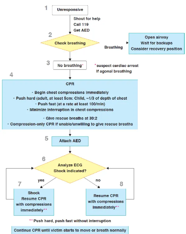
1.Checking Response and Calling EMS[Box 1]
The following are the procedures for responding in a situation where laypeople see someone collapse or who has collapsed (priority between EMS activation and CPR initiation).
- Check the safety of the surroundings.
- Speak loudly to the person patting lightly on the shoulder. If there is no response or no purposeful movement, the person should be determined "unresponsive".
- If the person is unresponsive, call for help loudly to bring attention from the surroundings.
- Ask people around to activate the EMS (call 119) and bring an AED (if available nearby).
The EMS operator is supposed to immediately dispatch an ambulance upon suspicion of cardiac arrest while talking with the caller.
Fig 1 BLS algorithm for lay rescuers
1)Recognition of cardiac arrest[Box 2, 3]
Rescuers should begin CPR if the victim is unresponsive and not breathing (ignoring occasional gasps).
checking for a respiration, and should check carefully if the victim has absent or
abnormal breathing (ie, only gasping). The rescuer should take no more than 10
seconds to check for breathing. Gasping is not a normal breathing, but a sign of
cardiac arrest. Typically it shows an irregular pattern of respiration and could be
observed with an adult suddenly collapsed. Health care providers should open
the airway while checking respiration.
Neither lay rescuers nor inexperienced (non-expert) healthcare providers
should check for a pulse. It is reasonable for experienced healthcare providers to
check for a pulse simultaneously with checking for breathing.
When not confident with feeling a pulse, resucuers should concentrate on
checking for breathing. Once recognizing no breathing, perform CPR immediately.
Delays in starting CPR because of checking for pulse should be avoided.
If the victim is breathing normally, keep the airway open and wait for help and the EMS personnel. In the meantime, continue observing the victim's breathing. If the victim has stopped breathing, begin CPR immediately. If the rescuer needs to go ask for help and leave the victim then when no other option exists, the victim should be placed in the recovery position.
There may be rare occasions where the victim has no breathing but has a pulse. The trained rescuer should open the airway and provide rescue breathing, while checking for the pulse frequently so as not to delay beginning chest compressions.
2.Starting CPR and chest compressions[Box 4]
All rescuers, trained or not, should provide chest compressions to victims of cardiac arrest. A strong emphasis on delivering high quality chest compressions remains essential: rescuers should push hard to a depth of at least 2 inches (or 5 cm) for adult, at least one-third of the anterior-posterior dimension of the chest for infants and children, compress at a rate of at least 100 compressions per minute, and minimize interruptions of chest compressions.
1)Initial sequence of CPR
Rescuers begin CPR with chest compressions rather than opening the airway and giving rescue breathings.
2.Starting CPR and chest compressions[Box 4]
All rescuers, trained or not, should provide chest compressions to victims of cardiac arrest. A strong emphasis on delivering high quality chest compressions remains essential: rescuers should push hard to a depth of at least 2 inches (or 5 cm) for adult,
at least one-third of the anterior-posterior dimension of the chest for infants and children, compress at a rate of at least 100 compressions per minute, and minimize interruptions of chest compressions.
8) Pulse check during CPR
For lay rescuers, interrupting chest compressions to perform a pulse check is not recommended, unless there is obvious sign (normal breathing or purposeful movement) that clearly shows ROSC. Healthcare professionals should continue CPR without checking a pulse if there is no monitor available. It is reasonable to check a pulse if an organized rhythm is visible on the ECG monitor.
9)Changing rescuers
It may be reasonable for another rescuer to take over after a period of no longer than 1 to 2 minutes, to prevent deterioration of the quality of compressions. Rescuer should be aware that quality of chest compression may deteriorate faster during compression-only CPR (Class Ⅱb). Switching should be done with the minimal interruptions of the compressions.
3. Airway and Ventilation[Box 4]
If rescue breathing is possible, rescuers use a compression to ventilation ratio of 30:2. The victim's airway needs to be kept open during rescue breathings.
1) Opening the Airway
For unresponsive adults and children, open the airway using the head tilt-chin lift maneuver when assessing breathing or giving ventilations. The trained rescuer should use the jaw thrust maneuver if necessary. Use a head tilt–chin lift maneuver if the jaw thrust does not open the airway. As the jaw lift maneuver (a jaw thrust performed with the thumb put into the oral cavity) can be harmful, it requires careful attention to adaptive decision making and practice.
Use manual spinal motion restriction rather than immobilization devices for victims with suspected spinal injury.
2) Tidal Volume and Ventilation Rate
For all victims, give each breath to achieve visible chest rise. Avoid hyperventilation during CPR. For mouth-to-mouth ventilation for adult victims using exhaled air or bag-mask ventilation with room air or oxygen, give each breath over about 1 second to
achieve chest rise. In infants and children, a reduction in minute ventilation to less than baseline for age is reasonable to provide sufficient ventilation to maintain adequate ventilation-to-perfusion ratio during CPR while avoiding the harmful effects of hyperventilation.
3) Barrier Devices
The risk of disease transmission in out-of-hospital is very low and initiating rescue breathing without a barrier device is reasonable. If available, rescuers may consider using a barrier device. However, safety precautions should be taken both in the in-hospital and out-of-in-hospital situations if the victim is known to have a serious infection (eg, human immunodeficiency virus (HIV) infection, tuberculosis, hepatitis B virus infection, or severe acute respiratory syndrome (SARS) ). Healthcare providers on duty must always follow standard precautions when performing CPR.
(1)Bag-valve-mask
When two or more experienced rescuers are present, ventilation using a bag-valve-mask is reasonable. Holding the bag-valve-mask to the victim's face with both hands can ensure a better mask seal.
4.Chest compressions and ventilation during CPR
1)Compression-ventilation ratio
A compression-ventilation ratio of 30:2 is reasonable for adults, children and infants under cardiac arrest whose airway is not secured. For experienced healthcare providers performing 2-rescuer CPR in children and infants, a compression-to-ventilation ratio of 15:2 is reasonable.
When advanced airway such as a tracheal tube is in place, compressions should not be interrupted for ventilations.
2)Interruptions of chest compression
Interruptions to chest compressions during CPR must be minimized. Legitimate reasons for the interruption of CPR include: the need to ventilate, the need to assess the rhythm or to assess ROSC, and the need to defibrillate. During these procedure interruptions of chest compressions must be minimized.
Chest compressions alone are recommended for untrained laypersons responding to cardiac arrest victims. Performing chest compressions alone is reasonable for trained lay-persons if they are unable or unwilling to give rescue breathings. CPR with rescue breathings is preferred for patients with cardiac arrest caused by choking, drowning or airway obstruction, for infants and children, for adults whose cardiac arrest is not witnessed, and for patients under extended period of resuscitation attempt.
5.AED[Box 5]
When the AED arrives during CPR, connect it to the victim immediately. Some AEDs automatically turn on when the lid is opened, and others need to be turned on manually. In the case of the latter type of device, push the power button first.
1) Placement and size of paddles/pads
It is reasonable to place paddles or pads on the exposed chest in an anterior-lateral position. Acceptable alternative positions are the anterior-posterior (for paddles and pads) and apex- posterior (for pads). In large-breasted individuals it is reasonable to place the left electrode pad (or paddle) lateral to or underneath the left breast, avoiding breast tissue. Consideration should be given to the rapid removal of excessive chest hair prior to the application of pads/paddles but delays in shock delivery must be minimized. Pediatric pads should be applied for preschool-age children. Adult pads can be substituted if pediatric pads are unavailable and no other choice is left. Pediatric pads must not be used for an adult.
2) Shock and resumption of chest compression
[Box 7]
When the AED starts analyzing, keep off the victims. Following the audio instructions from the AED, push the shock button to deliver the electric shock. Immediately after the shock is delivered, resume chest compressions rather than checking the pulse or analyzing the rhythm.
3) Implantable cardioverter-defibrillator or pacemaker
In the case of victims with implantable cardioverter-defibrillators or pacemakers, avoid placing the pads directly over the implanted device, and deliver the shock promptly. Although some reports suggest that the ideal pad position should be at least 8cm away from the device, such pad placement should not delay electrical shock.
The precordial thump may be considered for patients with monitored, unstable ventricular tachycardia if a defibrillator is not immediately available. It should not be used for unwitnessed out-of-hospital cardiac arrest.
6.Continuing BLS
Rescuers should continue CPR until sufficient circulation is restored in the victim, or EMS providers or other responders who are able to provide advanced life support take over the care of the victim. If an AED is present, follow its instructions, and analyze the ECG and deliver the electric shock if needed. Immediately after the shock delivery, resume CPR by giving chest compressions.
7.Foreign body airway obstruction
In responsive adults and children >1 year of age with FBAO, rescuers should activate emergency response system and perform back blows, abdominal thrusts, and/or chest thrusts for obstruction relief. These techniques should be applied repeatedly in rapid sequence until the obstruction is relieved. If the choking infants are still responsive but can not make effective, strong cough, rescuers should hold the infants with their head down and perform back blows and chest thrusts.
If the victim with FBAO becomes unresponsive, the rescuer should immediately begin CPR. Citizen rescuers can begin CPR starting with chest compressions like usual CPR. The experienced healthcare providers start CPR with rescue breathing.
For unresponsive victims of FBAO, finger sweep may be applied when solid material is visible in the oral cavity.
■3 Science behind recommendations
1.Checking Response and Calling EMS
1) Dispatcher recognition of cardiac arrest and telephone instruction in CPR.
One before-after trial demonstrated significant increase from 15% to 50% in cardiac arrest recognition after the implementation of a protocol requiring that EMS dispatchers assess absence of consciousness and quality of breathing (normal/not normal) (LOE D3 3). Many descriptive studies (LOE D44-12 13) using a similar protocol to identify cardiac arrest report a sensitivity in the order of 70%, ranging from 38% 9 to 97% 13 and a high specificity from 95% 8 to 99% 10.
One case-control trial (LOE D314), one before-and-after trial (LOE D315), and four observational studies (LOE D416-19) describe agonal gasps or abnormal breathing as a significant barrier to cardiac arrest recognition by emergency medical dispatchers. Two before-and-after trials (LOE D320, 21) improved the recognition of abnormal breathing using education or counting of breaths. Information spontaneously provided by the caller about the quality of breathing and other information such as facial color or describing the victim as “dead” can aid in identifying cardiac arrest cases (LOE D314, 20, 21).
One descriptive study (LOE D422) suggests that in cases where the victim’s problem is “unknown” to the EMS dispatcher, inquiring about the victim’s level of activity (standing, sitting, moving, or talking) helps to identify cases who are not in cardiac arrest. Two descriptive studies (LOE D419, 23) suggest that confirming the absence of a past medical history of seizure may increase the likelihood of recognizing cardiac arrest among victims presenting with seizure activity. A case-control study (LOE D324) suggests that asking about regularity of breathing may help to recognize cardiac arrest among callers reporting seizure activity.
EMS dispatchers should inquire about a victim’s absence of consciousness and quality of breathing (normal/not normal) when attempting to identify cardiac arrest victims. If the victim is unresponsive, it is reasonable to assume that the victim is in cardiac arrest when callers report that breathing is not normal. Dispatchers should be specifically educated about identification of abnormal breathing in order to improve cardiac arrest recognition. The correct identification of cardiac arrest may be increased by careful attention to the caller's spontaneous comments and by focused questions about seizures. Three studies (LOE 212, 14, 25) provide evidence that dispatcher telephone CPR instructions may improve survival from sudden cardiac arrest (SCA). In one randomized trial (LOE 126) compression-only dispatcher telephone CPR instruction produced survival to discharge at least equivalent to compression plus ventilation dispatcher telephone CPR instruction. Five additional simulation studies (LOE 527-31) demonstrated simplified chest compression-only telephone instructions in CPR reduces barriers to achieving reasonable quality bystander CPR.
Using video instruction via cell phone enhanced performance of dispatcher in instructing CPR32 to the callers. It is reported that real-time instruction via cell phone video caused some delay in the initiation of chest compressions, it improved depth and rate of compressions33. Meanwhile, another study reported that it took a much longer time before airway opening and initial rescue breathing even though improvements were seen in both of these procedures. In addition, despite the increased accuracy of hand position and compression rates, there was no definitive improvement in compression depth and tidal volume between the cases with and without video instructions.
Dispatchers should assertively provide compression-only CPR instructions to untrained rescuers for adults with suspected SCA without any delay(Class Ⅰ). If dispatchers
suspect asphyxial arrest, it is reasonable to provide instructions on rescue breathing followed by chest compressions to trained rescuers(Class Ⅱa). When performing quality improvement efforts, it is reasonable to assess the accuracy and timeliness of dispatcher recognition of cardiac arrest and the delivery of CPR instructions(Class Ⅱa).
2) Recognition of Cardiac Arrest
(1) Opening Airway at the Initial Assessment
Observe movements of the victim's chest and abdomen when assessing breathing. Traditionally, rescuers first opened the airway using the head tilt-chin lift maneuver, and then leaned down and placed the ear close to the victim's mouth and nose, and performed “Look, Listen, Feel", checking the chest movements for breathing. However, a simplified breathing asessment can lead to a quicker start of CPR and improved rate of CPR carried out by laypeople. Therefore this guideline suggests that the initial assessment for breathing by laypeople exclude head tilt-chin lift, and be simplified into observation of chest and abdominal movements.
For a victim who is unconscious but has normal breathing, airway opening is crucial. Hence, even in training using simplified chest compression-only CPR, teaching airway opening assuming a victim with normal breathing is reasonable.
The recovery position is used for unresponsive adult victims who have normal breathing. Although healthy volunteers report compression of vessels and nerves in the dependent limb when the lower arm is placed in front36, 37, the ease of turning the victim into this position may outweigh the risk. If rescuers are obliged to temporarily leave the scene to call for backups, it is reasonable to place the victim in the recovery position (Class IIb).
(2)Assessment of Breathing
Early recognition is a key step in the initiating early treatment of cardiac arrest and relies on using the most accurate method of determining cardiac arrest.
There is a high incidence of agonal gasps after cardiac arrest(LOE 415-17, 40) and lay rescuers tend to consider this to be "breathing" and fail to recognize cardiac arrest. Several studies have shown that lay rescuers do not easily master the techniques of breathing assessment and they are often unable to recognize agonal gasp. It is necessary for lay rescuers to always consider the possibility of cardiac arrest unless the victim is "breathing normally".
Recognition of cardiac arrest by determining the presence or absence of a pulse is unreliable. Pulse checking by lay rescuers during CPR has been de-emphasized in the 2005 guidelines for CPR and ECC by Japan, AHA, and ERC. Its importance has decreased in cases performed by healthcare providers as well. It should not take more than 10 seconds even for a healthcare provider to check a pulse.
There are no studies assessing the accuracy of checking the pulse to detect human cardiac arrest. There have been nine LOE D5 studies demonstrating that both lay rescuers and healthcare providers27-32 have difficulty mastering the pulse check and remembering how to perform it(LOE 541, 42, 45-51). Three LOE D5 studies support the ability of healthcare providers to perform the pulse check; two evaluated the direct ear to chest method in infants, and the third supported an alternative technique for the carotid pulse check when tested by dental students on healthy volunteers(LOE 552, 53). In one study the technique of simultaneous pulse check and breathing check by professional rescuers increased the diagnostic accuracy(LOE 554).
Two RCT studies(LOE 551, 55) conducted in infants and children with non-pulsatile circulation during extracorporeal membrane oxygenation (ECMO) demonstrated that doctors and nurses in a pediatric tertiary care institution, who were blinded to whether the child was receiving ECMO support or not, commonly assessed pulse status inaccurately and often took longer than 10 seconds. In these pediatric studies, healthcare professionals were able to accurately detect a pulse by palpation only 80% of the time. They mistakenly perceived a pulse when it was non-existent 14-24% of the time, and failed to detect a pulse when present in 21-36% of the assessments. Although some of the children in this study were pulseless, all children had circulation (ie, none were in cardiac arrest) so other signs typically associated with pulseless arrest (delayed capillary refill, poor color) were absent in this population.
(4)Signs of Circulation
In the past, students were taught to recognize cardiac arrest by looking for the absence of signs of circulation, such as movement. No studies were found which measured the sensitivity and specificity of that approach for diagnosing cardiac arrest. A study(LOE 44) showed that CPR guidance by EMS dispatchers was impeded by callers mentioning “signs of life.” Mere recognition of a pulse by itself is not a credible way to determine whether or not the victim is in cardiac arrest. Agonal gasps, which are often seen after cardiac arrest, should not be considered normal breathing.
(5)Etiology of Cardiac Arrest
For lay rescuers there is insufficient evidence to recommend any diagnostically reliable method to differentiate sudden cardiac arrest of cardiac origin from one of non-cardiac origin. Except for obvious external causes of cardiac arrest (e.g. gunshot wound,
drowning), professional rescuers should rely on rhythm analysis from cardiac monitors or AEDs and other diagnostic tests to determine the cause of cardiac arrest.
In one registry study (LOE 256), cardiac arrest was more likely to be due to a cardiac cause in victims above the age of 35 years and due to a non-cardiac cause up to the age of 35 years. Two other registry studies (LOE 357, 58) do not demonstrate diagnostically useful cut-off ages. An additional registry study (LOE 259) demonstrated that 83% of cardiac arrests under the age of 19 are of non-cardiac origin. One prospective study (LOE 260) and one retrospective study (LOE 361) showed that identification of the cause of cardiac arrest by healthcare providers can be inaccurate, leading to an underestimation of non-cardiac etiology cardiac arrest, in particular, failure to diagnose exsanguination.
▲Knowledge Gaps
・How accurately do rescuers identify cardiac arrest outside of the hospital by using a useful advanced technology to assist with diagnosing cardiac arrest?
・Which specific factors improve diagnostic accuracy?
・What is the accuracy of the pulse check performed by health professionals in cardiac arrest patients?
・Is there an association between the time required to successfully detect a suspected cardiac arrest victim’s pulse and resuscitation outcome?
・ How does opening the airway in the process of breathing assessment for cardiac arrest recognition influence recognition of respiratory arrest?
2.Starting CPR
1)Sequence of CPR initiation
In the basic life support sequence for the lone rescuer, the choice is between starting with airway and breathing or starting with chest compressions. Because of the importance of initiating chest compressions as soon as possible, the need for initial breaths is questioned. The AHA and Japanese CPR guidelines 2005 recommended starting CPR with opening airway and giving 2 breaths rather than with compressions. On the other hand, the European Resuscitation Council Guidelines 2005 recommended starting CPR with 30 compressions rather than with 2 breaths. These guidelines were based on a consensus of experts rather than clear evidence.
Evidence from one observational, adult manikin LOE 5 study shows that starting with 30 compressions rather than 2 ventilations leads to a shorter delay to first compression. There is no published human or animal evidence to determine if starting
CPR in adults or children with 30 compressions rather than 2 breaths leads to improved outcomes.
Bystander rescuers are likely to hesitate to perform mouth-to-mouth ventilation for a variety of reasons. The common reasons for unwillingness are fear of disease transmission, anxiety and panic, and lack of knowledge of performing CPR (LOE 560, 61) The complex procedure of ventilation might reduce the rate of performing bystander CPR. To avoid any delay or hesitation by rescuers before starting CPR, it is reasonable to start CPR with chest compressions (Class Ⅱa). If rescuers are not capable of adding rescue breaths, they may continue chest compressions alone (Class Ⅱb).(LOE 560, 62) During the first few minutes after non-asphyxial cardiac arrest, the blood oxygen content remains high, and myocardial and cerebral oxygen delivery is limited more by the diminished cardiac output than by a lack of oxygen in the lungs. Ventilation is, therefore, initially less important than chest compressions. (LOE 560, 63, 64)
All rescuers may start CPR with chest compressions rather than with ventilations for treatment of adult victims of cardiac arrest (Class Ⅱa). It is reasonable for skilful rescuers with barrier devices including BVM to start CPR with ventilation for pediatric patients and when suspected cause of cardiac arrest is asphyxiation, drowning, or airway obstruction (ClassⅡa).
2) Chest compressions
Cardiac arrest victims should be placed supine, with the rescuer kneeling beside the victim’s thorax.
CPR should be performed on a firm surface when possible. Air filled mattresses should be routinely deflated during CPR. There is insufficient evidence for or against the use of backboards during CPR. If a backboard is used, rescuers should minimize delay in initiation of chest compressions, minimize interruption of chest compressions, and take care to avoid dislodging of catheters and tubes during backboard placement. One case series (LOE 490) and four manikin studies (LOE 591-94) demonstrated that chest
compressions performed on a bed are often too shallow. No studies have examined the risks or benefits of moving the patient from a bed to the floor to perform CPR.
3)Locating hand position for chest compressions
No randomized controlled human trials support use of an alternative to the hand position recommended in 2005 (“The rescuer should compress the lower half of the victim’s sternum”) when performing external chest compressions for adults or children in cardiac arrest. During transesophageal echocardiography of humans receiving chest compressions with placement of the hands on the lower half of the sternum, the area of
maximal compression was most often over the base of the left ventricular and the aortic root, a location that potentially impedes forward flow of blood (LOE 4). Hand position for chest compression to achieve optimal hemodynamics is unclear.
One of the instructions for locating the recommended hand position for chest compression is "place hands in the center of the chest". One adult manikin study using this recommended hand position instruction showed a reduction in hands-off time (time spent not doing chest compressions) but also a loss of hand-placement accuracy. In four other LOE 5 adult manikin studies,65-68 however, locating the recommended hand position for chest compression using the instruction “place hands in center of the chest” resulted in a significant reduction in hands-off time and no significant reduction in accuracy compared with locating the rib margins and xiphisternum.
Some studies claim that the inter-nipple line is an unreliable landmark for hand placement. In one study of CT scans, the inter-nipple line was 3 cm superior to the lower third of the sternum (LOE 569). One LOE 5 study70 of adult surgical patients
demonstrated that if the rescuer’s hands are placed on the inter-nipple line, hand deviation to or beyond the xiphisternum occurs in nearly half the cases, sometimes into the epigastrium.
One study that compared the length from the inter-nipple line to the xiphisternum in 30 infants with the finger position achieved by 30 adults demonstrated that the recommended method of locating finger position for chest compression in infant cardiac arrest can cause pressure on the xiphisternum or abdomen(LOE 571).
For adults receiving chest compressions, it is reasonable for rescuers to place their hands on the lower half of the sternum(Class Ⅱa). No reliable study suggests any specific method for locating this position quickly and with accuracy. Nor was there any study directly comparing hand position in the center of the chest with the inter-nipple line in terms of accuracy and required time. It is reasonable to teach this location in a simplified way, such as, “place the heel of your hand in the center of the chest with the other hand on top.” This instruction should be accompanied by a demonstration of placing the hands on the lower half of the sternum(Class Ⅱa).
4)Chest compression depth
Three adult human LOE 4 studies72-74show that the measured compression depth
during adult human resuscitation is often less than the recommended lower limit of 4 cm (1.5 inches). One adult human LOE 4 case series, 75two adult human studies with
retrospective control groups (LOE 376, 77), and one LOE 5 study78 suggest that
ROSC. These findings are supported by three swine studies (LOE 579-81) showing
improved survival with deeper compression depths, and one adult human study (LOE 482) showing that improved force on the chest produced a linear increase in systolic blood
pressure. However, one swine study (LOE 583) reported no improvement of myocardial
blood flow with increased compression depth from 4 cm to 5 cm although coronary perfusion pressure (CPP) improved from 7 to 14 mm Hg. No human studies directly compared the effectiveness of compression depth of 4-5 cm (1.5-2 inches) with alternative compression depths.
It is reasonable to compress the sternum at least 2 inches/5cm for all adult cardiac arrest victims. There is insufficient evidence to recommend a specific upper limit for chest compression depth.
Evidence from anthropometric measurements in three LOE 5 case series84-86 showed that
in children the chest can be compressed to 1/3 of the A/P chest diameter without causing damage to intrathoracic organs. One LOE 5 mathematical model based
on neonatal chest CTs87 suggests that 1/3 AP chest compression depth is more effective
than . compression depth and safer than 1/2 AP compression depth.
A good quality LOE 574 adult study found that chest compressions are often inadequate
and a good quality LOE 4 pediatric study85 showed that during resuscitation of patients
>8 years of age, compressions are often too shallow, especially following rescuer changeover.
Rescuers should push hard to a depth of at least 2 inches (or 5 cm) for adult.
In infants and children, rescuers should be taught to compress the chest by at least 1/3 the A-P dimension(Class Ⅱa).
5)Chest Compressions in Children
There are no outcome studies comparing 1- versus 2-hand chest compressions for children in cardiac arrest. Evidence from 1 LOE 5 randomized crossover child manikin study (LOE 588) showed that higher chest-compression pressures are generated by healthcare professionals using the 2-hand technique. Two LOE 5 studies89, 90 reported no increase in rescuer fatigue comparing 1-hand with 2-hand chest compressions delivered by healthcare providers to a child-sized manikin. Either a 1- or 2-hand technique can be used for performing chest compressions in children (Class Ⅱa).
Although 2005 CoSTR recommended adding circumferential squeeze of the chest when performing chest compression in infants with the 2-thumb technique, recent review of literature failed to find convincing evidence for or against the need for a circumferential squeeze of the chest when performing the 2-thumb technique of external chest compression for infants. There are insufficient data showing that chest compression technique with circumferential squeeze is more effective than other techniques in infants.
6)Chest decompression
There are no human studies specifically evaluating ROSC or survival to hospital discharge with or without complete chest wall recoil during CPR. One LOE 4 out-of-hospital case series91 documented a 46% incidence of incomplete chest recoil by professional rescuers using the CPR technique recommended in 2000, and two in-hospital pediatric case series demonstrated a 23% incidence of incomplete recoil that was more common just following switching providers of chest compressions (LOE 485, 92). Another LOE 4 study93 electronically recorded chest recoil during in-hospital pediatric cardiac arrests and found that leaning on the chest occurred in half of chest compressions.
Animal studies (LOE 5) demonstrate significant reductions in mean arterial pressure, coronary perfusion pressure, cardiac output, and myocardial blood flow with only small amounts of incomplete chest recoil94, 95. Some adult and pediatric studies conducted in and out of hospitals showed that 23-50 % of chest compressions contained incomplete chest recoil (LOE 485, 91-93). Chest recoil can be increased significantly with simple techniques; for example, lifting the heel of the hand slightly but completely off the chest during CPR improved chest recoil in a manikin model. However, these alternative techniques may also reduce compression depth (LOE 491、LOE 596).
While allowing complete recoil of the chest after each compression may improve circulation, there is insufficient evidence to determine the optimal method to achieve the goal without compromising other aspects of chest compression technique. Rescuers should allow complete chest wall recoil after each compression (Class Ⅱb) with careful attention to depth of compression (Class Ⅲ).
7)Chest compression rate
It is reported that chest compressions of at least 100 times per minute to a recommended depth achieves optimum blood flow (LOE 597). The number of chest compressions during a certain period (e.g. 1 minute) given to cardiac arrest patients depends on two factors: the time interval between compressions (ie the compression rate) and the duration of any interruptions in compressions. One LOE 4 study of in-hospital cardiac arrest patients98 showed that chest compression rates >80/min were associated with ROSC. Some animal studies and one clinical study showed that interruption of chest compressions during CPR decreased ROSC rates and survival rates. An observational study of 506 patients with out-of-hospital cardiac arrest showed improved survival to hospital discharge when at least 60 chest compressions were delivered in each minute with compression rates between 100 and 127 per minute, but
there was not an association between compression rate and survival (LOE 4) 99. This study suggests that it is important to maximize the number of chest compressions delivered in a minutes.
It is reasonable for lay rescuers and healthcare providers to perform chest compressions for adults at a rate of at least 100 compressions per minute(Class Ⅱa). There is
insufficient evidence to recommend a specific upper limit for compression rate. Pauses should be minimized to maximize the number of compressions delivered per minute (Class Ⅰ).
8)Duty cycle
The term duty cycle refers to the time spent compressing the chest as a proportion of the time between the start of one cycle of compression and the start of the next. Duty cycle is one of the factors that determine coronary blood flow. (A 50% or greater duty cycle decreases blood flow.)(J-LOE 5100) In cardiac arrest animals, there was no significant difference in neurological outcomes 24 hours later between duty cycles of 20& and 50% (J-LOE 5101). A mathematical model of mechanical CPR showed significant improvements in pulmonary, coronary, and carotid flow with a 50% duty cycle when compared with compression-relaxation cycles in which compressions constitute a greater percentage of the cycle (J-LOE 5102). At duty cycles ranging between 20% and 50%, coronary and cerebral perfusion in animal models increased with chest compression rates of up to 130 to 150 compressions per minute (J-LOE 597, 103, 104). In a manikin study, duty cycle was independent of the compression rate when rescuers increased the rate progressively from 40 to 100 compressions per minute (J-LOE 5105). A duty cycle of 50% is mechanically easier to achieve with practice than cycles in which compressions constitute a smaller percentage of cycle time (J-LOE 5106).
It is reasonable to use a duty cycle (ie, ratio between compression and release) of 50%.
9)Feedback for chest compression quality
Chest compression frequency, rate and depth provided by lay responders (LOE 4107), hospital teams (LOE 472), and EMS personnel (LOE 474, 108) were insufficient when compared with recommended methods. Ventilation rates higher than recommended during CPR will impede venous return (LOE 5) 109.
Two studies in adults (LOE 2) 110, 111and one study in children (LOE 2) 112 showed improved end-tidal CO2 measurements and consistent chest compression rates when feedback was provided from audio prompts (metronomes or sirens). Studies where
devices measuring chest compression depth and rate provided real-time feedback during CPR (LOE 376, 93, 113, 114、LOE 477, 115) showed these devices were effective in improving CPR quality in terms of chest compression depth, rate, and chest decompression.
Two manikin studies (LOE 5) 116, 117 demonstrated the potential for overestimating compression depth when using an accelerometer chest compression feedback device if compressions are performed (with or without a backboard) on a soft surface. No studies to date have demonstrated a significant improvement in long term survival related to the use of CPR feedback/prompt devices during actual cardiac arrest events (LOE 3) 76.
If more than one rescuer are present, it is reasonable for providers and EMS agencies to monitor and improve the CPR quality, ensuring adherence to recommended
compression and ventilation rates and depths(Class Ⅱa). Real-time chest
compression-sensing and feedback/prompt technology (i.e. visual and auditory prompting devices) may be useful adjuncts during resuscitation efforts (Class Ⅱb).
10)Pulse Check during CPR
A study in manikins (LOE 548) confirmed a low ability (<50%) of EMS providers to correctly identify the presence of a carotid pulse as an indication to stop further chest compressions. A palpable pulse is usually absent immediately after defibrillation during out-of-hospital cardiac arrest (LOE 5135, 136). AED algorithms that recommend that rescuers check for a pulse immediately after a shock delivery are not useful and will lead to delay in resumption of chest compressions following shock delivery (LOE 5 135-137). Three studies show that measurement of thoracic impedance through the AED electrode pads may be an indicator of return of circulation(LOE 5138-140).
Two studies in adults(LOE 545, 46) and two RCT studies in children with non-pulsatile circulation(LOE 551, 55) showed that even health care providers commonly made inaccurate assessments of the presence or absence of a pulse and often took unacceptable long time.
For lay rescuers, interrupting chest compressions to perform a pulse check is not recommended (Class Ⅲ), unless there is obvious reaction (normal breathing or purposeful movement) that clearly shows ROSC. Healthcare professionals should continue CPR without checking a pulse if there is no monitor available. (Class Ⅰ).It is reasonable to check a pulse if an organized rhythm is visible on the monitor.
Quality of chest compressions including the rate and depth may potentially deteriorate with fatigue of rescuers. Two studies involving health care provider (LOE
5124-127)and lay person (LOE 5128) demonstrated that rescuers were not able to perform
chest compressions with adequate depth after 1 min conventional CPR of 15:2 ratio. In many cases, rescuers were not aware of the fatigue-related deterioration of the CPR quality.. One study128 demonstrated that CPR of 30:2 ratio by lay person resulted in no deterioration in the quality of chest compressions during CPR.
One LOE 5 manikin study129 demonstrated that skillful paramedics were able to continue chest compressions for 10 minutes while maintaining the quality recommended in the guidelines. However, one LOE 4 47 human study on in-hospital cardiac arrest, continuous chest compressions for 3 min by healthcare professionals, with feedback on performance to the rescuers, demonstrated that the mean depth of compression deteriorated after 90 to 180 sec. In additions, many other LOE 5 studies confirmed a time-related deterioration in depth of compressions by health care providers. These reports suggest that it is reasonable for rescuers to switch chest compressions every 1 to 2 minutes in order to avoid deterioration of the quality of chest compressions (especially the depth) due to fatigue. Animal(LOE 563, 130-133) studies and a human study (LOE 5134) demonstrated that interruption of chest compressions during CPR were associated with lower rate of ROSC and survival.
During chest compression-only CPR, quality of compressions were reported to deteriorate earlier, within 60-90 seconds after starting compressions135, 136,137, than during CPR with 15:2 (LOE 5135)or 30:2 (LOE 4137、LOE 5136)of compression to ventilation ratio. When performed by paramedics, quality of compressions did not deteriorate during >10 minutes of CPR with compression to ventilation ratio of 15:2 or 30:2, or with chest compressions alone129.
In most of the above studies, the earlier deterioration in quality of chest compressions during compression-only CPR is considered to be related to the failure to “rest” during rescue breathings. It is considered that fatigue of rescuers and the consequent deterioration in quality of CPR are more prominent in compression-only CPR than CPR with compression to ventilation ratio of 30:2, and lest prominent in CPR with compression to ventilation ratio of 15:2.
When performing chest compressions, it may be reasonable for another rescuer to take over after a period of no longer than 1 to 2 minutes, to avoid deterioration in the quality of compressions (Class Ⅱb). Rescuer should be aware that quality of chest compressions may deteriorate earlier during compression-only CPR than during CPR with compressions and ventilations (Class Ⅱb). Switching the provider of chest compressions should be done with minimal interruptions of compressions. (ClassⅠ)
12)Alternative compression techniques
A few case reports (LOE 4) 138-145 documented limited benefit of cough CPR during initial seconds to minutes of cardiac arrest in patients who remained conscious in a controlled, monitored setting of electrophysiology testing with patient instruction prior to the onset of anticipated cardiac arrest.
Use of cough CPR may be considered only for patients maintaining consciousness during the initial seconds to minutes of VF or pulseless VT cardiac arrest in a witnessed,
monitored, hospital setting (such as a cardiac catheterization laboratory) (Class Ⅱb). Even in these cases, however, prior instruction on the cough CPR procedure to the patient is required.
(2)CPR in prone position
Six case series that included 22 intubated hospitalized patients documented survival to discharge in 10 patients who received CPR in a prone position(LOE 5146-151).
CPR with the patient in a prone position is a reasonable alternative for intubated hospitalized patients who cannot be placed in the supine position (ClassⅡa).
▲Knowledge gaps
What is the optimal hand position for maximizing cardiac output?
How effective is the simple method of teaching hand placement in terms of skill retention?
Does a chest compression rate of more than100/min increase long term survival from cardiac arrest?
What is the minimal number or count of chest compressions to be delivered each minute to enhance survival?
What is the relationship between chest compression rate and depth?
Does a chest compression depth greater than 5 cm improve survival?
What is the chest compression depth beyond which complications increase?
What is the optimal technique to facilitate complete chest recoil and maximize survival?
Does any adjunctive methods to enhance chest decompression improve survival?
Does a use of CPR feedback/prompt devices improve survival?
3. Airway and Ventilation
1) Opening the Airway
Evidence from a case series of drowning victims (LOE 4152) and prospective clinical studies in patients under anesthesia evaluating clinical (LOE 5[Cheng, 2008, 573] [Guildner, 1976, 588][Safar, 1959, 760]) or radiologic (LOE 5[Greene, 1961, 570] [Morikawa, 1961, 265][Ruben, 1961, 271]) measures of airway patency reported
the head tilt-chin lift maneuver as feasible, safe and effective. Prospective clinical studies evaluating clinical (LOE 5172) or radiologic (LOE 5153, 154) measures supported the chin lift maneuver in children under anesthesia, while other prospective clinical studies failed to prove the effect compared to neutral position (LOE 5155-157). In five studies of the effectiveness of the jaw thrust maneuver to open the airway of patients who received general anesthesia, three were supportive (LOE 5155, 158, 159), one was neutral (LOE 5157) and one opposing (LOE 5160).
A LOE 5161 study in anesthetized childrenrecommended the jaw lift with the thumb in the mouth. However, other studies reported harm to the victim (LOE 5181, 182) or rescuer (LOE 4152) from inserting digits into the mouth in attempts to clear the airway.
Maintaining a patent airway and providing adequate ventilation is a priority in CPR (Class I). For unresponsive adults and children, it is reasonable to open the airway using the head tilt-chin lift maneuver when assessing breathing or giving ventilations. If a healthcare provider suspects a cervical spine injury, trained rescuers may open the airway using a jaw thrust without head extension (Class IIb). Use a head tilt-chin lift maneuver if the jaw thrust does not open the airway. As the jaw lift maneuver can be harmful, careful attention to adaptive decision making and practice is required.
Use manual spinal motion restriction rather than immobilization devices for victims with suspected spinal injury (Class IIb).
2) Tidal Volumes and Ventilation Rates
Evidence from 4 LOE 572, 74, 109, 162 adult studies showed that hyperventilation was common during resuscitation from cardiac arrest. In an animal study hyperventilation during resuscitation from cardiac arrest decreased cerebral perfusion pressure, ROSC, and survival compared with lower ventilation rates. One LOE 5163 animal study found that increasing respiratory rate during conditions of reduced cardiac output improved alveolar ventilation but not oxygenation, and reduced coronary perfusion pressure.
In human studies (LOE 5164-166), tidal volumes of 600 mL using room air were sufficient to maintain oxygenation and normocarbia in apneic patients. When tidal volumes less than 500 mL were used, supplementary oxygen was needed to achieve satisfactory oxygenation. These studies, however, concern not in cardiac arrest victims but anesthetized patients, and therefore the results are not directly applicable to patients with cardiac arrest. As the difference in oxygenation shown in these studies is small, the clinical significance of the 100mL difference in tidal volume remains unclear from the view point of oxygen delivery. What should be taken into consideration is not the uniform tidal volume of 600mL but the difference in body size between the Japanese and the Westerners who were the subjects of the studies. On the other hand, in a human study with 8 subjects (LOE 4167), expired air resuscitation led to hypoxia and hypercarbia. Although it is reasonable to avoid hyperventilation, no sufficient data that suggest the optimal value of tidal volume have been provided from any report. There are no data to identify the optimal minute ventilation (tidal volume or respiratory rate) for infants or children with an advanced airway during CPR. One LOE 5168 animal study
showed that reducing tidal volume by 50% during CPR resulted in less hyperventilation without affecting ROSC.
The 2005 CoSTR recommended giving each breath over an approximately 1-second inspiratory time. Studies of mechanical models (LOE 5169-171) found no clinically important difference in tidal volumes when a 1- or 2-second inspiratory time was used. Considering the interruption in chest compressions during ventilation, the inspiratory time should be shorter.
Thus, for mouth-to-mouth ventilation for all victims using exhaled air or bag-mask ventilation with room air or oxygen, it is reasonable to give each breath over about a 1-second inspiratory time to achieve chest rise(Class Ⅱa). It is reasonable to avoid hyperventilation in patients regardless of the cause of the cardiac arrest(Class Ⅲ).
3) Barrier Devices
No human studies have addressed the safety, effectiveness, or feasibility of using barrier devices to prevent patient contact during rescue breathing. There are only a very small number of cases reported (LOE 5) 172-176177-182 where performing CPR has been implicated as a cause of disease transmission. One systematic review found that in the absence of high-risk activities, such as intravenous cannulation, there were no reports of transmission of hepatitis B, hepatitis C, human deficiency virus or cytomegalovirus during either training or actual CPR.
The recommendations and guidelines by the Centers for Disease Control and Prevention propose or advocate the use of barrier devices to protect the rescuer from transmitted disease. Three LOE 5 studies showed183-185 that barrier devices can decrease transmission of bacteria in controlled laboratory settings.
The risk of disease transmission is very low and initiating rescue breathing without a barrier device is reasonable. If available, rescuers may consider using a barrier device. Safety precautions should be taken if the victim is known to have a serious infection (eg, human immunodeficiency virus (HIV), tuberculosis, hepatitis B virus, or severe acute respiratory syndrome (SARS).
In addition, healthcare providers on duty must always follow standard precautions when performing CPR.
(1)BVM
It is recommended to use a BVM for ventilation when two or more experienced rescuers perform CPR (Class Ⅱa). Holding the mask to the victim's face with both hands can ensure a better mask seal (LOE 5 186).
▲Knowledge gaps
・What is the effectiveness of airway maneuvers by bystanders during standard and chest compression–only CPR?
4.Chest compression and ventilation during CPR
1)compression-ventilation ratio
Any recommendation for a specific CPR compression-ventilation ratio represents a compromise between the need to generate blood flow, and the need to supply oxygen to the lungs and remove CO2 from the blood. At the same time any such ratio must be taught to would-be rescuers, so effect of the compression-ventilation on skills acquisition and retention must be considered.
In adults cardiac arrest (out-of-hospital and in-hospital), 30:2 compression to ventilation ratio without an advanced airway was recommended in 2005 CoSTR. However it was recommended on indirect evidence only. The actual number of chest compressions per minute is determined by the compression to ventilation ratio. To increase the number of actual compressions, reduce interruptions of chest compressions and simplify instruction for teaching and improving skill retention, optimal compression ventilation ratio was examined in many studies.
Evidence from several human studies (LOE 3187-190, LOE 499, LOE 5191) in adults and 23 additional studies (LOE 5: animal, manikin, and computer models) provide conflicting information about the optimal compression-ventilation ratio to maximize ROSC and survival to hospital discharge when CPR is administered by lay rescuers or by professional rescuers to patients with cardiac arrest in any setting.
In 2005, a single compression-ventilation ratio of 30:2 for the lone rescuer of an infant, child, or adult victim was recommended192.After implementation of this new guidelines, two studies (LOE 3214, 216) demonstrated improvement of survival compared to survival with use of the previous 15:2 C:V ratio. However, other studies (LOE 3187, 189, 193)failed to show any beneficial effect of the new guidelines on survival.
Animal studies (LOE 5194, 195) showed improved survival with a C:V ratio above 30:2. However, a C:V ratio of more than 100:2 was associated with low ROSC rate and reduced arterial partial pressure of oxygen. 196 The mathematical studies (LOE 5) suggested that the optimal C:V ratio was around 30:2 for healthcare professionals and near 60:2 for lay rescuer and was a function of body weight in children (LOE 5197, 198). Other theoretical studies have recommended ratios of 15:2 or 50:5199or around 20:1. 200
There is insufficient evidence that any specific compression ventilation ratio is associated with improved outcome in patients with cardiac arrest. A compression - ventilation ratio of 30:2 is still recommended until additional high level of evidence emerges.
There are insufficient data to identify an optimal compression-to-ventilation ratio for CPR in infants and children similarly as for adults. In five animal studies (LOE 5) 63,
130, 201-203 chest compressions without ventilations were sufficient to resuscitate animals
decreasing the frequency of ventilation was detrimental in the first 5-10 minutes of resuscitation of VF-induced cardiac arrest.
Two studies of asphyxial arrest in pigs (LOE 5)205, 206 showed that ventilations added to chest compressions improved outcome compared with compressions alone. Thus, ventilations are more important during resuscitation from asphyxia-induced arrest than during resuscitation from VF. Most cardiopulmonary arrests in infants and children are of respiratory origin207-211 One prospective, population-based, observational study (LOE 2212)showed that in children aged 1–17 years who had cardiopulmonary arrests of non-cardiac causes, conventional CPR produced more favorable neurological outcome than did compression-only CPR.
Many manikin studies (all LOE 5) showed that the CPR performance, quality and rescuer’s fatigue were not significantly different with differing C:V ratios, 128, 129, 195, 213, 214 while others showed mixed results among various C:V ratios from 5:1 to 60:2. 80, 143, 241-245
A compression-ventilation ratio of 30:2 is reasonable for an adult victim of cardiac arrest whose airway is not secured. (Class Ⅱa).A compression-to-ventilation ratio of 30:2 is reasonable for the lone rescuer performing CPR in infants and children (Class Ⅱa). Because of the high incidence of non-cardiac arrest and the importance of ventilation in infants and children, a compression-to-ventilation ratio of 15:2 is reasonable for
healthcare providers performing 2-rescuer CPR in infants and children (ClassⅡa). When advanced airway such as a tracheal tube is in place, compressions should not be interrupted for ventilations (Class Ⅱa).
2)Interruption of chest compressions
Interruption of chest compressions decreases the coronary perfusion pressure and coronary flow. After resuming chest compressions, several compressions are necessary before the coronary flow recovers to its previous level. (ERC Guideline2005)
Three simulation studies (LOE 565, 128, 215)with manikins demonstrated that prolonged interruption of chest compression were common during CPR. In two observational studies(LOE 472, 74) and secondary analyses of two randomized trials (LOE 5118, 134), interruptions of chest compressions were common both in and out of hospital. No chest compressions were provided for 24% to 49% of total arrest time. Interruption of chest compressions was associated with a decreased rate of successful defibrillation (LOE 5134). Five animal studies (LOE 563, 130-133)and one human study (LOE 5134) confirmed that interruption of chest compressions during CPR reduced ROSC, survival and post resuscitation myocardial function.
Interruptions of chest compressions during CPR must be minimized. Although interruption of chest compressions is inevitable when giving synchronized ventilations, assessing the rhythm or pulse and giving defibrillatroy shocks, effort should be made to minimize the duration of interruption.
3)Compression-only CPR
Any recommendation regarding the use of compression only CPR vs. standard CPR is dependent not only on the skill level and ability of the provider (eg untrained layperson, trained layperson, professional rescuer) but also the patient (eg age and etiology of arrest) and the clinical settings (eg number of providers, phases of prehospital care).
There are no human studies that have compared compression-only CPR with standard CPR using a 30:2 ratio of compressions to ventilations. Multiple mathematical and educational studies (LOE 525-27, 128, 197, 216-219) showed some supporting evidence favoring a high compression to ventilation ratio or compression-only CPR. Some animal models of sudden ventricular fibrillation cardiac arrest (LOE 563, 130, 203) demonstrated benefits of compression-only CPR compared with conventional CPR. Additional animal studies (LOE 5225-231) demonstrated neutral evidence, while other animal studies (LOE
566, 113) show advantages to adding ventilations to chest compressions. One animal study
(LOE 5)220 showed that blood oxygenation deteriorated with compression-only CPR compared with standard CPR in asphyxial arrests.
Evidence from one interventional human trial (LOE 1237) and eight observational studies (LOE 28, 15, 99, 238-241; LOE 3242) documented consistent improvement in survival to hospital discharge when compression-only CPR compared with no CPR is administered by untrained or trained bystanders to adults with an out-of-hospital witnessed cardiac arrest.
One clinical study ( LOE 2221 )suggested that CPR with continuous chest compressions without ventilation was associated with better outcome compared to chest compressions with ventilation. Five LOE2 107, 222-225 and one LOE3 226 studies failed to demonstrate a difference in survival when chest compression-only CPR was compared to CPR with ventilation. One LOE2 clinical study227 suggested that continuous chest compressions without ventilation was associated with poor outcome compared to chest compressions with ventilation for OHCA of non-cardiac origin.
Four human studies (LOE 2268,; LOE 3270, 271) demonstrated that provision of continuous chest compressions by trained professional (EMS) providers led to an improvement in survival to hospital discharge compared to standard CPR. Lower methodological rigor limits the ability to determine whether those improvements in survival were attributable to the provision of continuous chest compressions without pauses for ventilation or due to other factors. Three additional studies (LOE 1228、 LOE 2229、LOE 5230)failed to consistently show improvement in survival to hospital discharge when compression-only CPR was compared with conventional CPR administered by professionals to adult patients with an out-of-hospital cardiac arrest.
Evidence from one LOE 2 large pediatric prospective observational investigation212 showed that children in cardiac arrest of non-cardiac etiology (asphyxial arrest) have a higher 30-day survival with more favorable neurological outcome if they receive standard bystander CPR (chest compressions with rescue breathing) compared with chest compression-only CPR. Standard CPR and chest compression-only CPR were similarly effective and better than no bystander CPR for pediatric cardiac arrest from cardiac causes. Of note, the same study showed that more than 50% of children with out-of-hospital cardiac arrest did not receive any bystander CPR. Compression-only CPR was as ineffective as no CPR in the small number of infants and children with asphyxial cardiac arrest.
All rescuers may start CPR with chest compressions rather than with ventilations for treatment of adult victims of cardiac arrest (Class Ⅱa). After 30 chest compressions, if rescuers are unwilling or unable to give rescue breathings, continuing CPR with chest compressions alone is reasonable for all rescuers. Providing chest compressions with ventilations are reasonable for trained lay persons who are willing and able to give CPR with ventilations to cardiac arrest victims, if they are able to ensure that interruptions of chest compressions are minimized (Class Ⅱa).
Professional rescuers are recommended to perform 30:2 CPR with minimal interruptions of chest compressions (ClassⅠ). Performing chest compressions alone is reasonable for trained laypersons if they are unable to deliver rescue breathings with minimal interruptions of chest compressions (ClassⅡa).
All rescuers should perform chest compressions for all patients in cardiac arrest (Class Ⅰ ). Chest compressions alone are recommended for untrained laypersons responding to cardiac arrest victims (ClassⅠ). Performing chest compressions alone is reasonable for trained laypersons if they are unwilling or unable to secure airway and give rescue breathings to cardiac arrest victims (Class Ⅱa). Chest compressions alone is not recommended for patients with cardiac arrest caused by choking, drowning or airway obstruction, for infants and children, for adults whose cardiac arrest is not witnessed, and for patients under extended period of resuscitation attempt: Rescuers should provide CPR with compressions and rescue breathings (Class Ⅱa).
4) Passive Ventilation
No study was identified that reported effect of compression-only CPR on outcome when airway was secured by lay rescuers with or without passive oxygen delivery. Furthermore, no study was identified that compared outcomes of any passive airway or ventilation technique with no airway or ventilation technique during chest compression–only CPR. In a LOE 5228 prospective, randomized study on adult cardiac arrest patients, constant-flow insufflation with oxygen did not improve outcomes (ROSC, survival to admission, and survival to ICU discharge) compared with conventional mechanical ventilation during CPR. In another LOE 5231 study, adults with witnessed VF arrest had improved neurologically intact survival with passive oxygen insufflation