HAL Id: tel-01374196
https://tel.archives-ouvertes.fr/tel-01374196
Submitted on 30 Sep 2016
HAL is a multi-disciplinary open access archive for the deposit and dissemination of sci-entific research documents, whether they are pub-lished or not. The documents may come from teaching and research institutions in France or abroad, or from public or private research centers.
L’archive ouverte pluridisciplinaire HAL, est destinée au dépôt et à la diffusion de documents scientifiques de niveau recherche, publiés ou non, émanant des établissements d’enseignement et de recherche français ou étrangers, des laboratoires publics ou privés.
Viruses of hyperthermophilic archaea : entry and egress
from the host cell
Emmanuelle Quemin
To cite this version:
Emmanuelle Quemin. Viruses of hyperthermophilic archaea : entry and egress from the host cell. Microbiology and Parasitology. Université Pierre et Marie Curie - Paris VI, 2015. English. �NNT : 2015PA066329�. �tel-01374196�
THESE DE DOCTORAT DE L’UNIVERSITE PIERRE ET MARIE CURIE Spécialité : Microbiologie
Pour obtenir le grade de
DOCTEUR DE L’UNIVERSITE PIERRE ET MARIE CURIE
V
IRUSES
O
F
H
YPERTHERMOPHILIC
A
RCHAEA
:
E
NTRY
I
NTO
A
ND
E
GRESS
F
ROM
T
HE
H
OST
C
ELL
Présentée par
M. Emmanuelle Quemin
Soutenue le 28 Septembre 2015 devant le jury composé de :
Université Pierre et Marie Curie – Paris VI Ecole doctorale Complexité du Vivant ED515 7, quai Saint-Bernard, case 32
75252 Paris Cedex 05
Unité de Biologie Moléculaire du Gène chez les Extrêmophiles Département de Microbiologie - Institut Pasteur 25, rue du Dr. Roux 75015 Paris
Prof. Guennadi Sezonov Prof. Christa Schleper Dr. Paulo Tavares Dr. Claire Geslin Dr. Jacomine Krijnse-Locker Dr. David Prangishvili Dr. Mart Krupovic Président du jury Rapporteur de thèse Rapporteur de thèse Examinateur Examinateur Directeur de thèse Superviseur de thèse
V
IRUSES
O
F
H
YPERTHERMOPHILIC
A
RCHAEA
:
A mes parents, A ma grand-mère,
Viruses Of Hyperthermophilic Archaea:
Entry Into And Egress From The Host Cell
RESUME ... 1
ABSTRACT ... 3
Key words ... 3
INTRODUCTION ... 9
The third domain of life. ... 9
Highly diversified archaea. ... 11
Unique archaeal virosphere. ... 12
Sulfolobus, a model for hyperthermophilic archaea. ... 17
Cell surface characteristics. ... 18
Cell surface appendages. ... 19
Insights into the biology of hyperthermophilic archaeal viruses. ... 22
SSV1. ... 23
SIRV2. ... 24
Virus-host interactions in Archaea: state-of-the-art. ... 25
CHAPTER 1 ... 31
Insights into the biology of archaeal viruses by high-throughput approaches... 31
CHAPTER 2 ... 37
Virus-host interactions in Archaea - the best is yet to come. ... 37
CHAPTER 3 ... 49
Unique spindle-shaped viruses in Archaea. ... 49
CHAPTER 4 ... 57
One update on the architecture of SSV1 virions. ... 57
CHAPTER 5 ... 85
The egress of SSV1 or how to bud from an archaeon. ... 85
CHAPTER 6 ... 107
Unravelling the early stages of SIRV2 infection. ... 107
DISCUSSION ... 119
Successful spindle-shaped archaeal viruses. ... 119
Architecture of spindle-shaped virions: the case-study of SSV1. ... 120
SSV1 as a model for lipid-containing viruses infecting archaea... 121
SIRV2 as a model for non-enveloped viruses infecting archaea. ... 127
Concluding remarks and future perspectives. ... 130
REFERENCES ... 133
ACKNOWLEDGMENTS ... 145
~ 1 ~
RESUMELes archées sont principalement connues pour leur capacité à croître et survivre dans des conditions extrêmes de température, pression, pH, etc. qui sont hostiles à l’homme. Néanmoins, il est désormais clair que les archées sont aussi présentes de manière ubiquitaire dans divers environnements. L’étude détaillée des différents aspects de la biologie de ces microorganismes a amené à des découvertes pour le moins inattendues comme celle de la virosphère associée aux archées qui est unique. En effet, plusieurs virus infectant les archées ont été isolés et présentent une incroyable diversité tant au niveau morphologique que génomique et ne ressemblent aucunement aux virus connus de bactéries ou d’eucaryotes. Récemment, l’analyse en détails du cycle viral a mis à jour de nouveaux mécanismes d’interactions avec la cellule hôte. Au cours de mes travaux de thèse, nous nous sommes intéressés aux systèmes virus-hôtes présents dans les milieux hyperthermiques et acidophiles en sélectionnant les virus fusiforme et filamenteux SSV1 et SIRV2 en tant que modèles d’étude. Tout d’abord, nous avons défini une nouvelle classification des virus fusiformes basée sur l’analyse comparative des protéines structurales et des génomes viraux. L’ensemble des virus considérés forme un réseau global malgré le fait qu’ils ont été isolés dans des environnements distincts ; qu’ils infectent des hôtes qui sont distant phylogénétiquement parlant et que certains de leurs virions présentent une certaine pléomorphicité. Ensuite, la caractérisation en détails de l’architecture des virions fusiformes de SSV1 a révélé qu’ils étaient enveloppés, composés de protéines de capside glycosylées et contenaient le complexe nucléoprotéique. Finalement, nous nous sommes concentrés sur la manière dont les virus d’archées interagissent avec la cellule hôte. Alors que les virions de SIRV2 semblent utiliser une stratégie pour l’entrée qui est similaire aux bactériophages dits flagellotrophiques ; on observe que les virions de SSV1 emploient un mécanisme de sortie qui rappelle le bourgeonnement des virus eucaryotes enveloppés. L’ensemble de ces recherches participent à une meilleure compréhension de la biologie des archées ainsi que de leurs virus et permettent de définir des cibles intéressantes pour de futures études.
~ 3 ~
ABSTRACTAlthough, archaea were initially regarded as exotic microorganisms capable of growing in conditions which are hostile to humans, it became clear that they are ubiquitous and abundant in various environments. Detailed studies focusing on different aspects of archaeal biology have led to many unexpected discoveries, including the unique virosphere associated with archaea. Indeed, highly diverse viruses characterized by uncommon virion shapes and mysterious genomic contents have been isolated that typically do not resemble viruses of either bacteria or eukaryotes. Recent analysis of the sequential events of the viral cycle resulted in major breakthroughs in the field. In the framework of my PhD studies, I have focused on two model hyperthermo-acidophilic virus-host systems, the spindle-shaped SSV1 and rod-shaped SIRV2, both infecting organisms of the genus Sulfolobus. Initially, we defined structure-based lineages for all known spindle-shaped viruses isolated from highly divergent hosts and residing in very different environments. Then, we provided insights into the architecture of spindle-shaped viruses by showing that SSV1 virions are composed of glycosylated structural proteins and contain a lipid envelope. Finally, we focused on virus-host interplay. Whereas SIRV2 virions appear to use a similar entry strategy as flagellotrophic bacteriophages, SSV1 virions employ an exit mechanism reminiscent of the budding of eukaryotic enveloped viruses. Collectively, these studies shed light on the biology of archaeal viruses and help to define interesting targets that should be the focus of intensive research in the next future.
Key words
archaea – hyperthermohpiles – spindle-shaped viruses – rod-shaped viruses – viral entry – viral egress.
~ 7 ~
~ 9 ~
INTRODUCTIONThe third domain of life.
The classification of living organisms into three cellular domains, namely the Bacteria, the Archaea and the Eukarya, was initially proposed by Carl R. Woese based on ribosomal RNA gene sequences (Woese and Fox, 1977). This phylogenetic approach also indicated that the third domain of life, the Archaea, could be subdivided into two kingdoms: the Euryarchaeota and the Crenarchaeota (Woese et al., 1990). Although Euryarchaeota encompass methanogens, extreme halophiles, thermoacidophiles and a few hyperthermophiles; Crenarchaeota exclusively include thermophiles and hyperthermophiles. Subsequently, the possibility to sequence uncultivated organisms by high-throughput methods led to the proposal of other phyla (Figure 1). Historically, the first additional division was called Korarchaeota and included a large group of deep-branching unclassified archaea. These microorganisms have been detected in several geographically isolated terrestrial or marine thermal environments and remain uncultured (Barns et al., 1994; Barns et al., 1996; Elkins et al., 2008). Members of the Thaumarchaeota were initially classified as mesophilic crenarchaea and later on, their ecological importance together with peculiar genomic features were recognized (Brochier-Armanet et al., 2008). Indeed, thaumarchaea are highly diversified and widely distributed in oceans and soils where they are abundant and significantly contribute to the global cycles of carbon (Thauer, 2011) and nitrogen (Pester et al., 2011). The unprecedented parasitic lifestyle of Nanoarchaeum equitans argued in favor of a novel and early-diverging archaeal phylum, the ‘Nanoarchaeota’ (Huber et al., 2000). Alternatively, Nanoarchaeum equitans, the obligate symbiot of Ignicoccus hospitalis, might also be part of a fast-evolving lineage within the Euryarchaeota (Brochier et al., 2005). ‘Candidatus caldiarchaeum subterraneum’ has as well been proposed to represent a separate phylum tentatively named ‘Aigarchaeota’ based upon specific genomic characteristics (Nunoura et al., 2010). Interestingly, the Thaumarachaeota, ‘Aigarchaeota’, Crenarchaeota and Korarchaeota have a common set of genes involved in cytokinesis, membrane remodeling, cell shape determination and protein recycling. The fact that these genes are also shared with eukaryotes led to the hypothesis that they are related to, and even emerged from, the so-called ‘TACK’ superphylum (Guy and Ettema, 2011). In-depth phylogenetic analyses and tree reconstruction even placed Eukarya as a sister group of the ‘TACK’ superphylum (Raymann et al., 2015). In support of this hypothesis, phylogenomic analyses of recently
~ 10 ~
Figure 1: Unrooted Bayesian tree of the archaeal domain based on a concatenation of 57 ribosomal proteins present in at
least 89 of 99 genomes (5838 unambiguously aligned amino acid positions); for details on the procedure for dataset assembly see [3•]. We used Phylobayes 3.3 [56] to recode the alignment according to the Dayhoff-6 amino acid categories and to infer a tree with the CAT+Γ model to take into account evolutionary rate site variations among sites. The scale bar indicates the average number of substitutions per site. Numbers at branches represent posterior probabilities as inferred by Phylobayes 3.3. Most relationships are well supported, but some need further investigation, in particular (i) the monophyly of Desulfurococcales, (ii) the relationships among Class I methanogens, (iii) the relationships among Class II methanogens and their link with Halobacteriales, (iv) the phylogenetic position of ARMAN (in particular the grouping of some of them with Nanoarchaeum equitans) and (v) the relationship among main archaeal phyla.
Reproduced with permission from Brochier-Armanet et al., 2011: Phylogeny and evolution of the Archaea: one hundred
~ 11 ~
described ‘Lokiarchaeota’ concluded to a common ancestry between eukaryotes and this deeply-rooting clade of the ‘TACK’ superphylum. The genomes reported, although only partially assembled, stem from the most abundant and uncultured organisms found in deep marine biosphere (Spang et al., 2015). Future research is likely to provide significant insights into the classification and biology of the closest relative to eukaryotes known to date.
Highly diversified archaea.
Metagenomic approaches have proven to be powerful in identifying and characterizing novel lineages of uncultivated archaea thereby increasing our comprehension of the diversity of life on our planet. Environmental surveys revealed that archaea are ubiquitous and present in almost all ecosystems examined until now including humans. Although archaea have never been shown to cause any disease, several reports described human-associated species from the gut (Miller et al., 1982), vagina (Belay et al., 1990), oral cavity (Kulik et al., 2001; Lepp et al., 2004) and skin (Probst et al., 2013). The potential activation of innate and adaptive immune systems by archaea as well as the overall impact of the human microbiome have been the focus of recent studies (Dridi et al., 2011; Bang and Schmitz, 2015). Various environments around the world have been sampled by scientists and sequences of genomes isolated from very different ecosystems are now available. The massive amount of data generated by metagenomic methods revealed that archaea are globally distributed. Prokaryotes form a significant fraction of the total biomass and in subsurface sediments archaea represent up to 87% of the biomarkers used to assess the presence of living cells (Lipp et al., 2008). In fact, the relative abundance of prokaryotes in soil and freshwater is known to vary depending on locations (Simon et al., 2000; Keough et al., 2003; Ochsenreiter et al., 2003; Tringe et al., 2005; Jorgensen et al., 2012; Jorgensen et al., 2013; Urich et al., 2014). In oceans, up to 20% of total microbiota is made of ammonia-oxidizing archaea and bacteria involved in the process of nitrification (Pester et al., 2011). Importantly, the copy number of archaeal amoA genes – encoding a subunit of the key ammonia monooxygenase enzyme – from crenarchaea was found to be 3,000 times more abundant than their bacterial homologues (Leininger et al., 2006). Activities of methanogens have been extensively investigated in deep-sea marine sediments (DeLong, 2005), hot spots of anaerobic oxidation of methane (Knittel et al., 2005), peatland ecosystems (Galand et al., 2005) or even petroleum hydrocarbon-contaminated aquifer (Kleikemper et al., 2005). Interestingly, methanogenic archaea are the only organisms capable of methanogenesis identified so far and other
non-~ 12 non-~
classical energy metabolisms have been detected in metagenomes (Pester et al., 2011). Hence, members of the third domain of life significantly contribute to Nitrification and Carbon cycles (Offre et al., 2013). The predominance of archaea in marine plankton might also have a major impact in the global biogeochemistry of Earth, especially in the deep Ocean and cold marine water (DeLong et al., 1994). In addition, several species have been described as alkalophiles, acidophiles, halophiles, barophiles, hyperthermophiles or psycrophiles depending on optimal growth requirements. They are found in habitats with high or low pH, salinity close to saturation, high atmospheric pressure, and temperature above 80°C or down to -20°C. Although such ecological niches were initially regarded as sterile, they are now known to be almost exclusively inhabited by archaea (Alves et al., 2013; Eme et al., 2013; Gittel et al., 2014; Jaakkola et al., 2014). It has also become clear that archaea cannot only be seen as extremophiles but are highly diversified microorganisms in terms of metabolism and incredibly successful in colonizing almost all environments possible.
Unique archaeal virosphere.
Archaeal viruses belong to 15 families or equivalent groups and infect members of 16 archaeal genera, nearly exclusively hyperthermophiles and extreme halophiles. In comparison, bacterial viruses belong to 11 families and infect members of 179 bacterial genera (Ackermann and Prangishvili, 2012). A certain proportion of the viruses infecting archaea which have been described up to date resemble bacteriophages: (i) head-tail viruses belong to the well-studied families Myoviridae, Siphoviridae and Podoviridae from the order Caudovirales, (ii) icosahedral viruses with internal envelope structures are part of the Sphaerolipoviridae and Turrivirdae (Tectiviridae and Corticoviridae in bacteria) and (iii) pleomorphic viruses form the Pleolipoviridae (Plasmaviridae in bacteria). Direct observations suggested that the majority of virus-like particles (VLPs) found in hypersaline environments are non-tailed (Santos et al., 2012; Brum et al., 2013), however geographical and temporal screens for viral diversity in liquid and solid samples came to opposite conclusions (Atanasova et al., 2012; Atanasova et al., 2015). Indeed, all early isolated haloarchaeal viruses were similar to tailed bacteriophages (Reiter et al., 1988) and this holds true for the great majority of haloviruses characterized so far (Dyall-Smith et al., 2003; Kukkaro and Bamford, 2009). Beside the predominant myo-, sipho- and podoviruses, the first archaeal virus with ssDNA genome displayed enveloped, pleomorphic virions with protein spikes extending from the membrane surface (DeLong et al., 1994; Pietila et al., 2009; Pietila et al., 2010).
~ 13 ~
Halorubrum pleomorphic virus 1 (HRPV1) represented a novel viral group together with all lipid-containing, pleomorphic viruses: the ‘Pleolipoviridae’ family (Roine et al., 2010; Pietila et al., 2012). For example, a detailed biochemical characterization indicated that His2 is a pleolipovirus (Pietila et al., 2012) although it had been initially assigned to the floating ‘Salterprovirus’ genus with the spindle-shaped virus His1 (Bath et al., 2006). Another family, the Sphaerolipoviridae, comprises tail-less, icosahedral viruses with a selectively acquired lipid membrane underneath the outer protein capsid. On top of the original members, the haloarchaeal viruses SH1, PH1, Haloarchaea hispanica icosahedral virus 2 (HHIV-2) and SNJ1, the family was expanded to include bacteriophages P23-77 and IN93 which infect Thermus thermophiles. Beside an overall similar virion morphology and structure of capsid, all members share a conserved block of core genes arranged in the same order, i.e. a gene for a putative genome packaging ATPase in close proximity to the genes encoding the small and the large MCPs (Pawlowski et al., 2014).
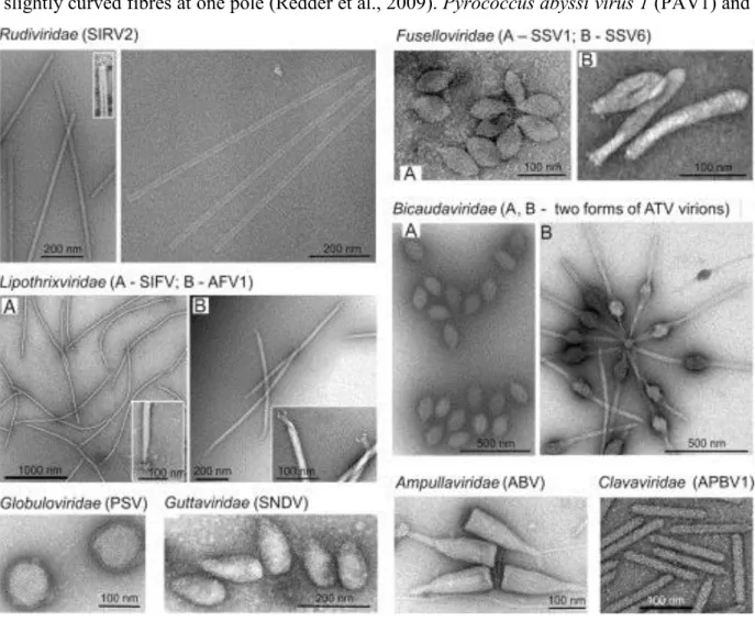
There are considerably fewer viruses characterized for methanogenic archaea: PG (Bertani and Baresi, 1987), φF1, φF3 (Nolling et al., 1993), ψM1 (Jordan et al., 1989), its deletion variant ψM2 (Pfister et al., 1998) and the prophage ψM100 (Luo et al., 2001). All the viruses listed above, except defective ψM100, have virions which display the typical morphology of siphoviruses. Notably, a VLP with morphology similar to fuselloviruses (see below) was found in cultures of Methanococcus voltae A3 (Wood et al., 1989). Further analyses of the diversity of VLPs in natural environments that contain predominantly archaea, i.e. extreme geothermal ecosystems, revealed that head-tail viruses are rather rare (Prangishvili et al., 2013). Electron microscopy on enrichment cultures from two hot springs of Yellowstone National Park shed light on the unexpected diversity reporting 12 different morphotypes (Rachel et al., 2002). Surveys in various locations – Iceland, Japan, USA – provided insights into the exceptional diversity of viruses infecting hyperthermophilic members (Rice et al., 2001; Rachel et al., 2002; Geslin et al., 2003; Bize et al., 2008; Garrett et al., 2010; Mochizuki et al., 2010; Quax et al., 2010). In fact, cultured archaeal viruses isolated from geothermal environments exhibit unique morphologies described hereinafter (Figure 2). They all, except two with single-stranded (ss) DNA, contain double-stranded (ds) DNA genomes and infect members of the genera Pyrococcus, Thermococcus, Thermoproteus, Pyrobaculum, Aeropyrum, Sulfolobus and Acidianus (Pina et al., 2011; Prangishvili, 2015).
Viruses with fusiform virions, either tail-less, tailed or two-tailed, are common in archaea-dominated environments and constitute a large fraction of described archaeal viruses. Within
~ 14 ~
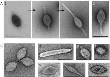
the family Fuselloviridae, members share a unique spindle-shaped morphology with spindle- or lemon-shaped particles (60x100 nm) decorated by sticky terminal fibres at one of the two pointed ends (Wiedenheft et al., 2004). Exceptions are viruses Sulfolobus spindle-shaped virus 6 (SSV6) and Acidianus spindle-shaped virus 1 (ASV1), whose virions tend to be more pleomorphic resembling a thin cigar or a pear, respectively (Redder et al., 2009). In addition, they seem to differ from the other fuselloviruses in the presence of three or four thicker and slightly curved fibres at one pole (Redder et al., 2009). Pyrococcus abyssi virus 1 (PAV1) and
Thermococcus prieuri virus 1 (TPV1) infect hyperthermophilic euryarchaea and also produce enveloped, lemon-shaped particles with a tail-like protrusion terminated by fibres (Geslin et al., 2003; Gorlas et al., 2012). Single-tailed fusiform viruses, Sulfolobus tengchongensis spindle-shaped virus 1 (STSV1) and STSV2 discovered in China are distantly related to the Bicaudaviridae (Xiang et al., 2005; Erdmann et al., 2014). This family has been established
Figure 2: Transmission electron micrographs of representative members of eight families of viruses of the
Crenarchaeota.
Reproduced with permission from Krupovic et al., 2012: Chapter 2 – Postcards from the Edge: Structural Genomics of
Archaeal Viruses.
~ 15 ~
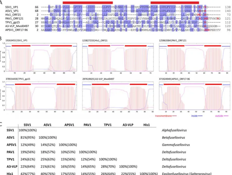
based on one known representative, Acidianus two-tailed virus (ATV) (Prangishvili et al., 2006). Notably, the two-tailed fusiform virions are extruded from host cells as tail-less particles and in conditions close to natural habitat, i.e. 85°C, two long tails are being developed from the spindle-shaped body. At the two identical ends, tubes are formed with a thin periodic filament inside and terminate in an anchor-like structure (Haring et al., 2005c). Exclusive to archaea, fusiform viruses comprise several isolates which remained unclassified for a long time. They are associated with a broad range of hosts which are highly diverse in terms of metabolism and belong to phylogenetically distant groups. Using structural markers, we defined that the two viral lineages: the Bicaudaviridae or Fuselloviridae families depend on the unique helix-bundle fold in the major capsid protein (MCP) or the presence of two hydrophobic domains, respectively. Importantly, we show that most of the isolates display the hallmark of the Fuselloviridae making it the most prominent and evolutionary successful family among the viral groups which have been described in Archaea up to now (Krupovic et al., 2012). The results of our in-depth comparative analysis have been published and can be found in the Chapter 3 of the manuscript.
In environments dominated by archaea, linear viruses are also very abundant and all of them have dsDNA genomes, a property not previously observed for any linear virus. They are part of the Ligamenvirales order which includes the two families Rudiviridae and Lipothrixviridae (Prangishvili and Krupovic, 2012). The discrimination between the rudiviruses and lipothrixviruses was initially claimed based on different principles of virion architecture and is now supported by comparative genomic data. Rudiviruses have non-enveloped virions with a length proportional to the size of the linear genomes (23x610-900 nm). They contain a superhelix formed by dsDNA genome and copies of a single glycosylated, basic DNA-binding protein. At the two ends of the tube-like structure are anchored three short tail fibres made of a minor structural protein of 100 kDa (Prangishvili et al., 2013). Members of the Lipothrixiviridae exhibit a variety of terminal appendages. In particular, there can be up to six terminal filaments forming the terminal appendage in the case of Sulfolobus islandicus filamentous virus (SIFV) (Arnold et al., 2000) or even synthesis of complex structures such as ‘claws’ in the case of Acidianus filamentous virus 1 (AFV1) (Bettstetter et al., 2003) or ‘bottle brush’ for AFV2 (Haring et al., 2005b). Consistently, they significantly differ in the structures of virion core, genomic properties and replication mechanisms sustaining a classification into four different genera (Pina et al., 2011). Historically, Thermoproteus tenax virus 1 (TTV1), TTV2, TTV3 were the first studied and belong to the α-lipothrixviruses (Reiter et al., 1988).
~ 16 ~
Other morphotypes are completely atypical and their unique characteristics justified the establishment of novel viral families by the International Committee for the Taxonomy of Viruses (ICTV). Acidianus bottle-shaped virus (ABV) is the only representative of the Ampullaviridae family and virions exhibit a complex architecture. The viral particles resemble a bottle with three structural elements: the ‘stopper’ or pointed end, the nucleoprotein core and the inner core. The dsDNA-containing nucleoprotein is folded into a cone-shaped core that is further encased in an envelope. There is a narrow, pointed end and a broad end with 20 short, thick filaments which insert into a disc and are also interconnected at their bases (Haring et al., 2005a). Members of the Guttaviridae are Sulfolobus neozealandicus droplet-shaped virus (SNDV) and Aeropyrum pernix ovoid virus 1 (APOV1). Virions of SNDV display a droplet-shaped body (90x180 nm) made of a core surrounded by a 7-nm-thick coat. Despite an overall droplet form, the surface seems to be helically ribbed and appears ‘bearded’ by multiple, long, thin fibres covering about half of the particles from the apex (Arnold et al., 2000). Interestingly, virions of APOV1 (70x55 nm) are about 1.5 times smaller than SNDV particles and do not exhibit any attached fibre (Mochizuki et al., 2011). The Globuloviridae includes Pyrobaculum spherical virus (PSV) and Thermoproteus tenax spherical virus 1 (TTSV1) infecting anaerobic and hyperthermophilic archaea of the genera Pyrobaculum and Thermoproteus (Haring et al., 2004; Ahn et al., 2006). The spherical virion (100 nm) of PSV contains multimers of a CP and host derived lipids enclosing a superhelical nucleoprotein (Haring et al., 2004). The first virus to be isolated from the order Desulfurococcales was Aeropyrum pernix bacilliform virus 1 (APBV1), family Clavaviridae. Viral particles have a rigid bacilliform topology (140x20 nm) with one end pointing and the other, round (Mochizuki et al., 2010). In the Spiraviridae, the ssDNA is bound by several copies of MCP folding into a superhelix with two levels of organization. Indeed, the non-enveloped, hollow, cylindrical virions of Aeropyrym coil-shaped virus (ACV) have appendages at both ends and are based on a rope-like fiber of two intertwined halves of a single nucleoprotein complex (Mochizuki et al., 2012). Sulfolobus turreted icosahedral virus (STIV) was the first icosahedral virus with an archaeal host identified. Viral particles are composed of circular dsDNA genome enclosed within an internal membrane and have been classified in the Turriviridae family (Rice et al., 2004; Maaty et al., 2006). Another icosahedrally symmetric, membrane-containing archaeal virus has been isolated, STIV2 for which the host-attachment structures are significantly different (Happonen et al., 2010).
~ 17 ~
To conclude, archaea, and hyperthermophilic members in particular, are infected by a range of viruses with unique morphotypes. The unexpected and unprecedented diversity of reported particle shapes is linked with unique aspects of the cellular biology of archaea (Prangishvili, 2015). Fusiform and filamentous viruses are the most abundant VLPs found in hyperthermic and hypersaline environments where archaea outnumber bacteria and thus have been quite extensively studied in comparison with the rest of the archaea-specific virosphere. The fusellovirus Sulfolobus spindle-shaped virus 1 (SSV1) and rudivirus Sulfolobus islandicus rod-shaped virus 2 (SIRV2) infect hosts of the genus Sulfolobus and have become model systems to study the biology of viruses infecting archaea.
Sulfolobus, a model for hyperthermophilic archaea.
One of the most impressive features of archaea is their capacity to sustain temperatures up to 122°C (Kashefi and Lovley, 2003). Thermophiles and hyperthermophiles have been the focus of pioneering research from Wolfman Zillig’s laboratory (Albers et al., 2013). Numerous sampling campaigns provided insights into the diversity of archaeal species — often associated with various genetic elements — present in major solfataric fields, i.e. acidic springs, water and mud holes (Zillig et al., 1993). The members of the genus Sulfolobus are characterized by: (i) an overall spherical shape of cells with lobes; (ii) facultative autotrophy and growth on sulfur or simple organic compounds; (iii) non-classical cell wall structure devoid of peptidoglycan; (iv) pH requirement ranging from 0.9 to 5.8; (v) conditions of temperature between 55 and 80°C (Brock et al., 1972). Several species and strains have been isolated from different geographical locations including Naples, Italy; Kamchatka, Russia; Lassen Volvanic National Park and Yellowstone National Park, USA (Guo et al., 2011). Interestingly, the structure of populations showed intra-species diversification and local adaptation without any correlation with temperature or pH (Whitaker et al., 2003; Grogan et al., 2008). Comparative analyses of genomes from all Sulfolobus islandicus strains available concluded that there is a strong conservation of gene synteny with distinguishable biogeographical patterns of differentiation (Jaubert et al., 2013). In general, members of the Sulfolobus have been extensively studied and several aspects of their biology are now well understood (Stetter, 1999; Urbieta et al., 2014). For example S. solfataricus and S. acidocaldarius were used to conduct the first studies on the cell cycle in Archaea (Bernander and Poplawski, 1997; Hjort and Bernander, 1999). Detailed characterization has recently revealed that homologues to the endosomal sorting complex required for transport (ESCRT)
~ 18 ~
in Eukayotes are involved in cell division in Archaea as well (Samson et al., 2008; Lindas and Bernander, 2013). Sulfolobus species are relatively easy to cultivate under laboratory conditions, in comparison with other archaeal members, and have emerged as a model of choice for investigating adaptation to geothermal environments.
Cell surface characteristics. It was realized very early that the cell surface of archaea and
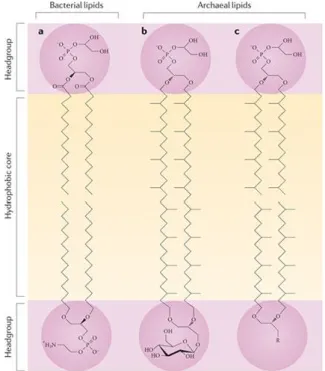
bacteria differ substantially. The first noticeable characteristic is the presence of polar lipids composed of hydrocarbon chains of 20 to 40 carbons in length and usually saturated (Figure 3). The isoprenoid moieties are ether-linked to the glycerol-1-phosphate (G-1-P) backbone. By contrast, in bacteria and eukaryotes the fatty acid derived chains are ester-linked to G-3-P
Figure 3: The lipids found in the archaeal membrane are fundamentally different from those found in eukaryotic and
bacterial membranes. In eukaryotes and bacteria, the glycerol moiety is ester-linked to an sn-glycerol-3-phosphate backbone, whereas in archaea the isoprenoid side chains are ether-linked to an sn-glycerol-1-phosphate moiety. The sn1 stereochemistry of the glycerol backbone is a truly archaeal feature, as ether lipids occur in minor amounts in eukaryotes and bacteria. The common bilayer-forming lipids in bacteria are phophatidylglycerol (upper lipid) and phosphatidylethanolamine (lower lipid) (see the figure, part a). Part b of the figure shows the structure of monolayer-forming tetraether lipids; for example, the glycophospholipid from the thermoacidophilic archaeon Thermoplasma acidophilum, in which the hydrophobic core consists of C40C40 caldarchaeol. Part c of the figure shows a bilayer formed of archaeal diether lipids, which can be found, for example, in Halobacteriales. The hydrophobic core consists of C20C20 archaeol isoprenoids. The headgroups of phospholipids can be a range of polar compounds — for example, glycerol, serine, inosine, ethanolamine, myo-inositol or aminopentanetetrols. Glycolipids also exhibit a range of sugar residues — for example, glucose, mannose, galactose, gulose, N-acetylglucosamine or combinations thereof.
Reproduced with permition from Albers and Meyer, 2011: The archaeal cell envelope.
~ 19 ~
(De Rosa et al., 1986; De Rosa and Gambacorta, 1988; Leininger et al., 2006). Notably, Sulfolobus cells were shown to almost exclusively contain glycerol dialkyl glycerol tetraether (GDGT) lipids organized in a covalently-bound bilayer resembling a monolayer (Langworthy et al., 1974; 1976; Langworthy, 1977; Chong, 2010). In nature, a variety of archaeal lipid species is found (Koga and Morii, 2005) and their biosynthesis pathways are just starting to be investigated (Jain et al., 2014; Villanueva et al., 2014). The polar head groups are identical between the three domains and it is generally assumed that the bipolar lipids found in archaea play an important role in the survival and adaptation of these microorganisms to extreme environments (Chong, 2010). On the basis that they are more chemically stable than their bacterial and eukaryotic lipids, archaeosomes, i.e. liposomes made of archaeal lipids, are being developed as potential next-generation adjuvants and drug delivery systems (Krishnan and Sprott, 2008). In vivo, the monolayer-like membrane is involved in the maintenance of cell homeostasis in combination with specific properties of the cell wall. Indeed, only a subgroup of archaea contains pseudomurein and the cytoplasmic membrane is normally surrounded by a proteinaceous, quasi-crystalline surface (S-) layer (Figure 4). In Sulfolobus, the cell wall is composed of two conserved polypeptides instead of one-component systems found in many other groups (Albers and Meyer, 2011). The outer layer assumes a three-fold symmetry based on dimers of the large protein SlaA and is anchored by membrane-bound stalks made of small peptide SlaB (Veith et al., 2009). The S-layer is proposed to contribute to cell shape, osmotic balance and protection from harsh environmental conditions. S-layer proteins (Peyfoon et al., 2010), as well as other surface-exposed proteins, undergo extensive post-translational modification by the N- and O-glycosylation pathways (Meyer and Albers, 2013; Jarrell et al., 2014).
Cell surface appendages. Several appendages and membrane-associated components have
been identified at the surface of Sulfolobus, like flagella and pili which, at first glance, appear similar to their bacterial counterparts (Figure 5). However, detailed studies showed that the archaeal flagella, the archaella (Albers and Meyer, 2011), resemble bacterial flagella only in terms of function. A single locus is responsible for ‘flagellation’ in the vast majority of species; the fla operon contains seven genes which are conserved and essential for biosynthesis and function of the apparatus (Lassak et al., 2012a). FlaB encodes archaellins, subunit components homologous to bacterial pilins which maturate through proteolytic
~ 20 ~
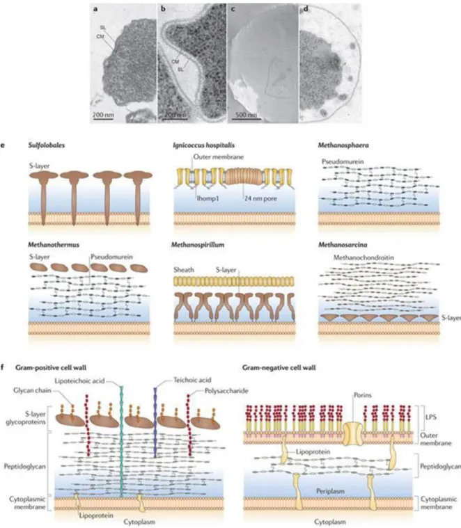
Figure 4: a,b | Electron micrographs of ultra-thin sections of the euryarchaeote Methanocaldococcus villosus (a) and the
crenarchaeote Metallosphaera prunae (b). c,d | Electron micrographs of a freeze-etched cell (c) and a thin-section cell (d) of Ignicoccus hospitalis144. e | Schematic side view of cell wall profiles from different archaea. Pseudoperiplasmic space is shown in blue. f | Schematic of bacterial cell walls. Gram-positive bacteria have a thick, amorphous, multilayered coat of peptidoglycan, teichonic and lipoteichonic acid as their cell wall and in some cases have surface-layer (S-surface-layer) glycoproteins as the outermost surface-layer above the peptidoglycan (also known as murein), for example, in Bacillus stearothermophilus20, 21. Gram-negative bacteria have an outer asymmetric bilayer membrane composed of two leaflets, an outer one containing lipopolysaccharides (LPSs), and an inner one containing mainly phospholipids, a gel-like periplasm containing peptidoglycan and the cytoplasmic membrane. CM, cytoplasmic membrane; SL, S-layer. Reproduced with permition from Albers and Meyer, 2011: The archaeal cell envelope.
~ 21 ~
cleavage. FlaG and FlaF are membrane proteins of unknown function and FlaH is an ATP- binding protein. In addition, FlaI (Reindl et al., 2013) and FlaJ are homologous to PilB and PilC, which correspond to the motor ATPase and basal membrane protein of the type IV pili in bacteria, respectively. Another key component is FlaX, the central protein required as a priming subunit during assembly in S. acidocaldarius (Banerjee et al., 2012). Structures of archaella from Halorubrum salinarium and S. shibatae are overall similar and display a thin filament with a right-handed helix around a central core (Cohen-Krausz and Trachtenberg, 2008). Given the assembly model and the structural characteristics, the archaellum is more
related to bacterial type IV pili as opposed to flagella, despite the fact that they play the same role in motility, attachment to surface and biofilm formation (Ellen et al., 2010). In bacteria, the type IV pili are key structures mediating a variety of biological processes including adhesion to surface, cell-cell interactions, conjugation, twitching motility, and pathogenicity (Craig et al., 2004). In archaea, thin, flexible 10-nm filaments of variable lengths have also
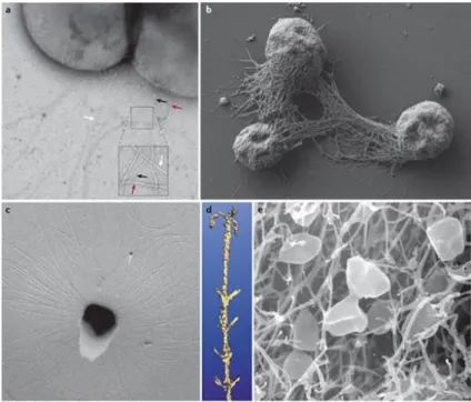
Figure 5: a | A transmission electron micrograph of negatively stained of Sulfolobales acidocaldarius cells showing
flagella (~14 nm in diameter, red arrows) pili (~10–12 nm, white arrows) and threads (~5 nm, black arrows). b | A scanning electron micrograph of Methanocaldococcus villosus157 cells grown on a surface, exhibiting bundles of flagella that act in cell–cell connections and surface adherence. c | Electron micrograph of a platinum-shadowed SM1 euryarchaeal coccus. d | Three-dimensional model of the hamus structure as visualized by surface rendering of a de-noised data set, obtained by cryo-electron tomography. The hook is 60 nm in width. e | Scanning electron micrograph of Pyrodictium spp. cells growing in a net of cannulae.
Reproduced with permition from Albers and Meyer, 2011: The archaeal cell envelope.
~ 22 ~
been observed and resemble pili in their appearance (Ng et al., 2008; Ajon et al., 2011). For example, the UV-pilus is encoded by the ups operon and is strongly induced by UV irradiation. Its biosynthesis involves the potential secretion ATPase, two pre-pilins, a putative transmembrane protein and a protein of unknown function (Frols et al., 2008). Another operon critical for surface adhesion has also been identified: the aap operon (Lassak et al., 2012a). Notably, archaeal homologues to the ATPase PilB which provides the energy necessary for pilus assembly in bacteria has been characterized. However, no ATPase involved in retraction could be identified by in silico approaches, suggesting that pili and flagella are unable to retract in Archaea. Other reported structures seem to be unique to the third domain of life (Figure 5). In S. solfataricus, a large number of sugar binding proteins have been identified with a class III signal peptide. These proteins require the bas operon for their functional surface localization. It has been proposed that BasEF would form the core of the assembly machinery at the membrane while BasABC would participate in cleavage of pilin signal peptides and correct assembly of binding proteins into a macromolecular complex (Zolghadr et al., 2007). The ‘bindosome’ would serve as a platform dedicated for sugar uptake from the environment in addition to pore-like openings of S-layer which are suggested to only permit passage of nutrients and other small molecules (Ellen et al., 2010). Although never reported for Sulfolobus, cannulae and hamus are fascinating appendages associated with surface. Cannulae of hyperthermophilic Pyrodictium abyssi are hollow tubes with a diameter of ~25 nm and associate in a network facilitating intercellular communication, nutrient exchange or transport of genetic material (Rieger et al., 1995). Hamus found at the surface of the psychrophilic archaeon SM1 plays a role in surface attachment, biofilm formation and anchoring; the filaments are 7-8 nm in diameter forming a complex helix with three hooks present every 4 nm (Moissl et al., 2005).
Insights into the biology of hyperthermophilic archaeal viruses.
Fusiform and filamentous VLPs are highly abundant and widely distributed in archaea-dominated habitats. The two groups of viruses are represented by fusellovirus SSV1 and rudivirus SIRV2 which have been among the first archaeal viruses to be isolated from geothermal environments where they can infect Sulfolobus cells. SSV1 has a rather broad host range (Schleper et al., 1992), whereas SIRV2 can only infect a limited number of strains of S. islandicus (Bize et al., 2009). The two viruses serve as model systems for the study of hyperthermophilic archaeal viruses.
~ 23 ~
SSV1. The genome of SSV1 was shown to be present in S. shibatae B12 in two forms: as a
linear form within the host chromosome and as free, circular episomal copies in the cytoplasm (Yeats et al., 1982). UV irradiation is a strong stimulus to enhance the production of lemon-shaped particles encasing the circular form of the viral genome (Martin et al., 1984). SSV1 was initially called SAV-1 due to misclassification of its natural host as a strain of S. acidocaldarius. It is a temperate virus and infection results in a lysogenic cycle leading to growth recovery of cultures even after stimulation (Schleper et al., 1992). The capacity to integrate into the cellular genome at a specific site within a tRNA-Arginine gene (Reiter et al., 1989) has been used to establish one of the first genetic systems in Archaea (Schleper et al., 1992). As a result, the viral tyrosine recombinase has been extensively studied (Muskhelishvili et al., 1993; Serre et al., 2002; Letzelter et al., 2004; Zhan et al., 2012). Development of genetic tools has also allowed systematic analysis of the functions of viral open reading frames (ORFs) and effects of their deletions on virus fitness (Stedman et al., 1999; Iverson and Stedman, 2012). In particular, the integrase gene has been shown to be non-essential for infection (Clore and Stedman, 2007). Interestingly, unlike the situation found in bacteriophages, upon viral genome integration, the integrase gene is partitioned in two fragments (Reiter et al., 1989). Several isolates are now known to be similar to SSV1 in morphology, genomic content, replication strategy, etc. (Stedman et al., 2003; Wiedenheft et al., 2004; Redder et al., 2009); nevertheless, SSV1 remains to be a model to understand the biology of spindle-shaped viruses. Using genome-wide microarray, it was shown that there is a tight regulation of gene expression timing, reminiscent of bacteriophages and eukaryotic viruses. The transcription starts with a small UV-specific transcript and continues with early and late transcripts towards the end of the viral cycle (Frols et al., 2007a; Fusco et al., 2013). Interestingly, there was no marked difference detected in the transcriptome of the host S. solfataricus, in line with the postulated egress of SSV1 by budding through the cytoplasmic membrane without lysis of the host (Martin et al., 1984). Most of the particles released are uniform in size (60x100 nm) although up to 1% of viral population can be larger, the maximum length being about 300 nm (Reiter et al., 1988). Recently, the structure of SSV1 was examined by cryo-electron microscopy (cryo-EM) and 3D image reconstruction. A model of SSV1 structure has been proposed despite the fact that resolution was severely limited by particle size, lack of global symmetry, structural heterogeneity, and a small number of particles considered (Stedman et al., 2015). In particular, the presence of an actual lipid
~ 24 ~
membrane encasing the virion body could not be verified and remained controversial up to now. Thus, one of the main objectives of my PhD was to perform a comprehensive biochemical characterization of SSV1 virions which is described in the Chapter 4. Briefly, we showed that SSV1 is a lipid-containing virus composed of glycrosylated proteins and host-derived lipids encasing the nucleoprotein filament (Quemin et al., 2015). These findings provide insights into the architecture of unique archaeal viruses and are used as a foundation for ongoing studies targeting the interactions of SSV1 with its host Sulfolobus (Quemin et al., in preparation). We recently obtained significant insights into the assembly and release strategy utilized by SSV1 virions which are presented in the Chapter 5.
SIRV2. The non-enveloped, linear dsDNA virus SIRV2 is the type-species of the Rudiviridae
family. The stiff, rod-shaped particles (950x26 nm) were observed for the first time in the culture of S. islandicus strain KVEM10H3 (Zillig et al., 1993). There is a central cavity of 6 nm in diameter and three 28-nm-short appendages protruding from both ends. The viral particles consist of a tube-like superhelix which length correlates with the genome size, i.e. SIRV1 virions are 70 nm shorter than those of SIRV2 and have genomes of 32.3 versus 35.8 kbp in SIRV2. Recently, the structure of SIRV2 virions using cryo-EM and 3D image reconstruction became available and revealed a unique DNA topology in the A-form which has only been previously observed in bacterial spores and in vitro (DiMaio et al., 2015). Interestingly, SIRV1 is also known to have a very high mutation rate of 3.10-3 substitutions per nucleotide per replication cycle, whereas SIRV2 is considered to be invariant (Prangishvili et al., 1999). The genome of SIRV2 is covalently closed at the termini and carries inverted terminal repeats. It contains 54 ORFs (Peng et al., 2001). Transcription of the viral genome has been shown to start simultaneously at multiple sites and spread over the two strands in a uniform pattern through the course of infection (Kessler et al., 2004). Microarray analysis (Okutan et al., 2013) and RNA-seq approach (Quax et al., 2013) revealed that transcription is limited to the two distal termini of the viral genome immediately after infection and then spreads over the totality of ORFs within 2 hours. The host cell machinery is extensively reprogrammed with more than half of host genes having a different level of expression. The genes involved in cell division, chromosome maintenance, and stress response were down-regulated while anti-viral defense mechanisms, i.e Clustered Regularly Interspaced Short Palindromic Repeats (CRISPR) – CRISPR associated proteins (Cas) and toxin/antitoxin systems, were massively activated (Quax et al., 2013). The results on
~ 25 ~
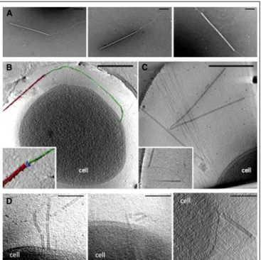
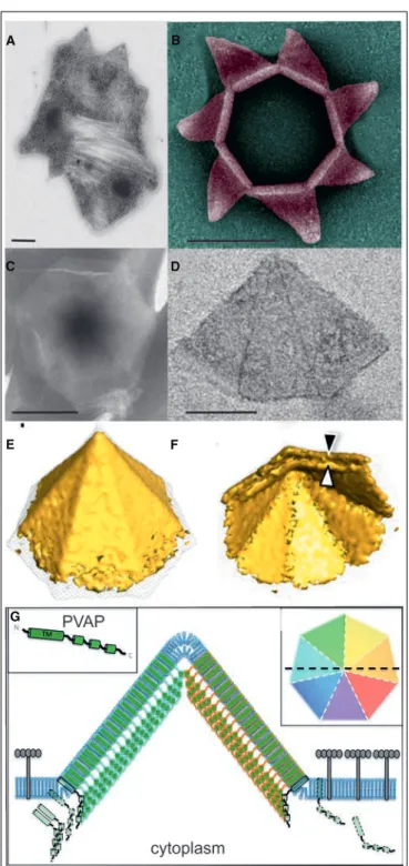
transcription are consistent with the lytic cycle of SIRV2. Indeed, the virus orchestrates an elaborated mechanism leading to degradation of host chromosome and remodeling of the surface which results in cell death. The progeny is assembled in the cytoplasm occupying the entire space and released after formation and opening of specific virus-associated pyramids (VAPs) which are anchored in the plasma membrane and protrude through the S-layer (Bize et al., 2009; Quax et al., 2011; Daum et al., 2014). As opposed to the egress mechanism of SIRV2, prior to the advent of my PhD project literally nothing was known about the early stages of the infection cycle, i.e. adsorption to the host cell surface and entry of the viral genome into the cell interior. In order to gain insights into the entry of SIRV2 virions, we have utilized a number of different assays to assess the binding kinetics, reversible and irreversible adsorption, receptor saturation, etc. as well as transmission electron microscopy and whole-cell electron tomography (Quemin et al., 2013). The recently published article is included in my PhD thesis and reported in the Chapter 6.
The cases of SSV1 and SIRV2 exemplify the uniqueness of the virosphere specific to Archaea. Not only they display morphotypes which have never been associated with bacteria or eukaryotes but also reveal uncommon principles for virus biology and infection. Although, data have been accumulating on the diverse architectures of virions (Prangishvili et al., 2013) and original genomic content (Krupovic et al., 2012), the exploration of virus-host interactions in the third domain of life is still in its infancy. The aim of the work performed during the course of my PhD was to provide insights into the infection cycle of archaeal viruses. The global strategies employed for entry, assembly and egress have been investigated by a combination of electron microscopy approaches, biochemistry, cellular and molecular biology techniques. The data presented here on the molecular mechanisms of virus-host interactions for both lipid-containing, fusiform SSV1 and non-enveloped, filamentous SIRV2 allows comparison with bacterial and eukaryotic virus-host systems.
Virus-host interactions in Archaea: state-of-the-art.
Members of the third domain of life, the archaea, were initially regarded as exotic microorganisms capable of growing in conditions which are hostile to humans. Among other intriguing features, they are now known to host unique viruses classified into exclusive viral families. Several studies have permitted the isolation of highly diverse viruses characterized by atypical virion shapes and mysterious genomic contents. The research undertaken in the
~ 26 ~
past thirty years has improved our appreciation of the virosphere associated with archaea. However, the study of archaeal viruses imposes serious constraints and the collection of virus-host systems found in laboratories is far from representing the situation observed in natural environments. The isolation and characterization are indeed limited due to the need of culturing cells under extreme conditions of temperature, pH, salinity, pressure, etc. which are complicated to set up in laboratory. Another restriction comes from the viruses themselves which tend to be produced in low titers rendering analysis by classical techniques often challenging.
Using high-throughput approaches, one can neglect some of these factors and overcome the major difficulties linked to the research on archaeal viruses. In the Chapter 1, the editorial outlines the recent insights that have been obtained on the infection cycle of hyperthermo-acidophilic virus-host models, namely SSV1, SIRV2, and STIV (Quemin et al., 2014). We put a particular emphasis on data covering structural genomics, whole-genome microarrays or RNA-sequencing, as well as large-scale proteomic analysis of infected cells. In fact, comparative genomics defined the structure and/or function of more than 10% of the ORFs identified in viral genomes. Additional insights came from screens for interactions or whole-transcriptome analyses in the case of SIRV2. Viral and host gene expression through the course of infection varies and a tight timing of transcription has been described for SSV1 with early, middle and late genes. Considering the proteome, STIV infection was shown to induce significant differences in protein levels and, more importantly, in post-translational modification profiles. Together these studies highlight the rapid development of high-throughput methods in the field of archaeal viruses and help to define interesting targets that should be the focus of intensive research in the near future.
Moreover, recent studies trying to decipher the sequential events of the viral life cycle have led to major breakthroughs in the field. The review proposed in the Chapter 2 has been written during the framework of my PhD. It summarizes the available information concerning the virus-host interplay in Archaea with a focus on hyperthermo-acidophilic virus-host systems (Quemin and Quax, 2015). We discuss the possibility that appendages, which are observed to decorate virion termini in various families and can even form complex structures, are required during the entry process of these viruses. In the same line, novel strategies employed for egress have been recently described and are reported in great detail. The molecular mechanisms of virus-host interactions in archaea are also compared to the ways bacterial and eukaryotic viruses interact with their respective hosts. Together with the harsh
~ 27 ~
environmental conditions, the characteristics of archaeal cell surface, i.e. cytoplasmic membrane and S-layer, might render the delivery of viral nucleic acids and the release of viral progeny quite difficult. Therefore, the host specificities in terms of ecology and biology could have compelled viruses to adapt and employ uncommon strategies that we are just starting to discover and understand.
~ 29 ~
~ 31 ~
CHAPTER1
703 ISSN 1746-0794 Future Virol. (2014) 9(8), 703–706
REVIEW
part of 10.2217/FVL.14.52 © 2014 Future Medicine LtdEDITORIAL
Hard out there: understanding
archaeal virus biology
1Institut Pasteur, Unité Biologie Moléculaire du Gène chez les Extrêmophiles, Département de Microbiologie,
75015 Paris, France
*Author for correspondence: [email protected]
KEYWORDS
• Archaea • hyperthermophiles
• Sulfolobus rod-shaped virus 2
• virus evolution • virus–host interactions
Each of the three domains of life, Archaea, Bacteria and Eukarya, is associated with a specific virosphere. Despite the fact that archaeal viruses represent only a minute portion of the characterized virosphere, they have recently gained wider attention, mainly due to the unexpected morpho-logical properties of their virions and the unprecedented molecular mechanisms employed throughout their life cycles. Archaeal viruses are currently classified into 15 different families [1,2]. Especially remarkable are the viruses of the hyper-thermohilic archaea; these viruses are extremely diverse morphologically and include members with lemon-shaped, droplet-shaped and bottle-shaped viri-ons [1]. Furthermore, the viral genomes encode proteins with little to no significant similarity to proteins in public databases and often possess unique structural folds [3]. Although classical biochemical and genomic studies have yielded important information on the architectures of sev-eral hyperthermophilic archaeal viruses, as well as on the functions of some viral pro-teins, the molecular mechanisms underly-ing different aspects of the infection cycle
remain poorly understood for most of these viruses.
Studies on bacterial and eukaryotic viruses have benefited from the availabil-ity of well-established genetic tools that have been developed for the respective hosts and, more generally, from the broad knowledge base on the host biology. This, unfortunately, has not been the case for most of the archaeal virus–host systems. The assays that are considered trivial when thinking about bacterial or eukary-otic viruses (e.g., the plaque test used for virus particle enumeration) present dif-ficulties in the case of hyperthermophilic archaeal viruses. Indeed, the cultivation of hyperthermophilic acidophiles, such as Sulfolobus, which, for optimal growth, requires 80°C and pH 2–3, might be challenging. Similarly, live-cell imaging at physiological temperatures, which is widely used to investigate virus–host interaction in eukaryotes, is normally also off the table when dealing with hyperthermophiles. Consequently, the scientific inquiries into the properties of hyperthermophilic archaeal viruses have been, for a long time, limited by the lack of adequate tools. Emmanuelle RJ Quemin1, David Prangishvili1 & Mart Krupovic*,1
Future Virol. (2014) 9(8)
704
EDITORIAL Quemin, Prangishvili & Krupovic
future science group Given all of these difficulties, one might
wonder why anyone would bother with stud-ying archaeal viruses in the first place. The major incentives are the following. First, the morphological diversity of hyperthermophilic archaeal viruses is astonishing [1]. Whereas sampling of the bacterial virosphere seems to have reached convergence (i.e., no truly new morphotypes of bacterial viruses have been discovered for decades), virions with unique, previously unseen morphologies are constantly being discovered in the Archaea. It has been suggested that the archaeal virosphere more closely reflects the ancient diversity of viruses on our planet [1]. Consequently, exploration of the archaeal virus diversity provides an exclu-sive opportunity to learn about the ancient viral architectures that might not have been retained in other cellular domains. Second, the molecular mechanisms underlying virus–host interactions in Archaea combine components that are specific to archaeal viruses with those that are shared with viruses infecting other cellular domains. Thus, in addition to uncov-ering new Archaea-specific features that are sometimes breathtakingly elegant (as in the case of the recently discovered pyramidal egress structures [4,5]), these studies allow us to better understand the origin and evolution of the mechanisms underlying the infection processes of viruses infecting eukaryotic hosts (see below). Third, due to their ability to with-stand harsh environmental conditions, hyper-thermophilic archaeal viruses contain consid-erable appeal for developing various bio- and nano-technological applications. Furthermore, the enzymes encoded by these viruses can be potentially employed for molecular biology applications.
During the past few years, many mod-ern high-throughput techniques have been adapted for studying archaeal viruses, and new genetic tools have been developed for an increasing number of archaeal hosts and their viruses [6–8]. These newly developed/adapted approaches and genetic tools, in combination with the more classical biochemical techniques, have recently yielded valuable information on the biology of some archaeal viruses. Two hyperthermophilic viruses infecting Sulfolobus species have been investigated from different perspectives and served as models for under-standing the biology of archaeal viruses. These include Sulfolobus turreted icosahedral virus
(STIV) and Sulfolobus islandicus rod-shaped virus 2 (SIRV2). These two viruses funda-mentally differ from each other in virion mor-phology, genomic content and viral cycle [1]. STIV is a prototype member of the family Turriviridae. The STIV virion consists of an icosahedral protein capsid that covers the lipid membrane vesicle enclosing the circular dsDNA genome [9]. Such a virion architecture is commonly found in bacterial and eukaryotic viruses [10]. SIRV2 is the type organism of the family Rudiviridae, which comprises viruses with elongated rod-shaped particles contain-ing linear dsDNA genomes [11]. The termini of SIRV2 virions are decorated with terminal protein fibers that mediate the attachment of the viral particles to the pili-like appendages at the host cell surface [12]. Interestingly, despite profound morphological and genomic differ-ences, both STIV and SIRV2 utilize a unique virion release mechanism involving the forma-tion and opening of large pyramidal structures at the surface of the host cell [4,5].
Even though high-throughput approaches generate impressive amounts of data, the com-prehensive picture of the viral infection cycle can only be unraveled using a combination of different high-throughput and more clas-sical techniques targeted at particular aspects of the infection cycle and specific viral pro-teins. Indeed, clues obtained in the course of high-throughput studies have proved to be instrumental for identifying prominent play-ers in the viral life cycles and designing tar-geted studies in order to understand the func-tions of these proteins. For example, RNA sequencing analysis of SIRV2-infected cells has revealed that ORF83a/b transcripts are dominant, starting within the first minutes of infection and remaining abundant throughout the infection [13], predicting an important role for P83a/b. The x-ray structure of the P83a/b homolog from rudivirus SIRV1, which was solved during the structural genomics pro-ject, revealed a helix-turn-helix DNA-binding motif, suggesting that the protein might be involved in the processing of viral DNA [3]. Subsequent yeast two-hybrid analysis has showed that P83a/b interacts with the subu-nit of the host-encoded PCNA, a processivity factor for DNA polymerase [14]. These results indicate that P83a/b might be responsible for recruiting the PCNA for viral genome replica-tion. Consequently, the complementary results
“…exploration of the archaeal virus diversity provides an exclusive opportunity to learn about
the ancient viral architectures that might not have been retained in
705 Hard out there: understanding archaeal virus biology EDITORIAL
future science group www.futuremedicine.com obtained from different studies illuminated a
key role of P83a/b in SIRV2 propagation, pro-viding a framework for further inquiries into the molecular mechanisms of its action.
An important step forward in understand-ing the biology of archaeal viruses has also been obtained for the example of STIV by the com-bination of different approaches. In this case, large-scale proteomic analysis of infected cells by 1D and 2D differential gel electrophoresis coupled with protein identification by mass spectrometry and activity-based protein profil-ing has been used to investigate the interaction between STIV and two Sulfolobus solfataricus strains (P2 and P2-2-12) that significantly differ with respect to their susceptibility to STIV [15,16]. In the highly susceptible P2-2-12 strain, only ten cellular proteins were changed in abundance. By contrast, 71 host proteins representing 33 different cellular pathways were affected during the infection of the poorly sus-ceptible P2 strain [15,16], shedding some light on the basis of the different susceptibilities to infec-tion of closely related Sulfolobus strains. Most notably, among the highly upregulated proteins were components of the antiviral CRISPR-Cas system and cell division proteins that are homologous to the eukaryotic endosomal sort-ing complexes required for transport (ESCRT) machinery [17,18], suggesting that the latter proteins play an important role in the STIV infection cycle. In eukaryotes, the ESCRT machinery is employed as the major escape route for many enveloped viruses, including impor-tant human pathogens, such as retroviruses,
filoviruses, paramyxoviruses and herpesviruses [19]. Importantly, a recent study has confirmed a critical role of the archaeal ESCRT proteins dur-ing the late stages of STIV infection, specifically during the maturation of the virion membrane and possibly the opening of pyramidal portals located at the host cell envelope and involved in the release of viral progeny [20].
To conclude, a combination of different high-throughput approaches with more con-ventional biochemical and microscopic tech-niques has helped us to uncover the secrets of the enigmatic archaeal viruses. Even though studies on viruses thriving in extreme environ-ments remain challenging, they are also highly rewarding. We have learned a great deal about the inventiveness of these viruses and new sur-prises are certainly expected in the future. The detailed understanding of archaeal viruses and their interactions with their hosts will enable comparisons with the bacterial and eukaryal virus–host systems, which should eventually reveal the general tendencies underlying the functioning of the virosphere.
Financial & competing interests disclosure The authors have no relevant affiliations or financial involvement with any organization or entity with a finan-cial interest in or finanfinan-cial conflict with the subject matter or materials discussed in the manuscript. This includes employment, consultancies, honoraria, stock ownership or options, expert testimony, grants or patents received or pending, or royalties.
No writing assistance was utilized in the production of this manuscript.
References
1 Prangishvili D. The wonderful world of
archaeal viruses. Annu. Rev. Microbiol. 67, 565–585 (2013).
2 Pawlowski A, Rissanen I, Bamford JK,
Krupovic M, Jalasvuori M.
Gammasphaerolipovirus, a newly proposed bacteriophage genus, unifies viruses of halophilic archaea and thermophilic bacteria within the novel family Sphaerolipoviridae. Arch. Virol. 159, 1541–1554 (2014).
3 Krupovic M, White MF, Forterre P,
Prangishvili D. Postcards from the edge: structural genomics of archaeal viruses. Adv. Virus Res. 82, 33–62 (2012).
4 Bize A, Karlsson EA, Ekefjard K et al. A
unique virus release mechanism in the Archaea. Proc. Natl Acad. Sci. USA 106, 11306–11311 (2009).
5 Brumfield SK, Ortmann AC, Ruigrok V et al.
Particle assembly and ultrastructural features associated with replication of the lytic archaeal virus Sulfolobus turreted icosahedral virus. J. Virol. 83, 5964–5970 (2009).
6 Iverson E, Stedman K. A genetic study of
SSV1, the prototypical fusellovirus. Front. Microbiol. 3, 200 (2012).
7 Jaubert C, Danioux C, Oberto J et al.
Genomics and genetics of Sulfolobus islandicus LAL14/1, a model hyperthermophilic archaeon. Open Biol. 3, 130010 (2013).
8 Wirth JF, Snyder JC, Hochstein RA et al.
Development of a genetic system for the archaeal virus Sulfolobus turreted icosahedral virus (STIV). Virology 415, 6–11 (2011).
9 Veesler D, Ng TS, Sendamarai AK et al.
Atomic structure of the 75 MDa extremophile Sulfolobus turreted icosahedral virus
determined by CryoEM and x-ray crystallography. Proc. Natl Acad. Sci. USA 110, 5504–5509 (2013).
10 Krupovic M, Bamford DH. Double-stranded
DNA viruses: 20 families and only five different architectural principles for virion assembly. Curr. Opin. Virol. 1, 118–124 (2011).
11 Prangishvili D, Koonin EV, Krupovic M.
Genomics and biology of rudiviruses, a model for the study of virus–host interactions in Archaea. Biochem. Soc. Trans. 41, 443–450 (2013).
12 Quemin ER, Lucas S, Daum B et al. First
insights into the entry process of
hyperthermophilic archaeal viruses. J. Virol. 87, 13379–13385 (2013).
13 Quax TE, Voet M, Sismeiro O et al. Massive
activation of archaeal defense genes during viral infection. J. Virol. 87, 8419–8428 (2013).
“The detailed understanding of archaeal
viruses and their interactions with their
hosts will enable comparisons with the bacterial and eukaryal