RESEARCH OUTPUTS / RÉSULTATS DE RECHERCHE
Author(s) - Auteur(s) :
Publication date - Date de publication :
Permanent link - Permalien :
Rights / License - Licence de droit d’auteur :
Bibliothèque Universitaire Moretus Plantin
Institutional Repository - Research Portal
Dépôt Institutionnel - Portail de la Recherche
researchportal.unamur.be
University of Namur
Epidermal morphogenesis during progressive in vitro 3D reconstruction at the
air-liquid interface
Frankart, Aurélie; Malaisse, Jérémy; De Vuyst, Evelyne; Minner, Frédéric; Lambert de
Rouvroit, Catherine; Poumay, Yves
Published in: Experimental dermatology DOI: 10.1111/exd.12020 Publication date: 2012 Document Version
Early version, also known as pre-print
Link to publication
Citation for pulished version (HARVARD):
Frankart, A, Malaisse, J, De Vuyst, E, Minner, F, Lambert de Rouvroit, C & Poumay, Y 2012, 'Epidermal morphogenesis during progressive in vitro 3D reconstruction at the air-liquid interface', Experimental dermatology, vol. 21, no. 11, pp. 871-5. https://doi.org/10.1111/exd.12020
General rights
Copyright and moral rights for the publications made accessible in the public portal are retained by the authors and/or other copyright owners and it is a condition of accessing publications that users recognise and abide by the legal requirements associated with these rights. • Users may download and print one copy of any publication from the public portal for the purpose of private study or research. • You may not further distribute the material or use it for any profit-making activity or commercial gain
• You may freely distribute the URL identifying the publication in the public portal ?
Take down policy
If you believe that this document breaches copyright please contact us providing details, and we will remove access to the work immediately and investigate your claim.
Epidermal morphogenesis during progressive in vitro
3D reconstruction at the air-liquid interface
Aurélie Frankart*, Jérémy Malaisse*, Evelyne De Vuyst, Frédéric Minner, Catherine Lambert de Rouvroit, and Yves Poumay
URPHYM (Research Unit for Molecular Physiology), Cell and Tissue Laboratory, NARILIS, University of Namur (FUNDP), Namur, Belgium
Corresponding author: Professor Yves Poumay,
Cell and Tissue Laboratory, URPHYM-NARILIS, University of Namur (FUNDP),
61 Rue de Bruxelles, B-5000 Namur, Belgium. E-mail: [email protected]
Tel: +32-81-724257 Fax: +32-81-724261
ABSTRACT
Keratinocyte monolayers, cultured in immersed conditions, constitute a frequently used in vitro model system to study keratinocytes behaviour in response to environmental assaults. However, monolayers lack the keratinocyte terminal differentiation and the organisation of the epidermal tissue which are observed in vivo. Advancements of in vitro techniques were used to reconstruct three-dimensional equivalents that mimic human epidermis in terms of layering, differentiation and barrier function. Here we update a published method and illustrate the progressive morphogenesis responsible for in vitro reconstruction. The analysis of cell proliferation, expression of differentiation markers, and barrier efficacy demonstrate the excellent similarity of the reconstructed tissue with normal human epidermis. Availability of epidermal tissue during its reconstruction phase in culture appears crucial for studies intending to challenge the barrier function.
INTRODUCTION
By creating an impermeable cornified layer on its surface, the epidermis provides a barrier against loss of body fluid and environmental assaults such as solar radiation, infections, or chemical aggressions (1). Primary cultures of keratinocytes constitute an in vitro model to characterize this cell type’s behaviour. However, keratinocyte monolayers in immerged culture conditions represent an incomplete epidermal model regarding terminal differentiation and production of the barrier. Indeed, complete epidermal morphogenesis requires exposure of stratified keratinocytes to the air-liquid interface (1). Advancements in keratinocyte culture were used to reconstruct three-dimension epidermal equivalents that mimic human epidermis in terms of tissue architecture, differentiation and barrier function (2).
While the pioneering work of Rheinwald and Green produced stratified colonies of human primary cultured keratinocytes (3), the differentiation program was abnormal in such conditions (4), creating a need for cultures that would more closely mimic normal expression of differentiation markers. Chemically defined culture media permitted keratinocyte culture in absence of serum and feeder cells, in the presence of low calcium concentration (5), inspired by data collected with mouse keratinocytes (6). However, calcium concentration above 1 mM is required for intercellular anchoring junctions and stratification of keratinocytes. Thus, Prunieras and co-workers developed a well-differentiated epidermis in culture (7) and demonstrated full differentiation of keratinocytes from basal to cornified layers by exposing cells to the air-liquid interface (8,9). By sorting primary keratinocytes expressing high levels of integrin 6, highly proliferative clones capable of epidermal reconstruction can be obtained from single cells (10).
Currently, several commercial human skin substitutes or reconstructed epidermis represent reproducible models with controlled environment, often used in cutaneous toxicological studies. However, the cost of such tissues and the need for tailor-made culture parameters led us to publish methods for an open-source reconstruction of a fully differentiated epidermis. Here we update detailed description of our method intended to obtain human epidermis that exhibits typical in vivo morphology 11 days after seeding keratinocytes on polycarbonate filters (11). We also illustrate progressive morphogenesis during reconstruction of the epidermis and concomitantly analyzed cell proliferation, expression of differentiation markers, and efficacy of the barrier.
MATERIALS AND METHODS Culture media
The basal medium for culture settings, KGM-2 (Clonetics), is supplemented with SingleQuot KGM-2 (Clonetics) according to manufacturer’s instructions in order to contain as final concentrations 50µg/ml bovine pituitary extract, 10ng/ml EGF, 5µg/ml insulin, 5.10-7 M hydrocortisone, 5µg/ml transferrin and 0.15 mM Ca++.
EpiLife medium (Cascade Biologics) supplemented with HKGS (Cascade Biologics) was used according to manufacturer’s instructions for growth of keratinocytes and contains 0.2 % bovine pituitary extract , 0.2 ng/ml EGF, 5µg/ml insulin, 5.10-7 M hydrocortisone, 5µg/ml transferrin and 0.06 mM of Ca++. The medium used on
the first day of tissue reconstruction is composed of complete EpiLife medium supplemented by CaCl2 in order
to reach 1.5 mM Ca++. After exposure to the air-liquid interface, modified complete EpiLife culture medium is
used, supplemented with 1.5 mM Ca++, 50 µg/ml vitamin C (Sigma-Aldrich), and 10 ng/ml keratinocyte growth
factor (KGF; R&D Systems).
Human keratinocytes isolation and reconstruction of the epidermis
Human primary keratinocytes were isolated from superficial normal adult skin collected from plastic surgery (Dr B. Bienfait, Clinique St. Luc, Namur-Bouge, Belgium) as described (12). Briefly, keratinocytes were isolated by the trypsin float technique and proliferating primary cultures initiated in complete KGM-2 medium. Before reaching confluence, proliferating keratinocytes were harvested by trypsinization and plated for sub-culture in complete EpiLife medium, containing supplements (HKGS). When a 60% density was reached, cells were trypsinized for preservation in liquid nitrogen (12). For tissue reconstruction, third-passage proliferating keratinocytes were used and our previously published protocol (11) was slightly improved. Cell suspensions containing approximately 2 million cells, thawed after preservation in liquid nitrogen, were diluted in complete KGM-2 medium in one 175 cm2 culture flask for 24h, and then in complete EpiLife medium renewed every two
days. When keratinocytes covered 60-70 % of the flask area, cells were harvested by trypsinization and centrifuged for 10 min at 1000 rpm at 4°C. The pellet was resuspended at a density of 300 000 cells/ml in ice-cold EpiLife medium containing high (1.5mM) calcium concentration. Polycarbonate culture inserts (0.63 cm2 of
area containing 0.4 µm diameter pore size; Millipore) were placed in six-well plates containing 2.5 ml of cold medium. Each insert received 500 µl of keratinocyte suspension (300 000 cells/ml) corresponding to about 250 000 cells/cm2. After 24h of incubation at 37°C in a humidified atmosphere containing 5% CO
2, cells were
exposed to the air-liquid interface by removal of the medium in the upper compartment. The 2.5 ml of medium under the filter were replaced by 1.5 ml of complete EpiLife medium, modified for tissue reconstruction (see above paragraph) and renewed every two days.
Histological analysis and immunolabelling of epidermal differentiation markers
Reconstructed human epidermis (RHE) were fixed after 1, 3, 5, 7, 9 and 11 days of culture in 4% formalin-acetic acid solution, dehydrated and embedded in paraffin. Incubation in toluene prior to paraffin embedding induces detachment of the polycarbonate filter from the insert, releasing tissue. Tissue sections (6 µm thick) were prepared perpendicular to the filter and laid over microscopic slides before staining with haematoxylin-eosin. Tissue sections for immunolabelling were deparaffinized, rehydrated and rinsed with water. For keratin 14 or filaggrin labelling, sections were immersed in 10 mM citrate buffer pH 6 at 90°C for 20 min, then washed twice in PBS before blocking in PBS containing 0.2% bovine serum albumin and 0.02% Triton X-100 for 1h followed by incubation for 1h at room temperature with the appropriate antibodies. Mouse monoclonal primary antibodies
were used to label keratin 14 (Santa Cruz, dilution 1:50), keratin 10 (Dako, dilution 1:100), involucrin (Sigma-Aldrich, dilution 1:200) and filaggrin (Thermo Scientific, dilution 1:75). For immunofluorescence, tissue sections were washed in PBS containing 0.2% bovine serum albumin and 0.02% Triton X-100 before incubation with Alexa 488-conjugated anti-mouse IgG (Molecular Probe Invitrogen, dilution 1:100) for 1h. Coverslips were mounted in Mowiol and tissue sections observed under confocal microscopy (Leica) and phase-contrast microscopy and pictures were finally overlapped.
Labelling of cycling keratinocytes: BrdU incorporation
Tissues were incubated for 24 h with 10 µM bromodeoxyuridine (BrdU) before fixation and embedding in paraffin. Tissue sections were deparaffinized and incubated for 30 min in 10 mM citrate buffer pH 6 at 90°C. DNA was denatured by incubation of sections for 30 min in 2N hydrochloric acid and then neutralized using 0.1 M sodium tetraborate pH 8.5. Incorporated BrdU was detected using a monoclonal primary antibody (BD Pharmingen, dilution 1:25) followed by Alexa-488 anti-mouse secondary antibody (Molecular Probe Invitrogen Invitrogen, dilution 1:100). Coverslips were mounted in Mowiol for observation under confocal microscopy. Percentage of BrdU-positive cells in RHE was calculated as (number of BrdU-positive cells/total number of cells in basal layer) x 100 for each condition.
Trans-epithelial electrical resistance measurements and permeability to Lucifer Yellow
Trans-epithelial electrical resistance (TEER) measurements of RHE were performed from day 1 to 21 of culture using Millicell-Electrical Resistance System (Millipore). Tissues were placed in six-well multiplates containing 3.5 ml of culture medium and overlaid with 450 µl of culture medium for the time required to measure electrical resistance.
For determination of RHE permeability to Lucifer Yellow fluorescent dye, a 1 mM solution was laid over the tissue for 2 hours at 37°C before fixation and embedding in paraffin for tissue processing. Sections were analysed using fluorescence microscopy.
RESULTS
Epidermal morphogenesis benefits from a transiently elevated keratinocyte proliferation and results into the formation of an efficient barrier at the air-liquid interface
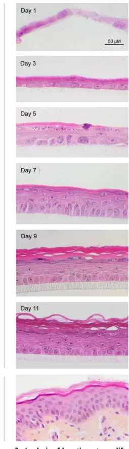
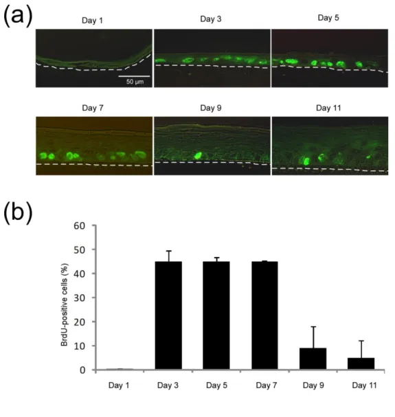
Histological observations, as well as measurements of keratinocyte proliferation by BrdU incorporation were performed through epidermis reconstruction process, on RHE obtained after 1, 3, 5, 7, 9 and 11 days of culture (Figures 1 and 2). One day after seeding keratinocytes, the tissue exhibits one or two not yet proliferative flattened cell layers (Day 1) (Figures 1 and 2). After exposure to the air-liquid interface, a suprabasal, probably protective layer, composed of rather acidophilic keratinocytes can be observed just above the highly proliferative basal layer (Day 3) (Figures 1 and 2). After five days, the proliferation rate of basal keratinocytes remains high (Figure 2) allowing keratinocytes to build RHE composed of four to five cell layers (Figure 1). Tissue differentiation leads to progressive formation of typical epidermal layers. Indeed, the presence of keratohyalin granules easily identified at Day 5 defines a granular layer (Figure 1). After 9 and 11 days of culture, keratinocyte proliferation in the RHE decreases while the tissue’s morphology becomes very similar to the normal human epidermis (Figures 1 and 2). Basal layer is composed of columnar keratinocytes anchored to the
polycarbonate filter and covered by spinous and granular layers. Superficially, cornified flattened keratinocytes are acidophilic, showing no nucleus and superficial desquamation. TEER values increase progressively to reach a plateau by Day 15, illustrating the production of an effective barrier (Figure 3a). While penetration of Lucifer Yellow is observed in the immersed cells at Day 1, it is impeded from Day 3 as a consequence of exposure to the air-liquid interface (Figure 3b).
Localization of differentiation markers during epidermal morphogenesis
In order to monitor the different steps of the epidermal differentiation program, immunofluorescent staining of several markers was performed on sections of the RHE after 1, 3, 5, 7, 9 and 11 days of culture on polycarbonate filters (Figure 3c). Expression of keratin 14 (KRT 14), a marker of basal non-differentiated cell phenotype, is already observed 1 day after seeding. Through the complete reconstruction process, KRT 14 is localized in the basal layer as in normal human skin. The keratin 10 (KRT 10), considered as a marker of suprabasal layers with keratin 1, is rapidly highlighted in one or two cell layers after 3 or 5 days of growth. With thickening of the RHE, the number of cell layers expressing this protein is progressively increased, but the basal layer consistently remains negative. Expression of involucrin (IVL) or filaggrin (FLG), considered as later markers of epidermal differentiation, is observed as soon as on the third day of epidermal morphogenesis. The labelling is located within the upper layers of the RHE similarly to the labelling of the skin. Involucrin labelling appears more basally than in vivo during the differentiation process since its expression seems initiated earlier in the suprabasal layers, whereas the expression of filaggrin is found in upper layers only. These results illustrate that quite normal epidermal differentiation is rapidly initiated after exposure of keratinocytes to the air-liquid interface.
DISCUSSION
Progress in the field of tissue engineering has resulted in reconstruction of three-dimensional epidermal equivalents that mimic epidermis in terms of tissue architecture and function. When keratinocytes are grown at the air-liquid interface on a filter and are fed with a culture medium supplemented with calcium and vitamin C, they stratify as a normal epidermal tissue composed of basal, spinous, granular and cornified cell layers (11). Successful reconstruction of this multilayered tissue largely depends on the hyperproliferative status of keratinocytes harvested from growing monolayers. Such rapidly cycling cells create, when seeded on filter, a proliferative basal layer (Figure 2) which is responsible for tissue morphogenesis during the culture period at the air-liquid interface. The control of this proliferation is not totally defined, but a recent study has highlighted that tissues exposed to the air-liquid interface exhibit higher rates of basal cell proliferation than submerged tissues, suggesting that epidermal morphogenesis might be regulated by exposure of keratinocytes to higher oxygen tension (13,14). Accordingly, hyperbaric oxygen treatment has been shown to enhance keratinocyte stratification in a skin equivalent model (15). Basal epidermal proliferation is also induced in response to disruption of the barrier function. Indeed, a damaged barrier increases epidermal DNA synthesis, likely in order to restore a functional barrier (16,17). Keratinocytes seeded on filter following the procedure described herein exhibit an important decrease in their basal proliferation after the seventh days of culture. Histological analysis (Figure 1) suggests that this decrease happens when the tissue as acquired characteristics observed in vivo. In other words, our observations suggest that some homeostatic feed-back regulation slows down basal proliferation when a functional barrier is reconstructed.
Exposure of keratinocytes to air-liquid interface increases the expression of genes involved in the making of an epidermal protection against environmental assaults responsible for oxidative damage or disruption of the barrier (13). For instance, topical exposure to organic solvents or detergents results in a transient burst of free fatty acids, sphingolipids, and cholesterol synthesis, which finally leads to the replenishment of lipids within the cornified layer (16). These properties of keratinocytes might explain the rapid formation of a “protective layer” composed of acidophilic, possibly cornified, cells as soon as two days after exposure of the tissue at the air-liquid interface (Figure 1, day 3). Concomitantly, an increase in TEER indicates the progressive setting of a protective barrier (Figure 3a).
In the literature, the differentiation process associated with epidermal reconstruction in vitro has revealed abnormalities. For instance, in contrast to in vivo observations, involucrin is expressed in cells located immediately above the basal layer in pioneering culture conditions (4). Conversely, labelling of several differentiation markers at the end of tissue reconstruction revealed correct localisation of KRT 14, KRT 10, IVL, and FLG after 11 days on polycarbonate filter (11). In vivo-like expression of K1 and K2 in the suprabasal layers was observed in organotypic epidermis cultured on de-epidermized dermis (18). Here, we have monitored the localisation of those markers during the whole process of tissue morphogenesis (Figure 3c). KRT 14 is mainly restricted to the basal layer as found in normal skin, whilst KRT 10 appears strictly suprabasal. Although IVL and FLG can be found just above the basal layer as soon as on Day 3, their localisation becomes progressively more restricted to the granular layer, similarly to what is observed in normal skin (4).
Our data illustrate the excellent similarity of the RHE with normal epidermis at the end of tissue reconstruction (2). The availability of fully differentiated RHE is suitable for in vitro cutaneous toxicology or permeation studies (2,11,19), but not for studies of barrier establishment. Our data also demonstrate that experimental conditions suspected to alter tissue morphogenesis and homeostasis should be investigated in the course of the reconstruction phase. Thus, availability of epidermal tissue at different stages in culture appears crucial for studies intending to challenge the barrier function.
ACKNOWLEDGEMENTS
AF developed improvements in production of RHE, while JM developed analytical approaches of cell
proliferation and barrier formation. ED contributed to collection of TEER and Lucifer Yellow data. AF, JM and FM performed morphological analysis. AF, JM, CLDR and YP conceived the research, interpreted data, and wrote the paper. Technical help from D. Van Vlaender, K. De Swert, and V. De Glas is gratefully acknowledged. Contract grant sponsors: Henkel AG & CoKGaA, Düsseldorf; Pierre Fabre Dermo-Cosmétique, Toulouse; FNRS and FRFC: contract grants numbers: 1.5.033.06F and 2.4.522.10F to YP.
CONFLICT OF INTEREST
The authors have no conflict of interest to declare.
1. Simpson CL, Patel DM, Green KJ. Deconstructing the skin: cytoarchitectural determinants of epidermal morphogenesis. Nat Rev Mol Cell Biol 2011: 12: 565-580.
2. Coquette A, Poumay Y. The Reconstructed human epidermis models in fundamental research. In: Meyer M, Handschel, Wiesmann (eds). Fundamentals of tissue engineering and regenerative medicine 2009. Springer, Berlin, pp 967-976.
3. Rheinwald JG, Green H. Formation of a keratinizing epithelium in culture by a cloned cell line derived from a teratoma. Cell 1975: 6: 317-330.
4. Banks-Schlegel S, Green H. Involucrin synthesis and tissue assembly by keratinocytes in natural and cultured human epithelia. J Cell Biol 1981: 90: 732-737.
5. Boyce ST, Ham RG. Calcium-regulated differentiation of normal human epidermal keratinocytes in chemically defined clonal culture and serum-free serial culture. J Invest Dermatol 1983: 81: 33s-40s. 6. Hennings H, Michael D, Cheng C, Steinert P, Holbrook K, Yuspa SH. Calcium regulation of growth and
differentiation of mouse epidermal cells in culture. Cell 1980: 19: 245-254.
7. Prunieras M, Regnier M, Woodley D. Methods for cultivation of keratinocytes with an air-liquid interface. J Invest Dermatol 1983: 81: 28s-33s.
8. Rosdy M, Clauss LC. Terminal epidermal differentiation of human keratinocytes grown in chemically defined medium on inert filter substrates at the air-liquid interface. J Invest Dermatol 1990: 95: 409-414. 9. Harriger MD, Hull BE. Cornification and basement membrane formation in a bilayered human skin equivalent
maintained at an air-liquid interface. J Burn Care Rehabil 1992: 13: 187-193.
10. Fortunel NO, Cadio E, Vaigot P, Chadli L, Moratille S, Bouet S, Roméo PH, Martin MT. Exploration of the functional hierarchy of the basal layer of human epidermis at the single-cell level using parallel clonal microcultures of keratinocytes. Exp Dermatol 2010: 19: 387-392.
11. Poumay Y, Dupont F, Marcoux S, Leclercq-Smekens M, Herin M, Coquette A. A simple reconstructed human epidermis: preparation of the culture model and utilization in in vitro studies. Arch Dermatol Res 2004: 296: 203-211.
12. Minner F, Herphelin F, Poumay Y. Study of epidermal differentiation in human keratinocytes cultured in autocrine conditions. Methods Mol Biol 2010: 585: 71-82.
13. Koria P, Andreadis ST. Epidermal morphogenesis: the transcriptional program of human keratinocytes during stratification. J Invest Dermatol 2006: 126: 1834-1841.
14. Koria P, Brazeau D, Kirkwood K, Hayden P, Klausner M, Andreadis ST. Gene expression profile of tissue engineered skin subjected to acute barrier disruption. J Invest Dermatol 2003: 121: 368-382.
15. Dimitrijevich SD, Paranjape S, Wilson JR, Gracy RW, Mills JG. Effect of hyperbaric oxygen on human skin cells in culture and in human dermal and skin equivalents. Wound Repair Regen 1999: 7: 53-64. 16. Proksch E, Holleran WM, Menon GK, Elias PM, Feingold KR. Barrier function regulates epidermal lipid
and DNA synthesis. Br J Dermatol 1993: 128: 473-482.
17. Proksch E, Feingold KR, Man MQ, Elias PM. Barrier function regulates epidermal DNA synthesis. J Clin Invest 1991: 87: 1668-1673.
18. Virtanen M, Sirsjö A, Vahlquist A, Törma H. Keratins 2 and 4/13 in reconstituted human skin are reciprocally regulated by retinoids binding to nuclear receptor RAR. Exp Dermatol 2010: 19: 674-681.
19. Götz C, Pfeiffer R, Tigges J, Blatz V, Jäckh C, Freytag EM, Fabian E, Landsiedel R, Merk HF, Krutmann J, Edwards RJ, Pease C, Goebel C, Hewitt N, Fritsche E. Xenobiotic metabolism capacities of human skin in comparison with a 3D epidermis model and keratinocyte-based cell culture as in vitro alternatives for chemical testing: activating enzymes (phase I). Exp Dermatol 2012: 21: 358-363.
FIGURE LEGENDS
Figure 1: Histological analysis of epidermal reconstruction. Perpendicular sections were performed on RHE
1, 3, 5, 7, 9 or 11 days after seeding of keratinocytes on polycarbonate filters, or on normal human skin. Sections were stained with haematoxylin and eosin after fixation of RHE with formaldehyde and embedding in paraffin. They were observed in an Olympus AX70 microscope (bars 50 µm).
Figure 2: Analysis of keratinocyte proliferation during epidermal reconstruction. BrdU incorporation was
allowed for 24h in RHE cultured for 1, 3, 5, 7, 9 or 11 days before fixation and tissue embedding in paraffin. (a) Proliferative cells were detected using specific monoclonal antibody to BrdU followed by secondary Alexa 488 anti-mouse IgG. Sections were observed under confocal microscopy (bars 50 µm). The dotted lines delineate the polycarbonate filter of RHE. In panel (b), results are expressed as the percentage of BrdU-positive cells and represented as the mean +/- SD from three independent tissues.
Figure 3: Analysis of barrier efficiency and localization of differentiation markers during epidermal reconstruction. (a) Trans-epithelial electrical resistance (TEER) measurements were performed on RHE until
Day 21. Data represent measurement for three different tissues at each time point. (b) Permeability of RHE to Lucifer Yellow was tested on Day 1, 3 or 14. (c) Immunofluorescent staining of histological sections perpendicular to the surface of RHE cultured for 1, 3, 5, 7, 9 or 11 days, and of normal human skin were performed using primary antibodies specific for keratin 14 (KRT 14), keratin 10 (KRT 10), involucrin (IVL) and filaggrin (FLG), followed by detection using Alexa 488-conjugated secondary antibodies. Sections were observed under confocal and phase-contrast microscopies (bars 50 µm). The dotted lines delineate the polycarbonate filter of RHE or the interface between epidermis and dermis in human skin.