HAL Id: tel-00683646
https://tel.archives-ouvertes.fr/tel-00683646
Submitted on 29 Mar 2012
HAL is a multi-disciplinary open access archive for the deposit and dissemination of sci-entific research documents, whether they are pub-lished or not. The documents may come from teaching and research institutions in France or abroad, or from public or private research centers.
L’archive ouverte pluridisciplinaire HAL, est destinée au dépôt et à la diffusion de documents scientifiques de niveau recherche, publiés ou non, émanant des établissements d’enseignement et de recherche français ou étrangers, des laboratoires publics ou privés.
équines d’Iran : impact épidémiologique de
l’environnement et du climat
Farzaneh Ahmadnejad
To cite this version:
Farzaneh Ahmadnejad. Circulation du virus West-Nile dans les populations équines d’Iran : impact épidémiologique de l’environnement et du climat. Sciences agricoles. Université de Grenoble, 2012. Français. �NNT : 2012GRENS001�. �tel-00683646�
THÈSE
Pour obtenir le grade de
DOCTEUR DE L’UNIVERSITÉ DE GRENOBLE
Spécialité : "Modèles, méthodes et algorithmes en biologie, santé et environnement"
Arrêté ministériel : 7 août 2006 Présentée par
Farzaneh
AHMADNEJAD
Thèse préparée au sein du Laboratoire "Techniques de l'Ingénierie Médicale et de la Complexité - Informatique, Mathématiques et
Applications de Grenoble" (TIMC-IMAG) - École Doctorale "Ingénierie pour la santé, la Cognition et
l’Environnement" (EDISCE)
West Nile Virus Circulation in Equine
Population from Iran: Epidemiological
Impact of Environment and Climate
Thèse soutenue publiquement le 25/01/2012, devant le jury composé de :
M. Christophe PEYREFITTE Rapporteur
Docteur Service de Santé des Armées, Fondation Mérieux
M. Stephan ZIENTARA Rapporteur
Directeur de Recherche ANSES
M. Emmanuel DROUET Examinateur
Professeur UJF Grenoble1
M. Jacques DEMONGEOT Examinateur
Professeur UJF Grenoble1
M. Vahid OTAROD Examinateur
Chef de Service, Iran's Veterinary Organization
Mme Sylvie LECOLLINET Examinateur
Ingénieur de Recherche, ANSES
M. Philippe SABATIER Directeur de thèse
A
AC
CK
KN
NO
OW
WL
LE
ED
DG
GM
M
EN
E
NT
TS
S
This doctoral thesis would not have been possible without the support of many people. First of all, I would like to acknowledge my committee members:
Philippe Sabatier, who provided me, supported and taught me much valuable lessons throughout my study. The opportunity to work as a student with him was very rewarding and unforgettable.
Vahid Otarod, without whose support I would not have obtain the samples and start epidemiological work.
Sylvie Lecollinet and Stephan Zientara who provided me with technical support and laboratory facilities in France and accepted to review and examine my PhD thesis.
Christophe Peyrefitte, for accepting to evaluate my work and his helpful comments to improve the work.
Emmanuel Drouet for agreeing to chair my thesis defence and Jacques Demongeot for accepting to review and examine my PhD thesis.
My deepest gratitude also goes to Fereshteh Firouzi for her help and assisting me during my study and travels.
A warmly thanks to Mohammad Hosein Fallah who provided me with technical support and laboratory facilities in Iran.
A very special thanks goes out to Philippe Pourquier (ID Vet Company) for providing, gracefully, the ID SCREEN® competitive ELISA kits for the detection of West Nile Virus anti-pr-E antibodies in the sera and also to Agnes Leblond, Francis Geiger and Marc Grandadam for their helps in the preliminary study.
My heartfelt appreciation goes for Benoit Durand for statistical advices and review of the articles.
I express sincere appreciation to Amanollah Fathnia and Ali Ahmadabadi for their teaching me using Geographical Information System and other related softwares for the data analysis. I would like to thank Steeve Lowenski, Josiane Maingault, Mehdi Kamyabi, Saeid
Rahimizadeh, Alireza Gholami, Alireza Zavareh, Iman Vazirian, Alireza Janani, Saeid
Jodairy-Eslami, Hassan Amiri, Ali Moradi-Joshaghan, Hossein Najafi, Celine Bahuon, Matthieu Chaumien, Dominique Bicout and Martine Meyer, for help with laboratory analyses, their technical assistance and helpful consultation.
I place special thanks to Mr. Amini and Ms. Elahi Rad regarding bird data.
My deep gratitude goes to Sirous Zeinali, Ahmad Fayaz, Gholamreza Ebadi and Mohammad Reza Siavashi for their official support.
I would like to sincerely thank Hooshng Ghaemi, for providing partially meteorological data.
I would like to acknowledge the Service de Coopération et d’Action Culturelle (SCAC) of the Embassy of France in Tehran for partly financial support and also to The Center for International Scientific Studies and Collaboration (CISSC) Ministry of Science, Research and Technology of Iran.
Thanks also go out to all the veterinarians who were involved in field support for this study. Special thanks to Iman Vazirian for editing and improving my thesis and all the help he gave to me.
I am thankful to my friends, Elham Mazaheri-Tehrani and Nargess Miandehi for all the moral support they provided and for always being there.
Lastly, I would like to thank my family for their support, encouraging me and be patient. In addition, they have always encouraged me to reach higher and this work is a result of their encouragement.
I dedicate this thesis to the spirits of my father and older brother who always encouraged me and I lost them at the beginning and the end of this project, also to the other members of my dearest family.
T
T
A
A
B
B
L
L
E
E
O
O
F
F
C
C
O
O
N
N
T
T
E
E
N
N
T
T
S
S
GENERAL INTRODUCTION ... 1
CHAPTER 1. ENVIRONMENTAL AND CLIMATIC CONTROL OF WEST NILE VIRUS
TRANSMISSION ... 5
1. INTRODUCTION ... 7
2.
GEOGRAPHIC DISSEMINATION OF WNV ... 8
2.1. Worldwide circulation of WNV ... 8
2.2. Geographical distribution of the lineages ... 11
2.3. WNV Circulation in the Middle East ... 13
2.4. WNV Circulation in Iran and Neighboring Countries ... 17
3.
WEST NILE VIRUS TRANSMISSION ... 19
3.1. Epidemiological cycle ... 20
3.2. Vectorial transmission ... 21
3.3. Birds reservoirs ... 23
3.4 Roles of the other vertebrates ... 24
4.
ENVIRONMENTAL AND CLIMATIC RISK FACTORS FOR WNV CIRCULATION25
4.1. Direct impact of atmospheric parameters ... 26
4.2. Indirect impact through terrestrial parameters ... 28
CHAPTER 2.
WEST NILE VIRUS CIRCULATION IN EQUINE POPULATIONS ... 31
1.
INTRODUCTION ... 33
2.
MATERIAL AND METHODS ... 33
2.1 Study area ... 34
2.2 Target and study population ... 34
2.3 Treatment of samples ... 35
2.4 Serological analyses ... 35
2.5 Statistical analyses ... 39
3.
RESULTS ... 39
4.
DISCUSSION ... 44
CHAPTER 3. IMPACT OF ENVIRONMENTAL AND CLIMATIC FACTORS ON WEST
NILE CIRCULATION ... 48
1.
INTRODUCTION ... 50
2.
MATERIAL AND METHODS ... 50
2.1 Study area ... 50
2.2 Environmental Factors ... 52
2.3 Statistical Analysis ... 58
3.
RESULTS ... 58
4.
DISCUSSION ... 65
GENERAL DISCUSSION ... 69
1.
SPATIAL HETEROGENEITY OF THE SEROPREVALENCE IN EQUINES ... 71
2.
VARIABILITY OF THE VECTORIAL TRANSMISSION ... 73
3.
VICINITY OF WETLANDS AND ATTRACTIVITY FOR BIRDS ... 76
4.
KEY ROLE OF THE MESOPOTAMIAN MARSHLANDS ... 77
5.
PREVENTING NEW EMERGENCES OF WNV OUTBREAKS ... 80
CONCLUSION ... 82
REFERENCES ... 84
APPENDICES ... 107
L
L
IS
I
ST
T
O
OF
F
F
FI
IG
G
UR
U
RE
ES
S
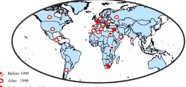
Figure 1. Occurrence of West Nile virus epidemic and/or epizootic in the world ... 11
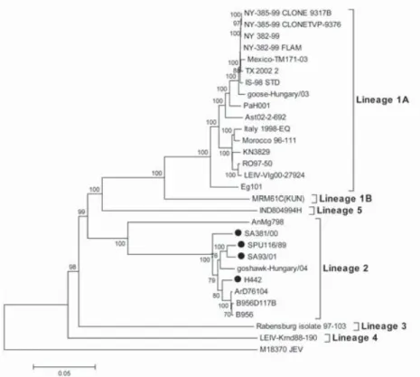
Figure 2. Phylogenetic tree of West Nile Virus (WNV) complete nucleotide sequences (Botha et al. 2008). ... 12
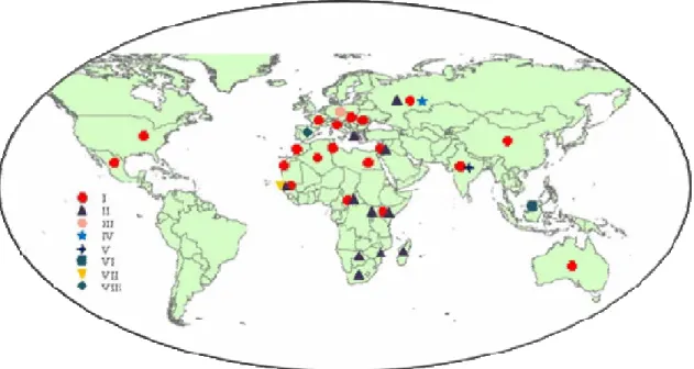
Figure 3. Geographical distribution of different genetic lineages of West Nile virus. ... 13
Figure 4. Map of Iran showing the location of 13 communities sampled for West Nile Virus antibodies in 1976 (Saidi et al. 1976). ... 18
Figure 5. Transmission cycle of West Nile virus ... 21
Figure 6. Effects of climatic parameters on mosquito life cycle (Reisen 1995). ... 27
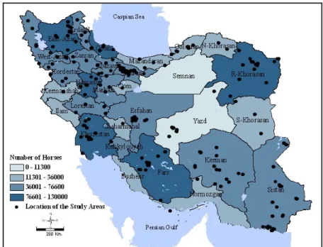
Figure 7. Geographic distribution of equine population by province and geographic location of the collected samples, Iran, 2008-2009. ... 34
Figure 8. Principle of Competitive ELISA method. ... 36
Figure 9. Anti-WNV seroprevalence determined by PRNT in equines, Iran, 2008-2009. ... 41
Figure 10. Distribution of IgM positive animals in Iran, 2008-2009... 42
Figure 11. Distribution of four ecological zones of Iran (Heshmati 2007). ... 51
Figure 12. Thiessen polygons analysis. ... 53
Figure 13. Spectral reflectance of green vegetation (Rahman 2004). ... 54
Figure 14. Flowchart of NOAA/AVHRR data processing and calculation of NDVI values for sampling places. ... 56
Figure 15. Important studied wetlands and sampling sites. ... 57
Figure 16. Annual mean temperature and geographical distribution of WNV infected (bold oval) and uninfected (un-bold oval) stables in Iran. ... 61
Figure 17. Distribution of positive stables (thick line) and of negative stables (thin line) according to the annual average temperature. ... 61
Figure 18. Distribution of positive stables (thick line) and of negative stables (thin line) according to the distance to the nearest wetland. ... 61
Figure 19. Distribution of positive stables (thick line) and of negative stables (thin line) according to the annual average humidity. ... 61
Figure 20. Distribution of positive stables (thick line) and of negative stables (thin line) according to the annual average precipitation. ... 61
Figure 21. Distribution of positive stables (thick line) and of negative stables(thin line) ... 62
Figure 22. Annual mean NDVI in Spring in Iran, and the geographic distribution of WNV infected (triangle) and uninfected (stars) stables. ... 62
Figure 23. Distribution of positive stables (thick line) and of negative stables (thin line) according to the normalized local NDVI differences. ... 63
Figure 24. Distribution of positive stables (thick line) and of negative stables (thin line) according to the normalized seasonal NDVI differences. ... 63
Figure 25. Receiver operating curve of the multivariate logistic model of the presence of anti-WNV seropositive animals in Iranian stables, 2008-2009. ... 64
Figure 26. Predicted risk areas of WNV infections in equines, Iran. ... 64
Figure 27. Elevation in Iran(http://www.fao.org/countryprofiles/Maps/IRN/02/lg/index.html). ... 72
Figure 28. Annual Temperature (left) and Precipitation in Iran (right) (http://www.fao.org/countryprofiles/maps.asp?iso3=IRN&lang=en). ... 72
Figure 29. Spatial distribution of four species of Culex spp. (Zaim et al. 1985). ... 74
Figure 30. Geographical distribution of Common Coot in Iran in breeding and wintering place (Mansoori 2008)... 77
Figure 31. Geographical location of wetlands in: (a) Middle-East; (b) Mesopotamian plain; (c) Khuzestan province, Iran. ... 78
Figure 32. Main flyways of migratory bird around the world ... 79
Figure 33. Sensitivity to the surveillance methods for WNV circulation in USA (CDC 2001). ... 81
L
LI
IS
ST
T
O
OF
F
T
TA
AB
BL
L
ES
E
S
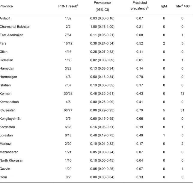
Table 1. Anti-WNV seroprevalence determined by PRNT in equines, Iran, 2008-2009. ... 40
Table 2. Logistic model of anti-WNV seropositivity determined by PRNT in equines, Iran, 2008-2009. ... 43
Table 3. Effect of animal kind and geographical coordinates on West Nile virus seropositivity, Iran, 2008-2009. ... 43
Table 4. Risk factors of West Nile virus infection included for statistical analysis. ... 58
Table 5. Statistical results for mean climatic factors in WNV infected and un-infected stables ... 59
Table 6. Multivariate logistic model of the presence of anti-WNV seropositive animals in Iranian stables, 2008-2009. ... 59
G
T h e s i s b y F a r z a n e h A h m a d n e j a d - U n i v e r s i t é d e G r e n o b l e ( 2 0 1 2 ) 2 Despite considerable success in controlling communicable diseases, emerging and reemerging diseases have caused and still cause detrimental effects in different continents. Zoonotic infectious diseases include a large portion of this group. They involve the transmission of the etiologic agents to humans from an ongoing reservoir life cycle in animals or arthropods.
The zoonoses are a group of diseases that can be passed from animals to humans. This may be through direct contact with infected animals, contamination of water and food, infected aerosols, or by the bite of an infected arthropod vector which is an important route of transferring diseases to human (Hubalek 2003).
West Nile (WN) infection is an arboviral zoonotic disease, transmitted by mosquitoes, which is not restricted by international borders and involves all continents; causing illness and deaths in humans and horses.
This virus belongs to Flaviviridae family, genus Flavivirus and is maintained in a transmission cycle involving bird and mosquito species. Although humans and horses are considered as incidental hosts, and infection is often inapparent or mild, recently it has been shown that the infection can lead to severe disease or even deaths (Gubler et al. 2007)
Since the increase in the frequency and severity of outbreaks in humans and animals, particularly horses, in the Middle East, European and African countries, and also its introduction in 1999 in USA and rapid spread throughout the western hemisphere, more research has turned toward the biological and ecological aspects of WN virus (WNV), especially when it was shown that the isolated strain in USA was very close to the isolated strain in the Middle East (Lanciotti et al. 1999, Deubel et al. 2001, Ruiz et al. 2004, Zeller & Schuffenecker 2004).
The Middle East, because of its geographical situation, is extremely prone to the introduction of diseases and the spread of endemic infections and zoonotic diseases. This part of the world is a crossroad between different continents. WN disease or circulation of the virus has been reported from different countries in the region; such as Egypt, Israel, Lebanon, Iraq and the United Arab Emirates (UAE) (Hubalek & Halouzka 1999b, Chowers et al. 2005, Alfaresi & Elkoush 2008).
Iran is located at the eastern most edge of the Middle East region and the first report of circulation of the virus goes back around forty years ago when sera from residents of Khorasan and Khuzestan provinces were found positive for WNV neutralizing antibodies
T h e s i s b y F a r z a n e h A h m a d n e j a d - U n i v e r s i t é d e G r e n o b l e ( 2 0 1 2 ) 3 (Naficy & Saidi 1970). There are other studies, all before 1980, indicating the presence of antibodies against WNV in healthy children from the Caspian area (Saidi S. 1974) and in six of 7 provinces in the country (Saidi et al. 1976). According to these evidences, Iran has been included in the category of infected countries with WNV; but after that time, there are no more facts relating to the situation of the virus and its presumptive detrimental effects in the country.
The present work aims to document the epidemiology of WNV in Iran, by: (i) assessing the spatial circulation of WNV; (ii) analyzing the impact of the environmental and climatic factors on WNV circulation. Chapter 1 explores the environmental conditions, particularly the distribution of the habitats of mosquitoes and hosts, and the climatic conditions, particularly temperature and rainfalls, which are favorable to the contacts between hosts and vectors. Indeed, these factors affect the abundance and activity of mosquitoes and hosts, but also the force of infection of infective species (Thomson & Connor 2000, Mellor 2004, WHO 2004, Reisen et al. 2006). Chapter 2 assesses the circulation of WNV in Iran. For this purpose, we considered equines, facilely captured and sampled, as sentinel hosts for the virus circulation survey in the country. We study the WNV seroprevalence and spatial distribution of seropositivity in equines from different Iranian regions. Moreover, Chapter 3 analyses the effects of environmental and climatic factors on the spatial circulation of WNV. Iran has diverse ecological conditions, resulting in biodiversity and consequently differential epidemiological transmission patterns. For this purpose, we have to identify ecological and climatic parameters related to WNV circulation in Iran. This analysis allows us to explain the relation between epidemiology of WNV and ecology of infected areas by an “oasis effect”.
C
C
h
h
a
a
p
p
t
t
e
e
r
r
1
1
.
.
E
E
N
N
V
V
I
I
R
R
O
O
N
N
M
M
E
E
N
N
T
T
A
A
L
L
A
A
N
N
D
D
C
C
L
L
I
I
M
M
A
A
T
T
I
I
C
C
C
C
O
O
N
N
T
T
R
R
O
O
L
L
O
O
F
F
W
W
E
E
S
S
T
T
N
N
I
I
L
L
E
E
V
V
I
I
R
R
U
U
S
S
T
T
R
R
A
A
N
N
S
S
M
M
I
I
S
S
S
S
I
I
O
O
N
N
T h e s i s b y F a r z a n e h A h m a d n e j a d - U n i v e r s i t é d e G r e n o b l e ( 2 0 1 2 ) 7
1. Introduction
First described in Uganda in 1937 (Smithburn 1940), West Nile virus (WNV) is responsible for infections in humans and horses, the virus has a transmission cycle involving mosquitoes as vectors and birds as amplifying hosts (Work et al. 1955, Taylor et al. 1956). For a long time, WN disease was endemic in the Middle East and Africa with sporadic outbreaks reported in Asia and Europe; so that it was not expected that the disease would reach the Western Hemisphere and cause death of many humans, horses and birds (Murgue et
al. 2002). In Europe, several outbreaks have recently occurred in horses and humans in
different countries including Romania (1996, 2010), Italy (1998, 2011), Russia (1999, 2007) and France (2000, 2003, 2004) (Hubalek & Halouzka 1999a, Murgue et al. 2002, Platonov et
al. 2008, Bagnarelli et al. 2011). Since its introduction in North America (CDC 1999), WNV
is recognized as the most widespread of the flaviviruses, with worldwide distribution.
Infection with WNV shows different effects on the hosts. Most infected hosts have no symptoms. A few percentages of infected individuals develop mild flu like symptoms. Neuroinvasive disease rarely occurs, however, is the acute form of WNV infection in humans and horses. It is estimated that about 20% of people who become infected will develop WN fever with mild symptoms, including fever, headache, myalgia, and occasionally a skin rash on the trunk of the body. About 1 of 150 WNV infections (<1%) result in meningitis or encephalitis. Case fatality rates among patients hospitalized during recent outbreaks have ranged from 4% to 14%. (Petersen & Marfin 2002). The infected birds may or may not become ill. Birds generally do not have symptoms of West Nile until the virus has caused encephalitis, or inflammation of the brain. Some bird species, like crows, showed a high susceptibility with death during WNV outbreaks in the US (Eidson et al. 2001).
Even if the epidemiology remains unclear, birds are known to take part in various aspects of WNV circulation (McLean et al. 2001, Malkinson & Banet 2002). Several studies have proved the essential role of birds as reservoir and spreader of WNV (Reed et al. 2003, Jourdain et al. 2007). Iran has "important wetlands", which support large breeding populations of many species of birds; in addition, each year, millions of birds coming from different countries migrate to these areas to pass the winter or use them as staging areas on their way to and from wintering areas. Iran appears to be a privileged area for the WNV transmission cycle. Likewise, the objective in this study is to identify the biotic and abiotic factors
T h e s i s b y F a r z a n e h A h m a d n e j a d - U n i v e r s i t é d e G r e n o b l e ( 2 0 1 2 ) 8 involved in the circulation, transmission maintenance and spreading, of WNV in the
environment.
2. Geographic dissemination of WNV
West Nile virus belongs to the Flaviviridae family, flavivirus genus. The viruses belonging to this family possess a single strand, positive-sense RNA genome. Within Flavivirus genus, based on cross-neutralization and molecular genetics, WNV is classified within the Japanese Encephalitis serological complex (Kuno et al. 1998). WNV is currently distributed in many parts of the world. The virus was first recognized in 1937 after it was isolated from the blood of a febrile woman in the West Nile District of Uganda (Smithburn 1940). Since then, it has been reported from the Middle East, Africa, Europe, Asia, Australia and from American countries (Hayes 2001).
2.1. Worldwide circulation of WNV
In South Africa, the first evidence of WNV was documented in 1958. The largest human outbreak of the disease was reported in 1974, which involved tens of thousands of human cases in the Karoo and Northern Cape Provinces (Murgue et al. 2002).
The first report of WNV in Europe was the detection of specific virus antibodies in Albania in 1958 (Bardos et al. 1959). In France, the first outbreaks of WNV occurred in the south, in 1962, with several nervous system abnormalities reported in the Carmargue region in humans and horses. The region is characterized by wetlands and marshes, with a great number of migratory and resident bird species and large populations of mosquitoes (Hubalek & Halouzka 1999a, Murgue et al. 2001b).
After 38 years, another outbreak of WN disease was reported in horse population in southern France in 2000. During this outbreak, 76 of 131 equines with neurologic disorders were laboratory-confirmed; most of the cases were located in a region with large marshes (Murgue et al. 2001b). In Italy, during the late summer in 1998, an outbreak of neurologic disease occurred among horses residing in a large wetland area. The case-fatality rate of this outbreak (43%) was similar to the outbreak in 2000 in France (34%) (Autorino et al. 2002). In 2008, the WNV infection re-emerged in Italy; the isolated strains of these outbreaks (1998/ 2008) felt in the same sub-cluster with the isolates from Romania and Russia (Savini et al. 2008). However, phylogenetic tree analysis of the complete genome sequence of WNV
T h e s i s b y F a r z a n e h A h m a d n e j a d - U n i v e r s i t é d e G r e n o b l e ( 2 0 1 2 ) 9 strains isolated in 2009 in Italy showed that the strains were closely related to the two WNV strains isolated from magpies in Italy in 2008. The 2008-2009 Italian WNV isolates had a higher degree of divergence from the eastern European strains isolated in Romania in 1996 and in Russia in 1999 and from the American/Israeli cluster (Barzon et al. 2009).
In 1996, 94 equines were affected in Morocco; 42 of which died. WN disease was reported in all age categories and it caused a higher mortality rate (45%) than the other almost concurrent outbreaks. There was a silent period until 2003, when an outbreak of WNV occurred again among stabled horses. Based on phylogenetic analyses, the virus isolates of Morocco in 2003 were similar to the 1996 isolate and to other strains responsible for equine outbreaks in the western Mediterranean basin. Recently an outbreak of WNV has been reported in horses in Centre region in Morocco (Schuffenecker et al. 2005, GAH 2010).
Since the outbreak of WNV in France, there have been several reports of West Nile virus activity in other European countries such as Spain (in the 1960s and the 1980s, 2010, 2011) and Austria (1960s and 70s). The activity or outbreaks of the virus have been detected in Portugal, Italy, Greece, Bulgaria, Romania, Hungary, Slovakia, Belarus, Ukraine, and Czech Republic (Hubalek & Halouzka 1999a, Samoilova et al. 2003, Esteve et al. 2005, Hubalek et al. 2006, Krisztalovics et al. 2008, Barros et al. 2011, Danis et al. 2011, Garcia-Bocanegra et al. 2011, Lelli et al. 2011).
In 1996, the most serious European outbreak of WNV occurred in Romania where the first activity of the virus dated back to the 1960s (Campbell et al. 2001). Between July and October 1996, 835 patients were hospitalized with suspected central nervous system infection. Among 509 patients with samples for testing, 393 (77%) were laboratory- confirmed to have WN virus antibodies, including 286 from Bucharest. There were 17 fatalities (4·3%) in patients older than 50 years (Tsai et al. 1998).
Currently there is no evidence that WNV causes disease in the UK, nevertheless in 2003, Buckley et al. reported the presence of virus-specific neutralizing antibodies to WNV (22.7 %) in the sera collected from 30 different species of wild or farm birds (resident and migrant birds) in the UK (Cambridgeshire, Dorset and south Wales), implying that the virus was being introduced to UK birds, possibly by mosquitoes. It seemed that the virus is being introduced into UK-resident birds, apparently without causing any obvious reduction in the bird population. In addition, other activities included surveillance for WNV infections in humans, and in dead birds, mosquitoes and horses, carried out between 2001 and 2005, have found no positive cases of UK-acquired WNV. Between 2001 and 2006, tissue samples were
T h e s i s b y F a r z a n e h A h m a d n e j a d - U n i v e r s i t é d e G r e n o b l e ( 2 0 1 2 ) 10 collected from over 1800 wild birds by the laboratories of SAC in Scotland and the Veterinary Laboratories Agency (VLA) in England and Wales. The samples were tested by VLA for the presence of WNV by virus isolation and PCR. No evidence of WNV infection was found (Buckley et al. 2003, Morgan 2006, Pennycott & Cert 2011).
In 1999, WNV found its way to North America as an important pathogen and has spread rapidly throughout the country, presenting a threat to both human and animal health. During this outbreak, among 62 human cases, 59 (95%) were reported as West Nile meningitis or encephalitis (neuroinvasive disease), 3 (5%) were reported as West Nile fever (milder disease) and seven patients died (CDC 2007).
The case fatality rate for horses in New York was 25% and thousands of birds of as many as 18 species died during this outbreak (Steele et al. 2000). In the later years WNV continued to cause disease in humans, horses, and a variety of bird species in New York City and within several years, it became firmly established in U.S and gradually made its way to Latin America, the Caribbean and Canada (Komar et al. 2003b, Mattar et al. 2005). In Canada, the first reports of WN virus activity were notified in 2001 when the virus was found in dead birds and mosquito pools in Southern Ontario and the first human and horse cases were confirmed in the following year, 2002 (Weese et al. 2003).
The source of virus introduction to North America is unknown; potential sources include an infected vector, human, bird, or other vertebrate hosts (Lanciotti et al. 1999). The close genetic relationship between WN virus isolates from Israel and New York suggests that the virus was imported into North America from the Middle East (Lanciotti et al. 1999).
Overall, we can classify epidemic and/or epizootic occurrence of WNV to two distinct periods: before 1990 and after 1990. The years after 1990 apparently have had suitable conditions for the circulation of the virus as a higher occurrence of disease among human, horse and bird population has been reported in these years (
Figure 1). Circulation of WNV, like other vector-borne-diseases, is so dependent on the climate and the difference between two parts of time can be related to the global climate change. A millennial study (1000-2000) of climate change has revealed that the 1990s was the warmest decade and 1998 the warmest year (Jones 2000).
T h e s i s b y F a r z a n e h A h m a d n e j a d - U n i v e r s i t é d e G r e n o b l e ( 2 0 1 2 ) 11
Figure 1. Occurrence of West Nile virus epidemic and/or epizootic in the world
2.2. Geographical distribution of the lineages
Evidence from phylogenetic studies suggests that there are at least eight genetic lineages of WN virus:
- Lineage 1: these strains have a broad geographical distribution, isolated from over 20 countries. The viruses within this lineage are divided into two clades:
- 1a: the viruses isolated from Europe, Africa (north, West, central), India, Asia, Middle East and the United States;
- 1b: the virus isolated in Australia, also known as Kunjin virus (KUNV).
Most virulent viruses in recent outbreaks belong to clade 1a; however, the lineage contains both virulent and attenuated viruses,
- Lineage 2: mainly comprised of virus isolates from endemic areas of west, central and east Africa and Madagascar; although some strains have been found outside Africa. These viruses often cause asymptomatic infections or mild disease, but virulent viruses also exist. One virulent virus has caused encephalitis in raptors in Central Europe (Hungary) (Berthet et al. 1997, Lanciotti et al. 1999, Savage et al. 1999, Lanciotti et al. 2002). Recently it has been shown that WNV lineage 2 may cause neurologic disease in horses in southern Africa and in Hungary (Venter et al. 2009, Kutasi et al. 2011),
Before 1990 After 1990
T h e s i s b y F a r z a n e h A h m a d n e j a d - U n i v e r s i t é d e G r e n o b l e ( 2 0 1 2 ) 12 - Lineage 3: includes the isolates from Culex pipens at the Czech Republic/ Austria border (Rabensburg virus),
- Lineage 4: the isolate from Dermacentor marginatus ticks in the Caucasus Mountain valley, Russia (LEIV-Krnd88-190) makes this lineage (Prilipov et al. 2002, Bakonyi et al. 2005),
- Lineage 5: The Indian isolates had been placed previously within lineage 1 as a separate cluster (clade 1c), but more analyses of genome sequences have classified them as lineage 5 (Bondre et al. 2007).
- Lineage 6: this lineage has been proposed for the Sarawak Kunjin virus strain (Malaysian isolate), which is markedly different from the other Kunjin viruses.
- Lineage 7: African Koutango virus (isolated in Senegal), which is closely related to WNV
- Lineage 8: this lineage has been proposed on the basis of WNV sequences detected in Cx. pipiens captured in southern Spain (Vazquez et al. 2010, Papa et al. 2011).
Figure 2. Phylogenetic tree of West Nile Virus (WNV) complete nucleotide sequences (Botha et al. 2008).
T h e s i s b y F a r z a n e h A h m a d n e j a d - U n i v e r s i t é d e G r e n o b l e ( 2 0 1 2 ) 13 Figure 3. Geographical distribution of different genetic lineages of West Nile virus.
The phylogenetic analysis of WNV strains indicates that co-circulation of different lineages can occur in the same country (Briese et al. 2002) (Figure 3). Distribution of several genetic lineages of the virus in different geographic locations indicates the potential importance of birds in viral dispersal (Berthet et al. 1997) and also adaptation of the virus with different geographical regions and hosts.
2.3. WNV Circulation in the Middle East
The Middle East is geographically well known as an area where intense migratory activity of birds can be observed during the year. This region is considered as a cross road for the bird species that stop over between Europe and Africa.
Egypt- The first documents about the virus activity and its characteristics are related to
the studies in Egypt, where the first epidemiological studies of WN fever have been conducted in the 1950s. Sero-epidemiological surveys in humans, mammals and birds as well as entomological studies carried out at that time characterized the ecology of the virus and the endemicity of WNV in Egypt (Melnick et al. 1951, Hurlbut et al. 1956, Murgue et al. 2002). The results from an endemic area in the Nile Delta suggested that WN infection is essentially
T h e s i s b y F a r z a n e h A h m a d n e j a d - U n i v e r s i t é d e G r e n o b l e ( 2 0 1 2 ) 14 the symptomatology of the infection, it appeared that the infection was mostly self-limited,
non-fatal febrile disease rarely associatedwith manifestations of encephalitis (Taylor et al.
1956). Another study was carried out in 1968, among 120 children, attending the fever hospital in Alexandria and complaining of fever. It was noted that although the sera were collected during June to October, all positive sera were obtained in August (Mohammed et al. 1970).
A serologic survey conducted in equines (horses, donkeys, and mules) in 1959, showed a high prevalence of WN antibodies (54%) and the first reported WNV infections in a horse occurred in a 12-year-old horse that died and the virus was isolated from the brain tissue. Thereafter experimental infections of equines were conducted in order to study the role of equines as reservoirs of WNV and the duration and magnitude of viremia; surprisingly only two equines, among eleven, developed low grade fever and showed very short-term viremia which suggested that the equines probably were dead-end hosts (Schmidt & Elmansoury 1963).
Natural and experimental studies among different hosts exposed to WNV led to a better understanding on which species are important hosts and produce high level of viremia to infect non-infected mosquitoes. Based on these studies, it was concluded that the main cycle of the virus was probably through mosquitoes and birds; non-migratory birds were considered as hosts responsible for the local dissemination of the virus. The first report of natural WNV-associated morbidity in birds was a sick fledgling pigeon in Egypt in the early 1950s. One study carried out in the mid-1950s in Egypt confirmed the role of passerine birds as important reservoirs in this region. Neutralizing antibodies were found in all species of birds tested from an endemic area in which Hooded Crows and House Sparrows had the highest viral neutralizing antibodies; which suggested these species could be used as natural sentinels for monitoring virus transmission. Experimental infection of birds with WN virus by mosquito bites demonstrated a high mortality rate of crows which had high circulating virus titers and could imply a high mortality in naturally infected crows; but it was also stated that many survived as indicated by the high percentage of antibody positive crows (Work et al. 1953, Work et al. 1955, Taylor et al. 1956, Malkinson & Banet 2002).
The first field studies that implicated mosquitoes as important vectors of WN virus were conducted in Egypt from 1952 to 1954. During this study, 17 isolates of WN virus were obtained from pools of mosquitoes (Culex spp.). In addition, a number of pools of other arthropods, including ticks, lice, fleas, mites and flies were tested for WN virus isolation but
T h e s i s b y F a r z a n e h A h m a d n e j a d - U n i v e r s i t é d e G r e n o b l e ( 2 0 1 2 ) 15 were all negative. The persistence of WNV in Egypt was explained by mosquitoes that remained active throughout the colder months (Taylor et al. 1956).
Israel- WN fever is endemic in Israel. The virus was first recognized in 1951; although
it had probably already been present in that country during previous years. In 1951, an outbreak occurred among the population located at 30 km south of Haifa in Israel where 41% of its 303 inhabitants were patients. None of the cases was fatal; children represented most of the cases that were infected with WNV, and one of them developed mild meningitis (Bernkopf et al. 1953).
The occurrence of WNV encephalitis cases among elderly population in Israel in 1957 was the first report that WNV could cause serious central nervous system infections and established a correlation between the age of patients and severity of disease. Encephalitis has been reported from several locations of Hadera area (army camps, urban / rural places and nursing homes), but the death only has been observed in nursing homes by a rate of 8.2% (4/ 49) in a group of elderly patients (Chowers et al. 2001, Murgue et al. 2001a, Weinberger et
al. 2001). There was another outbreak among young soldiers in 1980 with one patient with
meningitis and no fatal case.
In 2000, 417 laboratory-confirmed WN fever cases were diagnosed in Israel with 35 deaths, all in patients >50 years of age. The highest incidence was in central Israel; the most populated part. Men and women were equally affected, and their mean age was 54.5±23.8 years (range 6 months to 95 years). This outbreak was different from previously reported outbreaks in Israel because of high rates of encephalitis, countrywide spread, and higher number of hospitalizations (Weinberger et al. 2001). At the same time (2000), an outbreak of West Nile virus infection affected the horse population with 76 horses with developing neurological disorder and 15 deaths. (Steinman et al. 2002).
During the 2000 WNV epidemic in Israel, specimens from a 72-year-old woman were analyzed and the results indicated closer phylogenetical relationship of this isolate to 1996 Romanian and 1999 Russian than to 1998-99 Israeli or 1999 New York isolates (Briese et al. 2002). During the same outbreak, WNV isolated from four patients and their phylogenetic analyses revealed that at least two strains of WN virus circulated in Israel in 2000. This is most likely the result of the movements of migratory birds between Africa and Europe via the Middle East. One sub-clade 1a strain (2 isolates) was similar to WN virus isolates from mosquito, horse, and flamingo during the 1999 NY outbreak. The other sub-clade 1a strain (2
T h e s i s b y F a r z a n e h A h m a d n e j a d - U n i v e r s i t é d e G r e n o b l e ( 2 0 1 2 ) 16 isolates) was similar to the viruses isolated from Romanian (1997) and Russian (Volgograd, 1999) outbreaks, but different from 1996 Romanian isolate (Hindiyeh et al. 2001).
Israel is located on the flyway corridor of migrating birds to and from Africa and to Europe, so the virus may have been introduced in this manner. During 1997 and 1998 for the first time, WN virus was reported as the cause of neurologic illness and death among the affected goose flocks. In a study carried out between December 1998 and October 1999, sera were collected from goose farmers and veterinarians working with sick or healthy geese to detect the infection following contact with sick geese. There was a relation between positive serology and contact with flocks of sick geese. Moreover, some farmers who worked with sick geese had developed a flu-like illness, indicating close contact with sick geese as a risk factor (Bin et al. 2001).
Another study conducted in Israel revealed again the role of birds for WNV distribution. A flock of 1200 migrating storks arrived outside Eilat in August 1998. Their arrival was attributed to strong, hot westerly winds, which forced the storks to ground in Eilat. Six days after their arrival, three of 11 healthy storks were shown to have WNV-neutralizing antibodies, indicating previous exposure to WNV. The authors concluded that it is improbable that infection had occurred in Eilat because the infected birds were collected within 2 days after arrival. It is likely that hatching time of fledglings and their exposure to WNV had occurred in Europe near the nesting sites or along the route of migration (Malkinson et al. 2002).
The phylogenetic analyses indicated a close genetic relationship between IS98 isolate (Israeli WNV strain isolated from a white stork in 1998) and an isolate from goose in Hungary in 2003 (Figure 2). The place where the WNV outbreak occurred in Hungary, is located in the wetland areas that are good habitats for many bird species, supporting the concept of introducing neurovirulent WNV strain from central Europe to Israel (Bakonyi et
al. 2006).
Turkey- Serologic evidence for the presence of WNV infections dates back to the
1970s based on the detection of hemagglutination inhibiting (HI) antibodies. In one study, WNV prevalence of HI antibodies was 6% in humans and 1.5% in sheep. Another survey indicated a seroprevalence as high as 40% in randomly selected human sera in southeast Turkey (Ozkul et al. 2006).
T h e s i s b y F a r z a n e h A h m a d n e j a d - U n i v e r s i t é d e G r e n o b l e ( 2 0 1 2 ) 17 A serological survey, first reported in 2005, indicated the exposure of a wide range of mammals to WNV in Turkey. In this study, 35 of 259 (13.5%) horses and 18 of 88 (20.4%) humans had developed WNV-neutralizing antibodies. Geographical locations of horses showed they were stabled near a lake in a park; a place for migrating birds. The presence of possible human WN disease was suggested among human attending hospitals in southeastern part of Turkey in 2005 (Ozkul et al. 2006, Ergunay et al. 2007).
Other countries in the Middle East- A study conducted in 2005 among 500 healthy
blood donors indicated that WNV infection was not present in the UAE; nevertheless, another study in 2007 demonstrated that WNV had entered the UAE. In the later study among 750 equines, 144 (19.2%) animals had antibodies to WNV. In addition, one horse with antibodies to WNV had clinical signs consistent with WN encephalitis (Wernery 2007, Alfaresi & Elkoush 2008).
In Jordan, a serosurvey was conducted among patients, over 5 years of age, attending a health center in 1998. The seroprevalence of immunoglobulin G antibodies against WNV was 8%. Moreover, authors suggested that the significant risk factor was the proximity to wastewater treatment plants (Batieha et al. 2000).
In Oman, 19 clinical cases from a population of 240 horses were reported in 2003 and demonstrated that the disease occurred in only one stable (Al Suleiman 2003). West Nile virus antibodies have also been detected in human sera from Iraq, Lebanon and Syria (Hubalek & Halouzka 1999a).
2.4. WNV Circulation in Iran and Neighboring Countries
The first description of West Nile virus (WNV) in Iran was in the 1970s, when sera were obtained from residents of Khorasan (303 sera) and Khuzestan (60 sera) provinces. The sera were from people of age 8-70 years and were tested for the presence of neutralizing antibodies to WN. A total of 103 (28.4%) cases were antibody positive, of whom 91 (30%) and 12 (20%) were from Khorasan and Khuzestan, respectively (Naficy & Saidi 1970).
In another study reported in 1974, 100 sera from children 1 – 6 years of age, resident of the Caspian area were tested for the prevalence of antibodies against 15 different viruses. The sera were collected during May 1970 to September 1971 and analyzed by
T h e s i s b y F a r z a n e h A h m a d n e j a d - U n i v e r s i t é d e G r e n o b l e ( 2 0 1 2 ) 18 inhibition (HI) method for detecting WNV antibodies. Ten (10%) children had antibodies against WNV (Saidi S. 1974).
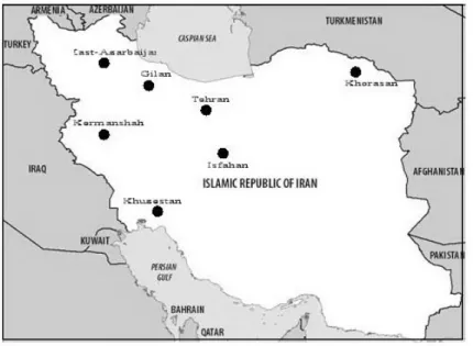
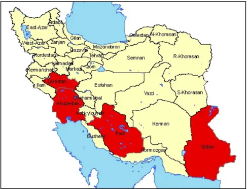
A study conducted in 13 different communities, located in 7 provinces (East- Azarbaijan, Gilan, Khorasan, Tehran, Isfahan, Kermanshah, Khuzestan), was reported in 1976. 186 (26.6%) of 698 human sera had antibodies against WNV. A positive reaction was detected in six of 7 provinces (except East -Azarbaijan province) with highest prevalence in the Dezful-Deigi area located in Khuzestan province (Figure 4) (Saidi et al. 1976).
Figure 4. Map of Iran showing the location of 13 communities sampled for West Nile Virus
antibodies in 1976 (Saidi et al. 1976).
A study of WNV infection in Iranian blood donors, who donated blood at the Tehran Blood Transfusion Center, was reported in 2010, in which 500 sera were examined by ELISA and Real-Time RT- polymerase chain reaction methods. All 500 donors were negative for WNV-specific IgM antibody and WNV-RNA, but 5% of them had IgG antibodies (Sharifi et
al. 2010).
WNV infection has been reported from Pakistan (eastern neighbor of Iran) and Russia. Russia is the northern neighbor of Iran; and some areas in Iran are considered as wintering places for migratory birds coming from Russia. Siberian Crane is one of these migratory birds that every year migrates from its breeding grounds in northern Siberia to wintering sites in Iran (Kanai et al. 2002). Ducks, geese and pelicans are among other birds coming from northern parts to the country.
T h e s i s b y F a r z a n e h A h m a d n e j a d - U n i v e r s i t é d e G r e n o b l e ( 2 0 1 2 ) 19 West Nile fever (WNF) is an endemic disease in Southern Russia and western Siberia-Altai territory. The virus was first isolated from soft ticks (Ornithodoros capensis) and hard ticks (Hyalomma marginatum, H. detritum, Rhipicephalus turanicus and R. muhsamae), also from birds in the delta of the Volga River of the Astrakhan region in 1963-1968 (Hubalek & Halouzka 1999a, Lvov et al. 2000, Fyodorova et al. 2006). Russia experienced a large outbreak of WNV in the south of the country (Volgograd, Astrakhan, and Krasnodar regions), in Summer and early of Autumn 1999 with a peak during 21-25 August. Volgograd City is located on the west bank of the Great Volga River, and the total number of suspected WNV cases was estimated to be 480 people in the region with at least 40 deaths. The majority of patients (50%) were over 50 years of age and thirty of the patients (75%) who died were >60 years of age (Lvov et al. 2000, Platonov et al. 2001). Also during the same period, 89 WNV cases with 5 deaths in Astrakhan Region and at least 38 WNV cases with 3 deaths in Krasnodar Region were reported (Platonov 2001).
During 2001-2004 in the Astrakhan region, sera sampled from 2,884 farming animals were investigated to indicate specific antibodies to West Nile virus. The antibodies were detected in all the examined species of animals (horses, cattle, camels, pigs, and sheep) (Vasil'ev et al. 2005).
The epidemiological and entomological studies indicated that the outbreak in the Volgograd area was similar to the epidemic in Romania in 1996 and in New York City in 1999. Phylogenetic analysis also supported this suggestion. Two different genotypes were isolated during the 1999 outbreak, one was similar to isolates of Israel in 1997-99 and New York in 1999, while the other one was similar to the Romania isolate in 1996 (Briese et al. 2002, Fyodorova et al. 2006). These strains belong to lineage 1, while the virus isolated in 1998, in Dermacentor marginatus ticks in the North-West Caucasus Mountain valley of Russia belongs to the lineage 4 (Bondre et al. 2007).
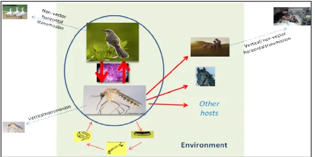
3. West Nile virus transmission
WNV is maintained in nature in an enzootic cycle between ornithophilic mosquitoes and birds. Mosquitoes become infected when they feed on infected birds (reservoir hosts) and the virus is transmitted to other birds when the mosquitoes bite again. This is defined as rural or "sylvatic cycle". In a secondary cycle, which is called urban, bridge mosquitoes (feeding
T h e s i s b y F a r z a n e h A h m a d n e j a d - U n i v e r s i t é d e G r e n o b l e ( 2 0 1 2 ) 20 on both birds and non avian hosts) become infected by feeding on an infected bird and then continue spreading WNV to other vertebrates (Hubalek & Halouzka 1999a).
3.1. Epidemiological cycle
Along with the maintenance of virus between mosquitoes and birds in nature, it is also supposed that the virus is transmitted between generations of mosquitoes. Natural transovarial transmission of WNV has been identified in Kenya in Culex univattatus. Also in laboratory studies, a female range of Culex and Aedes mosquitoes have been shown to vertically transmit the virus to offspring (Baqar S. 1993, Miller et al. 2000, Turell et al. 2001b). This mechanism is important as the virus can persist even during cold weather when there is no adult mosquito.
Vertical transmission is a general terms for transfer of pathogen from one generation to the next; while transovarial transmission means transfer of pathogens from parents to offspring through the ovary and eggs which occurs mainly in arthropods (Schmid-Hempel 1998). Lateral transmission between mosquitoes has also been documented. An infected mosquito and a non-infected mosquito were allowed to feed adjacently on a non-viremic host. After seven days, the non-infected mosquito had viral titers high enough to infect new hosts (Higgs et al. 2005). Presence of the virus in hibernating mosquitoes has been also observed (Nasci et al. 2001). This issue can lead to the circulation of the virus during the next warm season when mosquitoes become active.
Beside arthropod-borne transmission, vertical and direct transmission of WNV has been observed between vertebrate hosts without the involvement of arthropods. Transmission through transfused blood (Pealer et al. 2003), transplanted organs (Iwamoto et al. 2003) and transplacental (mother-to-child) transmission of WNV have occurred in humans (CDC 2002b). Transmission through breast-feeding is also likely (CDC 2002c). Laboratory-acquired WNV infections have been also reported among laboratory workers via percutaneous inoculation (CDC 2002a).
Based on recent studies, shedding of WNV RNA has been reported in the urine of some patients, some of whom had a shedding 1.6-6.7 years after the infection (Tonry et al. 2005a, Murray et al. 2010). It seems that shedding of WNV via urine, as a route of virus transmission, should be considered (Tonry et al. 2005a, Tonry et al. 2005b).
T h e s i s b y F a r z a n e h A h m a d n e j a d - U n i v e r s i t é d e G r e n o b l e ( 2 0 1 2 ) 21 The possibility of WNV transmission by aerosol among turkey handlers at a turkey trade and among animals raised the possibility of aerosol exposure. Studies showed the possibility of bird-to-bird transmission via saliva and/or fecal aerosols (Banet-Noach 2003, Komar et al. 2003a).
Ingestion of infected animals has also been considered as a possible source of transmission. By identification of WNV from a hawk in winter, and from farmed alligators concurrent positive RT-PCR for WNV in their horsemeat, authors concluded that animals probability acquired the infection by eating the infected animals (Garmendia et al. 2000, Petersen & Roehrig 2001) (Figure 5).
Figure 5. Transmission cycle of West Nile virus
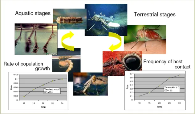
3.2. Vectorial transmission
WN virus has been identified in numerous mosquito species, including members of the genera Culex, Aedes and Ochlerotatus; mosquitoes belonging to the Culex species being the primary vector (Turell et al. 2001b). The ability of mosquito to become infected after feeding on infected hosts, to replicate the virus and to transmit infection to susceptible hosts is called "vector competence" (Reisen et al. 2005).
Female mosquitoes require a blood meal before they can lay eggs. The mosquito can take in as much as twice its body weight in blood, thus creating a relatively good chance of
T h e s i s b y F a r z a n e h A h m a d n e j a d - U n i v e r s i t é d e G r e n o b l e ( 2 0 1 2 ) 22 picking up the pathogens. When a mosquito bites an infected bird, the virus enters the mosquito’s bloodstream and finally reaches the salivary glands. After biting, they are capable of transmitting the virus within 10 to 14 days following feeding on an infected bird, so bites after that time are infectious (Jarratt et al. 2004).
The host preferences of mosquito species are also important determinants of transmission dynamics. For example, ornithophagic mosquitoes, such as Culex spp, typically feed on viremic birds, whereas Aedes prefer to feed on mammals. The species, such as Ae.
vexans, Cx. salinarius, Ochlerotatus. j. japonicus, and Oc. Triseriatus, known as bridge
vectors could become infected when feeding on an infected bird, and then transmit WN virus to another susceptible vertebrate host (Turell et al. 2001b).
Moreover, the distribution of principal vectors of WNV differs in different regions. In Egypt, Israel, and South Africa, the main vector is Cx. univittatus, while Cx. modestus was found to be an important vector for circulation of West Nile virus in the Volga Delta and in populated areas in Russia and in Europe, especially in wetlands of the Camargue in France.
Cx. triaeniorhychus and Cx. vishnui are the main vectors for WNV based on experimental
studies in India. The virus has been isolated from Cx. Vishnui complex in Pakistan (Hubalek & Halouzka 1999a, Lvov et al. 2000, Paramasivan et al. 2003, Balenghien et al. 2008). In the winter 2000 after the 1999 outbreak in the US, WNV was isolated from overwintering Cx.
pipiens mosquitoes collected in New York; providing the mechanisms for viral persistence
and re-emergence each spring (Nasci et al. 2001, Hayes 2005). The first isolation of an arbovirus from a hibernation mosquito (Cx. modestus Fic.) in Europe goes back in 1970 when Tahyna virus (family Bunyaviridae) was isolated from the mosquito in Camargue in winter (Chippaux et al. 1970).
During the large outbreaks occurring in urban populations of Romania, Volga delta in Russia, and U.S, the significant factor was the involvement of the common house mosquito,
Culex pipiens, which had not been considered before as an important vector. Proximity to
large rivers was a similar parameter between all the sites, presumably providing an attractive place for birds (Hayes 2001). Species in the Culex pipiens complex are considered the primary vectors of WNV in the New world because of their abundance in urban areas, occurrence of the disease during their peak and their experimentally confirmed competence. Unlike European Cx. pipiens, U.S. Cx. pipiens appears to bite readily both avian hosts and humans. It seems that a huge part of the U.S. mosquitoes are hybrids of human- biter and
T h e s i s b y F a r z a n e h A h m a d n e j a d - U n i v e r s i t é d e G r e n o b l e ( 2 0 1 2 ) 23 bird- biter forms and these hybrids caused recently epidemic condition in the U.S. (Fonseca et
al. 2004).
Some studies have indicated that exposure to infected mosquitoes is a risk factor for acquiring WNV infection. During the 1996 outbreak in Romania, the risk for WNV infection was the time spent outdoors and in flooded basements. In New York outbreak, human cases were clustered in areas with higher vegetation cover, indicating favorable mosquito habitat (Han et al. 1999, Brownstein et al. 2002).
Other arthropods have been also implicated as vectors of WNV, although their role in the ecology of the virus remains unknown. WNV has occasionally been isolated from hard and soft ticks, and experimental studies indicated that infected ticks (such as Ornithodoros
moubata, O. maritimus and O. erraticus) could transmit the virus to the hosts (Hubalek &
Halouzka 1999a, Lawrie et al. 2004).
3.3. Birds reservoirs
Since viremia in birds can reach high-level titers to infect the mosquito species, birds are considered as the main reservoir host for WNV in nature. However, all the birds do not develop high levels of viremia, and only some of them play an important role in the transmission cycle of WNV; such as passerines (including members of the crow family, Blue Jay and House Sparrow) and charadriiformes (such waders and gulls) (Hubalek & Halouzka 1999a).
Like vectors, several birds in different geographical regions have been implicated as reservoirs, and the virus has been isolated from a variety of wetland and terrestrial bird species. Although their infection usually can be clinically inapparent, that susceptibility to infection varies considerably among species and according to their geographical regions, for example deaths due to WNV has been observed in a pigeon in Egypt, in geese in Israel, in different species, especially corvids in U.S. and in sparrow hawk (Accipiter nisus) and several goshawk (Accipiter gentilis) fledglings (Lanciotti et al. 1999, Bernard et al. 2001, Erdelyi et al. 2007).
Even though vectors appear to be essential for transmission of WNV among avian species, it seems that birds have also the capability to transmit the virus by alternative modes of transmission. Studies have demonstrated the possibility of bird-to-bird transmission via saliva and/or fecal aerosols, ingestion of contaminated food (Komar et al. 2003a).
T h e s i s b y F a r z a n e h A h m a d n e j a d - U n i v e r s i t é d e G r e n o b l e ( 2 0 1 2 ) 24 Among different agents implicated in WNV circulation, infected migratory birds are important in spreading the virus into different, particularly naive, areas along their short or long migrations route (introducing hosts) which is followed by virus adaptation to local vectors and its subsequent amplification through the infection of local bird populations (amplifying hosts).
Migratory birds have long been suspected as the principal hosts for the spread of various pathogens into new regions. The studies show that the probable causes of introduction of WNV in the Middle East, Europe, and U.S. are the birds coming from endemic areas (Rappole et al. 2000, Malkinson et al. 2002, Jourdain et al. 2007).
There is a possibility that the migratory birds, which may be infected in their African wintering areas, transmit the virus northward, in the Middle East and Europe, during spring migration (Cabre et al. 2006). On the other hand, as suggested by the similarity between the Israeli isolate in 1998 and European isolate, the virus could have been introduced from central Europe to Israel by migratory birds (Malkinson et al. 2002).
3.4. Roles of other vertebrates
Horses, along with humans, are the most susceptible accidental hosts for WNV and like other hosts become infected by biting of infected mosquitoes; and since they are unable to generate a viral titer high enough to infect new mosquitoes, they are considered as “dead-end” hosts. Their infection ranges from asymptomatic to fatal encephalitis. The incubation period, or the time between being bitten by an infected mosquito and the time when clinical signs appear, ranges from three to15 days (Nash et al. 2001).
The estimated rate of clinical disease among infected horses with WNV is 10% with 38-42% mortality rates occurring in different geographical regions. While around 1% of infected humans develop neurological signs with an overall case fatality rate of approximately 10% which may reach 15-29% in people who are over 70 years old (Bunning
et al. 2002, Castillo-Olivares & Wood 2004, CFSPH 2009).
Few outbreaks of WN disease in horses were documented before the 1990s. The first neurological cases in horses infected with WNV were first described in Egypt and France in the 1960s with mortality rates ranging from 25% to 30% in France (Schmidt & Elmansoury 1963, Murgue et al. 2001b). Since then several outbreaks of neurological disease have been reported among horses in Morocco (1996, 2003, 2010), Israel (1998, 2000), Italy (1998,
T h e s i s b y F a r z a n e h A h m a d n e j a d - U n i v e r s i t é d e G r e n o b l e ( 2 0 1 2 ) 25 2008, 2009, 2010, 2011), France (2000, 2003, 2004), Argentina (2006) and U.S.A (1999 through 2009) (Dauphin et al. 2004, Morales et al. 2006, Macini et al. 2008).
Some animals with acute neurological disease die spontaneously, but many severely affected animals are euthanized which imposes economic burden for the countries. In contrast to the human disease, age does not appear to be a risk factor for development of severe neurological disease and death in horses. A study carried out among horses with encephalitis in New York State in 1999 and 2000, revealed that there was no significant difference in the age of horses that died or were euthanatized and horses that survived (Trock et al. 2001). During a serosurvey of WNV among equines in southern France, age had no effect on seropositivity (Durand et al. 2002). However, a study among previously uninfected horses in Texas revealed that older horses were at greater risk of death or euthanasia (Ward et al. 2006).
Although the role of the vertebrates other than birds in epidemiology and transmission of WNV is unclear, and they have been considered as accidental hosts, some studies have shown the role of some animals as potential sources of virus for mosquitoes. Several studies have shown the serological evidence of WNV exposure in a broad range of other mammalian hosts such as cats, dogs, feral swine, raccoons, foxes, bats, and squirrels (Komar et al. 2001, Dietrich et al. 2005, Docherty et al. 2006, Gibbs et al. 2006a). It seems that the lake frog (Rana ridibunda) appears to be a competent reservoir for WNV in Russia (Hubalek & Halouzka 1999a, van der Meulen et al. 2005).
4. Environmental and climatic risk factors for WNV circulation
The atmosphere plays a central role for the environmental conditions on earth. Climate refers to the conditions in the atmosphere. The environmental factors impacting public health are related to: (i) atmospheric parameters such as, temperature, precipitation, relative humidity, wind and solar radiation (abiotic); (ii) or terrestrial parameters, such as topography, fresh water ponds, rivers, lakes (abiotic) and hosts (mammals, reptiles, birds), natural predators, parasites, vectors, pathogens, vegetation (biotic) (WHO 1990).
Climate variation has two different effects on ecological processes: (i) direct effects on virus/hosts/vectors, for example, by selecting the variants sensitive, or controlling the physiology of organism such as metabolic processes and reproduction (Gubler et al. 2001,