HAL Id: tel-03144445
https://tel.archives-ouvertes.fr/tel-03144445
Submitted on 17 Feb 2021HAL is a multi-disciplinary open access archive for the deposit and dissemination of sci-entific research documents, whether they are pub-lished or not. The documents may come from teaching and research institutions in France or abroad, or from public or private research centers.
L’archive ouverte pluridisciplinaire HAL, est destinée au dépôt et à la diffusion de documents scientifiques de niveau recherche, publiés ou non, émanant des établissements d’enseignement et de recherche français ou étrangers, des laboratoires publics ou privés.
The role of chromatin remodelers in dopaminoceptive
neurons
Abdallah Zayed
To cite this version:
Abdallah Zayed. The role of chromatin remodelers in dopaminoceptive neurons. Neuroscience. Sor-bonne Université, 2019. English. �NNT : 2019SORUS437�. �tel-03144445�
THESE Presented at the SORBONNE UNIVERSITE
École doctorale : CERVEAU, COGNITION, COMPORTEMENT Spécialité : Neuroscience
Presented by Abdallah Zayed, MD
To obtain the degree of
DOCTEUR de L’UNIVERSITE SORBONNE
ROLE OF CHROMATIN REMODELERS IN DOPAMINOCEPTIVE NEURONS Date: 26th MARCH 2019
JURY:
Stéphanie Daumas, Examiner Geneviève Fourel, Examiner Giuseppe Gangarossa, Examiner Patricia Gaspar, President
Alban de Kerchove d’Exaerdre, Reviewer Laurence Lanfumey, Reviewer
Sébastien Parnaudeau, PhD supervisor François Tronche, PhD Supervisor
ACKNOWLEDGEMENTS
At the end of my thesis, I would like to thank all the people who made this thesis possible and an unforgettable experience for me.
First of all, I would like to express my deepest sense of gratitude to the director of the lab and my thesis co-director François Tronche. I have been very lucky to be his student; he supported me with patience, he offered his continuous advice, knowledge and encouragement throughout my thesis. I thank him for the systematic guidance, his constructive and enthusiastic critics and the great effort he put into training me in the scientific field.
I also thank my thesis co-director Sebastien Parnaudeau for his excellent guidance, extreme motivation, caring, patience, availability, immense knowledge and providing me with an excellent atmosphere for doing research. His guidance helped me throughout my research and in writing of this thesis. I could not have imagined having a better advisor and mentor for my PhD study.
Besides my advisor, I would like to thank the members of my mid-thesis committee:
Laurence Lanfumey, Giuseppe Gangarossa for their insightful comments and
encouragement, but also for the hard question which incented me to widen my research from various perspectives.
I would like to thank Patricia Gaspar for giving me the privilege to be the president of my thesis committee and to Alban de Kerchove d’Exaerdre and Laurence
Lanfumey for accepting to judge my work. Also, I would like to thank Giuseppe Gangarossa, Geneviève Fourel and Stéphanie Daumas for accepting to be part of
the jury.
I would like to express my very sincere gratitude to my office mate Soumee
Bhattacharya for her availability, advice and the important training she offered me in
all aspects of my thesis. Her wide experience and deep knowledge in molecular biology and behaviors has enhanced my knowledge and capacities.
I wish to thank Sheela Vyas and Jean-pol Tassin for the insightful comments and their continuous support during my thesis.
I would like to express my very sincere gratitude to our collaborators Phillipe Faure
and Fabio Marti for their contributions in the dopamine neurons electrophysiology.
Also, Peter Vanhoutte and the Estefani Saint Jour for their contribution in cell cultures.
I would like to thank all the people in the lab: Anne Claire Compagnion, Dorian
Battivelli, Cécile Vernochet, Carole Meirsman, Samah Karaki, Layal Maatouk, for
their positive attitude and collaboration.
I would like to thank Rose Katz for her continuous support and help during my time in France. The thesis would not have been possible without the financial contribution of the “Consulat Général de France à Jérusalem” and An-Najah National University in Palestine. I would like also to acknowledge the financial support of “Labex biopsy” during my last year of the thesis.
A big thank to my friends Walid Kayali, Fawwaz Awwad and Zyad Al-Nobani for being there all the way on.
Finally, I would like to thank my parents, my wife, my daughter and my brothers for their unconditional support and encouragement all these years.
To Eman and Elein To my parents To my brothers
Abbreviation
ACTH: Adrenocorticotropic hormone AVP: Arginine vasopressin
BAF: Brg1 Associated Factors BAP: Brahma Associated Protein
BDNF: Brain-derived neurotrophic factor BLA: Basolateral amygdala
Brg1: Brahma-related Gene 1
BST: Bed Nucleus of the Stria Terminalis
CaMKII: Calcium/calmodulin-dependent protein kinase II CBP: (CREB)-binding protein
CeA: Central Amygdala
CpG: Cytosine (C) linked by a phosphate bond to the base guanine (G) CPP: Conditioned Place Preference
CREB: Cyclic AMP response element protein CRH: Corticotropin releasing hormones CSDS: Chronic social defeat stress. DBD: DNA-binding domain
DST: Dexamethasone suppression test ERK: Extracellular signal-regulated kinases EPSCs: Excitatory postsynaptic currents
esBAF: Embryonic stem Brg1 Associated Factors GC: Glucocorticoid
GR: Glucocorticoid receptor
GRE: Glucocorticoid responsive element HDAC: Histone deacetylase
HATs: Histone acetyltransferases hBrm: Human Brahma
HPA: hypothalamo–pituitary–adrenocortical LBD: Ligand binding domain
LTD: Long Term Depression LTP: Long Term Potentiation
MSNs: Medium spiny neurons NAc: nucleus accumbens
nBAF: Neuronal Brg1 Associated Factors
npBAF: Neuronal progenitor Brg1 Associated Factors mPFC: Medial prefrontal cortex
PKA: Protein kinase A SN: Substantia nigra SWI: Mating type switching
PBAF: Polybromo-associated BAF PFC: Prefrontal cortex
PVN: Paraventricular nucleus VTA: Ventral Tegmental Area
Table of Contents
ACKNOWLEDGEMENTS ... 2
Abbreviation ... 5
Abstract ... 9
Part 1: Introduction ... 10
Chapter 1: Stress Response ... 10
I. Stress and the general syndrome of adaptation ... 10
II. hypothalamo–pituitary–adrenocortical axis and stress response ... 12
A) Description of HPA axis ... 12
B) Circadian and ultradian rhythms of glucocorticoid hormone ... 12
C) In response to stress ... 14
D) HPA axis dysregulation and depression ... 15
III. The glucocorticoid Receptor ... 16
A) Glucocorticoid receptor structure ... 17
B) Activation and regulation of GR ... 17
C) Role of GR in the brain ... 20
1) GR and social behavior, anxiety and depression. ... 20
2) GR and addiction ... 21
Chapter2: Chromatin remodelers and Epigenetics ... 22
I. Nuclear organization ... 22
II. Structure of chromatin ... 24
III. Epigenetics ... 26
A) DNA modifications (DNA methylation) ... 30
B) Histone modifications ... 30
1) Histone acetylation and deacetylation ... 30
2) Histone phosphorylation ... 31
3) Histone methylation ... 31
4) Ubiquitylation and sumoylation ... 32
5) Histones variants ... 32
C) Nuclear topology ... 34
IV. Chromatin remodeler complex (Nucleosome remodelers) ... 38
A) Different ATP-dependent chromatin remodelers complexes ... 38
B) SWI/SNF complex ... 38
C) SWI/SNF dependent nucleosome remodeling mechanism ... 40
D) Biological role of SWI/SNF complex (BAF) in mammal’s neurons ... 41
E) Brg1 and Brm; central catalytic ATPase subunits ... 44
1) Brg1 and Brm complexes and interacting proteins ... 45
2) Role of Brg1 and Brm in transcription control of nuclear receptors ... 45
a) Transcriptional activation ... 46
b) Connecting BAFs bridge GR and BRG1 ... 47
c) Transcriptional repression ... 48
V. Implication of Chromatin or nuclear protein complex in neurodevelopmental disorders and psychiatric diseases ... 49
A) Implication of SWI/SNF complex in neurodevelopmental disorders and psychiatric diseases ... 49
B) Implication of other nuclear proteins in neurodevelopmental disorders and psychiatric diseases ... 50
Chapter 3: Reward System, addiction, and depression ... 53
I. The reward system ... 53
A) The Brain structures of the reward system ... 54
1) Nucleus Accumbens ... 54
2) Prefrontal cortex ... 56
3) The amygdala ... 56
4) The VTA Dopamine neurons ... 56
B) Behavioral functions of VTA dopamine neurons ... 59
1) Dopamine receptors ... 60
II. Depression, addiction and the meso-cortico-limbic system ... 63
A) Depression and addiction ... 63
1) Depression ... 63
2) Addiction ... 65
B) Animal models ... 66
1) Depression model: the repeated social defeat stress ... 66
2) Behaviors related to addiction ... 67
C) Neuronal plasticity induced by social defeat and cocaine ... 68
1) Plasticity in the VTA ... 69
2) In the NAc ... 70
D) Molecular changes ... 72
E) Epigenetics changes ... 75
Rational of my thesis ... 81
Part II: Results ... 84
Abstract
Glucocorticoid (GC) hormone release is a key physiological response to stress exposure enabling the organism to cope with environmental challenges. Beneficial when working, a dysfunction of this adaptive response is associated to multiples pathologies including psychiatric disorders. GC act through the binding to their receptor, glucocorticoid receptor (GR), a ubiquitously expressed transcription factor. It has been shown that GR gene inactivation in dopaminoceptive neurons (GRD1Cre mice)
reduces dopamine neurons activity, decreases responses to cocaine and blocks social aversion induced by repeated social defeat.
GR can control genes expression through different mechanisms. Among others, it can interact with SWI/SNF chromatin remodeling complexes. These complexes include either Brahma (Brm) or Brahma-related gene 1 (Brg1) as an ATP catalytic core subunit. Chromatin remodeling complexes can move the DNA along the nucleosomes thereby opening the chromatin and favoring gene transcription. To examine the role of SWI/SNF complexes in stress-related behaviors, we used two different mouse models: the Brm constitutive mutant mice (Brm-/-) and the Brg1D1Cre mice in which Brg1 gene is
inactivated only in dopaminoceptive cells, as was the case for the above-mentioned GR gene mutant model.
We found that both mouse models showed a complete resilience to repeated social defeat as they exhibited similar social interactions as unstressed animals. Moreover, similarly to the phenotype of GRD1Cre mice, both Brg1D1Cre and Brm-/- mice showed
decreased responses to cocaine in behavioral sensitization and/or conditioned place preference paradigms. Social aversion induced by repeated social defeats correlates with a long-term increase of dopamine neurons firing in the ventral tegmental area (VTA). Whilst this increase is blocked in GR mutants, Brg1D1Cre mice on the contrary
showed a normal increase of dopamine neuron firing after social defeat despite their behavioral resilience. We therefore examined cell-signaling and immediate early genes induction in the mutated brain areas (including dorsal striatum, nucleus accumbens (NAc) and cortex) of our models and showed that while ERK signaling pathway is normally induced by an acute defeat or an acute cocaine injection, the phosphorylation of histone H3 (H3S10P) and the induction of c-Fos and Egr1 genes expression are reduced in the dorsal striatum and the NAc. Altogether, these results lead to further evidences for an involvement of chromatin remodelers in stress-related behaviors. The inactivation of Brm and /or Brg1 might result in a lack of molecular plasticity leading to a blockade of maladaptive behaviors.
Part 1: Introduction
Chapter 1: Stress Response
I. Stress and the general syndrome of adaptation
Biologists have always been impressed by the ability of living beings to maintain their own stability and to adapt to external world. In the late XIXth century, Claude Bernard
introduced the idea of the “inner world” stating that organs of the body work together to maintain a constant internal environment or “milieu intérieur”. In the beginning of XXth century, based on the work of Claude Bernard, Walter B. Cannon coined the concept of homeostasis to describe the maintenance within acceptable ranges of several physiological variables, enabling the organism to maintain its balance despite environmental challenges.
Cannon assumed that the brain coordinates with body systems, with the aim of maintaining a set of goal values for key internal variables. The body temperature is kept at 37,0 °C, the serum sodium level at 140 mEq/L, the blood glucose level at 90 mg/dL, and so forth. Any disturbances of the homeostasis either internal or external, by causing large enough deviations from the target values, lead to the arousal of the sympatho-adrenal system enabling what he called the “fight or flight” response. This observation brought attention to the role of the adrenal gland medulla and its release of adrenaline. In the 1930’s, Hans Selye built on Cannon’s work and described for the first time the hypothalamo-pituitary-adrenal (HPA) axis, introducing the role of adrenal cortex and steroid hormones in the observations made by Cannon (Selye, 1936). Both of them used the notion of “stress” and “strain”, an analogy with physics in which stress is a force that tends to deform a solid by compression, elongation, or torsion. The deformation produced by the force is strain. At relatively low levels of stress, deformation is elastic and may be considered analogous to homeostatic mechanisms, but when excessive physical forces are exerted, the strain might turn permanent. This notion was popularized by both researchers review articles and in “large audience” books during the 1930s’, although only the term « stress » remained in current
language, often used instead of strain and the notion of « stressors » became popular to describe forces.
Selye viewed all forms of stress as leading to a stereotyped pathological response pattern, including enlargement of the adrenal glands, shrinkage of the thymus gland (associated with atrophy of the lymph nodes and inhibition of inflammatory or immune responses), and ulcers or bleeding in the stomach or gastrointestinal tract. Based on these observations, he later framed the General Adaptation Syndrome theory to describe a non-specific response of the body to any demand placed upon it (Figure 1). The General Adaptation Syndrome theory relies on three steps. The alarm reaction is the immediate reaction to a stressor described by Cannon and triggering the fight or flight response. It involves different sets of brain areas (the cortex, the limbic system, the hypothalamus) and the autonomous nervous system. If the stressor lasts, the organism enters in the phase of resistance during which the body aims at controlling the physiological changes triggered during the alarm phase. This step involves the HPA axis and helps the organism to cope with the stressor. However, if the stressor can’t be erased, the energy coast of this homeostasis maintenance can become too high and lead to mental and physical pathologies and eventually to death.
Figure 1: General Adaptation Syndrome: 3 phases: 1) Alarm: immediate reaction to
a stressor triggering the fight or flight response that involves the activation of the sympathoadrenal system. 2) Resistance: If the stressor lasts, the body aims at controlling the physiological changes triggered during the alarm phase. This phase involves the HPA axis and the GCs 3) Exhaustion: if the stressor persists, homeostasis maintenance can become too high and lead to disease and death.
II. Hypothalamo–Pituitary–Adrenocortical axis and stress response A) Description of HPA axis
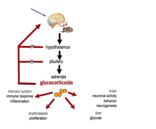
The stimulation of parvocellular neurons in the paraventricular nucleus (PVN) of the hypothalamus triggers the release of two neurohormones, the Corticotropin Releasing Hormone (CRH) and the vasopressin (AVP), into the blood vessels connecting the hypothalamus and the pituitary gland (the hypophysial portal blood). Both hormones stimulate the anterior pituitary gland to produce adrenocorticotropic hormone (ACTH), synthesized as part of a large pro-opiomelanocortin 241-amino-acid precursor. The hormonal secretion of ACTH leads to stimulation of melanocortin receptors located in the cortical part of the adrenals, which in turn synthetized GCs. ACTH, in turn, induces GCs synthesis and release from the adrenal glands, which are located at the top the kidneys. GCs regulate the function of multiple systems in living organisms e.g. neuronal activity, neurogenesis and behavioral responses, glucose metabolism in the liver, immune responses and inflammatory process, cardiovascular function, and erythropoiesis in bone marrow. Importantly, GCs can control the HPA axis activity through negative feedbacks by repressing the synthesis and the release of CRH in the hypothalamus, and of ACTH in the pituitary (Figure 2) (McEwen, 1979).
The activation of the HPA axis is under the control of different brain structures such as the prefrontal cortex (PFC), the hippocampus, and the BNST, the brainstem or the amygdala.
B) Circadian and ultradian rhythms of glucocorticoid hormone
The physiology of most organisms changes significantly between day and night. This represents a key adaptation to cope with the different environmental challenges the organism faces at different times of the day, such as changes in lighting conditions and temperature, food availability or the presence of predators. Most cells in multi-cellular organisms harbors their own cell-autonomous oscillators. The specialized suprachiasmatic nucleus in the ventral hypothalamus serves as the central pacemaker, which is known as the ‘central or master clock’ in mammals (Son et al., 2011) (Dickmeis, 2009), and GR signaling in peripheral cells, such as hepatocytes,
participates in the correct setting of peripheral cells with central pacemaker (Balsalobre et al., 2000).
Figure 2: Description of HPA axis
HPA axis activation stimulates GC secretion from the adrenal cortex which serves both to alert the organism to environmental or physiological changes and to maintain homeostasis. GCs control their own synthesis and secretion through a set of negative feedbacks at the forebrain, hypothalamic and pituitary levels.
Under the control of the suprachiasmatic nucleus, the CRH and the AVP are secreted in the portal blood system in a circadian manner leading to a similar activity of the adrenal gland and a circadian modulation of GCs levels. This daily rhythm in adrenal activity is not yet present at birth. In rats, a significant circadian variation in plasma corticosterone concentrations is seen only from three weeks of age; in humans, the rhythm is not fully established until two or three years of age.
Adrenal GC release is pulsatile in nature, marked by an ultradian release. Pulsatility occurs under basal conditions and is maintained in the face of acute or chronic stress.
Indeed, it appears that the magnitude of a stress response can depend on where the stressor occurs with respect to an ultradian pulse: stressors imposed on the rising phase of a pulse produce greater GC responses than those occurring during a falling phase. Pulsatility is an extremely important component of HPA function, as disruption of pulses (e.g., constant GC availability) disrupts GR signaling (Herman et al., 2016).
C) In response to stress
Acute stress efficiently drives HPA stress response, and feedback mechanisms effectively terminate the response after the stressor subsides. Generally, the HPA response begins with a pulse of ACTH, beginning within minutes and lasting a relatively short period of time (30-60 min). The GC response lags in time (due to the necessity for de novo GC production at the adrenal) and lasts substantially longer (90-120 min). The timing of both ACTH and GCs responses are dependent on stressor modality and intensity (Herman, 2013).
As studied in rodents, mainly by ablation approaches, many brain structures that innervate directly or indirectly the PVN are involved in the control of HPA axis regulation under stress. Their roles differ depending on the nature of the stress (hemorrhage, hypoglycemia, footshock, restraint) (Herman et al., 2016). The activity of CRH-releasing neurons of the PVN is controlled by a different sets of glutamate excitatory and γ-aminobutyric acid (GABA) inhibitory inputs.
GABA exerts an inhibitory tonus on CRH-releasing neurons, primarily via GABAA receptor binding, and this maintains low CRH neuronal activity (Radley and Sawchenko, 2011). GABAergic inputs also play a major role in limiting HPA axis responses to stress. Numerous hypothalamic nuclei send GABAergic projections to the PVN CRH neurons, including the medial preoptic nucleus, the dorsomedial hypothalamus, the BNST, and the lateral hypothalamus (Cullinan et al., 2008). Under stressful conditions, glutamatergic inputs to the PVN in areas such as the thalamus, the amygdala or the infralimbic cortex are activated, and GABAergic inhibition is suppressed, leading to HPA axis activation (Feldman and Weidenfeld, 1997). Noradrenaline contributes also to suppress GABA release, acting on a1 adrenoceptor located at presynaptic terminals (Han et al., 2002), and potentiates glutamatergic
inputs through a1 adrenoceptor activation (Plotsky, 1987). In addition to glutamate, the CRH-expressing PVN neurons are also activated by brainstem catecholamine-producing pathways from the nucleus of the solitary tract, which appears to promote HPA secretory activity following physical threat such as hemorrhage, hypotension and respiratory distress, and might play a role in ACTH responses to immune challenge as well (Herman et al., 2016). Additional HPA-excitatory information come from the Raphe nuclei, which send serotonergic projections to the PVN. Most studies indicate that serotonin (5-HT) stimulates ACTH and GCs secretion through the activation of 5HT2A and perhaps 5HT1A receptors in the PVN (Herman et al., 2016).
The importance of maintaining GC secretion within tolerable limits requires efficient mechanisms for inhibiting stress-integrative PVN neurons. This process appears to be accomplished by multiple mechanism. The negative feedback exerted by GCs, which inhibits the basal expression of CRH and AVP mRNA synthesis in the PVN, and pro-opiomelanocortin gene transcription in the anterior pituitary (Castro et al., 2010; Kellendonk et al., 2002). Also, the binding of GCs to its receptor (GR) in the PVN causes rapid synthesis and release of endocannabinoids that bind CB1 receptors on presynaptic terminals, inhibiting glutamate release and thereby reducing drive onto CRH neurons (Di et al., 2003).
D) HPA axis dysregulation and depression
HPA-axis dysregulation is the scientific term for the popular syndrome known as “adrenal fatigue.” It refers to any variation of HPA axis associated with a constellation of signs and symptoms including fatigue, sleep disruption, poor exercise tolerance and recovery, low libido, brain fog, weakened immune function, and reduced stress tolerance. HPA axis dysregulation is caused by many different aspects of the modern lifestyle, including poor diet, sleep deprivation, chronic stress, lack of (or too much) exercise, and inflammation. HPA-axis activity seems to be altered in several psychiatric (such as post-traumatic stress syndrome, major depression disorders or addiction) and neurological diseases such as Alzheimer’s or, Parkinson’s diseases (Hemmerle et al., 2012), with observed elevation of GC concentration in the saliva and plasma (Du and Pang, 2015; Carroll, 1982).
Concerning depression, up to 50% of patients (80% if severely depressed) display hyperactivity of the HPA axis, increased pituitary and adrenal gland volume, and chronic high levels of GCs in saliva, cerebrospinal fluid, blood plasma and urine. In this line, the dexamethasone suppression test that allows to detect a dysfunctional HPA axis feedback, was originally applied in the late 1960s as a laboratory diagnostic tool for major depression in adults (Zunszain et al., 2011). Clinically, as observed in Cushing’s syndrome, hyperactivity of the HPA-axis is highly correlated with significant increases in psychopathology, especially depression (Dorn et al., 1995). Dorn et al. found that 66% of Cushing’s patients exhibited psychopathology, consisting mainly of atypical and major depression as well as anxiety disorder and suicide ideation. 3 months following treatment for hypercortisolemia, this dropped significantly to 54% and further decreased to 24% 12 months after correction (Dorn et al., 1997), suggesting a direct causative link between elevated cortisol and psychopathology.
III. The Glucocorticoid Receptor
The physiological actions of GCs are mediated by two types of receptors; the Mineralocorticoid Receptor (MR) and the GR. Both belong to the steroid receptors family (together with the progesterone, the androgen and the two estrogens receptors present in mammals), a subfamily of the nuclear receptors superfamily that includes around thirty members. GR and MR have been originally identified as transcription factors, however, it is now clear that GCs for their long-known rapid physiological effects on neuronal activity act independent from gene transcription but on the MR and GR receptors that can both interfere with intracellular signaling cascades (such as the Jun kinase and Akt ones) and be activated at the cellular membrane to modulate glutamatergic synaptic neurotransmission (de Kloet et al., 2014).
Both MR and GR can be liganded by GC and aldosterone. They however differ in their affinity for these hormones. MR display a ten-fold higher affinity for GCs than GR. Concerning their cell distribution in the organism, whereas GR is almost ubiquitously expressed, MR has a more restricted expression profile. It is found in cells engaged in salt balance of the organism. In these cells, the expression of 11ß-HSD2 rapidly oxidizes GCs into inactive metabolites making them specifically responsive to aldosterone. However, in some cell type such as pyramidal cells of the hippocampus
both receptors are present, but the degradation enzyme is not. In these cells, MR is thus a bona fide GC receptor ( Oakley and Cidlowski, 2013; de Kloet et al., 2014).
A) Glucocorticoid Receptor structure
GR is a modular protein composed of three major domains: An N-terminal transactivation domain (NTD), a central DNA-binding domain (DBD), and a C-terminal ligand binding domain (LBD) (Kumar et al., 1999; Tronche et al., 1998) (Figure 3). In terms of both size and sequence homology the NTD represents the most variable domain among the nuclear hormone receptors. It contains a powerful transcriptional activation region; AF1/tau1/enh2 which is rich in acidic amino acid residues (Kumar et al., 2005). The NTD is highly immunogenic and contains the major known sites of phosphorylation in the GR.
The DBD, located centrally in the amino acid sequence of the GR, is the most conserved region among nuclear hormone receptors. It is composed of two highly conserved “zinc fingers”. The DBD contains amino acids that contact specific bases in GRE (Glucocorticoids Response Element) DNA sequences. The second zinc finger region stabilizes DBD:GRE interactions, and five amino acids in the second zinc finger, termed “D box”, play an important role in homodimerization at the GRE (Härd et al., 1990; Luisi et al., 1991). The DBD also interacts with certain other transcription factors, NF-AT, CREB or c-Jun for example. The C-terminal LBD is responsible for recognition and binding of steroid hormone ligands, chaperones and other proteins. In addition, the LBD contains a small but important ligand-dependent activation function (AF2) sub-domain located towards its C-terminal end which functions to complete binding surfaces for other proteins, e.g. coactivators and corepressors (Oakley and Cidlowski, 2013).
B) Activation and regulation of GR
In the cytoplasm of cells GR is present as part of a large multi-protein complex that includes chaperone proteins (hsp90, hsp70, and p23) and immunophilins of the FK506 family (FKBP51 and FKBP52; Figure 5; Grad and Picard, 2007; Pratt and Toft, 1997). These proteins maintain the receptor in a conformation that is transcriptionally inactive
but favors high affinity ligand binding. Upon binding GCs, GR undergoes a conformational change resulting in the dissociation of the associated proteins. This structural rearrangement exposes the two nuclear localization signals, and GR is rapidly translocated into the nucleus through nuclear pores.
Figure 3: GR domain structure and sites of post-translational modification. The
domains of GR and regions of the receptor involved in transactivation (AF1 and AF2), dimerization, nuclear localization, and hsp90 binding are pictured. Also depicted are the amino acid residues modified by phosphorylation (P), sumoylation (S), ubiquitination (U), and acetylation (A). Numbers are for human GR (Oakley and Cidlowski, 2013).
Once inside the nucleus, GR binds directly to GREs and regulates the expression of target genes (Figure 4, Freedman, 1992). Its specificity is however low and most GREs can also be bound by other steroid receptors (MR, PR and AR). The consensus GRE sequence, GGAACAnnnTGTTCT, is an imperfect palindrome that is composed of two 6-base pair half sites. GR binds this element as a homodimer or eventually heterodimers with MR, PR and AR, with each half site occupied by one receptor subunit. The GRE has been shown to mediate the GC-dependent induction of several genes and therefore is often referred to as an activating or positive GRE. However, genome-wide analyses have revealed that GR occupancy of the canonical GREs can also lead to the repression of target genes (Uhlenhaut et al., 2013). A negative GRE (nGRE) has also been recently described mediating GC-dependent repression of specific genes (Surjit et al., 2011).
Once bound to the GRE, the receptor undergoes additional conformational changes that lead to the recruitment of co-regulators and chromatin modifying enzymes that directly and indirectly modulate gene transcription rates by affecting the activity of RNA
polymerase II (Jenkins et al., 2001). Cofactors that mediate transcriptional activation include steroid receptor coactivators (SRC1-3), SWI/SNF, the histone acetyltransferases CBP/p300, and the nuclear methylase coactivator-associated arginine methyltransferase (CARM1). NCoR and SMRT are established corepressors that are recruited to GR bound to nGREs. The specific cofactors assembled and their subsequent activity are dictated by both the nature of the GC ligand and the specific GRE sequence bound by the receptor (Ronacher et al., 2009).
Figure 4: GR signaling pathways. GC-activated GR regulates gene expression in 3 primary ways: binding directly to DNA (A), tethering itself to other DNA-bound transcription factors (B), or binding directly to DNA and interacting with neighboring DNA-bound transcription factors (C). GR can also signal in a non-genomic manner through alterations in the activity of various kinases (Oakley and Cidlowski, 2013) GR can also regulate the transcription of target genes by physically interacting with other transcription factors (Figure 4). The association of GR with specific members of the STAT family, either apart from or in conjunction with GRE binding, has been shown to either repress or enhance the transcription of responsive genes (Nissen and Yamamoto, 2000; Rogatsky and Ivashkiv, 2006; Tronche et al., 2004). In contrast, the interaction of GR with the pro-inflammatory transcription factors, AP1 and NF-κB, antagonizes their activity and is considered to be a primary mechanism by which GCs
suppress inflammation (Yang-Yen et al., 1990). For some genes, the repression is accomplished by GR tethering itself to these DNA-bound proteins without itself directly interacting with the DNA (Chinenov et al., 2012).
C) Role of GR in the brain
As mentioned above, the GCs regulate brain functions and behavioral responses to environmental changes. Most of what we know about the brain function of GR in the brain is coming from rodent studies. Since constitutive GR knock-out mice die soon after birth due to respiratory failure arising from impaired lung development, the study of conditional knock-out has been crucial to study the role of GR in behavior. Here, we will mainly focus on anxiety and depressive-like behaviors as well as responses to drugs of abuse which constitute the main topics of my work.
1) GR and social behavior, anxiety and depression.
GR gene inactivation in the mouse central nervous system (GRNesCre mice) results in
reduction of stress related behaviors such as depression-like and anxiety-like behavior (Tronche et al., 1999) and behavioral responses to cocaine (Deroche-Gamonet et al., 2003). Loss of GR function in the nervous system impairs hypothalamus-pituitary-adrenal (HPA)–axis regulation, resulting in increased GC levels that lead to symptoms reminiscent of those observed in Cushing syndrome (Tronche et al., 1999).
Therefore, more refined models have been generated and studied. GR receptor inactivation in dopaminoceptive D1-expressing neurons (mainly the caudate putamen, the NAc and the medial prefrontal cortex; GRD1Cre mice) results in higher resistance to
chronic social defeat in terms of social interaction (Barik et al., 2013). Early life stress produces a decrease in GR mRNA in the brain, with a notable reduction in the amygdala that is associated with sustained alterations in anxiety, fear and sociability-like behaviors. Lentiviral-mediated restoration of the GR mRNA deficit, specifically within the adult central nucleus of the amygdala (CeA), reverses the enduring changes in anxiety and social behavior after early life stress (Arnett et al., 2015).
2) GR and addiction
In addition to its effects on anxiety and depressive-like behaviors, the absence of GR in the central nervous system in GRNesCre mice reduces the animals responses to
cocaine in locomotor sensitization and self-administration paradigms (Deroche-Gamonet et al., 2003). Conversely, the overexpression of GR in the forebrain confirms leads to an increase in cocaine sensitization (Wei et al., 2004). Dopaminergic transmission within the reward system is largely implicated in the addictive properties of drugs. Targeting GR along the dopamine pathway also alter responses to psychostimulant drugs. While GR gene inactivation in dopamine neurons (GRDATCre
mice) does not have any effect, GR inactivation in dopaminoceptive neurons specifically reduces the self-administration, sensitization and conditioned place-preference to psychostimulants (cocaine and amphetamine) but spear the responses to opiates (Ambroggi et al., 2009; Barik et al., 2010; Parnaudeau et al., 2014).
Chapter2. Chromatin remodelers and Epigenetics
As other steroid receptors, the GR has the peculiarity to be a transcription factors that control the expression of target genes in response to a specific hormone signal. As we saw, GR can act on intracellular signaling cascades such as MAP-Kinases and Akt/PI3A pathways and, when present at the membrane, can modulate synaptic transmission, but its best-known function is its transcription factor one. It is present at the promoters or enhancers of target genes when bound to GRE DNA segments but also when interacting, via protein-protein bounds, with other transcription factors themselves bound to DNA targets like; AP1, STAT3, STAT5 (Nissen and Yamamoto, 2000; Rogatsky and Ivashkiv, 2006; Yang-Yen et al., 1990). It can intervene on the recruitment of the general transcriptional machinery, just before RNA polymerization. It can also intervene earlier, at the chromatin level, to recruit modifier enzymes that act on chromatin shaping at the protein or DNA level and modulate thus chromatin structure and chromatin accessibility of other regulatory proteins and the general transcriptional machinery (McEwen et al., 1996).
The regulation of gene expression through chromatin modification is known since the 1960’s and if a vast amount of knowledge accumulated since those years demonstrated its importance for all biological functions (development, differentiation, adaptation), conceptualizing theories start to emerge. In eukaryotic cells, nuclear DNA is packaged into chromatin structures that can be modulated to generate transcriptionally active or repressed configurations in different cellular contexts and under changing environmental conditions. This remarkable plasticity is governed in part by multi-subunit protein complexes that enzymatically regulate chromosomal structure and activity. These complexes can either chemically modify the histone tails of nucleosomes, disrupt histone-DNA contacts through ATP hydrolysis or chemically modify nucleic bases in the DNA major groove.
I. Nuclear organization
In eukaryotic cells, chromatin is tightly packed in a highly organized fashion within a nucleus composed of the nucleoplasm and the nuclear envelope. There are sub-compartments in the nucleus containing factors involved in essential nuclear functions
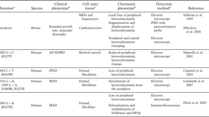
such as DNA replication, transcription, and RNA splicing (Prasanth et al., 2010). The nuclear envelope separates nuclear functions from cytoplasmic functions and at its inner surface it provides a docking site for chromatin. The major structural elements of the nuclear envelope are the inner nuclear membrane, the outer nuclear membrane, the nuclear pore complexes, and the nuclear lamina. The lamina is comprised of a complex meshwork of proteins closely associated with the inner nuclear membrane and attached to the periphery of nuclear pore complexes and to chromatin. The main constituents of the lamina are the type V intermediate filament proteins, the nuclear lamins. Lamins are also found, in lower concentrations, distributed throughout the nucleoplasm. The organization of lamins at the nuclear periphery as well as within the nucleoplasm is influenced by numerous lamin-binding proteins (Figure 5, Dechat et al., 2008). The importance of lamins in chromatin organization is most evident in cells derived from patients suffering from various laminopathies and in the LMNA-null mouse (Table 1).
Figure 5: Electron microscopic observation of fibroblastic nucleus. Low-magnification
views show peripheral heterochromatin and nucleoplasmic heterochromatic foci in the normal nucleus.
Taken together, these findings demonstrate that the mutations of lamin gene leads to global changes in the epigenetic organization of chromatin, which undoubtedly contributes to the phenotypes observed in different laminopathies, including defects in DNA repair and alterations in gene expression.
Table 1: Changes in heterochromatin organization caused by lamin A/G mutation. a The DNA base pair changes and the amino acid changes (in parentheses) are shown. bThe phenotypes are as follows: autosomal-dominant Emery-Dreifuss muscular dystrophy (AD-EDMD), familial partial lipodystrophy (FPLD), mandibuloacral dysplasia (MAD), and Hutchinson-Gilford progeria syndrome (HGPS). cThe cell types or tissue examined are listed. d Alterations in chromatin organization described. (H3K9me3) Histone H3 thrimethylated at Lys 9; (HP1) heterochromatin-associated protein 1; (H3K9me1) histone H3 monomethylated at Lys 9; (H3K27me3) histone H3 thrimethylated at Lys 27; (H4K20me3) histone H4 thrimethylated at Lys 20.
II. Structure of chromatin
Eukaryotic DNA is packaged into a complex macromolecular structure called chromatin that may facilitate compaction of the genetic material. The DNA is folded and condensed into chromatin in a dynamic manner since it still needs to be accessible to carry out key functions such as replication, transcription, and DNA repair. The first step of compaction is achieved by packaging the naked DNA into the nucleosome which consist of 174 base pairs of DNA wrapped around the histone proteins octamer containing two molecules of each core histone H2A, H2B, H3, and H4 (Luger et al., 1997). Nucleosomes are separated by approximatively 50 base pairs. Histone H1 is important for higher-order chromatin formation and compaction (Robinson and Rhodes, 2006). It stabilizes the interaction of DNA with the nucleosome and prevents
ATP-dependent remodeling of the chromatin from changing the position of nucleosome (Ramachandran et al., 2003), Figure 6).
Figure 6: Structure of the chromatin (Ramachandran et al., 2003). Nucleosome
consist of 174 base pairs of DNA wrapped around the histone proteins octamer containing two molecules of each core histone H2A, H2B, H3, and H4.
Histones are small basic proteins that can be covalently modified at their flexible N- or C-terminal tails (Kouzarides, 2007). Histone post-translational modifications affect their interactions with DNA, leading to alterations in chromatin structure and function (Choi and Howe, 2009; Strahl and Allis, 2000); Strahl and Allis 2000). For instance, certain histone post-translational modifications are associated with a more open and accessible chromatin conformation, while others may be found in highly compacted and transcriptionally inactive loci, hinting towards the “histone code” (Strahl and Allis, 2000). They will be described later in details.
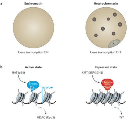
From the functional point of view, various chromatin structures are commonly divided into euchromatin and heterochromatin. Euchromatin describes regions within the nucleus that contain actively transcribed genes. Generally, active regions contain histones that are hyperacetylated at lysine residues (Figure 7).
Constitutive heterochromatin describes the highly condensed regions of the genome that are visible as bright nuclear areas following staining with DNA dyes such as 4′,6-diamidino-2-phenylindole (DAPI) and Hoechst. These regions often comprise repetitive DNA (such as satellite sequences surrounding centromeres) and are generally thought have a low density of genes (Strahl and Allis, 2000, Figure 7).
Figure 7: Euchromatin and heterochromatin (from Allis and Jenuwein, 2016).
Euchromatin is a region within the nucleus that contains actively transcribed genes. Heterochromatin describes the highly condensed regions of the genome with a low density of genes.
III. Epigenetics
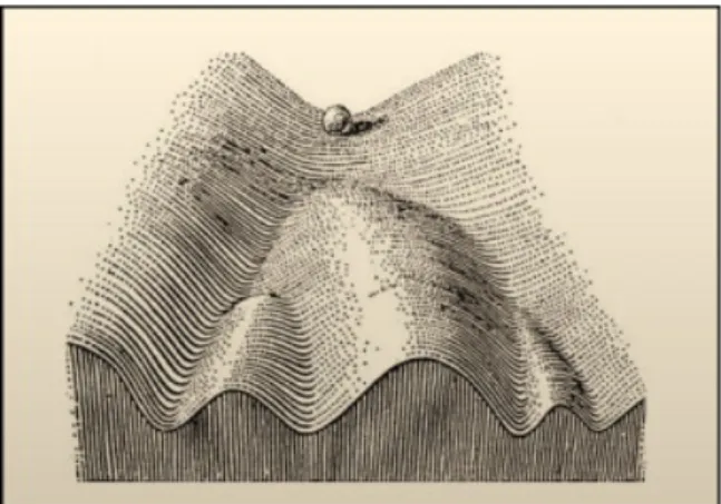
The term, “epigenetics,” was first used to explain the complex interactions between the environment and genome that are involved in development and differentiation in higher organisms. Historically, the word “epigenetics” was used to describe events that could not be only explained by genetic principles. Conrad Waddington (1905–1975), who is given credit for coining the term, defined epigenetics as “the branch of biology which studies the causal interactions between genes and their products, which bring the phenotype into being”. He forged the word from Aristoteles “Epigenesis” theory of development that was taken considering the influence of environment on embryo development and from “Genetics” since after T. Morgan it was clear that developmental genes existed and conditioned development. Epigenetics, in a broad sense, is a bridge between genotype and phenotype, a phenomenon that changes the final outcome of a locus or chromosome without changing the underlying DNA sequence. For example, even though the vast majority of cells in a multicellular organism share an identical genotype, organismal development generates a diversity of cell types with disparate, yet stable, profiles of gene expression and distinct cellular functions. Thus, cellular
differentiation may be considered an epigenetic phenomenon, largely governed by changes in what Waddington described as the “epigenetic landscape” rather than alterations in genetic inheritance (Goldberg et al., 2007; Figure 8).
Today, this term is used to refer to heritable alterations, through cell division, in higher organisms that are not due to changes in DNA sequence. By extension, it refers also to the mechanisms involved in changing gene expression, for instance long-term ones observed under environmental influences. Epigenetic modifications rather alter DNA accessibility and chromatin structure, thereby regulating patterns of gene expression. Today, a wide variety of illnesses, behaviors, and other health indicators already have some level of evidence linking them with epigenetic mechanisms, including cancers of almost all types, cognitive dysfunction, and respiratory, cardiovascular, reproductive, autoimmune, and neurobehavioral illnesses (Felsenfeld, 2014; Inbar-Feigenberg et al., 2013).
Figure 8: In 1957, Conrad Waddington proposed the concept of an epigenetic
landscape to represent the process of cellular fate decision-making during development. At various points in this dynamic visual metaphor, the cell (represented by a ball) can take specific permitted trajectories, leading to different outcomes or cell fates. Figure adapted from (Goldberg et al., 2007).
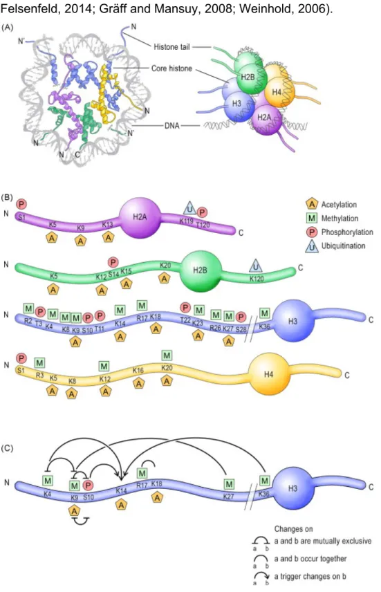
Histones are basic proteins consisting of a core and an N-terminus tail composed of a loosely-structured sequence of amino acids. Posttranslational histone modifications occur primarily on the N-terminus tail, and include acetylation, methylation, phosphorylation, ubiquitination and sumoylation (Fig. 9A).
DNA methylation occurs throughout the genome but is functionally most relevant when present in sequences rich in CpG dinucleotides. Because of their chemical properties, these modifications influence the condensation of chromatin, and thereby modulate the accessibility of DNA to the transcriptional machinery (Allis and Jenuwein, 2016; Felsenfeld, 2014; Gräff and Mansuy, 2008; Weinhold, 2006).
Figure 9: Epigenetic marks on histone tails and DNA. (A, left) View of the nucleosome
Schematic representation of the four-nucleosome core histones, H2A, H2B, H3 and H4. (B) Schematic representation of the N- and C-termini of the core histones and their residue-specific epigenetic modifications. (C) Crosstalk between epigenetic modifications on the H3 N-terminus tail. Adapted from (Gräff and Mansuy, 2008) As mentioned above, the regulation of gene expression involves chromatin modifications regulate the gene expression. For example, euchromatin in which most of active genes are present contain histones that are frequently hyperacetylated at lysine residues. As well the active genes are also enriched in methylated Lys4 and Lys79 histone H3 residues. And methylation of Arg2, Arg17 and Arg26 of H3, and Arg4 of H4, are associated with transcriptional activation (Daujat et al., 2002). Whereas, phosphorylation (notably Ser10 of H3) is associated with immediate early gene activation (Lachner et al., 2003). The hallmarks of constitutive heterochromatin include trimethylation at Lys9 of histone H3, a paucity of methylation at H3 Lys4 and trimethylation of H4 Lys20 (Kourmouli et al., 2004; Schotta et al., 2004).
An important feature of epigenetic marks that is essential for transcriptional regulation is their ability to crosstalk (Fig. 11C). Posttranslational histone modifications often act in concert, and multiple feed-forward and feed-back mechanisms involving the same nucleosome or histone, or distinct nucleosomes and histones have been identified. These cross-talks can enhance chromatin condensation when transcriptional silence is required, or chromatin opening when transcriptional activity is needed. The repertoire of DNA and histone modifications is controlled by specific enzymes that include DNA methyltransferases (DNMTs), histone acetyltransferases (HATs), histone deacetylases (HDACs), histone methyltransferases (HMTs), histone demethylases (HDMs), protein kinases (PKs), protein phosphatases (PPs), ubiquitin- and SUMO-associated enzymes (Figure 11B). These enzymes operate both independently and in synergy establishing a “histone code”, a highly dynamic and flexible chromatin marking that, in combination with chromatin-associated proteins, could stabilize the pattern of gene expression in response to given external stimuli. That way, epigenetic modifications at the level of the chromatin provide a focal point to bridge nuclear events to intracytoplasmic signaling cascades, and a potential molecular means to retain marks of prior transcriptional activity in the nuclear machinery.
Epigenetic processes are natural and essential to many organism functions, but if they occur improperly, there can be major adverse health and behavioral effects (Allis and Jenuwein, 2016; Felsenfeld, 2014; Weinhold, 2006).
A) DNA modifications (DNA methylation)
DNA methylation is defined as addition of a methyl group to cytosine within a CpG dinucleotide, thereby forming 5-methylcytosine (Bird, 1986). The term CpG refers to the base cytosine (C) linked by a phosphate bond to the base guanine (G) in the DNA nucleotide sequence. Most CpG dinucleotides in the human genome are methylated. Unmethylated CpGs are not randomly distributed, but are usually clustered together in ‘CpG islands’, which are in the promoter region of many genes (the region that facilitates transcription of a particular gene) (Bird, 1986; Ehrlich et al., 1982).
These modifications are achieved by two types of DNA methyltransferases. The first type, DNMT1 functions during DNA replication to copy the DNA methylation pattern from the parental DNA strand onto the newly synthesized daughter strand (Moore DL et al, 2012). DNMT3a and DNMT3b play a major role in de novo methylation, in which methylation can be performed on double-stranded DNA that is not methylated. DNA methylation is generally associated with gene silencing (Okano M et al,1999), and DNA demethylation is usually connected with gene activation (Goldberg et al., 2007). DNA demethylation can take place as either passive or active mechanisms. Passive DNA demethylation occurs in dividing cells. Active DNA demethylation can occur in both dividing and non-dividing cells (Moore et al., 2013).
B) Histone modifications
1) Histone acetylation and deacetylation
Acetylation of histone’s lysine group is highly dynamic and regulated by the opposing action of two families of enzymes, (HATs) and histone deacetylases (HDAC). Its link with gene expression is known for decades (Allfrey et al., 1964). The HATs utilize acetyl-CoA as cofactor and catalyze the transfer of an acetyl group to the ε-amino
group of lysine side chains. In doing so, they neutralize the lysine's positive charge and this action has the potential to weaken the interactions between histones and DNA. HDAC enzymes oppose the effects of HATs and reverse lysine acetylation, an action that restores the positive charge of the lysine. This potentially stabilizes the local chromatin architecture and is consistent with HDACs being predominantly transcriptional repressors (Bannister and Kouzarides, 2011).
2) Histone phosphorylation
Histone phosphorylation is a transfer of phosphate group from ATP to the hydroxyl group of the target amino-acid side chain. this modification adds significant negative charge to the histone that undoubtedly influences the chromatin structure. histone phosphorylation is highly dynamic process which is controlled by kinases and phosphatases that add and remove the modification, respectively (Oki et al., 2007).
3) Histone methylation
The initial publication that described histones modifications (Allfrey et al., 1964)not only observed that histones acetylation but also methylation was associated with changes in gene expression. Histone methylation mainly occurs on the side chains of lysine and arginine. Unlike acetylation and phosphorylation, however, histone methylation does not alter the charge of the histone protein. Furthermore, Methylation of lysine or arginine residues can occur in several modification states. Lysine residues can house either one (me1), two (me2) or three (me3) methyl moieties on their amine group, whereas arginine residues can carry one (me1) or two (me2) methyl groups on their guanidinyl group. These defined modification states can have different and profound implications on the function of chromatin. In general, H3K4, K36, and K79 methylation are found near active transcriptional units and H3K9 and H4K20 modifications are hallmarks of silenced or heterochromatic regions. Histone arginine methylation occurs on H3R2, R8, R17 and R26 and H4R3 and has roles in defining both active and repressed chromatin states (Ng et al., 2009; Turner, 2005).
For many years, histone methylation was strangely considered a stable, static modification. Nevertheless, a number of different reactions/pathways were suggested
as potential demethylation mechanisms for both lysine and arginine (Bannister and Kouzarides, 2011).
4) Ubiquitylation and sumoylation
All of the previously described histone modifications result in relatively small molecular changes to amino-acid side chains. In contrast, ubiquitylation results in a much larger covalent modification. Ubiquitin itself is a 76-amino acid polypeptide that is attached to histone lysines via the sequential action of three enzymes, E1-activating, E2-conjugating and E3-ligating enzymes (Hershko and Ciechanover, 1998). The enzyme complexes determine both substrate specificity (i.e., which lysine is targeted) as well as the degree of ubiquitylation (i.e., either mono- or poly-ubiquitylated). For histones, mono-ubiquitylation seems most relevant although the exact modification sites remain largely elusive. However, two well-characterized sites lie within H2A and H2B. For example, H2AK119ub1 is involved in gene silencing, whereas H2BK123ub1 plays an important role in transcriptional initiation and elongation.
Even though ubiquitylation is such a large modification, it is still a highly dynamic one. The modification is removed via the action of isopeptidases called de-ubiquitin enzyme and this activity is important for both gene activity and silencing (Bannister and Kouzarides, 2011).
Sumoylation is a modification related to ubiquitylation and involves the covalent attachment of small ubiquitin-like modifier molecules to histone lysines via the action of E1, E2 and E3 enzymes. Sumoylation has been detected on all four core histones and seems to function by antagonizing acetylation and ubiquitylation that might otherwise occur on the same lysine side chain (Nathan et al., 2006).
5) Histones variants
Histone variants are deposited onto chromatin by specific histone chaperones and also interact with other chromatin modifiers. Structural differences introduced by a core histone variant typically affect interactions between histone proteins within a nucleosome, hence their stability as well as the open/compact chromatin conformation. For example, histone variants H2A.Z and H3.3 are mainly linked with an open chromatin conformation and transcriptional activity, while macroH2A deposition
stabilizes the nucleosome and is often associated with a repressive chromatin state. Consequently, replacement of canonical histones with histone variants adds another level of complexity and a distinct way of modulating chromatin function (Skene and Henikoff, 2013).
Histone variants exchange play a potential mechanism of gene regulation in response to neural activity. For example; H3.3 dramatically accumulates in neuronal and glial chromatin with age and remains highly dynamic throughout life to control cell-type specific gene expression programs and physiological plasticity. Moreover, manipulations of H3.3 in neurons, which stall its incorporation and eviction from chromatin, reveal histone turnover as a critical mediator of neuronal activity-dependent gene expression, synaptic connectivity and cognition (Maze et al., 2015).
Histone Coding
The hypothesis of a histone code suggested by Strahl and Allis in 2000, proposes that the combination of histone modifications at a certain genomic locus determines the activity state of the underlying gene (Strahl and Allis, 2000). The modifications regulate accessibility for DNA binding and regulatory proteins, such as transcription factors, either by altering the charge on the histones and thus changing histone–DNA interactions or by recruiting structural proteins. By this means, histone modifications store epigenetic information. However, the histone code is heavily debated within the epigenetic field, and it has been argued that the regulation of gene expression by various histone modifications might simply reflect a cumulative effect rather than the interpretation of a combination (Henikoff, 2005). As an extension of the initial code, theoretical ‘modification cassettes’ have been suggested to have one specific output (Fischle et al., 2003). Combinations of modifications act when they are located on the same histone tail and on different histones within a nucleosome. For example, the ubiquitylation of H2B is necessary for the methylation on lysine 79 of histone H3 (Briggs et al., 2002). The histone code was thus expanded to a ‘nucleosome code’, suggesting that the presence of all post-translational histone modifications in one nucleosome regulate the underlying DNA sequence. Particular importance is attached to so-called binary switch modules in which modifications on two neighboring residues can influence each other. An example is the methylation of lysine 9 on histone H3, which is bound by heterochromatin protein 1 (HP1). During mitosis and chromatin
condensation, serine 10 is phosphorylated, which reduces HP1 binding to H3K9me. This binding is further reduced when H3 becomes acetylated at lysine 14, which strips HP1 from the mitotic chromosomes. In addition to the inhibition of HP1 binding to the lysine 9 methylated peptide, serine 10 phosphorylation interferes with the methylation of H3K9 by SUV39H1 (Garcia et al., 2005; Rea et al., 2000).
Genome-wide mapping of individual histone modifications made it possible to detect correlations between histone modification patterns and specific states of gene activity. For example, particular modifications including H3K4me2,3 (histone H3 lysine4 di- and trimethylation) and H3K36me2,3 are frequently located in actively transcribed regions. By contrast, modifications such as H3K27me3 and H4K20me3 are frequently mapped to regions where transcription is repressed (Barth and Imhof, 2010) (Figure 10).
A) Nuclear topology
Eukaryotic nuclei are well-organized structures with distinct nuclear compartments. The arrangement of these compartments inside the nuclear space is termed nuclear architecture (Figure 11).
The main resident of eukaryotic nuclei is the genetic material itself, packaged in the form of chromatin. Akin to the nucleus itself, chromatin is also organized into well-defined domains called chromosomes. The chromosomes themselves occupy distinct subvolumes of the nuclear space, termed chromosome territories and the position of gene loci within each chromosome is also subject to strict regulation. (Cremer et al., 1986) In the nervous system, nuclear architecture has been shown to be distinct among different types of cells and during different stages of differentiation or development are significantly determined and regulated by chromatin (Takizawa and Meshorer, 2008).
Figure 10: Distribution of histone modifications on active and silenced genes.
Modification patterns differ on actively transcribed and silenced genes, which is displayed in this figure as a schematic view on nucleosomes (a and c) or modification distribution over the gene (b and d). An active gene is shown in (a) and in (b). An external signal can lead to the activation of kinases (orange) in the nucleus, which can then phosphorylate (yellow circles) histones as well as transcription factors (TF, red) to elicit an appropriate physiological response. The transcription factors will then bind DNA at the promoter and facilitate the docking of RNA polymerase II (RNAPII, green). At this point, transcription can begin. Elongating RNA polymerase II, which is highly phosphorylated on its C-terminal domain, can interact with histone modifiers (pink), for example Set2, which methylates H3K36, and thus introduce active modifications into chromatin at this locus. In particular, nucleosomes within the promoters of actively transcribed genes carry high levels of active modifications such as acetylations and methylation of H3K4. At the transcriptional start-site there is a nucleosome-depleted region (NDR) within the promoter. Active modifications such as methylation of H3K79 are present in the body of these genes. Inactive genes, as shown in (c) and (d), have a fairly even distribution of silencing modifications, such as H3K9 methylation and H4K20 methylation, whereas H3K27 methylation is enriched in the promoter. These modifications can be bound by heterochromatic proteins (blue) and, thus, this chromatin area can condense, as seen in heterochromatin. Adapted from (Barth and Imhof, 2010).
Figure 11: Nuclear architecture. Examples of common nuclear bodies are depicted.
Neuronal immunofluorescence images are shown for the various nuclear domains (Takizawa and Meshorer, 2008).
Chromatin can exist in different ‘states’ as described earlier, including ‘open’ (eu-) and condensed (hetero-) chromatin. These are differentially defined by three characteristics: (1) loose or dense nucleosomal packaging euchromatin or heterochromatin, respectively, (2) specific types of post-translational histone modifications (such as acetylation), and (3) presence or absence of various chromatin regulatory proteins that either facilitate or repress transcription. For example, actively expressed genes in open chromatin show high levels of histone acetylation, with nucleosome-free intervals occupied by activator proteins (transcription factors) and the
RNA polymerase II initiation complex (Figure 12). Superimposed upon these types of nucleosomal organization is the 3-dimensional conformation of chromatin fibers and entire chromosomes, often described in terms such as ‘loopings’ or ‘globules’ and in toto referred to as the ‘3D genome’. This includes the ‘clustering’ of euchromatic and heterochromatic sequences that tend to assemble into alternating regions of approximately ~5 megabases (Mb). These ‘compartments’, positioned along the same chromosome, are able to interact with compartments from different chromosomes (Lieberman-Aiden et al., 2009). Euchromatic regions are termed ‘A compartments’ and are enriched with open/decondensed chromatin and correspond to much higher overall levels of transcription, whereas ‘B compartments’ harbor inactive and heterochromatic sequences (Rao SS et al, 2014)(Rao et al., 2014) (Figure 12). In most cell types, large clusters of heterochromatin are enriched at the nuclear periphery, in multiple pericentromeric foci in the nuclear interior and around nucleolar membranes (Padeken and Heun, 2014).
IV. Chromatin remodeler complex (Nucleosome remodelers)
The importance of histones and chromatin structure in the regulation of eukaryotic gene transcription has become much more widely accepted over the past few years. ATP-dependent chromatin remodeling complexes specifically recognize these histones modifications (acetylation, methylation, phosphorylation, ribosylation and ubiquitination), and through ATP hydrolysis unwrap, mobilize, exchange or eject the nucleosome, and subsequently recruiting a transcriptional apparatus to nucleosomal DNA (Cosma, 2002)
The mechanisms by which histones positioning and constitution contribute to the regulation of transcription both in vitro and in vivo have been clarify in the last decades (Vignali et al., 2000) However, chromatin itself is stable and shows limited mobility, and its dynamic properties are due to the action of chromatinmodifying and -remodeling complexes (Strahl and Allis, 2000). In this manner, chromatin structure simultaneously provides a packaging solution and a sophisticated apparatus for regulating gene expression.
A) Different ATP-dependent chromatin remodeler complexes
ATP-dependent chromatin remodeling complexes are large (>1 MDa) multi-components complexes (consisting of between 4 and 17 subunits) that are highly conserved within eukaryotes. They are characterized by the presence of an ATPase subunit belonging to the superfamily II helicase-related proteins (Singleton and Wigley, 2002). Proteins belonging to this class contain an ATPase domain that is itself comprised of two parts, the DExx and HELICc regions, which are separated by a linker. This class can be further classified into at least 4 different families (SWI/SNF, ISWI, NURD/Mi-2/CHD, and INO80) based on the additional presence of unique domains within or adjacent to the ATPase domain (Figure 13)(Tang et al., 2010).
A) SWI/SNF complex
SWI/SNF is a protein complex composed of a dozen of proteins. It was originally identified as a regulator of mating type switching (SWI) or as a requirement for growth on energy sources other than sucrose (SNF – sucrose non-fermenting) in yeast
(Neigeborn and Carlson, 1984; Peterson and Workman, 2000). The SWI/SNF complex is a large ATP-dependent chromatin remodeling complex that is highly conserved from yeast to human, which is essential for transcription regulation, genomic stability, DNA repair and many aspects of development (Kasten et al., 2011; Tang et al., 2010).
Figure 13: Classification of ATP-dependent chromatin remodeling complexes: The
ATPase subunit of all the remodeling complexes belongs to the superfamily II helicase group. The ATPase always contains a DExx and a HELICc domain, spaced by a linker. The remodelers are classified into different families based on the presence of additional domains on their ATPase subunits. The SWI/SNF family contains a HSA domain, involved in actin binding, and a bromodomain important for the binding of acetylated lysines. The ISWI family contains the SANT and SLIDE domains, important for histone binding. The CHD/NURD/Mi-2 family contains a tandem Chromo domain, also used for histone binding. The INO80 family, like the SWI/SNF family, comprises a HSA domain but it is also characterized by the presence of a longer insertion between the DExx and the HELICc domains (Tang et al., 2010).
In S. Cerevisiae, as in drosophila and Humans, there are two versions of the chromatin complex called SWI/SNF and RSC (for Remodeling the Structure of Chromatin). RSC is founded more in the cell comparing to SWI/SNF. RSC is essential for cell growth while SWI/SNF is not. The catalytic subunit of yeast SWI/SNF is the Swi2 or Snf2 protein and its paralog in RSC is the Sth1 subunit (Du et al., 1998).
In drosophila, the two forms of chromatin remodeler complexes are called BAP-complex (Brahma Associated Protein) and PBAP-BAP-complex (Polybromo-associated BAP) which both comprise the same ATPase subunit, named Brahma. In Human, similarly, these two complexes are called BAF complex (Brg1 Associated Factors) and PBAF complex (Polybromo-associated BAF) and can contain one of the two distinct