HAL Id: hal-01739143
https://hal.univ-lorraine.fr/hal-01739143
Submitted on 20 Mar 2018
HAL is a multi-disciplinary open access
archive for the deposit and dissemination of sci-entific research documents, whether they are pub-lished or not. The documents may come from teaching and research institutions in France or abroad, or from public or private research centers.
L’archive ouverte pluridisciplinaire HAL, est destinée au dépôt et à la diffusion de documents scientifiques de niveau recherche, publiés ou non, émanant des établissements d’enseignement et de recherche français ou étrangers, des laboratoires publics ou privés.
Extraction-Implantation Immédiate ; Données actuelles
Antoine Lascombes
To cite this version:
Antoine Lascombes. Extraction-Implantation Immédiate ; Données actuelles. Sciences du Vivant [q-bio]. 2012. �hal-01739143�
AVERTISSEMENT
Ce document est le fruit d'un long travail approuvé par le jury de
soutenance et mis à disposition de l'ensemble de la
communauté universitaire élargie.
Il est soumis à la propriété intellectuelle de l'auteur. Ceci
implique une obligation de citation et de référencement lors de
l’utilisation de ce document.
D'autre part, toute contrefaçon, plagiat, reproduction illicite
encourt une poursuite pénale.
Contact : [email protected]
LIENS
Code de la Propriété Intellectuelle. articles L 122. 4
Code de la Propriété Intellectuelle. articles L 335.2- L 335.10
http://www.cfcopies.com/V2/leg/leg_droi.php
ACADEMIE DE NANCY METZ UNIVERSITE DE LORRAINE FACULTE D’ODONTOLOGIE Année 2012 N° 3979
THESE
Pour le
DIPLOME D’ETAT DE DOCTEUR EN CHIRURGIE DENTAIRE
Par Antoine LASCOMBES Né le 3 janvier 1986 à Nancy (Meurthe‐et‐Moselle)ExtractionImplantation Immédiate ;
Données Actuelles.
Présentée et soutenue publiquement le Lundi 25 Juin 2012 Examinateurs de la thèse :Pr P. AMBROSINI Professeur des Universités Président
Dr J. PENAUD Maître de conférences des Universités Directeur
Dr C. SECKINGER Praticien Hospitalier Juge
Dr J. BALLY Assistant Hospitalier Universitaire Juge
2 A notre Président de thèse Pascal AMBROSINI Docteur en Chirurgie Dentaire Docteur de l’Université Henri Poincaré, Nancy‐I Vice‐Doyen au budget et aux affaires hospitalières Habilité à diriger des Recherches Professeur des Universités – Praticien Hospitalier Responsable de la Sous‐section : Parodontologie
A notre directeur de thèse Jacques PENAUD Docteur en Chirurgie Dentaire Docteur de l’Université Henri Poincaré, Nancy‐I Maître de conférence des Universités – Praticien Hospitalier Sous‐section : Parodontologie
4 A notre juge Cédric SECKINGER Docteur en Chirurgie Dentaire Praticien Hospitalier Odontologiste des Hôpitaux Spécialiste en chirurgie orale
A notre juge Julien BALLY Docteur en Chirurgie Dentaire Assistant hospitalier universitaire Ancien Interne en Odontologie Sous‐section : Chirurgie Buccale, Pathologie et Thérapeutique, Anesthésiologie et Réanimation
6 A notre juge Matthieu BOULANGEOT Docteur en Chirurgie Dentaire
A Albane, ma fille. J’espère qu’elle sera fière de moi. A mes parents, qui m’ont soutenu dans les épreuves et qui m’ont encouragé tout au long de mes études. A Marie, ma sœur, pour sa grande aide durant ces derniers mois et à Caroline qui sait être à l’écoute et qui est toujours de bons conseils. A Antoine et Matthieu, bienvenue dans la famille. A mes grands parents, merci pour votre affection et pour votre soutien. A mes cousins : Benjamin et Aurélie et leurs enfants, Sophie et Karl et leurs enfants, Marc, Adrien, Ariane, Thomas, Astrid. A mes amis : Marie et Marc chez qui je sais trouver une ambiance très appréciable. Clémence et Gael qui sont là quand il le faut. Le HALL, qui nous a permis de passer d’excellents moments durant ces années d’études. Loulou, Pipoo, Charloo et Anne, Greg, Jacques et son appart, Franky, Théo, Sophia, Carole, Marie, Hugues, Souny, Pauline et tous ceux que j’ai oubliés… A tous les membres du cabinet : Jean‐Luc, Annabel, Matthieu qui m’apprennent beaucoup par leur expérience, A Nathalie, Céline et Sonia qui apportent une très bonne ambiance dans ce lieu de travail. A Lucie et Amélie. J’espère que le plaisir d’y venir travailler durera le plus longtemps possible. Aux membres du jury : Merci de m’avoir aidé à finaliser mon cursus universitaire par cet ouvrage. J’espère devenir un confrère à la taille de vos espérances.
8 Introduction
1.1. Préambule
Au cours des 10 dernières années, l’implantologie a été en constante évolution. Des avancées importantes sont apparues tant en ce qui concerne les biomatériaux utilisés lors de ces interventions qu’en ce qui concerne les techniques opératoires.
De nombreuses études expérimentales précliniques et cliniques ont contribué au développement de cette thérapeutique.
Dans une large mesure, cette évolution ainsi que l’essor que prend l’implantation immédiate post extractionnelle (IIPE) sont le reflet d’un changement dans les attentes des patients.
Alors que par le passé les pathologies dentaires avaient souvent pour issue une extraction non compensée, on discute aujourd’hui du remplacement éventuel d’une dent lésée alors que celle ci est toujours en place. Il s’en suit donc une décision importante qui doit être prise en concertation entre le praticien et son patient, qui concerne le moment de l’implantation par rapport à l’extraction.
Aujourd’hui, alors que les besoins fonctionnels fondamentaux des patients représentés principalement par l’alimentation et la communication sont satisfaits les nouveaux protocoles de traitement doivent maintenant répondre à des impératifs de plus en plus exigeants tels que la réduction du temps de traitement, la diminution du coût et de l’inconfort ainsi que le résultat esthétique.
1.2. Historique
Les deux principales réalisations prothétiques visant à remplacer une ou plusieurs dents manquantes sont :o L’appareillage amovible : il consiste à remplacer une ou plusieurs dents
manquantes par un appareil en résine ou avec un renfort en métal maintenu en bouche sur les dents restantes par des systèmes d’attaches type crochets.
o La prothèse fixée type bridge qui est le remplacement d’une ou plusieurs
de délabrer du tissu dentaire afin de préparer les piliers du bridge ce qui est actuellement une alternative difficilement acceptable.
Jadis, pour des raisons fonctionnelles et esthétiques, le remplacement de la dent ou des dents extraites est devenu une nécessité. Si les prothèses conjointes et adjointes sont des solutions acceptables, le remplacement par des prothèses implanto‐portées est une proposition plus élégante et plus confortable, fonctionnel et esthétique. De plus, elles nécessitent moins de période d’adaptation du patient vis à vis de l’appareil amovible qui est parfois mal toléré voire non porté par le patient ce qui devient alors un échec du traitement de réhabilitation prothétique.
1.3. Définitions
1.3.1. Implantation différée
La restauration implantaire a commencé dans les années 1980 avec un protocole de restauration en deux temps appelé protocole d’implantation différée (ID). L’extraction était suivie d’une période de cicatrisation osseuse et muqueuse pendant plusieurs mois à laquelle faisait suite la pose de l’implant. L’implantation différée a été le protocole utilisé pendant de nombreuses années.
Cette approche est aujourd’hui toujours utilisée pour les cas d’édentements unitaires mais cette procédure conduit souvent à une importante résorption osseuse, elle même responsable d’un allongement du temps de traitement et d’une plus grande difficulté chirurgicale. Elle est également à l’origine d’inconforts, de phases d’adaptation et de désagréments esthétiques.
1.3.2. Implantation immédiate
L’implantation immédiate post extractionnelle associe dans le même temps opératoire l’extraction de la dent pathologique et la mise en place de l’implant. Elle est réalisée dans le site d’extraction qui n’a subi aucune cicatrisation des tissus osseux ni des tissus mous. La restauration implantaire, utilisant tout d’abord l’ID, a été perçue par le praticien et par le patient comme une avancée considérable sur un plan fonctionnel et esthétique.
10
1.4. Aperçu comparatif de l’IIPE et de l’ID
L’IIPE est souvent préférée à l’ID en raison de circonstances histologiques qui lui sont favorables mais aussi d’avantages fonctionnels et esthétiques. La technique d’IIPE permet de réduire les manipulations sur les tissus mous et de limiter la résorption des tissus durs ; cette dernière est en effet systématique et importante au cours des 6 premiers mois avec une perte moyenne de 40% de la hauteur et 60% de l’épaisseur de l’alvéole durant cette période. La perte osseuse est plus importante encore lorsque l’alvéole est endommagée (foyer infectieux, paroi osseuse absente). L’IIPE permettrait donc de disposer d’un volume osseux suffisant pour mettre en place un implant qu’il serait difficile d’implanter après les délais classiques de cicatrisation. De plus, l’extraction dentaire entraine un apport vasculaire important lié au desmodonte et à l’ouverture des espaces médullaires au niveau du site, ce qui optimiserait la cicatrisation.
Cette technique permet aussi de réduire le temps de traitement et le nombre d’interventions chirurgicales comparativement au protocole de l’ID.
L’IIPE a l’avantage de supprimer le retentissement fonctionnel durant la période pré implantatoire ainsi que le coté dysesthétique. Effectivement, on évite la prothèse transitoire durant la cicatrisation.
1.5. Objectifs de ce travail
L’objectif principal de ce travail est de présenter une analyse de la situation actuelle concernant l’IIPE.
Nous étudierons les données fondamentales disponibles à partir des travaux réalisés chez l’animal et chez l’homme qui ont permis d’évoluer vers l’IIPE.
Nous ferons une mise au point sur les techniques utilisées et les indications
2. Méthodologie
Ce travail a suivi une méthodologie précise basée sur la recherche d’articles publiées dans des revues référencées. • Recherche bibliographique o Mots clés Les mots clés dont la liste suit étaient utilisés en anglais de préférence mais également en français : Extraction dentaire Remodelage osseux (bone remodelling) Cicatrisation Parois alvéolaires Animal (chiens, rats, singes) Alvéole d’extraction Bone defect Dehiscence Implantation immédiate Ostéointégration Techniques Comblement o Les moteurs de recherche utilisés étaient : Pubmed, google scholar.
o La base de données des revues d’odontologie était celle du Service
Commun de Documentation (SCD) de l’Université Henri Poincaré (HUP).
12
• Nous avons analysé les publications des travaux de recherche chez l’animal puis
chez l’homme portant sur les phases biologiques de la cicatrisation de l’alvéole après extraction.
o Comblement naturel et cicatrisation de l’alvéole après extraction
o Remodelage osseux après extraction
o Résorption osseuse après implantation
• Chez l’animal et l’homme, nous avons étudié le remodelage osseux autour de
l’IIPE à partir des données de la littérature
o Remodelage osseux autour de l’implant : au niveau de la crête osseuse
dans son ensemble et des parois alvéolaires
o Différentes phases de la reconstruction osseuse
3. Plan
1. Introduction... 8 1.1. Préambule ... 8 1.2. Historique... 8 1.3. Définitions... 9 1.3.1. Implantation différée... 9 1.3.2. Implantation immédiate... 9 1.4. Aperçu comparatif de l’IIPE et de l’ID ...10 1.5. Objectifs de ce travail ...10 2. Méthodologie...11 3. Plan ...13 4. Revue de littérature et résultats ...14 4.1. Etudes sur le remodelage des alvéoles après extraction...14 4.1.1. Etudes animales ...14 4.1.2. Etudes cliniques chez l’homme ...24 4.1.3. Synthèse sur le remodelage des alvéoles après extraction...28 4.2. Etudes sur le remodelage des alvéoles après pose immédiate des implants ...29 4.2.1. Études du remodelage des alvéoles chez l’animal ...29 4.2.2. Etudes cliniques chez l’Homme...35 4.3. Attitudes thérapeutiques de l’IIPE...39 4.3.1. Le patient ...39 4.3.2. Les biomatériaux ...46 4.3.3. Le Clinicien ...47 4.3.4. Approche technique ...47 4.3.5. Synthèse sur les attitudes thérapeutiques ...61 4.4. Etudes cliniques comparatives entre l’IIPE et l’implantation différée ...62 5. Discussion et indications cliniques de L’IIPE ...64 5.1. Généralités ...64 5.2. Les facteurs de décision pour l’IIPE ...65 5.2.1. A quel moment implanter ?...65 5.2.2. La surface radiculaire de la dent extraite et la qualité de l’os du site concerné ...68 5.2.3. L’étiologie de l’extraction...72 5.2.4. L’anatomie de la lésion...73 5.2.5. La stabilité primaire de l’implant ...74 5.3. Circonstances cliniques favorables et défavorables à l’IIPE...74 5.3.1. Circonstance clinique favorable...75 5.3.2. Conditions défavorables à l’IIPE ...75 5.4. Avantages et inconvénients de l’IIPE...76 5.4.1. Avantages...77 5.4.2. Inconvénients...77 6. Conclusion ...7814
4. Revue de littérature et résultats
4.1. Etudes sur le remodelage des alvéoles après extraction
4.1.1. Etudes animales
Comblement naturel de l’alvéole d’extraction Dès 1967 dans une étude menée chez 32 jeunes rats suivis pendant 8 semaines, il est clairement reconnu que la crête osseuse alvéolaire subit une résorption osseuse après extraction dentaire alors que l’alvéole d’extraction se remplit avec de l’os nouvellement formé (55).En 1992, Iizuka a étudié histologiquement et à différents temps après l’extraction le comblement de l’alvéole d’extraction chez 12 rats (36).
• Quatre jours après l’extraction, l’alvéole est remplie par un tissu de granulation.
Du tissu osseux nouvellement formé est visible dans le fond de l’alvéole et à l’extérieur de l’alvéole. Des ostéoclastes sont présents dans les parois alvéolaires et dans le septum inter radiculaire.
• Huit jours après l’extraction, l’alvéole est recouverte par un épithélium. Elle est
remplie aux deux tiers par un os trabéculaire nouvellement formé.
• Quatorze jours après l’extraction, l’alvéole est presque comblée par un os
nouvellement formé qui devient mature. La moelle osseuse est visible entre les travées osseuses. Cet os trabéculaire comporte de nombreuses lignes d’apposition osseuse. Elles permettent la présence de logettes remplies d’ostéoclastes. Cette association est la preuve d’un authentique remodelage osseux.
Les flèches indiquent les nombreuses lignes d’apposition osseuse. Elles signifient le remodelage de l’os. Ref (36).
En 2003, Cardaropoli a décrit les modalités du comblement osseux des sites d’extractions dentaire en suivant les différentes phases histologiques chez le chien sur une période de 180 jours (16).
• Lors des 3 premiers jours, l’alvéole est remplie d’un caillot sanguin.
• Au 7ème jour, le caillot est organisé en une matrice provisoire.
• Au 14ème jour, l’alvéole est toujours occupée par la matrice provisoire tandis que
de l’os spongieux apparaît.
• Au 30ème jour, un tissu osseux occupe 88 % de l’alvéole. Il s’agit d’un os
minéralisé.
• Au 60ème jour, le tissu osseux est organisé de façon mature ; la moelle osseuse
occupe 75 % de l’espace osseux, démontrant ainsi l’organisation trabéculaire.
• Au 180ème jour, le tissu osseux tend à devenir plus porotique puisque la
proportion d’os minéralisé diminue à 15 % et celle de moelle osseuse augmente à 85 %.
Alveolar botie remodeling in ia rats 153
Figs. 7-12 are photomicrographs of extraction sockets from normal (Figs. 7, 9, 11) and osteopetrotic (Figs. 8, 10, 12) rats eight (Figs. 7-10)
or 14 (Figs. 11, 12) days after removal of first molars. In normal rats (Fig, 7) new bone formation (B) has almost filled the socket and the surface epithelium (E) is thick and well developed. In osteopetrotic mutants (Fig, 8) little new bone has formed in socket (R, position of tooth roots and I, original interradicular bone) but tbrmalion of new (periosteal) bone (P) outside socket is exuberant. At intermediate magnification newly forming bone (B) in the socket of normal rats (Fig. 9) is denser and of more regular pattern than the small amount of bone formed in mutants (Fig. 10). Bone in center of socket of normal rats by 14 days (Fig, 11) contains numerous reversal lines (arrows) indicating remodeling. In osteopetrotic rats (Fig. 12) reversal lines were rarely seen (arrow) except between the original socket wall and the new periosteal (P) bone formed outside the socket. Figs. 7, 8 = x 33; Figs. 9-12= x 134.
bone on periosteal surfaces of extrac-tion sites in the ia rats, compared to the extraction and non-extraction sites in the controls and the contralateral unex-tracted sites in ia rats was confirmed by von Kossa staining of undemineralized seetions (Figs. 13 and 14). The extensive formation of new periosteal bone at ex-traction sites in ia rats was also evident by fluorescence microscopy (not illus-trated). The new bone was extensively
labeled by the Ouorochrome markers, indicating mineralization new bone ma-trix at these sites.
Discussion
In normal rats, active bone resorption and new bone formation were observed in healing extraction sockets as de-scribed previously (16, 17). Thus, new bone formation was seen in both
resorp-tion dependent (extracresorp-tion socket) and independent (periosteal) sites. On the other hand, in osteopetrotic (ia) rats, a significant reduction in bone formation was seen in the socket but in periosteal sites (resorption independent) bone for-mation was exaggerated.
These results demonstrate that in ia rats, the primary defect, reduced resorp-tion, is expressed both in skeletal devel-opment and in extraction wound
heal- 16 a) J+1 : b) J+3 c) J+7 Alvéole remplie d’un caillot sanguin. d) J+14 e) J+30 f) J+60 g) J+90 h) J+120 i) J+180 Ref : (16)
En résumé, la cicatrisation d’une alvéole d’extraction évolue en commençant par la formation d’un caillot qui est secondairement remplacé par une matrice osseuse of animals, statistical analysis between
the units was considered unnecessary.
Results
All extraction sites healed uneventfully.
Gross histological observations
In specimens representing 1 day of healing, a coagulum was found to reside in most of the space previously occu-pied by the distal root of the fourth premolar (Fig. 2a). The marginal por-tion of the coagulum was covered with a layer of inflammatory cells, mainly neutrophilic granulocytes (Fig. 3). Also the gingival connective tissue adjacent to the extraction site harbored inflam-matory cells. The clot was comprised mainly of erythrocytes and platelets that were trapped in a network of fibrin (Fig. 4). Isolated neutrophils were seen to be present in the central and apical com-partments of the blood clot. Immedi-ately lateral to the hard tissue wall, i.e. the bundle bone of the socket, the severed PDL was found to contain large numbers of mesenchymal cells, fibers and a multitude of large, apparently dilated, vascular units. The principal fibers invested as Sharpey’s fibers in the bundle bone and were, in the central direction, found to be in direct contact with the coagulum in the socket (Fig. 5). In the marginal portion of the ‘‘ex-perimental unit’’ representing day 3 of healing (Fig. 2b), small segments of the coagulum had been replaced by a richly vascularized GT. In the center of the coagulum within zones A, B and C, areas could be identified in which erythrocytes had undergone lysis (coa-gulative necrosis; Fig. 6). The mem-branes of the blood cells in these compartments had lost their integrity and the area had a hyaline appearance. The severed PDL contained a large number of fibroblasts and vessels. The principal fibers (i) ran a course perpen-dicular to the surface of the hard tissue wall, (ii) were invested in the bundle bone, and (iii) made contact with the coagulum (Fig. 7).
After 7 days of healing (Fig. 2c), the wound in the experimental unit had undergone a marked change in compar-ison to the day 3 specimen.
The number of principal fibers (PF) of PDL that invested in the bundle bone was comparatively small (Fig. 8), but the PFs appeared elongated and were
included in a PCT that approached the center of the extraction socket. The PM (i) was comprised of newly formed blood vessels, immature mesenchymal cells, various types of leukocytes and collagen fibers and (ii) had apparently in part replaced the fiber bundles of the PDL as well as residues of the coagulum and GT (Figs 7 and 8).
In the central and apical zones of the socket, large areas of the coagulum exhibited signs of coagulative necrosis. Several marrow spaces within the bone walls, bundle bone, lining the socket
harbored osteoclasts. Such multinu-cleated cells were also seen within the Volkmann canals and indicated that the process of remodeling of this particular bone tissue was ongoing (Fig. 9).
After 14 days of healing, the margin-al portion of the extraction socket was covered by a connective tissue rich in vessels and inflammatory cells. This mesenchymal tissue was in part lined with epithelial cells.
The most conspicuous features char-acterizing this interval, however, was (i) the absence of a periodontal ligament
a b c
d e f
g h i
Fig. 2. Mesio-distal sections illustrating the extraction socket after different intervals of healing: (a) 1 day, (b) 3 day, (c) 7 day, (d) 14 day, (e) 30 day, (f) 60 day, (g) 90 day, (h) 120 day, (i) 180 day. H&E staining; original magnification! 16.
Bone healing dynamics 811 of animals, statistical analysis between
the units was considered unnecessary.
Results
All extraction sites healed uneventfully.
Gross histological observations
In specimens representing 1 day of healing, a coagulum was found to reside in most of the space previously occu-pied by the distal root of the fourth premolar (Fig. 2a). The marginal por-tion of the coagulum was covered with a layer of inflammatory cells, mainly neutrophilic granulocytes (Fig. 3). Also the gingival connective tissue adjacent to the extraction site harbored inflam-matory cells. The clot was comprised mainly of erythrocytes and platelets that were trapped in a network of fibrin (Fig. 4). Isolated neutrophils were seen to be present in the central and apical com-partments of the blood clot. Immedi-ately lateral to the hard tissue wall, i.e. the bundle bone of the socket, the severed PDL was found to contain large numbers of mesenchymal cells, fibers and a multitude of large, apparently dilated, vascular units. The principal fibers invested as Sharpey’s fibers in the bundle bone and were, in the central direction, found to be in direct contact with the coagulum in the socket (Fig. 5). In the marginal portion of the ‘‘ex-perimental unit’’ representing day 3 of healing (Fig. 2b), small segments of the coagulum had been replaced by a richly vascularized GT. In the center of the coagulum within zones A, B and C, areas could be identified in which erythrocytes had undergone lysis (coa-gulative necrosis; Fig. 6). The mem-branes of the blood cells in these compartments had lost their integrity and the area had a hyaline appearance. The severed PDL contained a large number of fibroblasts and vessels. The principal fibers (i) ran a course perpen-dicular to the surface of the hard tissue wall, (ii) were invested in the bundle bone, and (iii) made contact with the coagulum (Fig. 7).
After 7 days of healing (Fig. 2c), the wound in the experimental unit had undergone a marked change in compar-ison to the day 3 specimen.
The number of principal fibers (PF) of PDL that invested in the bundle bone was comparatively small (Fig. 8), but the PFs appeared elongated and were
included in a PCT that approached the center of the extraction socket. The PM (i) was comprised of newly formed blood vessels, immature mesenchymal cells, various types of leukocytes and collagen fibers and (ii) had apparently in part replaced the fiber bundles of the PDL as well as residues of the coagulum and GT (Figs 7 and 8).
In the central and apical zones of the socket, large areas of the coagulum exhibited signs of coagulative necrosis. Several marrow spaces within the bone walls, bundle bone, lining the socket
harbored osteoclasts. Such multinu-cleated cells were also seen within the Volkmann canals and indicated that the process of remodeling of this particular bone tissue was ongoing (Fig. 9).
After 14 days of healing, the margin-al portion of the extraction socket was covered by a connective tissue rich in vessels and inflammatory cells. This mesenchymal tissue was in part lined with epithelial cells.
The most conspicuous features char-acterizing this interval, however, was (i) the absence of a periodontal ligament
a b c
d e f
g h i
Fig. 2. Mesio-distal sections illustrating the extraction socket after different intervals of healing: (a) 1 day, (b) 3 day, (c) 7 day, (d) 14 day, (e) 30 day, (f) 60 day, (g) 90 day, (h) 120 day, (i) 180 day. H&E staining; original magnification ! 16.
Bone healing dynamics 811
of animals, statistical analysis between the units was considered unnecessary.
Results
All extraction sites healed uneventfully.
Gross histological observations
In specimens representing 1 day of healing, a coagulum was found to reside in most of the space previously occu-pied by the distal root of the fourth premolar (Fig. 2a). The marginal por-tion of the coagulum was covered with a layer of inflammatory cells, mainly neutrophilic granulocytes (Fig. 3). Also the gingival connective tissue adjacent to the extraction site harbored inflam-matory cells. The clot was comprised mainly of erythrocytes and platelets that were trapped in a network of fibrin (Fig. 4). Isolated neutrophils were seen to be present in the central and apical com-partments of the blood clot. Immedi-ately lateral to the hard tissue wall, i.e. the bundle bone of the socket, the severed PDL was found to contain large numbers of mesenchymal cells, fibers and a multitude of large, apparently dilated, vascular units. The principal fibers invested as Sharpey’s fibers in the bundle bone and were, in the central direction, found to be in direct contact with the coagulum in the socket (Fig. 5). In the marginal portion of the ‘‘ex-perimental unit’’ representing day 3 of healing (Fig. 2b), small segments of the coagulum had been replaced by a richly vascularized GT. In the center of the coagulum within zones A, B and C, areas could be identified in which erythrocytes had undergone lysis (coa-gulative necrosis; Fig. 6). The mem-branes of the blood cells in these compartments had lost their integrity and the area had a hyaline appearance. The severed PDL contained a large number of fibroblasts and vessels. The principal fibers (i) ran a course perpen-dicular to the surface of the hard tissue wall, (ii) were invested in the bundle bone, and (iii) made contact with the coagulum (Fig. 7).
After 7 days of healing (Fig. 2c), the wound in the experimental unit had undergone a marked change in compar-ison to the day 3 specimen.
The number of principal fibers (PF) of PDL that invested in the bundle bone was comparatively small (Fig. 8), but the PFs appeared elongated and were
included in a PCT that approached the center of the extraction socket. The PM (i) was comprised of newly formed blood vessels, immature mesenchymal cells, various types of leukocytes and collagen fibers and (ii) had apparently in part replaced the fiber bundles of the PDL as well as residues of the coagulum and GT (Figs 7 and 8).
In the central and apical zones of the socket, large areas of the coagulum exhibited signs of coagulative necrosis. Several marrow spaces within the bone walls, bundle bone, lining the socket
harbored osteoclasts. Such multinu-cleated cells were also seen within the Volkmann canals and indicated that the process of remodeling of this particular bone tissue was ongoing (Fig. 9).
After 14 days of healing, the margin-al portion of the extraction socket was covered by a connective tissue rich in vessels and inflammatory cells. This mesenchymal tissue was in part lined with epithelial cells.
The most conspicuous features char-acterizing this interval, however, was (i) the absence of a periodontal ligament
a b c
d e f
g h i
Fig. 2. Mesio-distal sections illustrating the extraction socket after different intervals of healing: (a) 1 day, (b) 3 day, (c) 7 day, (d) 14 day, (e) 30 day, (f) 60 day, (g) 90 day, (h) 120 day, (i) 180 day. H&E staining; original magnification! 16.
Bone healing dynamics 811 of animals, statistical analysis between
the units was considered unnecessary.
Results
All extraction sites healed uneventfully.
Gross histological observations
In specimens representing 1 day of healing, a coagulum was found to reside in most of the space previously occu-pied by the distal root of the fourth premolar (Fig. 2a). The marginal por-tion of the coagulum was covered with a layer of inflammatory cells, mainly neutrophilic granulocytes (Fig. 3). Also the gingival connective tissue adjacent to the extraction site harbored inflam-matory cells. The clot was comprised mainly of erythrocytes and platelets that were trapped in a network of fibrin (Fig. 4). Isolated neutrophils were seen to be present in the central and apical com-partments of the blood clot. Immedi-ately lateral to the hard tissue wall, i.e. the bundle bone of the socket, the severed PDL was found to contain large numbers of mesenchymal cells, fibers and a multitude of large, apparently dilated, vascular units. The principal fibers invested as Sharpey’s fibers in the bundle bone and were, in the central direction, found to be in direct contact with the coagulum in the socket (Fig. 5). In the marginal portion of the ‘‘ex-perimental unit’’ representing day 3 of healing (Fig. 2b), small segments of the coagulum had been replaced by a richly vascularized GT. In the center of the coagulum within zones A, B and C, areas could be identified in which erythrocytes had undergone lysis (coa-gulative necrosis; Fig. 6). The mem-branes of the blood cells in these compartments had lost their integrity and the area had a hyaline appearance. The severed PDL contained a large number of fibroblasts and vessels. The principal fibers (i) ran a course perpen-dicular to the surface of the hard tissue wall, (ii) were invested in the bundle bone, and (iii) made contact with the coagulum (Fig. 7).
After 7 days of healing (Fig. 2c), the wound in the experimental unit had undergone a marked change in compar-ison to the day 3 specimen.
The number of principal fibers (PF) of PDL that invested in the bundle bone was comparatively small (Fig. 8), but the PFs appeared elongated and were
included in a PCT that approached the center of the extraction socket. The PM (i) was comprised of newly formed blood vessels, immature mesenchymal cells, various types of leukocytes and collagen fibers and (ii) had apparently in part replaced the fiber bundles of the PDL as well as residues of the coagulum and GT (Figs 7 and 8).
In the central and apical zones of the socket, large areas of the coagulum exhibited signs of coagulative necrosis. Several marrow spaces within the bone walls, bundle bone, lining the socket
harbored osteoclasts. Such multinu-cleated cells were also seen within the Volkmann canals and indicated that the process of remodeling of this particular bone tissue was ongoing (Fig. 9).
After 14 days of healing, the margin-al portion of the extraction socket was covered by a connective tissue rich in vessels and inflammatory cells. This mesenchymal tissue was in part lined with epithelial cells.
The most conspicuous features char-acterizing this interval, however, was (i) the absence of a periodontal ligament
a b c
d e f
g h i
Fig. 2. Mesio-distal sections illustrating the extraction socket after different intervals of healing: (a) 1 day, (b) 3 day, (c) 7 day, (d) 14 day, (e) 30 day, (f) 60 day, (g) 90 day, (h) 120 day, (i) 180 day. H&E staining; original magnification ! 16.
Bone healing dynamics 811 of animals, statistical analysis between
the units was considered unnecessary.
Results
All extraction sites healed uneventfully.
Gross histological observations
In specimens representing 1 day of healing, a coagulum was found to reside in most of the space previously occu-pied by the distal root of the fourth premolar (Fig. 2a). The marginal por-tion of the coagulum was covered with a layer of inflammatory cells, mainly neutrophilic granulocytes (Fig. 3). Also the gingival connective tissue adjacent to the extraction site harbored inflam-matory cells. The clot was comprised mainly of erythrocytes and platelets that were trapped in a network of fibrin (Fig. 4). Isolated neutrophils were seen to be present in the central and apical com-partments of the blood clot. Immedi-ately lateral to the hard tissue wall, i.e. the bundle bone of the socket, the severed PDL was found to contain large numbers of mesenchymal cells, fibers and a multitude of large, apparently dilated, vascular units. The principal fibers invested as Sharpey’s fibers in the bundle bone and were, in the central direction, found to be in direct contact with the coagulum in the socket (Fig. 5). In the marginal portion of the ‘‘ex-perimental unit’’ representing day 3 of healing (Fig. 2b), small segments of the coagulum had been replaced by a richly vascularized GT. In the center of the coagulum within zones A, B and C, areas could be identified in which erythrocytes had undergone lysis (coa-gulative necrosis; Fig. 6). The mem-branes of the blood cells in these compartments had lost their integrity and the area had a hyaline appearance. The severed PDL contained a large number of fibroblasts and vessels. The principal fibers (i) ran a course perpen-dicular to the surface of the hard tissue wall, (ii) were invested in the bundle bone, and (iii) made contact with the coagulum (Fig. 7).
After 7 days of healing (Fig. 2c), the wound in the experimental unit had undergone a marked change in compar-ison to the day 3 specimen.
The number of principal fibers (PF) of PDL that invested in the bundle bone was comparatively small (Fig. 8), but the PFs appeared elongated and were
included in a PCT that approached the center of the extraction socket. The PM (i) was comprised of newly formed blood vessels, immature mesenchymal cells, various types of leukocytes and collagen fibers and (ii) had apparently in part replaced the fiber bundles of the PDL as well as residues of the coagulum and GT (Figs 7 and 8).
In the central and apical zones of the socket, large areas of the coagulum exhibited signs of coagulative necrosis. Several marrow spaces within the bone walls, bundle bone, lining the socket
harbored osteoclasts. Such multinu-cleated cells were also seen within the Volkmann canals and indicated that the process of remodeling of this particular bone tissue was ongoing (Fig. 9).
After 14 days of healing, the margin-al portion of the extraction socket was covered by a connective tissue rich in vessels and inflammatory cells. This mesenchymal tissue was in part lined with epithelial cells.
The most conspicuous features char-acterizing this interval, however, was (i) the absence of a periodontal ligament
a b c
d e f
g h i
Fig. 2. Mesio-distal sections illustrating the extraction socket after different intervals of healing: (a) 1 day, (b) 3 day, (c) 7 day, (d) 14 day, (e) 30 day, (f) 60 day, (g) 90 day, (h) 120 day, (i) 180 day. H&E staining; original magnification ! 16.
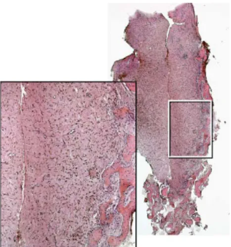
17 provisoire correspondant à un os primaire ; progressivement, cet os primaire devient organisé en un os spongieux qui devient mature avec ses travées d’os lamellaire minéralisé entre lesquelles se développe de la moelle osseuse. De l’os cortical se crée afin de recouvrir l’alvéole rendant au site d’extraction une solidité osseuse. 4.1.1.1. Remodelage des crêtes alvéolaires Une étude expérimentale conduite par Araujo chez 12 chiens en 2005 a permis d’évaluer d’une part la résorption des crêtes alvéolaires et d’autre part leur remodelage osseux dans les suites d’extractions dentaires réalisées sur la mandibule (1). L’évolution de la cicatrisation a été suivie morphologiquement et histologiquement pendant 1 à 8 semaines.
• Durant les semaines suivant l’extraction, un grand nombre d’ostéoclastes (flèches
blanches de la figure ci dessous) sont présents sur les versants externes des crêtes des parois alvéolaires tant vestibulaires que linguales. Cette forte activité ostéoclastique a pour résultat une réduction en hauteur et en épaisseur de ces crêtes. La réduction en hauteur de la crête alvéolaire est beaucoup plus prononcée du côté vestibulaire que du côté lingual.
Ref : 1 Après 1 semaine de cicatrisation. Crête alvéolaire linguale (a) et vestibulaire (b)
but the mucosa covering the sockets after 2, 4 and 8 weeks of healing were considered to be clinically healthy.
modest signs of inflammation. Thus, areas could be identified which were poor in their collagen content but rich in vascular structures and inflammat-ory cells.
The marginal portion of the lingual bone wall of the extraction socket was markedly wider than the corresponding portion of the buccal wall (Fig. 3). At Level A (Table 1) the lingual wall was 1.4! 0.2 mm (SD) wide while the corresponding width of the buccal wall was 0.6! 0.1 mm. The matching dimensions at Level B were 2.0! 0.3 and 1.3! 0.1 mm, respectively.
Both the buccal and lingual bone walls contained large numbers of
well-clasts) could occasionally be observed on the surface of this bundle bone. A severed periodontal ligament that included fibroblasts, distinctly orien-tated collagen fibers, vascular structures and inflammatory cells resided lateral to the bundle bone.
The crestal regions of the bone walls were comprised solely of bundle bone (Fig. 5a, b), the height of which was
more pronounced at the buccal
(X1 mm) than at the lingual wall (o0.5 mm). A large number of osteo-clasts were present on the outer surface of the crestal region of both bone walls. The internal portion of the extraction socket was occupied by coagulum, Fig. 3. Overview of the extraction site after
1 week of healing. Note the large amounts of provisional matrix and, in the center of the socket, remaining blood clot. BC, blood clot, B, buccal; L, lingual; PM, provisional matrix. H&E staining; original magnifica-tion " 16.
Table 1. Width of the bone tissue at the buccal and lingual walls of the extraction sites
1 week 2 weeks 4 weeks 8 weeks
buccal lingual buccal lingual buccal lingual buccal lingual
Level A 0.6 (0.1) 1.4 (0.2) 0.6 (0.1) 1.3 (0.3) 0.7 (0.2) 1.3 (0.1) 0.5 (0.1) 1.2 (0.1) Level B 1.3 (0.1) 2.0 (0.3) 1.1 (0.3) 1.9 (0.4) 1.1 (0.2) 1.6 (0.1) 1.1 (0.1) 1.7 (0.3) Level C 2.0 (0.0) 2.8 (0.8) 1.9 (0.4) 2.7 (0.7) 2.0 (0.4) 2.9 (0.3) 1.6 (0.1) 2.7 (0.5)
The measurements (mm) were made at different levels (A, B C; Fig. 1) and time intervals. Mean (SD).
Fig. 4. Higher magnification of outlined area in Fig. 3. The bundle bone covered the socket wall. Lateral to the bundle bone a severed periodontal ligament can be identi-fied. BB, bundle bone; PDL, severed periodontal ligament; original magnification
" 200.
Fig. 5. One week of healing. The crestal region of the lingual (a) and buccal (b) walls. The buccal bone crest is made exclusively of bundle bone while the lingual crest is comprised of a mixture of cortical bone and bundle bone. Note the presence of osteoclasts in the crestal regions of both walls (arrows). A, inner surface of the bone wall; BB, bundle bone; CB, cortical bone; O, outer surface of the bone wall; arrows, osteoclasts. H&E staining; original magnification " 50.