HAL Id: hal-03159234
https://hal.archives-ouvertes.fr/hal-03159234
Submitted on 4 Mar 2021
HAL is a multi-disciplinary open access archive for the deposit and dissemination of sci-entific research documents, whether they are pub-lished or not. The documents may come from teaching and research institutions in France or abroad, or from public or private research centers.
L’archive ouverte pluridisciplinaire HAL, est destinée au dépôt et à la diffusion de documents scientifiques de niveau recherche, publiés ou non, émanant des établissements d’enseignement et de recherche français ou étrangers, des laboratoires publics ou privés.
Effects of nickel oxide nanoparticles on survival,
reproduction, and oxidative stress biomarkers in the
marine calanoid copepod Centropages ponticus under
short-term exposure
Emna Djebbi, Delphine Bonnet, Olivier Pringault, Khawla Tlili, Mohamed
Néjib Daly Yahia
To cite this version:
Emna Djebbi, Delphine Bonnet, Olivier Pringault, Khawla Tlili, Mohamed Néjib Daly Yahia. Effects of nickel oxide nanoparticles on survival, reproduction, and oxidative stress biomarkers in the ma-rine calanoid copepod Centropages ponticus under short-term exposure. Environmental Science and Pollution Research, Springer Verlag, 2021, �10.1007/s11356-020-11781-1�. �hal-03159234�
1
Effects of nickel oxide nanoparticles on survival, reproduction, and
1
oxidative stress biomarkers in the marine calanoid copepod
2
Centropages ponticus under short-term exposure
3
Emna Djebbi1*, Delphine Bonnet2, Olivier Pringault3, Khawla Tlili4 and 4
Mohamed Néjib Daly Yahia5 5
1
Faculty of Sciences of Bizerte, Carthage University 7021, Zarzouna, LR18ES41 (Tunis El 6
Manar University), Tunis, 1082, Tunisia. 7
2
MARBEC, Univ. Montpellier, CNRS, Ifremer, IRD, Montpellier, France 8
3
Aix Marseille Univ, Université de Toulon, CNRS, IRD, MIO UM 110, 13288, Marseille, 9 France 10 11 12 4
LEBPAO, Faculty of Sciences of Tunis, University of Tunis, El Manar, FSB, 7021 Jarzouna, 13
Tunisia. 14
15 5
Department of Biological and Environmental Sciences, College of Arts and Sciences, 16
Qatar University. PO Box 2713, Doha, Qatar. 17
18 19 20
Environmental Science and Pollution Research
21 *Corresponding author 22 Emna Djebbi 23 Email: [email protected] 24 Tel: +21653257793 25 26 27
28 29 30 31 32 33 34 35
2
Abstract
36
Excessive use of nickel oxide nanoparticles (NiO NPs) in various industrial and
37
commercial products can lead to various negative effects in human and environmental
38
health due to their possible discharge into the environment. Nerveless, information
39
about their eco-toxicological effects on marine organisms are lacking. Copepods are
40
good eco-toxicological models because of their high sensitivity to environmental
41
stress and their key role in the marine food webs. In this study, 48 h acute tests were
42
conducted on the marine planktonic copepod Centropages ponticus to assess lethal
43
and sublethal toxicities of NiO NPs. The results revealed LC50 (48 h) of 4 mg/L for
44
adult females. Aggregation and settling of NiO NPs were observed at concentrations
45
≥2 mg/L. Exposure to sublethal concentrations (≥0.02 mg/L for 48 h) had significant
46
negative effects on reproductive success in C. ponticus. Egg production after 24 h and
47
48 h decreased by 32% and 46%, respectively at 0.02 mg/L and 70% and 82%,
48
respectively, at 2 mg/L. Hatching success was reduced by 70% and 79% at 2 mg/L for
49
eggs produced after 24 h and 48 h respectively. Antioxidant enzymatic activity
50
increased significantly with NiO NP concentration and time, indicating that NiO NPs
51
can cause oxidative stress in C. ponticus even under short-term exposure, while
52
significant inhibition of acetylcholinesterase activity at 2 mg/L after 48 h suggests
53
neurotoxic effects of NiO NPs.
54
Keywords Metal oxide nanoparticle, Copepods, Acute toxicity test,egg production,
55
egg hatching success, antioxidant enzymes.
3
Introduction
57
Nanoparticles (NPs) represent a serious environmental threat due to their toxicity and
58
persistence (Geissen et al. 2015). According to the British Standards Institution, these
59
compounds are defined as discrete pieces of material with all three external
60
dimensions in the nanoscale (size <100 nm). Size, shape, chemical composition and
61
structure of particle are main factors which determine the nanoparticle properties
62
(optical, electrical, and magnetic properties) (Khan et al. 2019). Based on their
63
chemical composition, NPs can be divided into various groups: e.g., metallic,
64
polymeric metallic oxide, semiconductor, and metallic oxide nanoparticles. Among
65
the metallic oxide group, nickel oxide nanoparticles (NiO NPs) have widespread use
66
in many industrial and commercial products (e.g., solar cells, lithium-ion batteries,
67
resistive random-access memory, and biosensors), due to their magnetic properties
68
(Tadik et al. 2015). There are also multiple medical and biotechnological applications
69
of NiO NPs, such as remediation of heavy metal contaminated water and
70
enhancement of biogas production from macrophytes of wastewater treatment plants
71
(Mahmoud et al. 2015; Salama et al. 2020). A recent study has reported an annual
72
production of NiO NPs of 20 tons only in the United states (Gomes et al. 2019) which
73
increases progressively the levels of NiO NPs exposure as stated by Avila-Arias et al.
74
(2019). The excessive use of NiO NPs leads to their possible widespread distribution
75
in terrestrial, estuarine, freshwater, and marine ecosystems through industrial and
76
domestic wastewaters and aerial deposition(Wiesner et al. 2006).Despite the number
77
of studies on NiO NPs, the environmental concentrations and distribution in field
78
environmental matrices (water, sediment, and biota) have not yet been explored.
79
Research investigating environmental nanoparticle concentrations in aquatic
80
ecosystems has shown that these compounds are detectable in aquatic matrices at
81
concentrations up to µg/L (Zhang et al. 2019). Overall, the behavior of nanoparticles
82
varies with the environmental matrix (water, sediment, soil, etc.). For NiO NPs,
83
aggregation behavior in seawater has been observed due to their low solubility and
84
high stability (Gong et al. 2016; Oukarroum et al. 2017).
85
There are numerous concerns about the risks posed by nanoparticle pollution to the
86
environment and human health (Ates et al. 2016; Oukarroum et al. 2017). Previous
87
laboratory studies have shown that NiO NPs have the potential to cross biological
88
membranes and to accumulate in target tissues, causing harmful toxic effects (Ates et
4
al. 2016; Oukarroum et al. 2017). In addition, Ates et al. (2016) showed that NiO NP
90
accumulation increases with exposure concentration and duration. Acute and chronic
91
exposures to NiO NPs have been associated with mortality effects on aquatic
92
organisms, depending on species sensitivity (Kovrižnych et al. 2014; Oukarroum et al.
93
2017). NiO NPs can limit cell division, as observed for the green algae Chlorella
94
vulgaris, and can also limit reproduction in zebrafish (Danio rerio) (Oukarroum et al.
95
2017). Moreover, NiO NPs have been shown to be associated with cytotoxic and
96
genotoxic effects even under acute exposure in algae, rats, and human cells (Ada et al.
97
2010; Oukarroum et al. 2017). The toxicity of nanoparticles is influenced by their
98
intrinsic characteristics (size, surface area, chemical composition, crystal structure,
99
shape, etc.) and their behavior (aggregation, dissolution) (Sukhanova et al. 2018).
100
Studies examining the toxicity mechanism of NiO NPs have found that they cause
101
toxicity in aquatic organisms by induction of oxidative stress through generation of
102
high levels of reactive oxygen species (i.e., hydrogen peroxide: H2O2, the superoxide
103
anion: O2-, and the hydroxyl radical: OH) and disruption of antioxidant enzymes such
104
as glutathione (GSH), as observed in algal cells (Manke et al. 2013; Oukarroum et al.
105
2017). Disruption of the antioxidant system can be associated with major risks, such
106
as neurotoxicity, lipid peroxidation, and apoptosis (Ada et al. 2010; Ates et al. 2016).
107
Despite the known toxicity of NiO NPs, marine species have received little attention,
108
as noted by Gong et al. (2019), in comparison with freshwater species (Oukarroum et
109
al. 2017; Sousa et al. 2018a, b). Thus, ecotoxicological studies of the effects of NiO
110
NPs on marine organisms are needed. Copepods may be interesting marine biological
111
models for such studies, considering their essential role in marine ecosystems as main
112
secondary producers in food webs and as vital links between phytoplankton and
113
higher trophic levels, to which they transfer carbon and energy (Jayalakshmi and
114
Santhanam 2019). In addition, copepods can represent a pathway for contaminant
115
entry for higher trophic links (Lauer and Bianchini 2010; Cailleaud et al. 2011).
116
Moreover, copepods are known as good bio-indicators of ecosystem pollution,
117
because of their sensitivity to several environmental disturbances (Bianchi et al. 2003;
118
Hussain et al. 2020). These organisms can develop adaptive responses to oxidative
119
stress using a sophisticated biochemical defense mechanism (Kim et al. 2014). The
120
marine calanoid copepod Centropages ponticus is a cosmopolite species and has been
121
used previously as an experimental model for ecotoxicological tests due to its short
5
life cycle, its small size, and its high sensitivity to contaminants such as metals
123
(Ensibi et al. 2015; Ensibi and Yahia 2017). In this study, we conducted acute toxicity
124
tests to investigate the lethal and sublethal effects of NiO NPs on C. ponticus under
125
short-term exposure. Indeed, acute tests mimic the effects of NPs under accidental and
126
short-term release into the environment. In addition, due to their lifestyle and ability
127
to swim, planktonic organisms, like copepods, should experience shorter time
128
exposure than benthic organisms for example. Moreover, due to their rapid
129
transformation behavior, NPs, such as NiO NPs, aggregate in marine waters and
130
deposit in the sediment which reduces their bioavailability for aquatic pelagic
131
organisms and raises the risk for benthic organisms (Miglietta et al. 2015). Finally,
132
results from acute tests allow selecting a range of NiO NPs concentrations to be used
133
in sublethal tests. Copepod reproductive traits and molecular responses (e.g., egg
134
production rate, hatching success, nauplius survival rate, biomarkers, and genomic
135
profiles) are reliable tools to evaluate the impacts of different types of environmental
136
contaminants (Zhou et al. 2018; Hussain et al. 2020). Egg production rate (EPR) and
137
hatching success (HS) were studied here as sub-lethal endpoints under different NiO
138
NPs concentrations. The response of the antioxidant defense system to NiO NPs was
139
investigated by determining the levels of some antioxidant enzymes such as catalase
140
(CAT), superoxide dismutase (SOD), and glutathione S-transferase (GST), and the
141
level of acetylcholinesterase (AChE) as a neurotoxicity indicator of oxidative damage.
142
This study is the first to focus on marine planktonic copepods, with the aim of
143
providing new knowledge to clarify the mechanisms behind NiO NPs toxicity in this
144
species.
145
Materials and methods
146
Copepod sampling and algal cultures 147
Copepods were captured in the Bizerte channel, north of Tunisia (37°16’1”N;
148
9°52’50”E), using a WP2 plankton net (200 µm mesh size) drawn by vertical tows.
149
Sampling was carried out during summer and autumn 2017. Copepods were placed in
150
cold boxes and immediately transported to the laboratory. Identification of C.
151
ponticus was performed based on their morphological and swimming characteristics,
152
using a Leica MZ125 stereomicroscope (Soler et al. 1988). Prior to exposure to
153
nanoparticles, adult females of C. ponticus were acclimated in seawater at 23 °C and
6
37 psu for 24 h, and fed the marine microalga Isochrysis galbana. Culture of this algal
155
species was performed in filtered seawater (0.2 µm) with f/2 enriched media at 20 °C
156
and on a 12-:12-h light: dark cycle. All glassware was soaked in 10% nitric acid
157
(NHO3) for 24 h and autoclaved at 121 °C for 15 min before use.
158
Nickel nanopowder preparation 159
Nickel oxide nanoparticles (product reference 637130) were purchased from Aldrich
160
Sigma Chemicals. A test solution was freshly prepared before the beginning of the
161
experiment and then diluted in filtered seawater. The NiO NPs were dispersed in
162
ultra-pure water to obtain a concentration of 100 mg/L and the suspension was then
163
sonicated for 20 min at 4 °C (40 KHz, 100W). The particle size of the NiO NPs was
164
≤50 nm with purity >99.8% and the particle morphology was cubic, according to the
165
supplier
166
(https://www.sigmaaldrich.com/catalog/product/aldrich/637130?lang=en®ion=TN). The
167
size of these nanoparticles (before its dispersion in ultrapure water or biological test
168
media (powder form) was confirmed (< 50 nm) by Sousa et al. (2018a) using
169
transmission electron microscopy. Right after dispersion in deionized water, Sousa et
170
al. (2018a) reported a main hydrodynamic size of 342 nm which exceeds 1000 nm
171
after 24 h and displays a negative zeta potential value.
172
Previous physical characterizations of other manufactured NiO NPs, different from
173
the one we used, in seawater showed that these NPs display negative zeta potential
174
value, non-uniform morphology and a size > 100 nm revealing their tendency to
175
aggregate in seawater (Gong et al. 2011; Ates et al. 2016; Gong et al. 2019).
176
Toxicity test procedures 177
Acute lethal toxicity test
178
To determine the lethal toxicity (median lethal concentration, LC50) of NiO NPs, 48-h
179
acute toxicity tests with adult females of C. ponticus were performed according to
180
ISO 14669:1999 revised 2015 and Zhou et al. (2016), with some modifications. All
181
glass beakers were washed with 10% NHO3 and rinsed three times with deionized
182
water. For each NiO NP concentration tested, triplicate groups of 10 adult females
183
were placed in 150 mL glass beakers containing 100 mL of the test solution. Five NiO
184
NP concentrations were tested: 0, 0.5, 1, 5, 10, and 50 mg/L. Photoperiod was
7
h light: dark cycle, salinity was 37 psu, and temperature was fixed at 23 °C. During
186
the experiment, the copepods were not fed. Mortality was recorded after 24 h and 48 h
187
of exposure. A copepod was considered dead if no swimming or appendage
188
movements were observed for 10 seconds.
189 190
Acute sub-lethal toxicity tests
191
Once the LC50 (48 h) for NiO NPs had been determined, acute sub-lethal toxicity tests
192
were performed using NiO NP concentrations below the LC50 value. Adult females of
193
C. ponticus were exposed to four sub-lethal concentrations: 50%, 5%, 0.5%, and
194
0.05% of the 48 h LC50 to study NiO NPs effect on reproduction and oxidative stress.
195 196
Reproduction
197
Reproductive toxicity tests were conducted on adult females according to Zhou et al.
198
(2016) with few modifications, and then EPR and HS were monitored over 48 h of
199
NiO NPs exposure. Three replicates were prepared for each concentration. Each
200
replicate comprised 3-4 ovigerous females of C. ponticus incubated in a 150 mL glass
201
beaker with 100 mL of filtered seawater. To prevent cannibalism, the females were
202
separated from their eggs using a Perspex® chamber with a 200 µm mesh false
203
bottom. The experiments were run at 37 psu and 23 °C in an incubator under a fixed
204
12-:12-h light:dark cycle. After 24 h, the females from each replicate were gently
205
transferred to a new glass beaker containing fresh test solution, to measure EPR after
206
48 h. The number of eggs spawned was counted every 24 h under a Leica binocular
207
microscope for each condition, and EPR was expressed as number of eggs spawned
208
per female per day (eggs/female/day). To monitor the effect of exposure to NiO NPs
209
on hatching success, the eggs spawned after 24 h and 48 h were then incubated for 48
210
h in the same solution of NiO NPs. The numbers of nauplii and unhatched eggs in
211
each treatment were determined at the end of the incubation period.
212 213
Oxidative stress
214
Prior to biomarker analysis, C. ponticus adult females were exposed for 48 h to the
215
same range of NiO NP concentrations as used in the reproduction tests. The
216
experiments were conducted in triplicate for each treatment and control, following the
217
protocol of Fossi et al. (2001). Each replicate received 150 individuals, which were
218
placed in a 1 L glass beaker filled with filtered seawater. The temperature and salinity
8
were 23 °C and 37 psu, respectively. A light regime of 12-:12-h light: dark cycle was
220
maintained. The experiment was run for 48 h and the organisms were not fed. A
221
sample of 75 copepods was taken every 24 h from each replicate per treatment and
222
immediately frozen at - 80 °C until measurement of biomarkers.
223 224 Biomarker analysis 225 Total protein 226
In order to determine the protein content in copepods, samples of C. ponticus were
227
homogenized at 4 °C using an ultratorax in a buffer solution containing tris base (20
228
mM), EDTA (1 mM), dithiothreitol (1 mM), sucrose (500 mM), and KCl (150 mM)
229
adjusted to pH 7.6 at a ratio of 1-4 (w/v) according to the protocol of Pinho et al.
230
(2005). The homogenate was then centrifuged at 9000g for 30 min at 4 °C. The
231
resulting supernatant was divided into several aliquots, which were stored at -80 °C
232
until total protein and biomarker analysis. Total proteins were quantified by the
233
Bradford method (Bradford 1976) using 50 µL sample protein and bovine serum
234
albumin as the standard. The assay was performed in a microplate reader using 96
235
well plates. Protein concentrations were expressed in mg/L after spectrophotometric
236
quantification at 595 nm.
237
Superoxide dismutase (SOD) activity measurement
238
Superoxide dismutase activity was measured based on the method of McCord and
239
Foridovich (1996). In brief, 1960 µL of Na2CO3/NaHCO3 (50 mM) (pH=10.2) were
240
combined with 10 µL of catalase bovine, 10 µL of sample, and 20 µL of epinephrine.
241
Total SOD activity was measured at 480 nm using a spectrophotometer, and was
242
expressed as SOD U/mg protein.
243
Catalase (CAT) activity measurement
244
Catalase is an enzyme that catalyzes the decomposition of H2O2 formed by SOD in
245
molecular water (H2O) and molecular oxygen (O2). Here, CAT activity was estimated
246
according to Clairborne (1985), where the rate of disappearance of H2O2 is monitored
247
by measuring the rate of decrease in H2O2 at 240 nm for 20 min. In brief, 50 µL of
248
sample were added to 50 µL of 30% H2O2 and 950 mL of 75 mM phosphate buffer at
249
pH=7. The results were expressed as CAT U/mg protein.
9
Glutathione S-transferase (GST) activity measurement
251
Glutathionine activity was determined by measuring the conjugation of
1-chloro-2-4-252
dinitrobenzene (CDNB) with GSH at 37 °C. The rate of GSH decrease is directly
253
proportional to the level of GST activity in the sample. A total of 50 µL of sample
254
was added to 50 µL of CDNB and 100 µL glutathione in 100 mM phosphate buffer
255
(100 mM) at pH 7.4 (Habig et al. 1974). The results were expressed as GST
256 nmol/min/mg protein. 257 258 Acetylcholinesterase measurement 259
Measurement of AChE activity was carried out according to Ellman et al. (1961),
260
with 3 mM acetylthiocholine iodide (AcSCh) as substrate and 0.1 Mm
261
dithiobisnitrobenzoate (DTNB) as reagent, at a controlled temperature of 20 °C.
262
Absorbance was recorded at 410 nm for 15 min using 20 µL of sample according to
263
Fossi et al. (2001). AChE activity was expressed in nmol/min/mg protein.
264 265
Data analysis 266
The results from the lethal and sub-lethal tests are presented as means ± their standard
267
deviation. Statistical analyses of the data were performed using SPSS (version 18).
268
The effects of the concentrations tested in each experiment were compared with those
269
of controls using two-way ANOVA (time and treatment), followed by post-hoc
270
Tukey’s test. Data were log-transformed, if necessary, to meet the ANOVA
271
assumption of normality and variance homogeneity. The level of significance was set
272
at p<0.05. The median lethal concentration (LC50) was calculated by probit analysis
273 using SPSS. 274 275
Results
276Acute lethal toxicity test and aggregation behavior 277
There was no mortality in the controls during the whole incubation period. The results
278
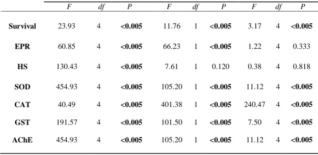
of two-way ANOVA indicated a strong influence of both time and concentration on
279
survival rate (p<0.05) (Table 1). They also showed a significant interaction between
280
exposure time and NiO NP concentrations (p<0.05). The survival rate at
281
concentrations ranging from 0.5 to 1 mg/L of NiO NPs was ≥ 90 % over the exposure
10
period. After 24 h, the survival rate was > 90% at a concentration of 5 mg/L and <50
283
% at a concentration ≥10 mg/L relative to the control groups (p<0.05). After 48 h of
284
incubation, NiO NPs at concentrations greater than 1 mg/L induced a significant
285
(p<0.05) reduction in survival rate, which decreased from 90±1.7 % at an NiO NPs
286
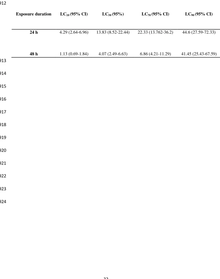
concentration of 1 mg/L to 0 % at 50 mg/L. The calculated LC50 value for 24 h and 48
287
h of exposure was 13.83 ± 2.3 mg/L and 4.07 ± 0.5 mg/L, respectively (Table 2).
288
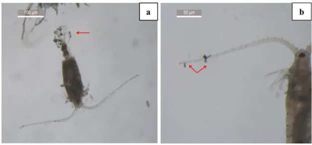
During the acute toxicity tests, black aggregates visible to the naked eye appeared at
289
the bottom of the beaker at NiO NPs concentrations ranging from 2 to 50 mg/L after
290
48 h of incubation. Microscope observations revealed similar aggregates of NiO NPs
291
on the exoskeleton of C. ponticus (Fig. 1a, b).
292 293
Effect on reproductive success 294
The effects of NiO NPs on reproductive success in C. ponticus were assessed by
295
monitoring EPR over 48 h and then determining HS (Fig. 2). For both parameters, no
296
significant (p>0.05) effect was observed at the lowest concentration of NiO NPs
297
(0.002 mg/L) over 48 h.
298
A significant (p<0.05) influence of time exposure on EPR was observed for all NiO
299
NPs treatments (Table 1). However, no significant (p>0.05) interaction was observed
300
between time exposure and NiO NPs concentration. For the control groups, EPR
301
varied between 11.3±0.6 eggs female-1 day-1 at 24 h and 8.3±1.1 eggs/female/day at
302
48 h. After 24 h, a significant reduction in EPR (p<0.05) was observed, to 7.5±1.5
303
eggs/female/day at 0.02 mg/L and 3.3±0.3 eggs/female/day at 2 mg/L of NiO NPs
304
(Fig. 2a). After 48 h, EPR showed a continuous decrease, reaching 1.5±0.2
305
eggs/female/day at the highest concentration (2 mg/L).
306
There was no significant effect of time exposure (p>0.05) on HS for the different
307
concentrations tested (Table 1). However, HS for eggs spawned after 24 h and 48 h
308
decreased significantly (p<0.05) with increasing NiO NPs concentrations ≥0.02 mg/L.
309
For the control groups, HS varied between 85.0±2.1% for eggs spawned after 24 h
310
and 81.3±4.0% for eggs spawned after 48 h. The lowest values of HS, observed at the
311
highest NiO NPs concentration tested (2 mg/L), were 24±7 % and 16±2 % for eggs
312
spawned after 24 h and 48 h, respectively (Fig. 2 b). For this endpoint measurement,
313
no significant interaction was observed between time exposure and NiO NPs
314
concentration (Table 1).
315 316
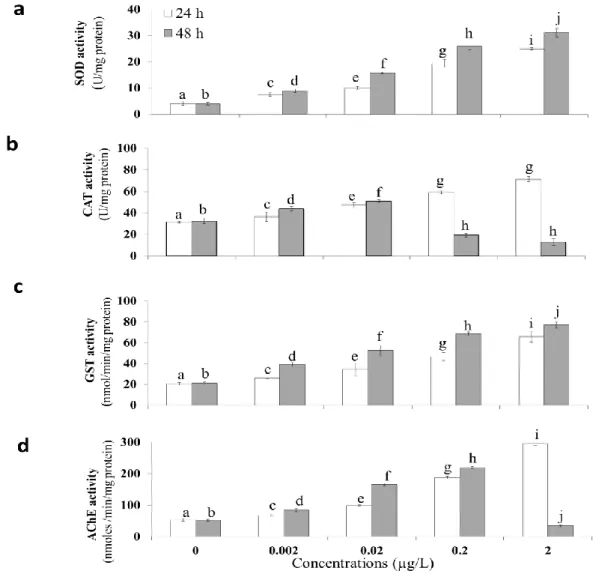
11 Effect of NiO NPs on oxidative stress 317
In order to assess the mechanisms behind NiO NPs toxicity at cellular level, some
318
oxidative enzymes (CAT, SOD, GST) and the neurotoxicity biomarker AChE were
319
measured in C. ponticus after exposure to 50%, 5%, 0.5%, and 0.05% of LC50 (48 h)
320
(Fig. 3). The activity of all enzymes analyzed was significantly (p<0.05) impacted by
321
time exposure and NiO NPs concentrations (Table 1). In addition, a significant
322
interactive effect was observed for exposure time and NiO NPs concentration
323 (p<0.05) (Table 1). 324 325 SOD activity 326
The level of SOD activity in C. ponticus after NiO NP exposure showed significant
327
stimulation (p<0.05) with increasing exposure time and NiO NP concentrations (Fig.
328
3a). The maximum SOD activity was 31±0.7 U/mg protein and was observed for the
329
highest NiO NPs concentration (2 mg/L) after 48 h. For the control groups, SOD
330
activity was 4.0±0.3 U/mg protein.
331
CAT activity
332
After 24 h of incubation, a significant (p<0.05) difference was observed between the
333
control groups and copepods exposed to NiO NPs concentrations (Fig. 3b). The level
334
of CAT activity was 71.3±2.5 U/mg protein at the highest concentration (2 mg/L),
335
compared with 31.4±0.8 U/mg protein in control groups. After 48 h, CAT activity
336
decreased significantly (p<0.05) at 0.2 mg/L (19.2±1.7 U/mg protein) and 2 mg/L
337
(12.8±3.0 U/mg protein) compared with the control groups (32.3±2.5 U/mg protein).
338
GST activity
339
After 24 h, exposure to NiO NPs led to a significant (p<0.05) increase in GST activity
340
in a dose-dependent manner (Fig. 3c). At 2 mg/L, GST activity reached 65.8±5.2
341
nmol/min/mg protein, in comparison with 20.4±1.7 nmol/min/mg protein for the
342
control groups. A significantly (p<0.05) higher level of GST activity was also
343
observed after 48 h relative to the values measured after 24 h of incubation.
344 345
Acetylcholinesterase activity
346
Exposure to NiO NPs for 24 h resulted in significant (p<0.05) stimulation of AChE
347
activity at all concentrations, with a minimum activity level of 67.4±4.4 nmol/min/mg
12
protein at 0.002 mg/L and a maximum of 296.4±6.4 nmol/min/mg protein at 2 mg/L
349
(Fig. 3d). After 48 h at NiO NPs concentrations ranging from 0.002 to 0.2 mg/L,
350
AChE activity was higher than the levels observed after 24 h. However, at the highest
351
NiO NPs concentration tested (2 mg/L), AChE activity was significantly reduced
352
(34.1±3.3 nmol/min/mg protein) in comparison with the control group (52.37.3±2.7
353 nmol/min/mg protein) (p<0.05). 354 355
Discussion
356Acute toxicity test and aggregation behavior of NiO NPs 357
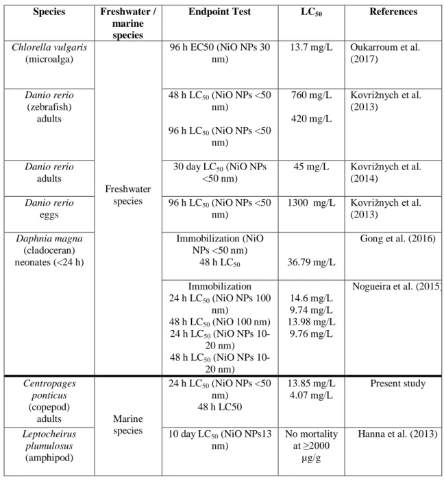
The results demonstrated the lethal acute toxicity of NiO NPs on adult copepod C.
358
ponticus under short-term exposure (up to 48 h), with an LC50 value of 13.83 mg/L
359
and 4.07 mg/L for 24 and 48 h, respectively. To our knowledge, this is the first study
360
to report on the lethal effect NiO NPs on marine planktonic copepods. Available data
361
regarding the lethal effect of NiO NPs on marine organisms in general are scarce and
362
the majority of previous research has focused on freshwater species (Table 3). To our
363
knowledge, only one previous study has investigated the mortality effect of NiO NPs
364
on the marine amphipod Leptocheirus plumulosus (Hanna et al. 2013). The results
365
from this study indicated that NiO NPs, tested at concentrations from 500 to 2000
366
µg/g dry weight, provoked no mortality in the amphipod over 10 days (Hanna et al.
367
2013).
368
In freshwater crustacean species including Daphnia magna, Gong et al. (2016)
369
observed a nine-fold higher value of LC50 (48 h) (36.79 mg/L for neonates) than found
370
here for C. ponticus (Table 3). However, similar values of LC50 (24 h) as the 48 h
371
LC50 found in the present study for C. ponticus were reported by Nogueira et al.
372
(2015) for the same life stage of D. magna exposed to NiO NPs (Table 3). Very high
373
LC50 values for NiO NPs have been recorded in other biological models at higher
374
trophic levels, such as in adult zebrafish under acute (LC50 (48 h) = 760 mg/L) and
375
chronic exposure (LC50 (30 days) = 45 mg/L) (Kovrižnych et al. 2013, 2014). Other
376
studies on the toxic effects of NiO NPs have shown that exposure of the freshwater
377
alga Chlorella vulgaris to a concentration of 13.7 mg/L, i.e., close to our LC50 (24 h),
378
reduces the viability of exposed cells to 50% after 72 h (Oukarroum et al. 2017).
379
Kanold et al. (2016) did not observe mortality effects of nickel nanoparticles (Ni NPs)
13
on the early life stage of the sea urchin Paracentrotus lividus exposed to
381
concentrations up to 3 mg/L of Ni NPs after 48 h, which correspond to 88 µg/L of
382
Ni2+.
383
In this study, NiO NPs displayed aggregation in seawater during the acute lethal and
384
sublethal tests at concentrations ranging from 2 to 50 mg/L, after 48 h. This in line
385
with findings observed by Sousa et al. (2018b) and Zhou et al. (2016), who observed
386
similar behavior for NiO NPs and Ni NPs at concentrations ranging from 4 to 10
387
mg/L after 72 h. Like other nanoparticles (titanium oxide (TiO2) NPs, silver (Ag)
388
NPs), NiO NPs tend to aggregate independently of the aqueous medium (freshwater,
389
seawater, deionized water, etc.), forming larger aggregates in seawater than in
390
freshwater due to seawater characteristics favoring aggregation (Ates et al. 2016). The
391
agglomerates increased in size with time varying from few hundred nanometers to
392
several microns both in seawater or freshwater (Wang et al. 2014; Ates et al. 2016;
393
Sousa et al. 2018a). Additionally, NiO NP aggregation does not exclude the
394
possibility of release of free dissolved Nickel ions (Ni2+) in the aqueous medium test,
395
which may also impact the physiology of the copepod (Gong et al. 2011; Sousa et al.
396
2018b). Unfortunately, NiO NPs dissolution behavior was not examined here to get a
397
clear idea of the amount of dissolved Ni2+ released into seawater test medium that can
398
act in NiO NPS toxicity. Previous studies indicated a low solubility of these NPs in an
399
aqueous medium test which is higher in freshwater (7%) than in seawater (0.14 %)
400
under short-term periods (Ates et al. 2016; Oukarroum et al. 2017; Gong et al. 2011,
401
2019). The availability of Ni2+ released into the seawater from NPs increased with
402
time and NiO NPs concentrations (Hanna et al. 2013; Zhou et al. 2016; Oukarroum et
403
al. 2017; Gong et al. 2011, 2019).Hanna et al. (2013) reported a slow dissolution of
404
NiO NPs under a long-term period, where NPs dissolved over several weeks in
405
seawater. The same authors reported a solubility of 21 % of NiO NPs after 28 days for
406
a concentration of 10 mg/L of NiO NPs which is higher than solubility observed in
407
others studies under short-term test (Gong et al. 2011, 2019). For 50 mg/L of NiO
408
NPs, a concentration which causes 100 % lethality in copepods after 48 h, a previous
409
study has reported a concentration of 110 µg/L of Ni2+ released in seawater after 96 h
410
(Gong et al. 2011). Copepods are therefore exposed to both particle and dissolved
411
forms of NiO NPs and toxicity can be linked to Ni2+ released ion from NPs(Sousa et
412
al. 2018b),to the particles themselves (Capasso et al. 2014)or to the combined effect
14
of Ni2+ and NPs aggregates (Gong et al. 2011, 2019). Further work needs to be done
414
to better explain the chemical mechanism involved in NiO NPs copepods toxicity
415
effects and to determine the real NiO NPs concentrations.
416
Settling of NiO NPs, as observed on the exoskeleton of C. ponticus in this study, has
417
also been observed for Chlorella vulgaris and the brine shrimp Artemia salina (Gong
418
et al. 2011; Ates et al. 2016). Biological surface coating by NPs, is considered as the
419
main mechanism of toxicity for no-ion releasing NPs or low soluble NPs such as NiO
420
NPs, deemed to alter the swimming behavior of aquatic organisms which could lead
421
to negative impacts in aquatic ecosystems (Gong et al. 2011; Noss et al. 2013;
422
Oukarroum et al. 2017; Gong et al. 2019). 423
According to Sukhanova et al. (2018), the lethal toxicity effect of NiO NPs on aquatic
424
organisms depends on the biological model (size, life stage, weight) and on
425
nanoparticle characteristics (size, shape, behavior). To our knowledge, the LC50 (48 h)
426
observed in the present study is lower than any LC50 value reported previously in the
427
literature, which suggests that copepods can be a good bio-indicator of the toxic
428
effects of NiO NPs. Comparison of our LC50 values with NiO NPs environmental
429
concentrations is not possible due to the lack of data in aquatic environments.
430
However, it is worth noting that mortality effect of NiO NPs on of C. ponticus was
431
observed at concentrations that were much higher than predicted environmental
432
concentrations, which do not exceed the µg range (Zhang et al. 2019). Thus, the
433
concentrations reported in this study may reflect the lethal effect upon accidental
434
exposure to NiO NPs.
435
Effect on reproductive traits 436
Our results showed that NiO NPs did not impact the reproductive performance (EPR
437
and HS) of C. ponticus at a concentration of 0.002 mg/L during 48 h of exposure.
438
However, EPR and HS were significantly affected at NiO NP concentrations ≥0.02
439
mg/L. Previous studies have also found that metallic nanoparticles (e.g., zinc oxide
440
(ZnO) NPs and Ni NPs) are associated with impairment of copepod reproductive traits
441
(Zhou et al. 2016; Parlapiano et al. 2017). Time exposure and concentration had a
442
strong influence on EPR (Table 1), which decreased with increasing NiO NPs
443
concentration and time exposure (by 32% and 46%, respectively, at 0.02 mg/L and by
444
70% and 82%, respectively, at 2 mg/L after 24 h and 48 h). Similar effects have been
15
observed in the crustacean species D. magna, with NiO NPs markedly reducing
446
offspring production at concentrations ≥0.2 mg/L after 21 days (Gong et al. 2016).
447
Despite the difference in incubation duration and probably frequent renewal of test
448
solution in that study, reproduction success in C. ponticus was impacted at much
449
lower concentrations (≥0.02 mg/L) than those tested by Gong et al. (2016). Likewise,
450
Zhou et al. (2016) did not observe any effect on EPR in the marine calanoid copepod
451
Acartia tonsa at Ni NPs concentrations up to 0.01 mg/L over 4 days.
452
The results in the present study showed significant concentration-related trends in HS,
453
which decreased with increasing NiO NPs concentrations. At the highest
454
concentration tested here (2 mg/L), HS was reduced by 70% and 79%, respectively,
455
for eggs produced after 24 h and 48 h. Zhou et al. (2016) also observed a negative
456
effect of Ni NPs on HS of the copepod A. tonsa at a concentration of 17 mg/L, where
457
only 9% of incubated eggs hatched. Similar responses to NiO NPs have been
458
observed in zebrafish, including a delay in egg hatching and a reduction in HS at
459
concentrations ranging from 100 to 800 mg/L (Kovrižnych et al. 2013). Our results
460
provide evidence of the impact of NiO NPs on copepod reproductive success at much
461
lower concentrations (0.02 mg/L) than those tested in previous studies on marine
462
organisms. To our knowledge, only one study has reported similar negative effects, on
463
reproduction of the freshwater crustacean D. magna, at similar NiO NPs
464
concentrations (0.045-0.14 mg/L) to those tested in the present study (Nogueira et al.
465
2015). Regarding NiO NPs solubility at the sublethal concentrations tested here
466
(0.002-2 mg/l) and according to our knowledge, there are no data in the literature on
467
NiO NPs solubility in seawater at the same concentrations range. The study of Sousa
468
et al. (2018b) reported a concentration of Ni2+of 250 µg/L released in deionized water
469
after 72 h from a concentration of 1.6 mg/L.Based on this finding and considering the
470
low solubility of NiO NPs in seawater in comparison to freshwater, we can suggest
471
that the concentration of Ni2+ released in seawater at concentration of 2 mg/L, under
472
our experimental conditions, could be less or close to 250 µg/L. This concentration is
473
well higher than the highest levels of nickel which are not expected to pose a
474
significant risk in species in saltwater under acute (74 µg/L) and chronic (8.3 µg/L)
475
exposure as fixed by US EPA Aquatic Life Criteria Water Quality Standards (EPA
476
2009). 477
16
Interestingly, the NiO NPs concentrations causing toxic effects on reproduction
478
physiology in C. ponticus and D. magna are close to those measured in contaminated
479
ecosystems, which are suspected to occur at µg range, suggesting that
480
environmentally realistic concentrations of NiO NPs are likely to impact reproduction
481
of both species. Reproduction is a crucial biological function in organisms to maintain
482
existence of species. Impairment of reproduction in copepods by NiO NPs, as
483
observed in this study, can result in alteration of marine community structure and in
484
modification of the food trophic web.
485
Oxidative stress 486
In order to understand the mechanisms of NiO NPs toxicity at cellular level, changes
487
in the activity of antioxidant enzymes and a neurotoxicity biomarker in C. ponticus
488
under short-term NiO NPs exposure were investigated. The results indicated a
489
significant modulation of the first-defense antioxidant enzymes SOD and CAT in C.
490
ponticus in response to sublethal exposure to NiO NPs. The stimulation of antioxidant
491
enzymes was possibly a defense mechanism for coping with the oxidative stress
492
generated by NiO NPs. This is in line with previous studies showing ability of
493
nanoparticles to generate oxidative stress in aquatic organisms under short-term
494
exposure (e.g. the brine shrimp Artemia salina, the copepod Calanus finmarchirus,
495
the fish Nile tilapia, etc.) (Abdel-khaled et al. 2015; Ates et al. 2016; Farkas et al.
496
2020).
497
Our results showed that antioxidant enzyme levels in C. ponticus were significantly
498
influenced by time exposure and NiO NPs concentration. Expression of SOD and
499
CAT enzymes exhibited similar patterns, with an increasing trend after 24 h that was
500
probably linked to production of reactive oxygen species (ROS), namely O2 and
501
H2O2,in C. ponticusas a consequence of NiO NPs exposure, as previously observed
502
in various freshwater microalgae and plants (e.g., Chlorella vulgaris,
503
Pseudokirchneriella subcapitata, Lemna gibba) (Oukarroum et al. 2015; Oukarroum
504
et al. 2017; Sousa et al. 2018b; Gong et al. 2019). After 48 h, SOD activity showed a
505
similar trend as at 24 h, while there was a significant reduction in CAT enzymatic
506
activity at the two highest concentrations of NiO NPs (0.2 and 2 mg/L). This decrease
507
might be linked to high production of H2O2 by SOD and limited capacity of CAT to
508
degrade it (Ighodaro and Akinloye 2018). Such antioxidant stress response was also
17
reported in the marine copepods Tigriopus japonicus exposed to sunscreens
510
containing Zinc oxide nanoparticle (ZnO NPs); with high ROS levels observed after
511
96 h at environmentally realistic concentrations of ZnO NPs(Wong et al. 2020). 512
In combination with the antioxidant response, exposure to NiO NPs also induced
513
activation of the multifunctional GST enzyme over the whole exposure period (48 h).
514
In line with our results, Farkas et al. (2020) reported that exposure to silver
515
nanoparticles (Ag NPs) triggered an increase in gene expressions of antioxidant
516
enzymes GST in the copepod Calanus finmarchirus after 96 h. GST enzyme plays a
517
key role in biotransformation of endogenous and exogenous toxic compounds present
518
in the cell and is also recognized as an indicator of lipid damage (Prione et al. 2016;
519
Dasari et al. 2017). NiO NPs has previously been reported as inductor of lipids
520
peroxidation at concentrations of 1 to 50 mg/L in Artemia salina and Gracilaria
521
lemaneiformis (Han et al. 2012; Ates et al. 2016). Results of the present study are in
522
accordance with the results observed for the copepod Eucyclop sp. exposed to
523
titanium dioxide nanoparticle (TiO2) combined to lead (Pb), where Glutathione
524
peroxidase (GPx), Glutathione reductase (GR), and CAT significantly increased after
525
contaminant accumulation confirming that the exposed copepods had suffered from
526
oxidative stress triggered by TiO2 and Pb(Matouke and Mustapha, 2018).
527
Neurotoxicity is one manifestation of oxidative stress damage induced by chemicals.
528
The enzyme AChE hydrolyzes the neurotransmitter acetylcholine (Ach) to acetate and
529
choline, in order to prevent constant stimulation of the synapse. The present study
530
showed a stimulating effect on AChE activity of NiO NPs (0.002-2 mg/L) after 24 h,
531
but we observed a significant reduction in AChE activity at 2 mg/L of NiO NPs after
532
48 h. Under metals exposure, AChE exhibited the same pattern in living organisms,
533
depending on time exposure; AChE increased during the earliest hours of metal
534
exposure and then decreased with time (Bainy et al. 2006; Emadeldeen 2014; Ensibi
535
and Yahia, 2017).
536
The high level of AChE activity in C. ponticus observed in this study may be related
537
to interactions between NiO NPs and acetylcholine receptors causing accumulation of
538
free unbound acetylcholine at cholinergic receptor sites. Some authors considered the
539
excess production of AChE activity as a compensatory mechanism related to an initial
540
reduction in this enzyme by pollutants (Bainy et al. 2006; Ferreira et al. 2012).
18
Inhibition of AChE activity is commonly used as an indicator of neurotoxicity in
542
living organisms such as copepods and cladoceran species under exposure to metal
543
and pesticide (Forget et al. 2003; Cailleaud et al. 2007; Ensibi et Yahia. 2017).
544
Inactivation of AChE activity at a concentration of 2 mg/L suggests that NiO NPs can
545
affect the brain of aquatic species and cause neurotoxicity through disturbance of
546
cholinergic neurotransmission. Similar inhibitory effects of metal nanoparticles
547
(silicon dioxide (SiO2), TiO2, aluminum oxide (Al2O3), and aluminum (Al) NPs) on
548
AChE have been observed previously and are reported to depend on nanoparticle
549
concentration (Wang et al. 2009). This reduction can be explained by the excess
550
production of AChE in C. ponticus ceased at a NiO NPs concentration of 2 mg/L.
551
Previous research has suggested, not confirmed, that modulation of AChE enzyme in
552
copepods can result in a disorder in swimming behavior, tetany, and paralysis(Forget
553
et al. 2003; Ferreira et al. 2012).Swimming behavior response of aquatic organisms,
554
such as fish and cladocerans (Daphnia sp.), under contaminant exposure, has been
555
reported to be strongly related to AChE activity. Further, it increases as the levels of
556
AChE enzyme increase while the opposite occurs when AChE enzyme levels
557
decrease, impacting, therefore, the survival of living organisms (Bonansea et al. 2016;
558
Ren and al. 2017). Therefore, alteration of swimming movement in copepods may
559
have severe impacts in the copepods community which affect other communities of
560
the ecosystem because it is associated with numerous vital functions like perception
561
and acquisition of food, mating and reproduction, predator-prey interaction,
562
respiration rate, and social behavior as stated by Mazzocchi and Paffenhofer (1999).
563
Changes in these biomarkers reflected the ability of NiO NPs to trigger oxidative
564
stress and highlighted the significant contribution of time exposure and NiO NPs
565
concentration in the response of C. ponticus to oxidative stress. Our results provide
566
evidence that oxidative stress is one of the main mechanisms of nanoparticle toxicity
567
in copepods under short-term exposure, as previously reported for other species
568
(Sousa et al. 2018b; Gong et al. 2019).
569 570 571 572 573
19
Conclusions
574
This is the first study to examine the potential toxic effects of NiO NPs on a marine
575
planktonic calanoid copepod. A LC50 (48 h) of around 4 mg/L was found for adult
576
females of C. ponticus, a concentration that can exceed the environmental
577
concentrations in contaminated environments. Results from sublethal tests clearly
578
indicated that NiO NPs caused negative effects on reproductive performance in C.
579
ponticus and led to oxidative stress at concentrations (0.02-2 mg/L), which are close
580
to environmentally relevant levels. C. ponticus resisted to the oxidative stress
581
generated by NiO NPs by increasing the levels of antioxidant enzymes in a
582
concentration and time-dependent manner. These results provide a better
583
understanding of the ecotoxicological risks of NiO NPs for marine organisms and
584
confirm that the calanoid copepod C. ponticus is a suitable marine model organism for
585
ecotoxicology studies. Future studies are required to determine the effects of NiO NPs
586
on the entire copepod life cycle, including impacts on juvenile development and
587
growth, and under long-term exposure. 588
589 590
20
Acknowledgements
591
The authors would like to thank the members of the Laboratory for Environmental
592
Biomonitoring (Faculty of Sciences of Bizerte; Tunisia), especially Professor
593
Mohamed Dalleli and Ms Wiem Saidani, for their help in biomarker analysis. We are
594
also grateful to the Institute of Fishing and Aquaculture (ISPA) (Bizerte, Tunisia),
595
especially Professor Mohammed Chalghaf, for algae supply. Special thanks to
596
Professor Abdelhak Othmani for providing nanoparticles. We would like to thank
597
Mary McAfee for the English correction and improvement.
598
This research is part of the PhD work of Ms Emna Djebbi, which is co-funded by the
599
University of Carthage (Bourse d’Alternance) and IRD-Tunisia (Institut de Recherche
600
pour le Développement).
21
References
602
Abdel-Khalek AA, Kadry M, Hamed A, Marie MA (2015) Ecotoxicological impacts of zinc metal in 603
comparison to its nanoparticles in Nile tilapia; Oreochromis niloticus. J Basic Appl Zool 72:113-125. 604
https://doi.org/10.1016/j.jobaz.2015.08.003. 605
Ada K, Turk M, Oguztuzun S, Kilic M, Demirel M, Tandogan N, Ersayar E, Latif O (2010) 606
Cytotoxicity and apoptotic effects of nickel oxide nanoparticles in cultured HeLa cells. Folia 607
Histochem Cytobiol 48:524-529. https://doi.org/10.2478/v10042-010-0045-8. Ates M, Demir V, 608
Arslan Z, Camas M, Celik F (2016) Toxicity of engineered nickel oxide and cobalt oxide nanoparticles 609
to Artemia salina in seawater. Water Air Soil Pollut 227:70.
https://doi.org/10.1007/s11270-016-2771-610
9.
611
Avila-Arias H, Loring F. Nies LF, Gray MB, Turco RF (2019) Impacts of molybdenum, nickel, and 612
lithium oxide nanomaterials on soil activity and microbial community structure. Sci Total Environ 652: 613
202-211. https://doi.org/10.1016/j.scitotenv.2018.10.189. 614
Bainy ACD, Medeiros MHG, Di Mascio P, de Almeida EA (2006) In vivo effects of metals on the 615
acetylcholinesterase activity of the Perna perna mussel’s digestive gland. Biotemas 19:35–39. 616
https://doi.org/10.5007/%X. 617
Bradford MM (1976) A rapid and sensitive method for the quantitation of microgram quantities of 618
Protein utilizing the principle of protein-dye binding. Anal Biochem 72:248-254. 619
https://doi.org/10.1016/0003-2697(76)90527-3. 620
Bianchi F, Acri F, Bernardi Aubry F, Berton A, Boldrin A, Camatti E, Cassin D, Comaschi A (2003) 621
Can plankton communities be considered as bio-indicators of water quality in the Lagoon of Venice? 622
Mar Bull Poll 46:964–971. https://doi.org/10.1016/S0025-326X(03)00111-5. 623
BonanseaRI, Wunderlin DA, Amé MV (2016) Behavioral swimming effects and acetylcholinesterase 624
activity changes in Jenynsia multidentata exposed to chlorpyrifos and cypermethrin individually and in 625
mixtures. Ecotox and Environ Safe 129:311-319. https://doi.org/10.1016/j.ecoenv.2016.03.043.
626
Cailleaud K, Maillet G, Budzinski H, Souissi S, Forget JL (2007) Effects of salinity and temperature 627
on the expression of enzymatic biomarkers in Eurytemora affinis (Calanoida, Copepoda). Comp 628
Biochem Phys A 174:841-849. https://doi.org/10.1016/j.cbpa.2006.09.012. 629
Cailleaud K, Budzinski H, Lardy S, Augagneur S, Barka S, Souissi S, Forget-Leray J (2011) Uptake 630
and elimination, and effect of estrogen-like contaminants in estuarine copepods: An experimental 631
study. Environ Sci Pollut R 18:226-236. https://doi.org/10.1007/s11356-010-0355-6. 632
Capasso L, Camatini M, Gualtieri M (2014) Nickel oxide nanoparticles induce inflammation and 633
genotoxic effect in lung epithelial cells. Toxicol Lett 226: 28–34. 634
https://doi.org/10.1016/j.toxlet.2014.01.040. 635
Claiborne A (1985) Catalase activity. In: Greenwald RA (ed) Handbook of methods for oxygen Radical 636
research. CRC Press, Boca Raton, Florida, pp 283–284. 637
Dasari S, Ganjayi MS, Oruganti L, Balaji H, Meriga B (2017) Glutathione s-transferases detoxify 638
endogenous and exogenous toxic agents-mini review. J Dairy Vet Anim Res 5:157-159. 639
https://doi.org/10.15406/jdvar.2017.05.00154. 640
Ellman GL, Courtney KD, Andres VJR, Feather-Stone RM (1961) A new and rapid colorimetric 641
determination of acetylcholinesterase activity. Biochem Pharmacol 7:88-90. 642
https://doi.org/10.1016/0006-2952(61)90145-9. 643
22
Emadeldeen HM (2014) Biochemical response of the cyclopoida copepod Apocyclops 644
borneoensis exposed to nickel. Jordan J Biol Sci 7:41-47. https://doi.org/10.12816/0008212. 645
Ensibi C, Pringault O, Hasnnaoui W, Yahia MND (2015) Effects of cadmium exposure on 646
reproduction and survival of the planktonic copepod Centropages ponticus. J Marine Sci Res Dev 647
5:159. https://doi.org/10.4172/2155-9910.100015 9
648
Ensibi C, Yahia MND (2017) Toxicity assessment of cadmium chloride on planktonic copepods 649
Centropages ponticus using biochemical markers. Toxicol Rep 4:83–88. 650
https://doi.org/10.1016/j.toxrep.2017.01.005. 651
EPA U (2009) National recommended water quality criteria. United States Environmental Protection 652
Agency, Office of Water, Office of Science and Technology. 653
Farkas J, Cappadona V, Olsen AJ, Hansen BH, Posch W, Ciesielski TM, Goodhead R, Wilflingseder 654
D, Blatzer M, Altin D, Moger J, Booth AM, Jenssen BM (2020) Combined effects of exposure to 655
engineered silver nanoparticles and the water-soluble fraction of crude oil in the marine copepod 656
Calanus finmarchicus. Aquatic Toxicology 277: 105582. 657
https://doi.org/10.1016/j.aquatox.2020.105582. 658
Ferreira G, Carvalho-Silva M, Gonçalves C, Silva J, Scaini G, Ghedim F, Deroza P, Zugno A, Pereira 659
T, Oliveira G, Kist L, Bogo M, Schuck P, Ferreira G, Streck E (2012) L-Tyrosine administration 660
increases acetylcholinesterase activity in rats. Neurochem Int 61:1370-1374. 661
https://doi.org/10.1016/j.neuint.2012.09.017.
662
Fossi MC, Minutoli R, Guglielmo L (2001) Preliminary results of biomarker responses in zooplankton 663
of brackish environments. Mar Pollut Bull 42:745-748.
https://doi.org/10.1016/S0025-326X(00)00214-664
9. 665
Fossi MC, Urban J, Maltese S, Mazzi L, Coppola D, Casini S, Panigada S, Lauriano G, Niño C, Rojas-666
Bracho L, Marsili L (2010) First assessment of biomarker responses and contaminant levels in 667
Balaenoptera Edeni skin biopsies of gulf of California (Mexico). J Plankton Res 32:1227–1229.
668
Forget LJ, Beliaeff B, Bocquené G (2003) Acetylcholinesterase activity in copepods (Tigriopus 669
brevicornis) from the Vilaine River estuary, France, as a biomarker of neurotoxic contaminants. Aquat
670
Toxicol 62:195-204. https://doi.org/10.1016/S0166-445X(02)00084-X. 671
Geissen V, Mol H, Klumpp E, Umlauf G, Nadal M, Ploeg MVD, Van de Zeea SEATM, Ritsema CJ 672
(2015) Emerging pollutants in the environment: A challenge for water resource management. Int Soil 673
Water Conserv Res 3:57-65 http://dx.doi.org/10.1016/jiswcr.2015.03.002. 674
Gomes SLL, Carlos P.Roca CP, Scott-Fordsmand JJ, Amorim MJ (2019) High-throughput 675
transcriptomics: Insights into the pathways involved in (nano) nickel toxicity in a key invertebrate test 676
species. Environ Pollut 245: 131-140. https://doi.org/10.1016/j.envpol.2018.10.123Get.
677
Gong N, Shao K, Feng W, Lin Z, Liang C, Sun Y (2011) Biotoxicity of nickel oxide nanoparticles and 678
bio-remediation by microalgae Chlorella vulgaris. Chemosphere 83:510–516. 679
https://doi.org/10.1016/j.chemosphere.2010.12.059. 680
Gong N, Shao K, Li G, Sun Y (2016) Acute and chronic toxicity of nickel oxide nanoparticles to 681
Daphnia magna: The influence of algal enrichment. NanoImpact 3-4:104-109. 682
http://dx.doi.org/10.1016/j.impact.2016.08.003. 683
Gong N, Shao K, Che C, Sun Y (2019) Stability of nickel oxide nanoparticles and its influence on 684
toxicity to marine algae Chlorella vulgaris. Mar Pollut Bull 149: 110532. 685
https://doi.org/10.1016/j.marpolbul.2019.110532. 686
23
Habig WH, Pabst MJ, Jakoby WB (1974) Glutathione S-transferases. The first enzymatic step in 687
mercapturic acid formation. J Biol Chem 249:7130-7139. 688
Han ZX, Zhang M, Lv CX (2012) Bioaccumulation and toxicity of NiO nanoparticles in Gracilaria 689
lemaneiformis. Advanced Mater Res 518–523: 942–945. 690
https://doi.org/10.4028/www.scientific.net/AMR.518-523.942. 691
Hanna SK, Miller RJ, Zhou D, Keller AA, Lenihan HS (2013) Accumulation and toxicity of metal 692
oxide nanoparticles in a soft-sediment estuarine amphipod. Aquat Toxicol 142-143: 441–446. 693
https://doi.org/10.1016/j.aquatox.2013.09.019. 694
Hussain MB, Laabir M, Daly Yahia MN (2020) A novel index based on planktonic copepod 695
reproductive traits as a tool for marine ecotoxicology studies. Sci Total Environ 727:138621. 696
https://doi.org/10.1016/j.scitotenv.2020.138621. 697
Ighodaro OM, Akinloye OA (2018) First line defence antioxidants-superoxide dismutase (SOD), 698
catalase (CAT) and glutathione peroxidase (GPX): Their fundamental role in the entire antioxidant 699
defence grid. Alexandria J Med 54:287-293. https://doi.org/10.1016/j.ajme.2017.09.001. 700
ISO 14669 (1999 revised 2015) Water quality determination of acute lethal toxicity to marine 701
copepod (Copepoda, Crustacea). International Organization for Standardisation, Switzerland. 702
Jayalakshmi T, Santhanam P (2019) A microcosm study on the impact of acidification on feeding, 703
survival, nauplii production rate, post-embryonic development and nutritional composition of marine 704
copepod. In : Santhanam P, Begum A, Pachiappan P (eds) Basic and Applied Zooplankton Biology. 705
Springer, Singapore, 395-428.https://doi.org/10.1007/978-981-10-7953-5_18.
706
Kanold JM, Wang J, Brümmer F, Siller L (2016) Metallic nickel nanoparticles and their effect on the 707
embryonic development of the sea urchin Paracentrotus lividus. Environ Pollut 212: 224-229. 708
https://doi.org/10.1016/j.envpol.2016.01.050. 709
Khan I, Saeed K, Khan I (2019) Nanoparticles: properties, applications and toxicities. Arab J Chem 710
12:908-931. https://doi.org/10.1016/j.arabjc.2017.05.011
711
Kim BM, Rhee JS, Jeong CB, Seo JS, Park GS, Lee YM, Lee JS (2014) Heavy metals induce oxidative 712
stress and trigger oxidative stress-mediated heat shock protein (hsp) modulation in the intertidal 713
copepod Tigriopus japonicus. Comp Biochem Physiol C Toxicol Pharmacol 166:65-74. 714
https://doi.org/10.1016/j.cbpc.2014.07.005. 715
Kovrižnych JA, Sotníková R, Zeljenková D, Rollerová E, Szabová E, Wimmerová S (2013) Acute 716
toxicity of 31 different nanoparticles to zebrafish (Danio rerio) tested in adulthood and in early life 717
stages – comparative study. Interdiscip Toxicol 6:67–73. https://doi.org/10.2478/intox-2013-0012. 718
Kovrižnych AJ, Sotníková R, Zeljenková D, Rollerová E, Szabová E (2014) Long-term (30 days) 719
toxicity of NiO nanoparticles for adult zebrafish Danio rerio. Interdiscip Toxicol 7:23–26. 720
https://doi.org/10.2478/intox-2014-0004. 721
Lauer MM, Bianchini A (2010) Chronic copper toxicity in the estuarine copepod Acartia tonsa at 722
different salinities. Environ Toxicol Chem 29:2297-303. https://doi.org/10.1002/etc.285. 723
Mahmoud AM, Ibrahim FA, Shaban SA, Youssef NA (2015) Adsorption of heavy metal ion from 724
aqueous solution by nickel oxide nano catalyst prepared by different methods. Egypt J Pet 24:27-35. 725
https://doi.org/10.1016/j.ejpe.2015.02.003.
726
Manke A, Wang L, Rojanasakul Y (2013) Mechanisms of nanoparticle-induced oxidative stress and 727
toxicity. Biomed Res Int 1:942916. http://dx.doi.org/10.1155/2013/942916. 728