HAL Id: hal-02448192
https://hal.archives-ouvertes.fr/hal-02448192
Submitted on 22 Jan 2020
HAL is a multi-disciplinary open access
archive for the deposit and dissemination of
sci-entific research documents, whether they are
pub-lished or not. The documents may come from
teaching and research institutions in France or
abroad, or from public or private research centers.
L’archive ouverte pluridisciplinaire HAL, est
destinée au dépôt et à la diffusion de documents
scientifiques de niveau recherche, publiés ou non,
émanant des établissements d’enseignement et de
recherche français ou étrangers, des laboratoires
publics ou privés.
The Clustering of Telomeres and Colocalization with
Rapl, Sir3, and Sir4 Proteins in Wild-Type
Saccharomyces cerevisiae
Monica Gotta, Thierry Laroche, Andrea Formenton, Laurent Maillet, Harry
Scherthan, Susan Gasser
To cite this version:
Monica Gotta, Thierry Laroche, Andrea Formenton, Laurent Maillet, Harry Scherthan, et al.. The
Clustering of Telomeres and Colocalization with Rapl, Sir3, and Sir4 Proteins in Wild-Type
Saccha-romyces cerevisiae. Journal of Cell Biology, Rockefeller University Press, 1996, 134 (6), pp.1349-1363.
�10.1083/jcb.134.6.1349�. �hal-02448192�
The Clustering of Telomeres and Colocalization with Rapl, Sir3, and
Sir4 Proteins in Wild-Type
Saccharomyces cerevisiae
Monica Gotta,* Thierry Laroche,* Andrea Formenton,* Laurent Maillet, ~ Harry Scherthan,*
and Susan M. Gasser*
*Swiss Institute for Experimental Cancer Research, CH-1066 Epalinges/Lausanne, Switzerland;*Department of Human Genetics, Universit~it Kaiserslauten, D-67663 Kaiserslauten, Federal Republic of Germany; and~Ecole Normale Sup6rieure de Lyon, 46, All6e d'Italie F-69364 Lyon, France
Abstract. We have developed a novel technique for combined immunofluorescence/in situ hybridization on fixed budding yeast cells that maintains the three- dimensional structure of the nucleus as monitored by focal sections of cells labeled with fluorescent probes and by staining with a nuclear pore antibody. Within the resolution of these immunodetection techniques, we show that proteins e n c o d e d by the SIR3, SIR4, and
RAP1 genes colocalize in a statistically significant man- ner with Y' telomere-associated D N A sequences. In wild-type cells the Y' in situ hybridization signals can be resolved by light microscopy into fewer than ten loci per diploid nucleus. This suggests that telomeres are
clustered in vegetatively growing cells, and that pro- teins essential for telomeric silencing are concentrated at their sites of action, i.e., at telomeres and/or subtelo- meric regions. As observed for Rap1, the Sir4p staining is diffuse in a sir3- strain, and similarly, Sir3p staining is no longer punctate in a sir4- strain, although the de- rivatized Y' p r o b e continues to label discrete sites in these strains. Nonetheless, the Y' F I S H is altered in a qualitative m a n n e r in sir3 and sir4 mutant strains, con- sistent with the previously r e p o r t e d p h e n o t y p e s of shortened telomeric repeats and loss of telomeric si- lencing.
S
EVERAL lines of evidence, including in situ hybridiza- tion with whole chromosome probes, suggest that the organization of chromosomes within the inter- phase nucleus is not random (for review see Cremer et al., 1993). Indeed, it is generally assumed that three-dimen- sional nuclear organization is likely to facilitate essential nuclear functions, such as transcription, the processing and transport of mRNA, replication, and recombination. Evi- dence for the organization of chromosomes in prophase nuclei was provided over a hundred years ago by Rabl's observation that salamander chromosomes are positioned in nuclei with centromeres clustered at one pole and the telomeres at the opposite pole (RAN, 1885). Work by Se- dat and his colleagues later lent support to this notion through the study of the polytene salivary gland chromo- somes of Drosophila melanogaster. They found that cen- tromeres, fused into the chromocenter, abut the nuclear envelope within a restricted area while telomeres tended to cluster at the opposite pole (Mathog et al., 1984; Hoch- strasser et al., 1986). A peripheral localization of telo- meres has been also reported in Trypanosoma (Chung etPlease address all correspondence to S.M. Gasser, Swiss Institute for Ex- perimental Cancer Research, Chemin des Boveresses 155, CH-1066 Epalinges/Lausanne, Switzerland. Tel.: 41 21 692 5886. Fax: 41 21 652 6933.
al., 1990), in plant cells (Shaw et al., 1992), and in vegeta- tively growing fission yeast (Funabiki et al., 1993), al- though in mammalian cultured cells this is clearly not the case (Vourc'h et al., 1993). Electron micrographs show, on the other hand, that the dark staining, inactive heterochro- matin of differentiated mammalian cells (Rae and Franke, 1972), as well as the inactive X chromosome (Walker et al., 1991), often occupy zones near the nuclear periphery.
In budding yeast the essential, abundant nuclear pro- tein, Repressor Activator Protein 1 (Rapll; Shore and Nasmyth, 1987) binds multiple sites within the (TGI_3) n se- quence at the ends of yeast telomeres, as well as many in- ternal chromosomal sites, where it plays roles both in the transactivation of abundant, constitutively expressed tran- scripts, and in the repression of the silent mating type loci (for review see Gilson and Gasser, 1995). We have previ- ously shown by immunofluorescence techniques that anti- Rapl antibodies stain brightly the ends of all the chromo- somes in a pachytene nuclear spread (Klein et al., 1992), confirming genetic and biochemical results that suggested a biological role for Rapl in telomere maintenance (Con- rad et al., 1990; Lustig et al., 1990; Wright et al., 1992).
1. Abbreviations used in this paper: DIG, digoxigenin; FISH, fluorescence in situ hybridization; IF, immunofluorescence; Rapl, repressor activator protein 1.
© The Rockefeller University Press, 0021-9525/96/09/1349/15 $2.00
The Journal of Cell Biology, Volume 134, Number 6, September 1996 1349-1363 1349
Both immunofluorescence and immunoelectron micros- copy studies also reveal intense anti-Rap1 staining in a limited number of foci in interphase nuclei of diploid yeast cells (6-8 per nucleus; Klein et al., 1992; Palladino et al., 1993a; Cockell et al., 1995). Assuming that the Rapl stain- ing reflects yeast telomeric DNA, this would indicate that the 64 telomeres are clustered together in groups of roughly 8. Quantitation of the immunoelectron micros- copy shows that over 70% of these clusters of Rapl stain- ing are found within a peripheral zone measuring one fifth of the nuclear radius, which comprises 50% of the spheri- cal nuclear volume. On the other hand, smaller signals from the silver-enhanced immunogold labeling, presum- ably reflecting individual molecules of Rap1, are distrib- uted in an apparently random fashion throughout the nu- cleus (Klein et al., 1992).
The localization of Rap1 in large foci is intriguing be- cause telomeres are sites of a Rapl-dependent chromatin- mediated gene repression in yeast (Gottschling et al., 1990). This repression reflects a telomere-proximal posi- tion effect and shares many characteristics with the repres- sion conferred on genes positioned near centromeric het- erochromatin in flies, which is clustered in the so-called chromocenter (for review see Sandell and Zakian, 1992). The focal Rapl staining pattern is disrupted in a number of mutants that affect telomere proximal silencing, namely those deficient for SIR3, SIR4, for histone NH2-termini, and two rap1 alleles that encode truncated forms of the protein, rap1-21 and rapl-17 (Palladino et al., 1993a; Cockell et al., 1995; Hecht et al., 1995). We have recently shown that antibodies recognizing the two histone-binding proteins, Silent Information Regulators 3 and 4 (Sir3p and Sir4p; Hecht et al., 1995), both of which are essential for chromatin-mediated repression, reveal a focal staining pattern like that observed with anti-Rapl (Palladino et al., 1993a; Cockell et al., 1995). As of yet, however, no direct colocalization of the proteins with telomeres has been demonstrated.
The observation that immunologically detectable Rapl foci are lost concomitantly with the loss of telomeric re- peats in the yeast mutant estl (Ever Shorter Telomeres 1, Lundblad and Szostak, 1989), supports the interpretation that the foci detected by anti-Rap1 antibodies reflect the numerous Rapl molecules bound to telomeric DNA (Pal- ladino et al., 1993a). Since both the Rap1 and Sir3p focal patterns of immunostaining are delocalized in the histone and sir mutants when telomeric silencing is abolished (Pal- ladino et al., 1993a, Cockell et al., 1995, Hecht et al., 1995), it became pertinent to ask whether under these conditions Rapl and Sir proteins are displaced from telomeric DNA. Alternatively, telomeres themselves might have a dis- persed subnuclear distribution. To distinguish between these two possibilities, a technique that would permit the simultaneous detection of proteins and DNA with the greatest possible resolution was required. Although elec- tron microscopy is optimal for high resolution immunolo- calization (see Klein et al., 1992), the detection of specific DNA sequences is not yet feasible on embedded yeast cells. Confocal microscopy with fluorescently labeled probes, on the other hand, is readily adapted to both im- munolocalization and in situ hybridization techniques (e.g., Ekwall et al., 1995). Although the limit of resolution
imposed by laser microscopy techniques (estimated at 200 nm) is significant in a small organism like S. cerevisiae,
yeast provides the possibility to correlate staining patterns with functional states by analysis in mutant strains.
We present here a novel double-labeling technique for
S. cerevisiae using specific probes for subtelomeric in situ hybridization and antibodies recognizing either Rapl, Sir3p, or Sir4p. Confocal sectioning and image reconstruc- tion, as well as the staining of nuclear pores, allows us to monitor the nuclear morphology. We show that in struc- turally preserved wild-type cells over 70% of the foci de- tected by FISH using a subtelomeric probe colocalize with Rapl and Sir4p foci and 54% with Sir3p loci. This sup- ports genetic evidence suggesting that these proteins func- tion largely at telomeres. Intriguingly, Sir4p is delocalized in a sir3- strain, and Sir3p is delocalized in a sir4- strain; in both sir- strains, Rapl staining no longer predomi- nantly colocalizes with telomeric DNA. Consistent with the idea that the loss of Sir proteins and Rapl from telo- meres modifies telomeric organization, we note that the subtelomeric hybridization is subtly altered in the sir mu- tants, although the lack of Sir3p or Sir4p does not result in a dispersion of telomeres throughout the nucleoplasm.
Materials a n d M e t h o d s Yeast Strains and Media
The congenic diploid strains GA192 (MATa/MATct, ade2-1/ADE2, trpl-
I/TRP1, his3-11,15/his3, ura3-1/ura3-52, leu2-3,112/LEU2, LYS2/lys2-6, canl-lOO/CAN1, sir3::TRPI/sir3::L YS2), GA202 (MA Ta/MA Ta, ade2-1/ ADE2, trpl-1/trpl, his3-11,15/his3, ura3-1/ura3-52, Ieu2-3,112/LEU2, lys2- 6/LYS2, canl-lOO/CANl, sir4::HlS3/sir4::HlS3), and GA225 (MAT a~ MATch, ade2-1/ADE2, trpl/trpl, his3-11.15/his3, ura3-1/ura3-52, canl-lO0/
canl-100) were described in Palladino et al. (1993a). An unrelated Sir + diploid strain was also used where indicated (RS453, described in Doye et al., 1994). For protein isolation the haploid BJ2168 (MA Ta, trpl. ura3-52, leu2, pep4-3, prbl-l122, prcl-407, gal2), and the same strain with a com- plete sir3::TRPl disruption (sir3 ; GA421) or a sir4::LEU2 disruption
(sir4-; a gift of David Shore, University of Geneva, Switzerland) were used. The sir3::TRP1, sir3::LYS2 and sir4::HIS3 alleles have been de- scribed elsewhere (Stone et al., 1991; Kimmerly and Rine, 1987). In all of these sir mutant strains silencing is abolished, and for simplicity, they are referred in the text as sir3- and sir4 , respectively. Standard genetic tech- niques and YPD medium supplemented with 40 mg/l adenine were used throughout (Rose et al., 1990).
Protein Techniques
For protein blots, crude nuclei from protease deficient strains were pre- pared as described by Verdier et al. (1990). After incubation in digestion buffer (10% glycerol, 50 mM NaC1, 20 mM Tris-HCl, pH 6.8, 5 mM MgC12, 0.1% Trasylol [Bayer, aprotinin], 1 m M phenylmethylsulfonyl flu- oride, 1/~g/ml pepstatin, 1 ~g/ml leupeptin, 0.05 mg/ml DNase I) for 2 h at
4°C, nuclei were dissociated in sample buffer (Laemmli, 1970). After SDS gel electrophoresis in 8% polyacrylamide gels, the proteins were Western blotted by standard techniques with affinity-purified Rapl, Sir3p. and Sir4p antibodies and peroxidase-coupled goat anti-rabbit Ig (Sigma Chem. Co., St. Louis, MO). The secondary antibody was detected by En- hanced ChemiLuminescence (Amersham Corp., Arlington Heights, IL), and multiple exposures were made for each blot to ensure that the signal was in a linear range.
Antibody Production, Affinity Purification, and Specificity
The preparation of the rabbit antisera against Rapl (Klein et al., 1992) and the full-length Sir3-13gal fusion (Cockell et at., 1995) were previously described. The anti-Sir4p antibody was raised against the carboxy-termi-
The Journal of Cell Biology, Volume 134, 1996 1350
nal 519 aa of the Sir4p protein expressed as a glutathione-S-transferase (GST) fusion protein in E. coli (in pGEX, Pharmacia, Dubendorf, Swit- zerland). Standard immunological methods were used (Harlow and Lane, 1988). All antibodies were affinity purified before use (Gasser et al., 1986), against the following E. coil expressed fusion proteins. For Rapl, aa 19-827 of Rapl were fused to phage T7 gene 10 protein (Gilson et al., 1993), and for anti-Sir antibodies full-length IacZ-SIR3 and lacZ-SIR4 fu- sions were used (Palladino et al., 1993a).
The specificities of the purified anti-Sir3p and anti-Sir4p antibodies were demonstrated by Western blot and immunofluorescenee on strains lacking the protein in question. As previously demonstrated with other anti-Sir4p antisera (Palladino et al., 1993a), anti-Sir4p signal migrates around Mr = 170,000 in a Sir + strain (Fig. 1, lanes 13-16), and is absent in the strain carrying a sir4 gene disruption (Fig. 1, lanes 17-18). The affinity- purified anti-Sir3p antibody reacts with two polypeptides in both Sir + and
sir4- strains, one of Mr = 120,000 and a more weakly reacting band of
Mr = 116,000 (Fig. 1, lanes 7-12). The larger polypeptide is absent in the
sir3::TRP1 strain (Fig. 1, lanes 9-10). The smaller, more weakly cross-
reacting band and several breakdown products do not appear to be de- rived from the SIR3 gene, as they are present in extracts of the sir3 deletion strain (Fig. 1, lanes 9-10). Although visible on Western blot, the cross- reaction contributes only a weak background immunofluorescence in a
sir3 strain (see Fig. 2 e); in view of the extensive homology between Sir3p
and Orcl, the ~120-kD subunit of the origin recognition complex (Bell et al., 1995), this cross-reacting band is probably the product of the ORC1 gene. In all strains tested, the affinity-purified anti-Rap1 recognizes a doublet migrating at M, = 116,000 (Fig. 1, lanes 1-6), of which the upper band ap- pears to be a hyperphosphorylated form of Rap1. We have previously shown that the anti-Rap1 interaction is competed by an excess of full- length bacterially expressed Rapl protein both on blots and in immunofluo- rescence (Klein et al., 1992). All other antibodies were purchased as fol- lows: anti-p62 monoclonal antibody (mAb414; Berkeley Antibody Corp., Berkeley, CA), Texas red-conjugated secondary antibody (Jackson lm- munoresearch Labs., West Grove, PA), Cy5-coupled reagents (Milan An- alytica, La Roche, Switzerland), and fluorescein-derivatized sheep anti- digoxigenin (DIG) F(ab) fragments (Boehringer Mannheim Corp., India- napolis, IN). Secondary antibodies are preadsorbed against fixed yeast spheroplasts before use, and no cross-reactivity among these reagents has been detected.
In Situ Hybridization
ProbesA 4.8-kb EcoRI/HindIII fragment containing the conserved core of the short (5.4-kb) Y' element was used for in situ hybridization, and was iso- lated from plasmid pEL42H10 kindly provided by E.J. Louis (Oxford University, Oxford, UK) (see Fig. 4). This probe extends from the middle of one Y' into the middle of an adjacent Y' element, spanning an inter-Y' TGI_3 repeat and recognizes all classes of Y' elements. Both the subtelo- meric X element, which has a very small "core X" domain of 500 bp that is highly conserved (Louis et al., 1994), and the TG~_3 repeat alone (which recognizes 300 bp at every telomere end) hybridize less efficiently than the Y' element. The probe called L Y S 2 consist of the 4.85-kb SalI frag- ment containing L Y S 2 from pDP6 (Fleig et aL 1986), and 13 kb of adja- cent D N A on Chr. II produced by PCR with appropriate primers. Probes for FISH are labeled by nick-translation (GibcoBRL BioNick System, Basel, Switzerland), using digoxigenin-derivatized dUTP (Boehringer Mannheim Corp.).
To estimate the number of telomeres carrying Y' elements in the two strain backgrounds studied here (GA225 and RS453), we used the 4.8-kb Y' probe to blot a pulsed field gel on which chromosomes of the two strains were separated (a gift of E.J. Louis). In both strains only two pairs of homologues completely lacked the Y' element (i.e., 8 of 64 telomeres). In both strains band intensities suggest that for two other chromosomes, either one homologue or one chromosomal end lacks Y' elements, allow- ing estimation of the maximum number of ¥ ' - f r e e ends as 10 or 16% (data not shown).
A second determination of Y' element lacking telomeres was by South- ern blot analysis of genomic D N A digested with restriction enzymes that do not cut within the Y' sequence (PvuIl, SmaI, ApaI, Nsil; Louis and Haber. 1992). Probing with the 4.8-kb Y' sequence revealed a minimum of 13 bands, several of which represent multiple telomeres, as judged by both the size and intensity of the signals. Reprobing with (TGI 3), and quantify- ing both sets of signals showed that in GA225, 83% of the (TGI_3), signal coincided with Y' containing bands, while only 17% was found in short fragments lacking Y' repeats; in RS453, 4% of (TGI_3), hybridization sig- nal did not coincide with Y' repeats.
Combined Immunofluorescence and In Situ
Hybridization on Yeast Cells
Cells were grown overnight to ~ 1 - 2 X 10⁷ cells/ml and were treated with 1,000 U/ml [3-glucanase (lyticase, see Verdier et al., 1990) and 0.1 mg/ml Zymolyase (20T, Seikagaku Corporation, Tokyo, Japan) for 10-12 min at 30°C in YPD medium plus 1.1 M sorbitol (YPD-S). The cells were not fully converted to spheroplasts, but retained part of their cell wall, which appears to help stabilize three-dimensional structure. Cells were washed twice in YPD-S and fixed for 20 min by incubation with 3.7% freshly dis- solved formaldehyde in YPD-S. Cells were recovered by centrifugation (1,000 g for 5 min), washed three times in ¥PD-S, spotted on slides and left to air dry for 5 min. Slides were immersed in methanol (6 rain) and in acetone (30 s) at -20°C. After rinsing in phosphate-buffered saline con- taining 0.1% Triton X-100 (PBS-T) and 1% ovalbumin, slides were incu- bated overnight at 4°C with the affinity-purified antibody diluted 1:2 in PBS-T. Slides were then washed in PBS-T and incubated with the appro- priate secondary antibody diluted to 0.025 mg/ml in PBS-T at 37°C for 1 h. In control studies we have also performed formaldehyde fixation (3.7% in YPD for 20 rain at 30°C) before cell wall digestion as described above. This resulted in an identical pattern of anti-Rap1 staining (data not shown), ruling out the possibility that spheroplasting before fixation in- duces the focal staining pattern. Although this protocol works for immu- nofluorescence alone, double IF/FISH labeling on prefixed cells was un- successful.
To perform in situ hybridization after IF, slides were fixed again in PBS containing 3.7% freshly dissolved formaldehyde for 20 min and incubated overnight in 4 x SSC (Sambrook et al., 1989), 0.1% Tween 20, 20 txg/ml of preboiled RNaseA at room temperature. The slides were then washed in water, sequentially immersed for 1 rain in 70%, 80%, 90%, and 100% eth- anol at -20°C, and air dried. After a 5-min denaturation at 72°C in the presence of 70% formamide and 2 x SSC, slides were again immersed for 1 rain sequentially in 70%, 80%, 90%, and 100% ethanol at -20°C, and air dried. The hybridization solution (50% formamide, 10% dextran sul- fate, 2 x SSC, 0.05 mg/ml labeled probe, and 0.2 mg/ml single-stranded salmon sperm DNA) was applied. After 10 rain at 72°C, the slides were in- cubated 40-50 h at 37°C and washed twice in 0.05 x SSC, once in BT buffer (0.15 M NaHC03, 0.1% Tween 20, pH 7.5; Scherthan et al., 1994) with 0.05% BSA, and immunodetection was performed in BT buffer with fluorescein-conjugated sheep anti-DIG F(ab) (Boehringer Mannheim) and the Texas red-conjugated goat-anti-rabbit IgG (to refresh immuno- fluorescence) diluted as described above, for 1 h at 37°C in a humid cham- ber. After three washes in BT buffer, slides were mounted in 1 x PBS, 50% glycerol, 24 I-Lg/ml 1,4 diazabicyclo-2,2,2,octane, pH 7.5, with 1 ~g/ml EtBr.
Confocal microscopy was performed on a Zeiss Axiovert 100 mi- croscope (Zeiss Laser Scanning Microscope 410) with a 63 x or 100 X Plan-Apochromat objective (1.4 oil). To detect EtBr and Texas red fluo- rochromes, a helium laser was filtered at 543 nm, while for the Cy5 fluoro- chrome the same laser was filtered at 633 nm. An argon laser at 488 nm was used to detect fluorescein fluorochrome. Under standard imaging conditions no signal from one fluorochrome could be detected on the other filter set. Standardized conditions for the pinhole size, for gain and offset (brightness and contrast), were used for image capture, and each image was the average of 16 scans. The subtracted background value ( ~ l 5 % of the maximum signal) is the signal level outside the cells. Image capture and background subtraction were done uniformly on all images to allow direct comparison. The surface topography profiles were analyzed with ImageQuant software v3.3 (Molecular Dynamics, Sunnyvale, CA).
Quantitation of Fluorescent Signals
The calculation of total loci in ceils labeled by either anti-Rap1 or Y' FISH alone was performed by scanning eight sequential 0.3-p.m focal sec- tions with separate traces indicating the boundary of the specific labeling (anti-Rap1 or Y' FISH) and of the EtBr-stained nuclear DNA. The spots per nuclear section were summed to give a total per nucleus. Signals that were clearly contiguous from one plane to the next were considered as one focus. The resolution in the Z-axis is lower than in the X-Y plane, which could contribute to an underestimation of foci, due to the apparent fusion of loci from plane to plane.
Quantitation of colocalization between antibody and FISH or anti-pore signals were carried out on computer graphic representations of the fluo- rescent signals from one focal section taken as near as possible to the equator of the field of cells (see Fig. 6). The threshold for contour tracing
Gotta et al. Telomere Clustering in Yeast 1351
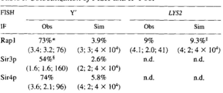
using Adobe Photoshop v3.0 software was set at the value of 140 out of a gray scale maximum of 255, after a normalization of each filter channel in- dependently to give the same maximum signal. This ensures that we ana- lyze colocalization of only the strongest sites of staining, although it can also result in the loss of some less intense foci (see Fig. 6, arrows). Texas red (indicating anti-Sir3p, -Sir4p, or -Rapl antibody) and fluorescein (Y' FISH) signals were printed as red or blue boundary traces, respectively. Three persons independently scored for 50% or more overlap of blue and red traced areas, which was called colocalization. In the case of Sir pro- teins and Rapl overlap with Y', there were very few borderline cases. Of the 371 cells analyzed, 15% were labeled by only IF or only FISH, and thus could not be used for analysis. In general we observe that the effi- ciency of Y' FISH is lower when performed after an immune reaction, and particularly after Sir3p or Sir4p immunostaining, e.g., on a given midsec- tion plane we observe on average only 2.1 Y' loci per nucleus after anti- Sir4p staining, which is only half the average number of Sir4p loci. There- fore, we calculated the percentage of Y' FISH coinciding with anti-Sir4p IF (see Table I), rather than the converse. In the case of Rapl, there is less interference between antibody and Y' FISH, and equal numbers of Rapl and Y' loci are detected per cell, of which overall 76% of IF signals coin- cide with Y' FISH, and 73% of FISH signals coincide with Rapl. In the case of Sir3p, 65% of the IF signals coincide with FISH, while 54% of FISH signals coincide with Sir3p signals. The fact that the values are lower for Sir3p appears to reflect a drop in the number of Sir3p foci detected be- fore and after FISH, probably due to a lower avidity of this antibody-anti- gen complex under FISH conditions. The coincidence of anti-p62 (nuclear pore) with anti-Rapl was calculated in a similar fashion, screening 70 cells containing 301 Rapl loci.
The calculation of the frequency of at least 50% surface overlap of a given number of randomly distributed spots (of either 0.2 ~ m or 0.25 ,~m in diameter) within a circle (nucleus) of 2 IJ, m in diameter was performed on a SUN computer. The number of red or blue spots was set by a Poisson distribution around a given number (2, 3, or 4), and their positions within the circle were generated randomly. The overlap criterion was tested on all red-blue combinations, in 104 simulations, and the percentage of red circles that colocalize with at least one blue circle was recorded. The re- sults of four such simulations were averaged for each combination of num- bers.
Results
Antisera were raised against the yeast nuclear proteins Rap1 (Klein et al., 1992), Sir3p, and Sir4p, and antigen- specific immunoglobulins were purified by binding and re- lease from antigen saturated strips of nitrocellulose before use for immunofluorescence or Western blots (see Materi-
als and Methods). Western blot analysis on sir3- and sir4-
strains indicates the loss of the major reactive bands at roughly Mr = 120,000 for Sir3p and Mr = 170,000 for Sir4p, in the appropriate mutants (Fig. 1, see Materials and Methods). The lower bands in the anti-Sir3p blot do not derive from the SIR3 gene; this binding is apparently of low affinity, as this cross-reaction does not contribute a significant background signal in immunofluorescence in a
sir3- strain (see Fig. 2 e). The affinity-purified anti-Rapl recognizes a doublet migrating at Mr = 116,000 (Fig. 1, lanes 1-6), which is efficiently competed by an excess of full-length bacterially expressed Rapl protein both on blots and in immunofluorescence (Klein et al., 1992).
On the autoradiographs shown in Fig. 1, each pair of lanes corresponds to two levels of loading of the nuclear proteins from each indicated strain (see figure legend) and serial exposures of the Western blots confirmed that the signals were in the linear range of the dose response. The blot suggests that Rapl levels do not to vary more than twofold in sir3- and sir4- strains (Fig. 1, lanes 1-6), and that the Sir3p protein level is also maintained within this range in the absence of Sir4p, and vice versa (Fig. 1, lanes
7-16).
Integrity and Three-Dimensional
Structure of the Nuclei Are Maintained through the Immunofluorescence Procedure
Immunofluorescence studies revealed strong anti-Rapl signals at each chromosomal end in spreads of pachytene- stage yeast cells and a limited number of brightly staining foci in vegetatively growing yeast cells (Klein et al., 1992, Palladino et al., 1993a). Immunofluorescence using anti- bodies against Sir3p or Sir4p showed punctate staining patterns, similar to that observed for Rapl, while anti-to- poisomerase II stained nuclear D N A uniformly (Palladino et al., 1993a). The protocol used in all these experiments involves digestion of the cell wall before formaldehyde fix- ation (see Materials and Methods), although fixing the cells before cell wall digestion results in a similar clustered
Figure 1. A n a l y s i s of affinity p u r i f i e d a n t i - R a p l , a n t i - S i r 3 p a n d anti-Sir4p a n t i b o d i e s b y W e s t e r n blot. C r u d e n u - clear p r o t e i n s w e r e i s o l a t e d f r o m a Sir + strain, BJ2168, a n d f r o m s t r a i n s carry- ing a sir3::TRP1 d e l e t i o n , o r a sir4:: LEU2 d i s r u p t i o n . T w o l a n e s o f e a c h i n d i c a t e d s t r a i n w e r e l o a d e d with ei- t h e r 100 or 200 Ixg o f a n u c l e a r - e n - r i c h e d f r a c t i o n (Sir + , 1-2, 7-8, 13-14; sir3-, 3-4, 9-10, 15-16; a n d sir4-, 5-6, 11-I2, 17-18). A f t e r S D S gel electro- p h o r e s i s , p r o t e i n s w e r e t r a n s f e r r e d to n i t r o c e l l u l o s e a n d p r o b e d w i t h affinity p u r i f i e d a n t i - R a p l (1-6), anti-Sir3p ( 7 - 12) o r a n t i - S i r 4 p (13-18) a n t i b o d i e s . S e c o n d a r y a n t i b o d i e s w e r e v i s u a l i z e d by E n h a n c e d C h e m i L u m i n e s c e n c e ( A m e r s h a m ) , a n d v a r i o u s e x p o s u r e s w e r e m a d e to e n s u r e t h a t t h e p e r o x i d a s e r e a c t i o n s h o w n w a s in a l i n e a r r a n g e . Sir3p m i g r a t e s with a M r a r o u n d 120,000. T h e b a n d at 110,000 a n d t h e l o w e r b r e a k d o w n p r o d u c t s in l a n e s 7-12 a p p e a r n o t to b e specific for SIR3. A l t h o u g h this c r o s s - r e a c t i o n is visible b y b l o t t i n g t e c h n i q u e s , its c o n t r i b u t i o n to i m m u n o s t a i n i n g in a sir3 d e l e t i o n s t r a i n is m i n i m a l (see Fig. 2 e). B o t h Sir3p a n d Sir4p (Mr = 170,000) m i g r a t e slightly m o r e slowly t h a n e x p e c t e d f r o m t h e i r p r e d i c t e d m o l e c u l a r m a s s e s . M a r k e r s (in ki- l o d a l t o n s ) a r e i n d i c a t e d at t h e left.
The Journal of Cell Biology, Volume 134, 1996 1352
appearance of R a p l staining (data not shown). The sphero- plasted yeast cells maintain their spherical shape through- out the labeling procedure, as shown by three-dimensional reconstitution of the cell nucleus from a series of focal sec- tions (Palladino et al., 1993a). Dimensions of the nucleus calculated from EtBr fluorescence give a diameter of 2.0 _ 0.2 ixm in the XY plane, and 2.4 --- 0.2 Ixm in the Z axis. This 20% distortion along the Z axis is also observed when cells are fixed before spheroplasting and reflects an inte- gral Z-stretch function of the confocal microscope used. Thus, the spheroplasting technique does not alter the shape of the yeast nucleus. On the other hand, if cells are treated with detergents before fixation, or if spheroplasts are protease-treated before FISH, cells become flattened, no longer maintaining this spherical shape (see Palladino et at., 1993b; Guacci et al., 1994) and the number of Z-sec- tions possible is reduced.
As an independent assay for nuclear integrity, we used a mouse monoclonal antibody raised against the human nu- clear pore protein p62, a homologue of yeast N s p l p (Wim- m e t et at., 1992). Immunofluorescence of N s p l p shows a ringlike staining at the nuclear periphery typical of nuclear pore staining and identical to our reactions with anti-p62 (Figs. 2 and 3). In our staining protocol, this staining is lost if cells are spheroplasted to completion before formalde- hyde fixation, or if the nuclear envelope is disrupted by de- tergents (data not shown). This confirms that the presence of a punctate/ringlike staining with anti-p62 correlates with a degree of nuclear integrity. In addition, this anti- body allows us to monitor the relationship of the immu- nofluorescent loci to the nuclear periphery as defined by the nuclear pore signal.
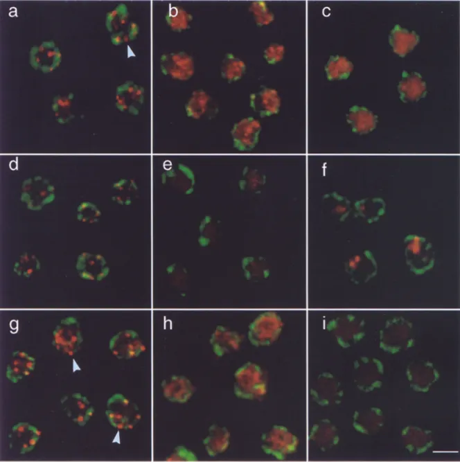
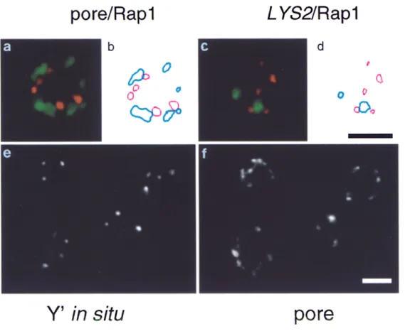
Fig. 2 shows that anti-Rapl, anti-Sir3p, and anti-Sir4p antibodies visualized by a Texas red-conjugated second- ary antibody reveal a limited number of brightly staining foci in a wild-type diploid strain (Fig, 2, a, d, and g). Dou- ble labeling by addition of the anti-p62 monoclonal anti- body attests the nuclear integrity under these immunoflu- orescence conditions. Quantitation of the coincidence of pore staining with either R a p l or Sir foci was performed on contour traced images such as those shown in Fig. 3, a and b. Only 13.6% of the Rap1 loci were found to have at least 50% overlap with nuclear pore signals, while 57.1% of the loci were found adjacent to, but having < 4 0 % sur- face overlap with a nuclear pore signal. Thus, while a ma- jority of the Rap1 signal is located near nuclear pores and hence near the nuclear periphery, a very low percentage coincide directly with pore complexes (arrows in Fig. 2, a, d, and g). The "juxtaposition" of R a p l and pore signals is statistically significant, yet due to the limited resolution of immunofiuorescence data, no conclusions can be drawn as to whether or not R a p l and/or Sir loci directly abut the nuclear envelope.
We next determined the localization of the three pro- teins in diploid cells deficient for both copies of SIR3 (GA192, Fig. 2, b, e, and h) or SIR4 (GA202, Fig. 2, c, f, and i). The punctate, ringlike nuclear pore staining (de- tected by a Cy5-conjugated secondary antibody, green in Fig. 2) suggests that nuclear integrity is maintained in both wild-type and mutant cells. As previously shown (Palla- dino et al., 1993a), Rap1 staining becomes diffuse in sir3- (Fig. 2 b) and sir4- (Fig. 2 c) ceils. Here we demonstrate
for the first time that Sir4p staining is also diffuse in sir3- cells (Fig. 2 h), and it is, as expected, absent in a sir4- strain (Fig. 2 i). In sir4- cells, anti-Sir3 staining no longer results in a discrete punctate pattern, but shows one of two morphologies. Either we see a diffuse distribution of Sir3p (data not shown), or we see one or two large, irregularly shaped aggregates (Fig. 2 f), clearly distinguishable from the foci in wild-type cells (Fig. 2 d). It is unclear what this staining pattern represents, but the signals do not coincide with telomeric D N A (data not shown). These double- labeling experiments make it unlikely that the discrete fo- cal patterns of Rap1 and Sir protein staining seen in wild- type cells, and their shift to a diffuse distribution in mu- tants, reflect either artefacts of the immunofluorescence protocol or loss of nuclear integrity during the assay. Subtelomeric Hybridization Patterns Reveal
Telomere Clusters
Do the staining patterns of Rap1, Sir3p, and Sir4p coincide with telomeres in interphase nuclei? To answer this ques- tion, we have combined immunofluorescence and in situ hybridization for the double labeling of S. cerevisiae dip- loid strains (see Materials and Methods). We aimed to de- tect both immunofluorescence and in situ hybridization signals, but also to maintain, as much as possible, the three-dimensional organization of the nucleus. For this reason we have omitted the protease treatment usually found in FISH protocols (e.g., Guacci et al., 1994). At- tempts to hybridize to the 300-bp TGI.3 repeat at the ends of all yeast chromosomes, showed irreproducible labeling efficiency in intact spheroplasts. Therefore, we chose to use the 5.4-kb, highly conserved Y' subtelomeric element, found immediately adjacent to the terminal TG~_3 repeat, as a probe. Although the highly conserved Y' element is not present at all chromosomal ends due to its strain-de- pendent distribution (Louis and Haber, 1992), a quantita- tive screening for the number of Y-containing telomeres in the two diploid strains used in this study showed that well over 80% of the telomeres have either the 5.4- or the 6.7-kb Y' element adjacent to the (TGv3), repeat (see Ma- terials and Methods; data not shown).
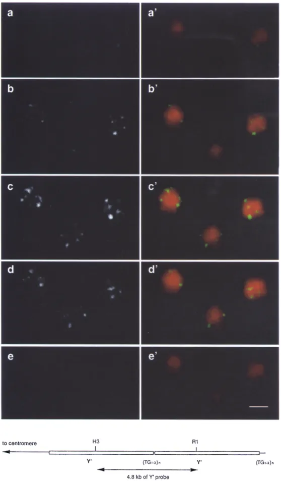
To perform in situ hybridization we adapted the fixation technique that we had optimized for immunofluorescence, knowing that it allows maintenance of the three-dimen- sional structure of the nucleus as monitored by confocal Z-series analysis. Under these conditions the average di- ameter of nuclear D N A is unchanged by in situ hybridiza- tion (2.0 -~ 0.2 I~m in the XY plane, and 2.4 _+ 0.2 Ixm in the Z axis), and nuclear pore staining is still detectable af- ter the FISH procedure (Fig. 3, e and .f). The average di- ameter of the nucleus measured after FISH by pore stain- ing is between 2.4 and 2.6 Ixm, similar to the diameter of nuclear D N A , with an additional 0.2 Ixm due to the pore signal itself. Fig. 4 shows five 0.3-txm sections of three wild-type yeast nuclei in which the D N A has been stained with ethidium bromide and the telomeres labeled with a Y' subtelomeric probe, detected by fluorescein-conju- gated anti-DIG F(ab) fragments. Because there are 32 chromosomes and 64 telomeres in these cells, of which nearly all contain Y' repeats (see Materials and Methods), we propose that telomeres are clustered in a limited hum-
Gotta et al, Telomere Clustering in Yeast 1353
Figure 2. Integrity of nuclei is maintained throughout immunolabeling as monitored by nuclear pore staining. To monitor nuclear integ- rity after the staining procedure, cells from a Sir + diploid (RS453, a, d, and g), a sir3- diploid (GA192, b, e, and h) and a sir4- diploid (GA202, c, f, and i) were stained with a mouse anti-p62 antibody detected by a Cy5-conjugated secondary antibody (in green), as de- scribed in Materials and Methods. Anti-p62 gives the characteristic punctate, ringlike staining in both wild-type and mutant strains. Cells were also stained with anti-Rapl (a, b, and c), anti-Sir3p (d, e, and ~ , and anti-Sir4p (g, h, and i) antibodies, visualized as red sig- nals through a Texas red conjugated secondary antibody. Sir3p staining is diffuse or concentrated in a few large regions of staining in sir4- cells (f), and is absent, as expected, in sir3- cells (e). Sir4p staining is diffuse in sir3- cells (h) and absent in sir4- cells (i). Shown are images taken on a Zeiss Axiovert 100 LSM confocal microscope using 100x Plan-Apochromat objective. The scanning parameters (pin- hole, brightness, and contrast) used were identical for all the images. Bar (i) 2 ~m. Arrows indicate Rapl or Sir4p signals adiacent to nu- clear pores.
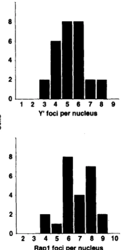
b e r of discrete loci, resulting in a staining p a t t e r n very sim- ilar to that o b t a i n e d with R a p l , Sir3p, and Sir4p antibodies. To calculate the total n u m b e r o f Y ' foci in each individ- ual cell, a series of eight 0.3-~m focal sections t h r o u g h f o u r fields o f cells was s c r e e n e d so that Y ' foci found in the dif- f e r e n t focal p l a n e s were s u m m e d (see M a t e r i a l s and M e t h - ods). T h e n u m b e r of Y ' foci p e r nucleus r a n g e d from t h r e e to eight, with an a v e r a g e of 5.3 +-- 1.3 (n = 28; Fig. 5). A similar o p e r a t i o n was p e r f o r m e d for ceils l a b e l e d only with a n t i - R a p l , and we o b t a i n e d a slightly larger n u m b e r
of Rap1 foci p e r nucleus (6.8 +- 1.4; n = 24; Fig. 5). This difference is likely to reflect either the r e d u c e d efficiency o b t a i n e d for F I S H as c o m p a r e d to i m m u n o f l u o r e s c e n c e , the fact that two pairs of h o m o l o g u e s d o not contain Y ' se- quences in these strains, o r possibly, the presence of R a p l foci that are not telomeric.
R a p l , Sir3p, and Sir4p Colocalize with Telomeres
R a p l , Sir3p, and Sir4p are essential for t e l o m e r i c silencing
The Journal of Cell Biology, Volume 134. 1996 1354
Figure 3. Graphic represen- tation of fluorescence signals for nuclear pore/Rapl and L Y S 2 / Rapl colocalization, and nuclear pore staining on cells subjected to FISH. Shown are representative cells from the double staining for Rapl (in red, a and c, de- tected by a Texas red-conju- gated secondary antibody) and for nuclear pore (in green, a, visualized by a Cy5- conjugated secondary anti- body), or for L Y S 2 FISH (in green, c, detected by fluores- cein-coupled anti-DIG F(ab) fragments). Also shown is the corresponding computer graphic representation (b and d, red for Rapl, blue for nuclear pore or LYS2, re- spectively) of signals abo~e a given threshold (see Materi- als and Methods). Similar traces were used for the quantitation of colocaliza- tion. e and f show Y' FISH (e) and nuclear pore staining (f) on cells subjected to the double immunofluorescence/ FISH protocol.
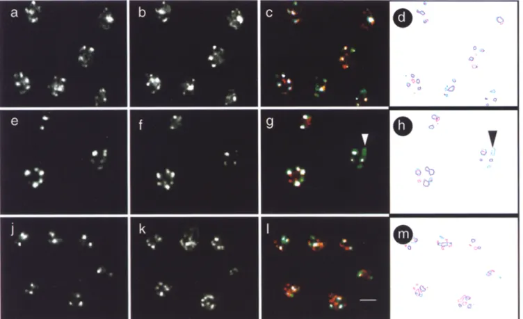
(Aparicio et al. 1991; Kyrion et al., 1993), yet R a p l , and possibly Sir proteins, are also involved in other nuclear processes (e.g., Devlin et al., 1991; Gottlieb and Esposito, 1989; Kennedy et al., 1995). It was thus important to ask whether the Rap1, Sir3p, and Sir4p foci coincide with te- lomeres as detected by in situ hybridization with the Y' probe. To perform combined immunofluorescence with ei- ther Rap1, Sir3p, or Sir4p antibodies and in situ hybridiza- tion on fixed yeast cells, we perform the standard labeling with affinity-purified Rap1, Sir3p, or Sir4p antibodies, and confirm by fluorescence microscopy that the reaction was successful. The formaldehyde fixation is then repeated, cells are treated with RNaseA and subjected to in situ hy- bridization. Fig. 6 displays the result of the double-labeling experiment in which Y' in situ hybridization on wild-type cells is detected by fluorescein fluorescence (a, e, and i), while Texas red fluorescence indicates antibody staining (Fig. 6 b, anti-Rapl; f, anti-Sir3p; and k, anti-Sir4p).
A focal staining pattern is obtained with both labeling techniques. To test for colocalization, we merged the two fully independent channels of fluorescence (Fig. 6, c, g, and l). In the merged image, the Y' in situ hybridization is green, the antibody signal red, and coincidence of the most intense signals is indicated in white (see Materials and Methods). Despite the fact that the presence of proteins cross-linked to D N A reduces in situ hybridization effi- ciency, quantitation of these results confirms a statistically significant colocalization of anti-Rapl, -Sir3p, and -Sir4p staining with Y' telomeric foci (see below).
To quantify the colocalization or direct coincidence of fluorescent signals in each of the combined antibody/
FISH, a graphic representation of the two signals was cre- ated directly from the computer using identically scanned images in all cases (see examples Fig. 6, d, h, and m; Mate- rials and Methods). Colocalization was scored on a single focal plane taken as close as possible to the equator of the nucleus, and in each case between 460 and 500 total signals were scored. The results for > 5 0 % of surface overlap (de- fined as colocalization) for loci from R a p l / Y ' , Sir4p/Y', and Sir3p/Y' labelings are summarized in Table I. For both R a p l - and Sir4p-staining, 73% and 74% of the Y' loci coincide with an antibody signal. In the case of Sir3p, fewer antibody complexes appear to resist the stringent conditions of hybridization (we score on average 1.6 Sir3p loci per nuclear section after FISH, compared to 3.0 foci per nuclear section before FISH), which reduces the chance that a Y' signal can colocalize with a Sir3p focus. The calculated colocalization of Sir3p on Y' is thus 54%, while the frequency with which Sir3p coincides with the Y' signals is 65% (summarized in Table I).
This frequency of signal colocalization could simply re- flect a stochastic event due to the small size of the yeast nucleus and the resolution of the signals. To rule out this possibility, we used a nontelomere proximal probe of 13 kb in length, which hybridizes near the L Y S 2 locus, 342 kb from the right telomere of Chr. II. The probe gives on av- erage 1.95 signals per diploid nucleus of a signal size roughly equivalent to that obtained with other probes (di- ameter of 0.2-0.25 txm; see Fig. 3, c and d). When identical parameters are applied to the images and colocalization of the L Y S 2 signal with R a p l staining is quantified by con- tour tracing (see Fig. 3, c and d, Table I), we obtain a value
Gotta et al. Telomere Clustering in Yeast 1355
Figure 4. Subtelomeric se-
quences are found in a lim- ited n u m b e r of loci. In situ hybridization of yeast diploid cells (RS453) was p e r f o r m e d as described in Materials and M e t h o d s with a D I G - d U T P labeled Y' sequence which is detected by fluorescein-con- jugated a n t i - D I G F ( a b ) frag- ments, a-e show the Y ' in situ hybridization, a ' - e ' show the merge b e t w e e n the Y' FISH signal and the D N A stained with the ethidium bromide. The five vertical panels show 0.3-~m sections of three fixed yeast cells d e m o n s t r a t i n g that the three- dimensional structure of the nucleus is m a i n t a i n e d after the in situ hybridization. Sim- ilar results were obtained with the wild-type strain GA225 (data not shown). A t the b o t t o m of the figure is a sketch of the X I I L yeast te- lomere with two short ( ~ 5 . 4 kb) Y ' elements. (TG1.3)n in- dicates the TG-rich repeat that encompasses ~ 3 0 0 bp at the end of the chromo- some and ~ 6 0 bp between the two copies of Y'. The po- sition of the probe used is un- derlined as a H i n D I I I (H3)- E c o R I (R1) fragment. Bar: (e) 2 Ixm.
The Journal of Cell Biology, Volume 134, 1996 1356
r,.) 8 8 4 2 O ~ I 2
JL
3 4 5 6 7 8 9 Y' foci per nucleusJL
3 4 5 8 7 8 9 1 0 Rap1 foci per nucleusFigure 5. Quantitation of Y' FISH loci and Rapl foci in yeast nu- clei. The wild-type diploid RS453 was labeled with either anti- Rapl or a DIG-dUTP labeled Y' probe in separate experiments. A series of confocal images such as those shown in Fig. 4 were made on 20 to 25 cells of each labeling. Quantitation of the sig- nals was carried out as described in Materials and Methods on six of the eight 0.3-1xm focal sections, since there were no foci in the first and eighth that were not already visible in the adjacent sec- tion. The graphs show the distribution of nuclei with respect to their number of either Y' foci (top) or Rap1 foci (bottom) per nu- cleus.
of 9% colocalization. The statistical frequency for more than 50% of surface overlap between two circles of 0.25 txm diameter and four other circles of 0.2 txm diameter, randomly distributed in a circle (nuclear section) of 2 Ixm in diameter, is 9.3% (Table I). For various numbers of 0.2 txm circles representing two classes of signals, randomly distributed on a 2-txm surface, the frequency of overlap varies between 2.6% and 5.8% (as indicated in Table I). This indicates that the frequencies of colocalization ob- served for R a p l and Sir foci with Y' signals (54-74%) are highly significant, while the value observed for L YS2 and Rap1 (9%) would be consistent with chance coincidence.
There are two parameters that reduce the efficiency with which colocalization can be scored. First, the pres- ence of the cross-linked antigen-antibody complex on the subtelomeric domain inhibits the efficiency of hybridiza- tion. The second is the threshold set for the contour trac- ing, which is unlikely to be optimal for all signals. In some cases, foci that are clearly visible by eye are not scored, be- cause the intensity of the signal is below the threshold value used (see example indicated in Fig. 6 h). Such highly restrictive threshold values are necessary when scoring colocalization, to avoid meaningless overlap between widely dispersed, low level signals.
Colocalization o f R a p l, Sir3p, and Sir4p with Telomeres Is Lost in sir M u t a n t s
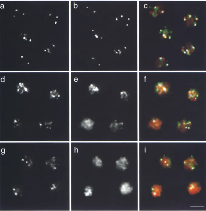
What happens to telomeric foci in sir3 and sir4 mutants when the silencing complex is disrupted allowing complete derepression of telomere proximal domains? Fig. 7 shows both the Y' in situ on wild-type, sir3- and sir4- cells (Fig. 7, a, d, and g), the anti-Rap1 on the same cells (Fig. 7, b, e, and h), and the merge of the two staining patterns (Fig. 7, c, f, and i). Although the Rap1 staining is diffuse in cells lacking Sir3p or Sir4p (7, e and h), Y' hybridization still appears clustered, and we routinely observe fewer foci than the 54 individual Y-containing telomeres. Y' FISH in the sir mutants analyzed, however, does not have the tightly defined foci observed in wild-type cells and reveals more heterogeneous Y' FISH pattern. In most sir- cells the hybridization signals appear extended, covering more of the nucleus, although the total fluorescent signal varies less than 10% from strain to strain (see Fig. 7, d and g). Despite this altered appearance, the Y' signals are not uni- formly dispersed through the nucleus like the IF signals for Sir3p, Sir4p, and R a p l in sir mutants (see Fig. 2). The qualitative change in the Y' hybridization signal is also ob- served when we perform FISH without previous antibody staining (Fig. 8).
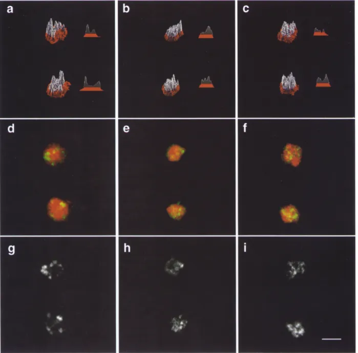
To visualize this qualitative difference of the in situ staining pattern, we have used a surface topology program that indicates the intensity at a given site in the cell as a to- pological peak (Fig. 8). The Y' signals (g-i and d - f in green) are superimposed on the EtBr-stained D N A (red in a-f). The foci in wild-type cells are well defined with high peaks of labeling and distinct valleys between them, while the intensity of Y' foci is lower and more dispersed in sir mutants (Fig. 8, a-c, see also the horizontal profile), perhaps reflecting a more extended chromatin state for the subtelomeric D N A in sir strains. This may reflect its organization in intact cells before the hybridization reac- tion, yet it could also be produced or exacerbated by the FISH procedure. Since the protocol is identical for both wild-type and mutant cells and the extended Y' pattern is highly reproducible, we favor the interpretation that it re- flects a physiological change in subtelomeric organization.
In this context is important to note that the anti-Rapl staining in sir3- and sir4- cells is no longer confined to Y' foci (see Fig. 7). Anti-Rapl gives a general nuclear stain- ing pattern, neither confined to nor excluded from the telo- meric signal. Immunofluorescence alone cannot distinguish between R a p l being completely diffuse in the nucleus and no longer telomere-bound, or the presence of two popula- tions of R a p l , one telomere-associated and the other dif- fuse. We favor the latter interpretation because conditions that interfere with R a p l - D N A interaction at telomeres, through either overexpression or mutation of the R a p l protein, result in chromosome loss rates up 20-fold above wild-type rates (Conrad et al., 1990; Kyrion et al., 1992), while the levels are only 2-5-fold elevated in sir3 and sir4 mutant cells (Palladino et al., 1993a). Telomere size dereg- ulation events are also much more dramatic in rap1 mu- tants, as compared to the modest 50-100 bp loss observed in sir3 and sir4 mutants (Conrad et al., 1990; Kyrion et al., 1992; Palladino et al., 1993a). Thus, it appears likely that in sir mutants, one subpopulation of R a p l remains telomere-
Gotta et al. Telomere Clustering in Yeast 1357
Figure 6. Y' FISH colocalizes with Rapl, Sir3p, and Sir4p foci. The wild-type diploid strain RS453 was subjected to immunofluores- cence with anti-Rapl (b), anti-Sir3p (f) and anti-Sir4p (k) antibodies which are detected by a Texas red-conjugated secondary antibody; cells were then hybridized with a DIG-dUTP labeled Y' subtelomeric probe (a, e, and/'), detected by a fluorescein-conjugated anti-DIG F(ab) fragment (Materials and Methods). c shows the merge of the Rapl staining (red) and the Y' probe (green), g shows the merge of the Sir3p staining (red) and the Y' probe (green), and I shows the merge of the Sir4p staining (red) and the Y' probe (green). A single fo- cal section near the equator of the cells is shown. Wave-length shifts are corrected automatically by the Zeiss confocal system. Coinci- dence of the two signals above a given threshold is shown in white (see Materials and Methods). d, h, and m show the respective com- puter graphic representation of the signals (red for the antibody staining, blue for the Y' FISH). The treatment of the images for allowing quantitation of colocalization is described in Materials and Methods. The arrows in g and h indicate an example in which coin- cidence is not scored because the intensity of the Rapl signal is below the threshold value used, even though the signal is visible. Any weak diffuse nuclear staining of the antibodies is not scored in this procedure. Bar: (/) 2 Ixm.
bound, while another population is released from a telo- meric or subtelomeric complex, producing the general im- munofluorescence pattern visible in Figs. 2 and 7.
Discussion
We have developed a combined immunofluorescence/in situ hybridization protocol for the budding yeast S. cerevi- siae, which maintains three-dimensional structure of the nucleus throughout the labeling procedure. We d e m o n - strate by confocal sectioning that the nuclei are not col- lapsed or flattened by the procedure, and that the nuclear pore complexes remain intact at the nuclear periphery. Using the double-labeling technique we demonstrate that Rap1, Sir3p, and Sir4p, three proteins essential for telo- mere proximal gene repression, are indeed localized in foci that correspond to their sites of action at subtelomeric and telomeric domains in wild-type cells. Within the limits of resolution offered by confocal microscopy, we calculate
that over 70% of the Rap1 loci are either adjacent to (57.1%) or coincident with (13.6%) nuclear pore signals, although there is also a significant fraction (29.3%) of R a p l foci that are more internal in the nucleus. These numbers agree well with a previous analysis of the posi- tioning of R a p l foci by immunoelectron microscopy (Klein et al., 1992).
The double in situ/immunofluorescence staining proto- col provides a powerful tool for the characterization of nu- clear protein localization and the positioning of specific c h r o m o s o m a l regions. However, by aiming to preserve three-dimensional structure and protein integrity, we also limit the sensitivity of the F I S H assay. Short, unique ge- nomic sequences ( < 10 kb) are difficult to detect reproduc- ibly in our double IF/FISH protocol, although they are quite readily detected in protocols that aim only to opti- mize in situ hybridization (Guacci et al., 1994). There can also be loss of immune complexes during the in situ hy- bridization, although these are minimal when high affinity antibodies (e.g., a n t i - R a p l ) are used.
The Journal of Cell Biology, Volume 134, 1996 1358
Table I. Colocalization ofFISH and IF Foci
FISH IF
Y ' LYS2
Obs Sim Obs Sim
R a p l 7 3 % * 3 . 9 % 9 % 9 . 3 % t (3.4; 3.2; 76) (3; 3; 4 × 10⁴) (4.I; 2.0; 4 1 ) (4; 2; 4 × 10⁴) Sir3p 5 4 % 8 2 . 6 % n.d. n.d. (1.6; 1.6; 160) (2; 2; 4 x 10⁴) Sir4p 7 4 % 5 . 8 % n.d. n.d. ( 3 . 6 ; 2 . 1 ; 9 6 ) ( 4 ; 2 ; 4 × 10⁴)
Shown are the observed (Obs) and computer simulated (Sim) percentages of colocal- ization between the loci identified by FISH (Y' or LYS2 probes) and the immunofluo- rescence (IF) signals of Rapl, Sir3p, or Sir4p. The observed colocalization corre- sponds to the fraction of FISH signals that have a surface overlap of more than 50% with a given 1F signal. In parentheses are indicated n, m, and T, where n, the number of IF loci per nuclear plane; m, the number of FISH foci per nuclear plane; and T, the total number of nuclei scored. For the computer simulation, Poisson-distributed num- bers of circles ranging from 2 to 4, as indicated in the brackets, were used to calculate the frequency of overlap that would occur by chance in 104 nuclei containing two classes of circles in randomly generated positions. For the simulation circles of 0.2 Ixm diam were used and the nuclear midsection circle was 2 tun in diameter. *The converse values for the percent of Rapl loci on Y' spots = 76%.
~For the simulation of the LYS2 probe, circles of 0.25 ~m diam were used, only to demonstrate the effect of slightly larger signals on the chance of colocalization (com- pare 5.8% with 9.3%). LYS2 signals were not on average larger than Y' signals, how-
e v e r .
~Tbe converse value for the percent Sir3p foci on Y' foci = 65%.
Rapl Staining Is Coincident with Telomeric D N A in Wild-Type Strains
By combining in situ hybridization, using the highly con- served Y' telomere associated sequence, with anti-Rap1 immunofluorescence, we find colocalization of 74% of the Rap1 foci identified in interphase nuclei of Sir + yeast cells with sites of Y' hybridization, while only 9% of the loci la- beled by a single copy internal sequence (LYS2) coincide with Rap1 when analyzed in an identical fashion. 9% is also the percentage of randomly distributed foci that show >50% surface overlap in computer simulation experi- ments using the same size and number of foci on a surface the size of a yeast nuclear midsection (see Table I). The DNA denaturation required for FISH is somewhat detri- mental for the maintenance of the antibody-antigen com- plex, and depending on the antibody, up to 30% of the im- munofluorescence staining can be lost during in situ hybridization. Thus, while the value of 74% colocalization is highly significant, the true value for Rap1 and telomere coincidence is likely to be even higher. Although two pairs of chromosomes in our strains lack Y' elements, and thus escape detection by hybridization, we found that other te- lomeric probes were less efficient either due to their length (i.e., the terminal TG1.3 repeat covers <400 bp) or lack of overall conservation (i.e., only 500 bp core of the X element is highly conserved, Louis et al., 1994). Mixtures of X and Y' probes produce FISH images very similar to those obtained with Y' alone on the strains used in this study (Scherthan, H., T. Laroche, and S.M. Gasser, unpub- lished results). We cannot, of course, rule out the possibil- ity that a fraction of the Rap1 foci might also reflect aggre- gates of this protein at sites other than telorneres (e.g., HAl
loci).
Subtelomeric FISH alone in the absence of anti-Rap1 confirms that telomeres are clustered in a number of foci significantly fewer than the number of chromosomal ends (5.3 detected per average diploid yeast nucleus). We have
also observed using unique telomere probes, that we can detect individual telorneres in these fixed yeast sphero- plasts, and that these also colocalize with Rap1 foci (Scherthan, H., and S.M. Gasser, unpublished results). We do not yet know the priniciple of telomere-telomere inter- action, nor whether the grouping of telomeres is a stochas- tic or regulated phenomenon. Ongoing FISH studies with telomere specific probes should provide answers to these questions.
Is There a Nuclear Envelope-Telomere Interaction?
The resolution of light microscopic techniques renders it impossible to judge whether the foci of telomeres, Rap1 or Sir proteins directly abut the nuclear envelope. Roughly 70% of telomeres (or Rap1 foci) have a perinuclear local- ization, and Rap1 foci do not appear to be directly associ- ated with nuclear pore complexes, since double-immuno- staining with anti-Rap1 and anti-pore antibodies show nonoverlapping spots around the nuclear periphery with only 13.6% direct coincidence (Figs. 2 and 3). In addition, we have performed Rap1 immunofluorescence on a strain carrying a mutation in nup133, which provokes the cluster- ing of nuclear pores on one side of the nucleus (Doye et al., 1994). In this strain the Rap1 staining has a wild-type focal pattern, suggesting that Rapl/telomere complexes are po- sitioned independently of the pore complexes (data not shown).
Although it is intriguing that human telomeres associate with the nuclear matrix isolated from cultured cell nuclei (de Lange, 1992), our attempts to purify the yeast nuclear lamina, which might associate with yeast telomeres have failed (see Cardenas et al., 1990; data not shown). More- over, we show here that despite its coiled-coil domain (Diffley and Stillman, 1989), Sir4 protein does not localize to the nuclear periphery in the absence of Sir3p, nor does it form a perinuclear ring, suggesting that Sir4p alone has no stable link to a peripheral nuclear substructure. If a structural element of the nucleus or a telosome compo- nent is responsible for the clustering of telomeres, further studies will be required to identify its components. While telomeres still appear clustered in the nuclei of sir3- or
sir4- cells, the resolution of FISH is inadequate to deter- mine whether or not the subnuclear positioning of these clusters is altered in the absence of Sir3p or Sir4p. Other methods of DNA detection with higher resolution will be required to resolve this question.
Sir3p, Sir4p, and a Subpopulation o f RapI Are Displaced from Telomeres in sir3 and sir4 Mutants
Genetic and biochemical evidence strongly suggest that Rap1, Sir3p, and Sir4p form a multiprotein complex essen- tial for telomeric silencing. Nonsense and missense muta- tions in the C O O H terminus of Rap1 abrogate telomeric repression (Kyrion et al., 1992; Liu et al., 1994) and over- expression of Sir3p suppresses some of these missense mu- tations (Liu et al., 1994). SIR3 overexpression also sup- presses certain sir4 point mutations (Marshall et al., 1987), and two hybrid studies show that domains of Sir3p and Sir4p interact with themselves, with each other, and with Rap1 (Moretti et al., 1994). In addition, full-length Sir4p and Rap1 can be coprecipitated from yeast nuclear ex-
Gotta et at. Telomere Clustering in Yeast t 3 5 9
Figure 7. Rap1 staining does not coincide primarily with subtelomeric FISH in sir3 and sir4 mutants. Wild-type (RS453, a-c), sir.q- (GA192, d-f) and sir4- (GA202, g-i) diploid cells were first stained with affinity purified anti-Rapl antibodies, which are detected by a Texas red-conjugated secondary antibody, and subsequently hybridized with a dig-dUTP labeled Y' probe, detected by fluorescein-cou- pled anti-DIG F(ab) fragments, a, d, and g show the FISH signal alone; b, e, and h show the anti Rap1 staining alone and c, f, and i show the merge of the FISH and the antibody staining patterns. Rap1 staining is diffuse in mutant strains (e and h) unlike the Y' probe (d and g). The total FISH signal integrated over twenty nuclei from each labeling varied <10% among strains. Bar: (i) 2 p.m.
tracts with either Rap1 or Sir4p antibodies (Cockell et al., 1995). We have shown that Sir3p and Sir4p immunolocal- ize to a limited n u m b e r of nuclear loci, much like those identified by anti-Rapl. Here we show unequivocally that Sir3p and Sir4p signals coincide with Y' repeats in wild- type cells, with colocalization values similar to those for R a p l and Y', all of which are significantly above any sto- chastic frequencies for overlap (see Table I). This rein- forces genetic evidence indicating that these proteins work together in a complex at telomeres.
Evidence that the tertiary complex is necessary for the proper localization of Sir3p, Sir4p, and at least a subpopu-
lation of R a p l , comes from immunofluorescence studies in either sir3- or sir4- strains. We see that R a p l has a gen- eral, dispersed nuclear staining in either mutant, while the normal focal pattern of Sir3p becomes diffuse in a sir4- strain. Likewise, Sir4p staining is diffuse in a sir3- strain. Because the net signal intensities are not higher in the mu- tant strains, this result does not reflect the "unmasking" of antigen that was undetected in wild-type cells. We confirm that nuclear integrity is maintained throughout these im- munolocalization studies by counter-staining with a m o n o - clonal antibody that recognizes nuclear pores and by con- focal Z-series analyses. The somewhat more diffuse Y'
The Journal of Cell Biology, Volume 134, 1996 1360
Figure 8. Y' in situ hybridization reveals a qualitatively altered pattern in sir3 and sir4 mutants. Shown are surface topography profiles of Y' signal in a focal plane (top). Wild-type (RS453, a, d, and g), sir3- (GA192, b, e, and h) and sir4- (GA202, c, f, and i) diploid cells were hybridized with a dig-dUTP labeled Y' probe, detected by fluorescein-coupled anti-DIG F(ab) fragments, in the absence of prior antibody staining. Genomic DNA was detected by EtBr staining and is shown in red. A single focal plane containing two cells is shown for each strain, taken from a Z-series analysis which demonstrated three dimensional integrity of the labeled spheroplasts, d - f show the merge of the FISH (green) and the EtBr stained DNA (red), while g-i show the FISH image alone, a-c show the surface topography profiles of the EtBr-stained DNA (red) and the DIG-dUTP FISH labeling (white) from the cells shown in d-f. The peaks represent the intensity of the signal. A transverse section is also shown for each labeled cell. All representations were calculated with the Carl Zeiss LSM software. The total FISH signal integrated over twenty nuclei from each labeling varied <10% among strains. Bar, 2 txm.
F I S H signals in sir3- and sir4- cells m a y reflect an altered, less c o m p a c t c h r o m a t i n structure, although o t h e r bio- chemical techniques will b e r e q u i r e d to e x p l o r e Y ' chro- matin structure in sir strains. M o r e i m p o r t a n t is the fact that the i m m u n o s t a i n i n g p a t t e r n s no l o n g e r coincide pri- m a r i l y with the Y ' signals. This is consistent with our inter- p r e t a t i o n that the loss of e i t h e r Sir3p or Sir4p disrupts the p r o p e r localization of a Sir3p/Sir4p c o m p l e x from its nor- mal site of action.
U n e x p e c t e d was the result that Rap1, which binds its consensus site in vitro with a K d = 10 -11 (Vignais et al., 1990), a p p e a r s d i s p e r s e d in the Sir-deficient nuclei while t e l o m e r e s , d e t e c t e d by the t e l o m e r e associated s e q u e n c e Y ' , are still m o r e or less c l u s t e r e d (Fig. 7). F l u o r e s c e n c e techniques are not sensitive e n o u g h to d i f f e r e n t i a t e be- t w e e n TGI_ 3 and Y ' repeats, h e n c e p r o b i n g with Y ' se- quences closely reflects the position of the relatively short TG1. repeat. O u r results d e m o n s t r a t e t h a t R a p l staining is
Gotta et al. Telomere Clustering in Yeast 1361