HAL Id: tel-01064224
https://tel.archives-ouvertes.fr/tel-01064224
Submitted on 15 Sep 2014HAL is a multi-disciplinary open access archive for the deposit and dissemination of sci-entific research documents, whether they are pub-lished or not. The documents may come from teaching and research institutions in France or abroad, or from public or private research centers.
L’archive ouverte pluridisciplinaire HAL, est destinée au dépôt et à la diffusion de documents scientifiques de niveau recherche, publiés ou non, émanant des établissements d’enseignement et de recherche français ou étrangers, des laboratoires publics ou privés.
DNA/polycation complexes in bulk and at interfaces as
advanced non-viral transfection vectors
Yulia Sergeeva
To cite this version:
Yulia Sergeeva. DNA/polycation complexes in bulk and at interfaces as advanced non-viral transfec-tion vectors. Chemical engineering. Université de Strasbourg, 2013. English. �NNT : 2013STRAE024�. �tel-01064224�
Thèse présentée pour obtenir le grade de
Docteur de l’Université de Strasbourg
Discipline: Chimie
par Yulia Sergeeva
Complexes ADN/polycation en solution et aux
interfaces en tant que vecteurs de transfection
non viraux de pointe
Date de soutenance prévue le 25 juin 2013
Membres du jury:
Directeur de Thèse: M. Gero Decher, Professeur, Strasbourg, France Rapporteur Interne: M. Vincent Ball, Professeur, Strasbourg, France
Rapporteur Externe: Mme. Karine Anselme, Directeur de recherche, Mulhouse, France Rapporteur Externe: M. Jean-François Berret, Professeur, Paris, France
3
Complexes ADN/polycation en solution et aux interfaces en
tant que vecteurs de transfection non viraux de pointe
Résumé de Thèse en Français (Summary of the Thesis in French)
La thérapie génique offre de nouvelles possibilités pour le traitement des maladies humaines graves telles que les maladies cardio-vasculaires, neurologiques et cancéreuses, la cicatrisation, les troubles inhérents, etc.... Le concept de cette approche repose sur la délivrance systématique d’acides nucléiques dans les cellules des patients et l’expression consécutive de protéines thérapeutiques (ADN) ou de gène dormant (siARN).
En général, les méthodes de transfert de gènes peuvent être regroupés en deux catégories principales: les méthodes de transfert de gènes virales et non virales. Bien que de nos jours l'approche la plus efficace pour le transfert d'ADN dans les différentes lignées cellulaires est basée sur la délivrance de gènes viraux, son application dans la médecine clinique est limitée en raison des effets secondaires possibles comme la mutagenèse ou la réponse inflammatoire.
Alternativement une approche non virale implique la formation de complexes ADN/macromolécules (dénommés polyplexes) par des interactions électrostatiques entre les groupes phosphates chargés négativement de l'ADN et les groupes chargés positivement des macromolécules telles que des polymères, des lipides ou des dendrimères. Même si une recherche intensive a été menée, l'efficacité de transfection de ces polyplexes reste faible.
Parmi les agents synthétiques de transfection existants, le jetPEI™ (poly(éthylène imine) linéaire, 22 kDa) est l'un des transporteurs les plus efficaces à l'heure actuelle. Les propriétés uniques de ce polymère telles que la densité élevée de charges positives, la biocompatibilité et le soi-disant effet “éponge à protons”, lui permettent de complexer l’ADN sous la forme de petites particules chargées positivement, de protéger l'ADN contre la dégradation dans
4
l'environnement cellulaire, de faciliter la libération endosomale et donc la réussie de la transcription. En dépit de leur efficacité supérieure, les vecteurs à base de PEI sont associés à une cytotoxicité élevée, en particulier pour les lignées cellulaires primaires.
Ce travail a été effectué dans le cadre d’un projet national à haut risque (Ship-In) dédié au développement de méthodes pour la reprogrammation de lignées cellulaires humaines afin d’induire des cellules pluripotentes. Les tâches de notre équipe ont été réalisées en collaboration avec le groupe du Professeur Stéphane Viville (IGBMC) et la société Polyplus Transfection™.
L'un des objectifs de ce travail a consisté à établir un protocole pour la transfection efficace d’une lignée cellulaire humaine primaire et à augmenter le rendement de transfection pour l’application d'un tel vecteur à base PEI pour un traitement médical.
L'efficacité de transfection des différents vecteurs non viraux a été testée sur la lignée cellulaire CPRE2 (fibroblastes dermiques humains primaires adultes).
Bien qu'au cours de ce travail des études approfondies ont été menées pour développer des vecteurs de pointe pour une transfection à long terme via une surface en testant divers films multicouches contenant de l'ADN et des agents de transfection comme le jetPEI™, le dendrimère poly(amido amine) (6ème génération), le chitosane, le collagène et le Chlorure de poly(méthacryloxyéthyltriméthyl ammonium), l'efficacité de transfection de ces systèmes s’est montrée très faible. Par conséquent, il a été décidé de focaliser nos efforts sur le développement d’un protocole robuste et reproductible pour la transfection efficace de la lignée cellulaire CPRE2 en solution.
A cet effet, les polyplexes à base de jetPEI™ ont été formés dans une solution à 0,15 M en NaCl avec de l'ADN plasmidique codant pour la protéine fluorescente verte (GFP). Les expériences ont été réalisées à une concentration constante de solution de jetPEI™ et la concentration de l'ADN a été variée en fonction du rapport azote sur phosphate N/P. Comme contrôle positif, la transfection du jetPEI™ dans des conditions standard a été
5
appliquée. L'efficacité de transfection est exprimée comme le pourcentage de cellules fluorescentes sur la population totale de cellules 48h après addition des polyplexes à la culture cellulaire.
Chose étonnante, les meilleurs résultats ont été obtenus pour la transfection avec des polyplexes préparés à N/P = 7,5. Une diminution de la concentration d'ADN conduit à des niveaux plus faibles de transfection alors que la viabilité des cellules augmente lentement.
Le traitement des cellules avec des polyplexes à N/P = 5 a conduit à une diminution de la viabilité des cellules par rapport à des cellules non traitées. La diminution de la dose d’ADN n'a pas eu d’effet dramatique sur la viabilité cellulaire. Dans les conditions d’efficacité de transfection maximale, la viabilité cellulaire était de 80%. La viabilité des cellules après la transfection avec des polyplexes formés à N/P de 10, 15 et 30 était respectivement de 82%, 84% et 78%.
Pour identifier les paramètres influençant les taux de transfection, la taille des polyplexes a été analysée par diffusion dynamique de la lumière (DLS). Les données ont été obtenues avec des échantillons fraîchement préparés dans du NaCl 0,15 M à 25 °C. Les résultats ont montré que la diminution de la concentration d'ADN conduit à la formation de particules plus petites. Les caractéristiques des complexes déterminées par DLS sont présentées dans le tableau 1. Rapport N/P ν(DNA), nmol Diamètre estimé, nm (distribution en nombre) Potentiel zeta (ζ) mV 5 3 777±15 34±4 7.5 2 575±61 36±4 10 1.5 472±63 31±2 30 0.5 220±12 27±6
Tableau 1. Taille et potentiel zêta (ζ) des complexes à base de PEI préparés à
6
Les micrographes de microscopie électronique à transmission ont révélé que les polyplexes présentent une structure sphérique et homogène. La taille des particules semble être beaucoup plus faible que celle mesurée par DLS. Ceci peut être dû à des différences dans le protocole qui sont discutées dans le texte principal. Le diamètre des polyplexes à N/P = 5 est compris entre 200 à 300 nm tandis que les polyplexes formés à N/P = 7,5 et 10 varie de 100 à 200 nm.
Figure 1. Micrographes TEM des polyplexes formés avec un rapport N/P de 5
(a) et de 7,5 (b).
Interaction des cellules primaires avec substrats fonctionnalisés par LbL
Comme la transfection via une surface nécessite la culture de cellules sur des films LbL contenant un agent de transfection et de l'ADN, l'étude du comportement des cellules sur des films LbL est donc d'une importance primordiale.
L'assemblage couche-par-couche est une approche facile et robuste pour la construction à façon de films multifonctionnels sur presque n’importe quelle surface avec un contrôle à l'échelle nanométrique de la composition du film. De plus, les propriétés des films telles que l'épaisseur, la charge de surface, la topologie et la mouillabilité peuvent être modifiées et optimisées en fonction des défis spécifiques. Du fait de sa simplicité et du nombre important des molécules
7
pouvant être incorporées dans un film, l’assemblage LbL est largement utilisé pour des applications en science des matérieux et dans les sciences de la vie, et en particuliers dans la conception de surfaces destinées à l'adhésion et la croissance cellulaire. Il est connu que le comportement, les fonctions et la viabilité des cellules sont définis par les interactions cellules-matériau qui peuvent être contrôlés par la chimie de surface, la topologie de surface et les propriétés physiques du matériau. La méthode couche-par-couche est l'exemple rare où ces trois paramètres peuvent être contrôlés indépendamment.
Dans cette étude, nous nous sommes focalisés sur l'influence de la composition chimique des films en investiguant les propriétés physiques telles que la stabilité et la mouillabilité des films multicouches sur la survie et la prolifération des cellules. Les polyélectrolytes suivants ont été utilisés pour l’assemblage LbL: la poly(éthylène imine) (PEI) ramifiée, l’hydrochlorure de poly(allylamine) (PAH), le chlorure de poly(diallyldiméthylammonium) (PDDA), le chitosane (Chit), le sulfate de dextran (DexS), l'alginate de sodium (Alg), l'hexamétaphosphate de sodium (PSP) et le poly(styrènesulfonate) de sodium (PSS). L'assemblage des multicouches a été suivi par ellipsométrie et microbalance à cristal de quartz avec mesure de dissipation (QCM-D).
Les films multicouches étudiés ont été assemblés par des interactions électrostatiques entre des polyélectrolytes de charges opposées. Toutefois, il est connu que les polycations libres peuvent induire des effets cytotoxiques via des interactions avec la membrane cellulaire ou des protéines de la matrice extracellulaire. Par conséquent, il est essentiel d'étudier la stabilité des systèmes LbL préparés dans le milieu de culture cellulaire dans les conditions physiologiques. Dans ce travail, la stabilité des films LbL a été analysée par QCM-D puisque cette technique permet de suivre simultanément in situ et en temps réel les changements structurels et de la masse á la surface du cristal. Les résultats obtenus ont révélé que le milieu de croissance cellulaire interagit étroitement avec les multicouches induisant l'adsorption rapide des composants du milieu ou change les propriétés physiques des multicouches. L'incubation
8
des couches pendant 96h n'a pas conduit à des changements dramatiques dans les courbes de fréquence et de dissipation suggérant que les films LbL étudiés garde leur composition dans des conditions physiologiques.
Pour l'analyse de l’effet des films LbL sur la survie/prolifération des cellules, des cellules CPRE2 ont été ensemencées sur des multicouches dans un milieu de croissance cellulaire. La population cellulaire a été comptée toutes les 24h et le quatrième jour, la viabilité des cellules a été analysée par cytométrie en flux.
Nos résultats ont montré que les cellules prolifèrent sur les couches contenant du PAH pendant 5 jours. Les meilleurs résultats ont été obtenus avec le film PEI(PSS/PAH)5 où la population cellulaire était proche de celle obtenue pour des cellules ensemencées sur une couche de gélatine. Cependant, les films PEI(DexS/PAH)5 et PEI(Alg/PAH)5 ont donné un nombre de cellules plus faible que sur du verre non modifié. La viabilité cellulaire après 4 jours était de 87%, 80% et 70% pour PEI(PSS/PAH)5 , PEI(DexS/PAH)5 and PEI(Alg/PAH)5 respectivement.
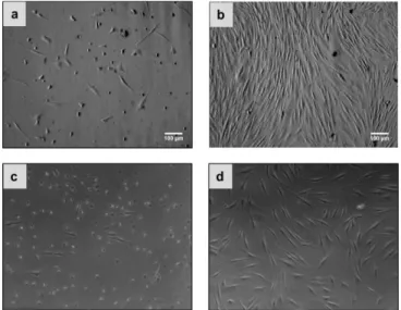
L’exposition des cellules aux multicouches contenant du PDDA a conduit à une faible adhésion cellulaire induit par un changement de la morphologie des cellules. Comme le montre la Figure 2, les cellules ne se propagent pas et montre une morphologie ronde. En outre, il a été constaté que les multicouches avec du chitosane entravent la croissance des cellules.
Figure 2. Micrographes de cellules CPRE2 ensemencées sur different
revêtements LbL 96h après ensemencement des cellules: a) PEI(PSS/PAH)5 b) PEI(PSS/PDDA)5 c) PEI(PSS/CH)5.
9
Après 24h, les cellules sont collectées et réensemencées sur une plaque recouverte de gélatine. Les données obtenues ont révélé que les cellules recueillies après l'incubation avec du PEI(Alg/CH)5, PEI(Alg/PDDA)5, PEI(PSS/CH)5 et PEI(DexS/CH)5 ont été en mesure de se fixer et de proliférer. En revanche, les cellules collectées après l'incubation avec du PEI(PSS/PDDA)5 n'étaient pas en mesure d'adhérer à la surface de la gélatine après réensemencement.
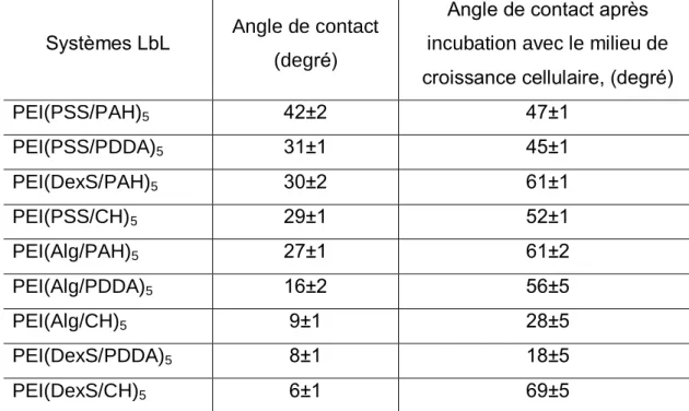
Pour mettre en évidence les raisons d'un tel comportement cellulaire préférentiel, les films multicouches ont été caractérisés par microscopie à force atomique et angle de contact avec de l'eau. Il a été constaté que les propriétés topologiques et de mouillage du revêtement ont radicalement changé après le contact avec le milieu de croissance cellulaire. Cependant, nos résultats mettent en évidence la faible influence de la topographie des multicouches étudiés sur le comportement cellulaire.
Systèmes LbL Angle de contact (degré)
Angle de contact après incubation avec le milieu de croissance cellulaire, (degré)
PEI(PSS/PAH)5 42±2 47±1 PEI(PSS/PDDA)5 31±1 45±1 PEI(DexS/PAH)5 30±2 61±1 PEI(PSS/CH)5 29±1 52±1 PEI(Alg/PAH)5 27±1 61±2 PEI(Alg/PDDA)5 16±2 56±5 PEI(Alg/CH)5 9±1 28±5 PEI(DexS/PDDA)5 8±1 18±5 PEI(DexS/CH)5 6±1 69±5
Tableau 2. Angles de contact des films LbL natifs et après incubation avec un
10
En outre, nos résultats ont montré que, pour certains systèmes, le comportement des cellules sur des films LbL pourrait être contrôlée non seulement par les propriétés de la couche la plus externe mais aussi par les propriétés de couche localisée dans le film (Figure 4). Cet effet a eu tendance à augmenter dans les expériences effectuées avec un milieu sans sérum.
Figure 3. Micrographes de cellules CPRE2 ensemencées sur des films LbL
PEI(PSP/PAH)5 (a et c) and PEI(PSP/PAH)5/PSP (b et d) dans un milieu contenant du sérum (a et b) et dans milieu sans sérum (c et d).
Le développement de méthodes pour suivre la transfection in-situ et pour détecter l'apparition de la cytotoxicité pourrait conduire à de nouvelles perspectives dans le domaine du transfert de gènes, qui est aujourd'hui principalement explorés par une approche par “essai-erreur”. Nous avons pour la première fois commencé à utiliser des microbalances à cristal de quartz avec mesure de dissipation (QCM-D) pour le suivi in situ de la transfection via des mouvements du cytosquelette (collaboration avec le groupe du Prof Sofia Svedhem, Chalmers University of Technology, Suède).
11
Acknowledgments
I would like to thank Professor Gero Decher for giving me an opportunity to work in his group and being supportive and attentive to me. Especially I appreciate that he gave me the opportunity and the freedom to do some discretionary work.
I would like to express my deepest gratitude to for Dr. Karine Anselme, Professor Jean-François Berret and Professor Vincent Ball for having accepted to act as referees for my thesis work.
I would like to express my gratitude to Professor Stéphane Viville, Dr. Philippe Tropel, Dr. Neuberg, Dr. Weill, Dr. Erbacher and Laura Jung for an excellent and fruitful collaboration over the course of the ”Ship-In” project.
I would like to sincerely thank Professor Bengt Kasemo, Professor Sofia Svedhem and Dr. Angelika Kunze for working together with me, for their guidance, help and support within our fruitful collaboration.
I am extremely grateful to Dr. Olivier Felix for enormous help and support during my research and preparation of this manuscript.
I would like to thank all my inspiring and talented colleagues, friends, fellows and relatives. There are many people who, in many ways, contributed to this work. I appreciate their help, and their contribution is a very important part of this thesis.
Most important, I would like to thank my family: my parents and husband for all their patience and understanding, for support and motivation. I could not have done it without you.
12
Table of Content
Résumé de Thèse en Français (Summary of the Thesis in French) ...3
ACKNOWLEDGMENTS ... 11
LIST OF ABBREVIATIONS ... 15
1. INTRODUCTION ... 17
1.1. Polyelectrolyte complexes (PEC) ... 17
1.1.1. Thermodynamics of complex formation ... 17
1.1.2. Characteristics of polyelectrolyte complex formation ... 18
1.2. Layer-by-Layer assembly... 23
1.2.1. Layer-by-Layer formation ... 24
1.2.2. The structure of polyelectrolyte multilayer films ... 26
1.2.3. LbL growth regimes ... 27
1.3. Biological applications of PEC ... 30
1.3.1. LbL films as substrates for cell adhesion ... 30
1.3.2. Gene delivery systems ... 35
1.3.2.1. Polycation/DNA complexes for gene delivery... 36
1.3.2.2. Formation of polycation/DNA complexes ... 37
1.3.2.3. Poly(ethylenimine) ... 38
1.3.2.4. Transfection mechanism ... 40
1.3.3. Layer-by-Layer assembly as a platform for surface-mediated transfection ... 41
1.4. Quartz crystal microbalance/Cell studies ... 45
2. MATERIALS AND METHODS ... 50
2.1. Materials... 50
2. 2. Methods ... 51
2.2.1. Dynamic light scattering (89) ... 51
2.2.1.1. Experimental setup (91) ... 53
2.2.2. ζ-potential (92) ... 54
2.2.2.1. Electrical double layer (EDL)... 54
2.2.2.2. Electrophoretic mobility ... 56
2.2.2.3. Experimental setup ... 57
13
2.2.3.1. Elliptically polarized light ... 58
2.2.3.2. Ellipsometry measurements ... 60
2.3.3. Substrate−film−air system ... 62
2.2.3.4. Experimental setup ... 63
2.2.4. Quartz Crystal Microbalance with enhanced dissipation (QCM-D) ... 64
2.2.4.1. Piezoelectricity and piezoelectric materials ... 64
2.2.4.2. Oscillation of quartz crystal at the resonance frequency ... 65
2.2.4.3. Energy dissipation kinetics ... 66
2.2.4.4. E4 QCM-D experimental setup ... 68
2.2.5. Flow cytometry (101, 102) ... 69
2.2.6. Contact angle measurements (103) ... 71
2.7. Experimental procedures... 73
2.7.1. Preparation of polyelectrolyte solutions ... 73
2.7.2. Cleaning procedures for the substrates ... 73
2.7.3. Polyplex preparation ... 74
2.7.3.1. Transfection experiments ... 74
2.7.3.2. Dynamic Light Scattering measurements ... 74
2.7.3.3. ζ-potential measurements ... 76
2.7.4. Nanobags preparation ... 76
2.7.4.1. Transfection experiments ... 76
2.7.4.2. Dynamic Light Scattering measurements ... 76
2.7.4.2. Dynamic Light Scattering measurements ... 77
2.7.4.3. ζ-potential measurements ... 77
2.7.5. Transmission electron microscopy (TEM) studies ... 78
2.7.6. Cell culture ... 78
2.7.7. Transfection experiments ... 78
2.7.8. Layer-by-Layer deposition ... 79
2.7.9. Cell seeding onto LbL films and viability studies ... 79
2.7.10. Layer-by-Layer deposition on QCM-D crystals... 81
2.7.11. QCM-D transfection studies ... 81
3. RESULTS AND DISCUSSION ... 82
3.1. Interactions of primary cells with LbL-coated substrates ... 82
3.1.1. Construction of LbL films on the model surfaces ... 83
3.1.2. QCM-D monitoring ... 85
3.1.3. Cell adhesion ... 91
3.1.4. Adsorption of proteins ... 100
3.1.5. Contact angle measurements ... 105
3.1.6. The Stability of LbL films ... 107
3.1.7. Surface topology ... 110
3.1.8. Conclusions ... 112
14
3.2.1. Nanobags containing DNA ... 116
3.2.2. lPEI/DNA complexes... 120
3.2.2.1. Transfection efficiency of lPEI/DNA polyplexes ... 120
3.2.2.2. Characterization of lPEI/DNA polyplexes... 122
3.3. QCM-D/Transfection studies ... 126
4. CONCLUSIONS AND PERSPECTIVES ... 135
ANNEX ... 137
15
List of abbreviations
AFM Atomic force microscopy
Alg Alginic acid sodium salt
BSA Bovine serum albumin
CH Chitosan
DexS Dextran sulfate
DLS Dynamic light scattering
dsDNA Double stranded DNA
pDNA Plasmid DNA
EDL Electrical double layer
ECM Extracellular matrix
FACS Fluorescence-activated cell sorting
FBS Fetal bovine serum
FTIR Fourier transform infrared spectroscopy
GAG Glycosaminoglycan
GFP Green fluorescent protein
HA Hyaluronic acid
HEP Heparin
hIPS Human induced pluripotent stem cells
HSA Human serum albumin
IgG Immunoglobulin G
LbL Layer-by-Layer
OWLS Optical waveguide lightmode spectroscopy
PAA Poly(acrylic acid)
PAH Poly(allylamine hydrochloride)
PEC Polyelectrolyte complex
PEG Poly(ethylene glycol)
PEI Poly(ethylenimine)
lPEI Linear poly(ethylenimine)
16
PGA Poly(glutamic acid)
PLGA Poly(L-glutamic acid)
PLL Poly(L-lysine)
PMAA Poly(methacrylic acid)
PSS Poly(sodium 4-styrenesulfonate)
PSP Sodium hexamethaphosphate
QCM-D Quartz crystal microbalance with enhanced dissipation RGD Arginine-glycine-aspartate sequence motif within fibronectin RMS roughness Root mean square roughness
siRNA Short interfering RNA
SAMS Self-assembled monolayers
17
1. Introduction
1.1. Polyelectrolyte complexes (PEC)The interactions between polyelectrolytes of opposite charges in aqueous medium lead to the formation of interpolyelectrolyte complexes. The process of complex formation is governed by the characteristics of individual polyelectrolyte components (properties of ionic sites, position of ionic sites, charge density, rigidity of macromolecular chains…) and the chemical environment (solvent, ionic strength, pH and temperature) (1-6). Polyelectrolyte complexes form in the bulk solution and at interface. The latter phenomenon leads to formation of polyelectrolyte thin films called “polyelectrolyte multilayers” and is discussed in the section 1.2.
1.1.1. Thermodynamics of complex formation
Macro-ions in aqueous solutions are surrounded by a electrical double layer (EDL) consisting of the small counter ions and co-ions. Since the average distance between the positive and negative ions is smaller than that between ions of the same charge, the ions involved in EDL are characterized by lower energy and limited translational freedom (lower entropy).
The interactions of two oppositely charged polyelectrolytes lead to the destruction of EDL of the macromolecules, and to the subsequent release of counter ions in the solution (Figure 1). The release of small counterions associated with the increase in entropy controls the shift of thermodynamical equilibrium towards the formation of the complexes. Although the decrease in the configurational and the translational entropy of complexed polyelectrolyte chains opposes complex formation, its contribution is small compared to the entropy of the counter ions release and thus the complexation is usually considered to be driven entropically (1, 6, 7).
18
Figure 1. Formation of polyelectrolyte complex (PEC) as a result of interactions
of two oppositely charged polyelectrolytes (1).
The dissociation of the complexes can be influenced by the ionic strength. Increase of the ionic strength leads to the screening of the electrostatic interaction between polyelectrolytes and therefore decreases the number of interpolyelectrolyte bonds within the PEC. However, at certain salt concentration the screening of electrostatic interaction may improve the complex stability. High salt concentration leads to the increase in complex dimensions and to reorganization of the complex (6-8).
1.1.2. Characteristics of polyelectrolyte complex formation
Since the composition and the properties of polyelectrolyte complexes depend on the degree of polyelectrolyte dissociation, they can be divided into four subclasses: the complexes containing a) strong polyacid - strong polybase, b) strong polyacid – weak polybase, c) weak polyacid – strong polybase and d) weak polyacid – weak polybase.
The properties of strong polyacid - strong polybase polyelectrolyte complexes were studied by Michaels et al. (9). It was demonstrated that mixing of poly(sodium 4-styrenesulfonate) (PSS) with
poly(4-vinylbenzyl-19
trimethylammonium chloride) results in the formation of equimolar complexes. The complex composition was independent of the solution pH and polyelectrolyte concentrations.
The composition of the complexes and the complex formation process depends on the position of charged groups in the polyelectrolyte chain (Figure 2).
Figure 2. The integral and pendant types of polyelectrolytes.
The complex formation and the properties of the complexes made of PSS and the series of integral or pendant types of polycations were studied by Tsuchida and co-workers (10). It was demonstrated that the use of pendant type of polycation leads to the formation of equimolar complexes independently of the order of mixing.
The addition of polyanion to a solution of polycation of integral type at the mixing ratio φ = 1 yielded the formation of equimolar complexes. However, further increase in the polyanion concentration led to the redissolution of the
20
complex at φ equal to 1/3. Moreover, the complexes solubility depends on the order of mixing. The addition of polycation to the polyanion resulted in the formation of water soluble complex at ratio 1/3, whereas equimolar water insoluble complexes were obtained at mixing ratio 1. Authors concluded that using pendant type of polycation after the formation of equimolar complex in excess of polyanion the cationic sides in the complexes are not accessible for further interactions with polyanion. In the integral type of polycation with cationic groups being slightly hindered, the polyanion chains are able to interact with the cationic groups of polycation to form soluble complexes.
The composition of complexes formed by weak polyelectrolytes is governed by their dissociation state (α) (2, 6). The conversion in the complex reaction (θ) defined as a ratio between actual number of interpolyeletrolyte salt bonds and their maximum number may be conveniently controlled by altering pH. The shift in free energy of complex formation (ΔGcs) due to the pH changes of the reaction mixture is given as (2):
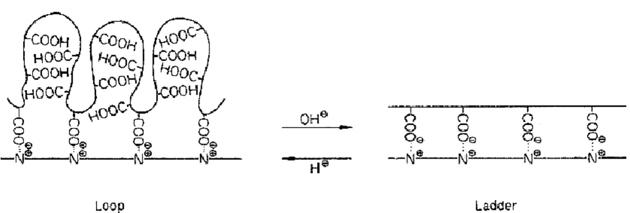
Tsuchida et al. suggested that the degree of dissociation of weak polyelectrolyte is affected by the presence of oppositely charged polyelectrolyte (8). It was demonstrated that the pKa of the poly(methacrylic acid) (PMAA) decreases in the presence of various polycations. Polycarboxylic acid in the complex undergoes reversible structural changes with respect to the solution pH. In the lower pH region it may take randomly coiled conformations and exhibit a looped shape due to the chains flexibility and weak electrostatic repulsions. At higher pH values the strong electrostatic repulsions between
RT G RT G G pH cs 3 . 2 ) , ( 3 . 2 ) ( ) ( ) , (
21
ionized carboxylic groups favor the increase in the rigidity of the chain backbones and the extended conformation of the polycarboxylic acid chains (Figure 3).
Figure 3. Reversible structural changes of PMAA with respect to the solution
pH (8).
Nakajima et al. reported that the rigidity of polyelectrolyte chains affects the complex composition (11). The studies on the complexes made of the polysaccharide chains revealed that the complexes prepared with more rigid polymer chains deviated from expected stoichiometry.
The formation of the complexes may favor the conformational changes of the macromolecular chains. The conformational transitions of poly(L-lysine) (PLL) in the complex with respect to the configuration (tacticity) of PMMA molecules were studied (12). It was described that iso-PMAA increases the number of the α-helical PLL chains in the complexes, whereas PMAA with lower stereo-regularity favor the helix-destructing effects.
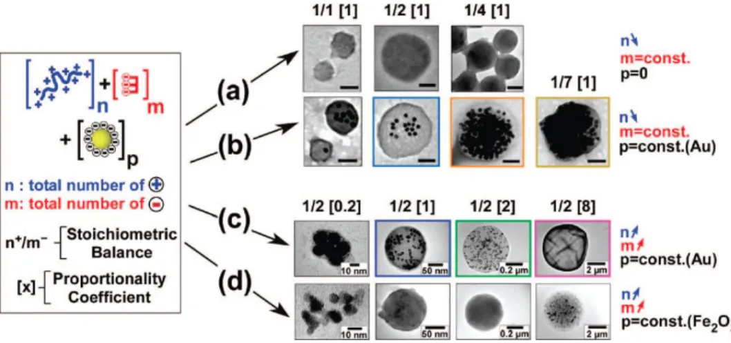
A new approach to control the flocculation process and therefore the parameters of forming complexes was demonstrated by Schneider and Decher in 2008 (13). The reported flocculation system was based on three components: trisodium citrate, poly(allylamine) and negatively charged citrate-stabilized gold nanoparticles.
22
Hybrid nanoparticle-filled aggregates or nanobags were formed after the injection of poly(allylamine) solution in the mixture of gold nanoparticles and trisodium citrate. It was established that nanobags parameters such as size, morphology and loading capacity can be easily controlled by tuning stoichiometric balance between the total number of positive and negative charges and the concentration of poly(allylamine) or citrate (Figure 4).
Figure 4. Transmission electron micrographs of individual flocs formed through
the flocculation of a three component system: poly(allylamine hydrochloride) , blue, a total of n positive charges; e.g., n+), trisodium citrate (red, a total of m negative charges; e.g., m-) and either gold (Au) or iron oxide (Fe2O3) citrate-stabilized nanoparticles (p) const; 1.2 nM in nanoparticles).
23 1.2. Layer-by-Layer assembly
Since its introduction in 1991, the Layer-by-Layer (LbL) deposition technique became a versatile method for the construction of nanostructured coatings, in general, for surface functionalization and engineering of multifunctional films (14, 15).
Layer-by-Layer buildup is based on intermolecular interactions, e.g. electrostatic interactions, hydrogen bonding, charge transfer, covalent bonding, hydrophobic interactions…, and subsequent formation of complexes between the macromolecules on the surface of the substrate (15).
Generally, LbL films are prepared under mild conditions and are assembled with no restriction to the size, shape or material used as a template (14, 15).
Due to its versatility, LbL assembly is attractive for engineering of the advanced biomaterials. Nanoparticles, clay platelets, enzymes, polypeptides, nucleic acids, proteins, polysaccharides, viruses, living cells can be incorporated into the film towards such purpose (Figure 5).
24
LbL assembly is an ideal tool to control the sustain release of bioactive molecules or drugs by varying their location in the film. Moreover, the concentrations of the film components can be controlled by the number of the corresponding layers. The LbL coatings are used for the creation of artificial photosynthesis membranes, bioreactors, biosensors, superhydrophobic surfaces and superstrong materals as well as for the improvement of biocompatibility of medical implants and for the design of the materials intended to control cellular adhesion and proliferation (15-18).
1.2.1. Layer-by-Layer formation
One of the important features of LbL assembly is its simplicity (14, 19).
As shown in Figure 6, the buildup of multilayers starts by bringing the substrate carrying a net charge in the contact with solutions of oppositely charged polyelectrolytes. The deposition of the polyelectrolyte layer normally leads to the overcompensation of the original surface charge (20). The alternation of the charge after each layer deposition allows to adsorb a polyelectrolyte of opposite charge and therefore to continue the film build up process (Figure 7).
After the substrate is rinsed to remove un-adsorbed or weakly adsorbed polymer chains, a polyelectrolyte carrying an opposite charge to the first layer is deposited. The rinsing of the substrate completes the formation of the first layer pair. Subsequently, additional layers can be deposited in the same manner yielding a multilayer film of desirable thickness and properties.
25
Figure 6. A simplified schematics of LbL deposition process (14).
LbL films can be assembled via dipping, spray-assisted assembly and spin-assisted assembly (15). The two latter methods have the advantage to reduce the deposition time of the layers and the volume of the solutions used for LbL build-up on large substrates. The time for the deposition of a single layer is about 15 min for dipping and about 10 sec for spray-assisted assembly.
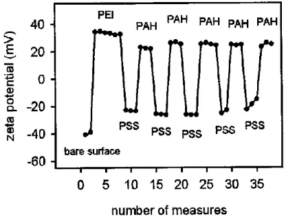
Figure 7. Evolution of the ζ-potential during the buildup of a PEI(PSS/PAH)5
26
The comparison of the layers deposited via dipping and spray-assisted assembly revealed that the sprayed films are thinner and smoother than the dipped ones (21). Therefore, changing the method of LbL assembly is another tool for tuning the properties of multilayer films.
1.2.2. The structure of polyelectrolyte multilayer films
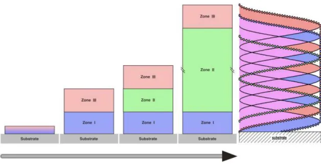
Often, the inner structure of LbL films represent a framework subdivided onto three zones (20) (Figure 8).
Zone I (precursor zone) consists of one or a few polyelectrolyte layers close to the substrate. The properties of these layers are defined by the characteristics of substrate surface e.g. charge density, substrate roughness, contact angle etc.
Figure 8. Schematic of the multilayer inner structure by the model of the three
zones (20).
The zone III (outer zone) is formed by one or few layers close to the surface of the film. The properties of this zone are defined by the properties of solutions or
27
the air. The external part of this zone consists of polyelectrolyte loops “dangling” from the film network into the solution.
The zone II (core zone) or “bulk film” frequently contains 1:1 stoichiometric polyelectrolyte complex. The zones I and III are formed during the first step in the deposition process. After these zones finalize their composition and thickness, the zone II forms and increases in thickness during the assembly process.
While zone I and zone III possess a small gradient of charge excess, the zone II is assumed to be electrostatically neutral due to the charge compensation of two oppositely charged polyelectrolytes. The borders between zones I and II and zones II and III are smooth. The thicknesses of the zones are defined by the substrate nature, the chemical structure of deposited polyion pair and conditions of the deposition process.
1.2.3. LbL growth regimes
There are two growth regimes for multilayered films. The LbL film is said to exhibit linear growth if its thickness and the amount of polyelectrolytes deposited on the surface of the substrate increases linearly with the layer pair number. The film possesses a somewhat fuzzy layered structure (Figure 8) as described above (see 1.2.2), and each polyelectrolyte layer forms an interpenetrating network with the closest neighbors.
Another growth regime, referred to as exponential, occurs, while it is possible for at least one of the components of the film to diffuse in and out of the film matrix at each layer deposition step.
One of the first examples of exponentially growing films was demonstrated by Elbert and co-workers. The film based on PLL and alginate (Alg) was constructed as a non-adhesive barrier for the cells on the top of the gelatin layer. At the physiologic pH the thickness of the dry film measured by
28
ellipsometry grown exponentially during the deposition of each polyelectrolyte layer (22).
Later, Picart et al. investigated the exponential growth of a multilayer film contained PLL and hyaluronic acid (HA) by optical waveguide lightmode spectroscopy, streaming potential measurements, atomic force microscopy and quartz crystal microbalances (QCM) (23). It was shown that (PLL/HA)n film build up proceeds via two different regimes. First regime corresponds to the formation and growth of isolated islands and islets on the top of the substrates, while the second stage of the films formation represents the coalescence of the islands to a homogeneous flat film. The authors suggested that the exponential growth of the film occurs due to the diffusion of free polyelectrolyte chains in and out the film, while the film is in contact with corresponding polyelectrolyte solution. The diffusion of PLL molecules during the deposition process was further confirmed by confocal laser scanning microscopy with fluorescently labeled PLL and HA molecules (24).
Based on these observations, the model of exponential growth regime was described by Lavalle et al. in 2002 (25). It was suggested that the films assembled with poly(glutamic acid) (PGA) and poly(L-lysine) contain two kinds of PLL chains: the first type of the chains strongly interacts with oppositely charged PGA and forms the multilayer network, whereas the second type of PLL macromolecules weakly binds to the film and thereby exhibits a certain mobility. Upon the formation of new PGA layer, the PGA macromolecules strongly interact with the PLL chains forming the top layer of the film (Figure 9). The mobile PLL chains then start to slowly diffuse out of film network, reach the film/solution interface, where they react with PGA chains from the solution forming PLL/PGA complexes. Further, these complexes interact with each other and form large entities. After most of the mobile PLL chains reach film surface, PGA chains diffuse into the film that leads to the surface charge overcompensation. While most of the free PGA chains diffuse out of the film after rinsing with buffer solution, the weakly bound PGA chains remain within the film. This process repeats during the deposition of the following PLL layer.
29
Exponential growth switches over to the linear regime after a certain number of deposition steps even if the diffusion of the polyelectrolyte still takes place. The switch from exponential to linear growth is thought to be due to the fact that polymer cannot reach the bottom of the film during the deposition time for this specific layer.
Figure 9. Schematic model of exponential growth regime of (PGA/PLL)n film (25).
30 1.3. Biological applications of PEC
1.3.1. LbL films as substrates for cell adhesion
Layer-by-Layer films were extensively exploited for the development of the substrates intended to control cell attachment and growth.
It was demonstrated that the cell adhesion can be manipulated by tuning the conditions of multilayer buildup, chemical composition, topology and rigidity of the films.
Richert et al. demonstrated that the cell adhesion properties of the multilayer films based on PLL and poly(L-glutamic acid) (PLGA) were defined by the pH of polyelectrolyte solutions used for film assembly. The films assembled at pH=4.4 possessed cell adhesive properties, while the architectures built at pH=10.4 prohibited cell attachment. It was suggested that the properties of multilayer coating are governed by their secondary structures and the differences in the film swelling properties (26).
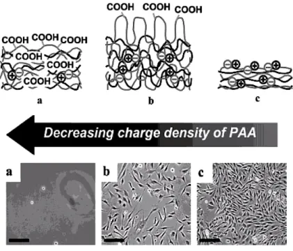
Mendelsohn and co-workers reported that the assembly of (PAA/PAH) layers at pH=6.5 supports cell growth, whereas the preparation of the films under acidic conditions (pH=2) results in the formation of bioinert coating (27). The drastic difference of the cell behavior was associated with the changes in the PAA charge density within the film (Figure 3). Highly ionically linked films attracted cells, while the weakly ionically linked multilayers tend to swell in physiological conditions developing highly hydrated surfaces, which resist fibroblasts attachment (Figure 10).
31
Figure 10. Schematics of the film structures and phase contrast micrographs of
murine NR6WT fibroblasts seeded onto (a) a (PAH/PAA)20 film prepared from the solutions of PAH and PAA at pH=2.0 (b) a (PAH/PAA)20 film prepared from the solutions of PAH and PAA at pH=7.5 and 3.5, respectively (c) a (PAH/PAA)50 film prepared from the solutions of PAH and PAA at pH=6.5 (27).
It was also demonstrated that the layers prepared at pH=6.5 and then treated at pH=2 possess cytophobic properties. Moreover, subsequent treatment of the same substrate at pH=6.5 led to the regeneration of the cytophilic properties of the film (28).
Other important parameters, which contribute to the cell-substrate interactions, are surface rigidity and topology.
Richert et al. demonstrated that the increase in the rigidity of the (PLL/HA)n films
by their chemical crosslinking improves the smooth muscle cell adhesion properties of LbL films. The crosslinking not only turns the layers cytophilic but also improves their resistance to enzymatic degradation (29).
32
smooth muscle cells can be controlled by the flexibility of the films (30). Since the (PAH/PAA)n films were prepared at different pH of polyelectrolyte building
solutions and therefore differed in their thickness, the thermal crosslinking of the multilayers yielded the formation of coatings of various rigidity and charge. The films prepared at pH 7.4 (solution of PAH)/4.6 (solution of PAA) were thicker and more flexible compared to films prepared at pH=7.4 for both polyelectrolyte solutions. It was found that cells phenotype was modulated with respect to the LbL mechanical properties (Figure 11).
Figure 11. Localization of total actin and smooth muscle R-actin in A7r5 cells
cultured on native and cross-linked (PAA/PAH) films. Cells were grown on native (A-C) and cross-linked (D-F) (PAH/PAA)4/PAH-coated coverslips. Actin filaments are stained with Phalloidin-Alexa 488 (green) and smooth muscle R-actin is labeled with a specific anti-R-R-actin antibody and Alexa 546-secondary antibody (B and E). Overlaid dual-labeled images (C and F); scale bar 10 μm (30).
33
Boudou et al. studied the correlation between the mechano-chemical properties of native and crosslinked (PLL/HA)n, (CH/HA)n and (PAH/PGA)n and the adhesion of skeletal muscle cells and NIH 3T3 fibroblasts. It was demonstrated that independently of the film composition and the presence of serum proteins, the cells better adhered, spread and proliferated faster on the crosslinked films forming stiffer surfaces (31).
The properties of LbL architecture can be altered by the incorporation of adhesive proteins into the films. Kirchhof et al. compared osteoblasts MG-63 cell adhesion onto the substrates coated with (CH/HEP)n multilayers with or without terminal layer of plasma fibronectin (32). The highest cell population was achieved on the LbL-coated substrates terminated with fibronectin, due to the presence of cell binding motifs such as RGD on the film surfaces. Moreover, the cell population was found to be higher on the coating with chitosan layer under that of fibronectin compared to the architecture with the layer of heparin preceded fibronectin layer.
Another possibility to improve cell adhesion properties of the LbL films is the post-functionalization of the terminating layer with bioactive molecules. One of the examples of such approach was demonstrated by Kinnane and co-workers (33). The authors reported the preparation of the anti-fouling poly(ethylene glycol) (PEG) films covalently assembled via Cu(I)-catalyzed cycloaddition of alkaline and the azide groups incorporated into PEG polymer chains and carboxylic groups in RGD. The prepared films were further modified by grafting the RGD sequence via click reaction with the free azide groups in the PEG. While the monkey kidney epithelial cells were poorly interacting with the native architectures, the cell adhesion and proliferation were enhanced on the substrates functionalized with RGD.
In another work, the effects of polyelectrolytes PAH and PAA in the solution or embedded into the film on the cell metabolism, proliferation and survival were studied. The experiments were carried out with rat aortic smooth muscle cells and osteosarcoma cells. It was shown that the solution of positively charged
34
PAH was much more toxic to both cell types compared to that of negatively charge PAA and caused necrotic cell death. By contrast, the low concentration PAA solutions were well tolerated by the cells, whereas at higher concentrations PAA induce the increase in cell metabolism (34). The incorporation of the polyelectrolytes into the films reduces their cytotoxic and cytostimulatory effects. It was observed that, regardless the thickness, the PAH-terminated films caused lower cell proliferation compared to those with the PAA outmost layer. Moreover, the thicker (PAA/PAH)n films yielded lower cell density than the thinner ones. The authors concluded that the electrostatic interactions between film components reduce their availability for cellular uptake or for the interaction with the cell membrane.
Layer-by-Layer assembly is successfully applied for the improvement of biocompatibility and antimicrobial properties of the implantable substrates (35). Kerdjoudj and co-workers described the preparation of cryopreserved vessels modified with LbL film with improved mechanical properties (36). The use of cryopreserved arteries for vascular tissue engineering meets certain limitations since the cryopreservation induces structural changes of the vessels, which generate the alteration in the biomechanical properties of the implanted vessels. Moreover, the cryopreservation provokes the loss of endothelial cells, leading to the direct interaction of blood components with extracellular matrix. These undesirable interactions induce thrombosis and/or restenosis after implantation. To improve the endothelial cell adhesion onto the vessel, the luminal surface of cryopreserved umbilical arteries was covered by (PAH/PSS)3/PAH films. The multilayer coating improves the mechanical properties of the defrosted vessels allowing to decrease the maturation time of vascular grafts. It was demonstrated that coated implants enhance cell adhesion and spreading (Figure 12). By contrast with the uncoated arteries, the cells seeded onto the multilayer-coated vessels exhibit flat and elongated morphology, keep their phenotype and form sub-confluent monolayer compared to the uncoated arteries. LbL-coated vessels maintain their functionality being
35
similar to that of fresh arteries and therefore present novel biocompatible grafts with improved biomechanical and anti-thrombogenic properties.
Figure 12. Scanning electron micrographs of untreated (A), covered with LbL
film (B) cryopreserved umbilical arteries with endothelial cells and fresh umbilical arteries (C) (36).
Recently, Davila and colleagues presented a new type of mechanotransductive film, where the layers made of RGD-grafted polyelectrolytes were assembled under the nonadhesive layers bearing phosphocholine (37). This LbL film supported cell adhesion upon the stretching by rendering RGD-ligands accessible to the cells and inducing cell adhesion through specific interactions. The effect was found partially reversible upon the relaxation due to the film restructuring that led to the only a partial remasking of RGD peptides.
1.3.2. Gene delivery systems
The primary challenge of gene therapy is a development of a safe and efficient method to deliver exogenous genetic material to the target cells and induce the production of the therapeutic proteins correcting or modulating damaged cellular functions. The transfer of the exogenous nucleic acids – transfection – is one of the stages in gene therapy process (38-41).
Due to their properties such as large size, negative charge and hydrophilicity, nucleic acids (DNA or siRNA) cannot easily cross the cellular membrane and
36
need to be incorporated into a system – vector – that facilitates cell binding and internalization and protects genes from degradation. Depending on the nature of the vector, the methods of gene therapy can be divided on viral and non-viral approaches.
Currently, viruses are the most efficient particles for injecting DNA into the target cells. However, the broad use of viruses that were genetically gutted of their genetic material and replaced with therapeutic genes (42) is limited due to the possible side-effects such as immunogenicity and oncogenic effects via random transgenic insertion of viral genetic material into the host chromosome followed by disruption of normal gene expression (43).
Methods of non-viral gene delivery present a promising alternative to viral vectors and include two main groups: physical and chemical methods.
Physical methods employ physical force that induces the defects in the cell membrane giving an opportunity to nucleic acids enter the cell, and include gene gun (mechanical forces), electroporation (electric forces), hydrodynamic (hydrodynamic forces) and ultrasound-facilitated gene transfer methods as the main gene delivery techniques (39, 44).
1.3.2.1. Polycation/DNA complexes for gene delivery
A common approach for delivering nucleic acid by chemical methods includes the formation of interpolyelectrolyte complexes (polyplexes) through electrostatic interactions between negatively charged phosphate groups of nucleic acid and positively charged groups of macromolecules (45, 46).
The general requirements for synthetic delivery system include 1) the ability of a macromolecule to effectively condense nucleic acid to particles with a size appropriate for cellular uptake 2) the enhancement of the complex binding capacity to the cell surface by masking DNA negative charge 3) protection of
37
DNA from degradation in extra- and intracellular environment and 4) delivery of genetic material to the site of action.
Currently, most of the non-viral delivery systems bear amine-based positively charged groups and include various polymers, dendrimers, lipids and peptides (47-50). The composition of the polyplexes is characterized by N/P ratio - the molar ratio between polycation groups containing nitrogen and negatively charged phosphate groups of DNA.
The transfection efficiency of the non-viral vectors is defined by many factors such as type of cell line, chemistry of nanoparticles, surface charge, size, stability of the aggregates, their solubility in the cell culture conditions and ability of the polycation to protect the nucleic acid from degradation into the cell environment (47-49).
1.3.2.2. Formation of polycation/DNA complexes
The mechanism of the complex formation is similar to that described for the synthetic polyions (see section 1.1.2). The mixing of aqueous solutions of nucleic acid and polycation leads to the formation of soluble PECs via cooperative interactions between electrostatically complementary chains.
In general, the molecular mass and the contour length of polycations are much lower than those of recombinant DNA molecules and therefore the polycations can be considered as “a guest” and DNA as “a host” (45, 46)
At low N/P ratios, while the concentration of DNA is higher than that of polycation, the water-soluble negatively charged nonstoichiometric complexes form. The charge of the complex increases with the content of polycation. At certain critical polycation concentration the two types of PECs form: nonstoichiometric complexes that stay in the solution and stoichiometric complexes formed in the excess of polycation macromolecules that either
38
precipitate or aggregate. The fraction of stoichiometric complexes increases with polycation concentration.
At N/P 1, while the molar concentrations of DNA and polycation are equal, all chains of nucleic acid are involved in the formation of stoichiometric complexes (46).
Sedimentation studies showed that DNA incorporated into PEC possesses a more compact structure. The investigation of DNA condensation process with poly(L-lysine) covalently coupled to glycoprotein asialoorosomucoid was performed by Golan and co-workers (51). It was demonstrated that depending on the composition of the polyplexes (N/P ratio), the complexes exhibited either toroid or short rod shape with thickness similar to that of chromatin fibers.
1.3.2.3. Poly(ethylenimine)
Poly(ethylenimine) (PEI) is considered as one of the most effective synthetic gene transfer agents (52).
Depending on the synthesis, linear or branched PEI is obtained. Both derivatives can be used as transfection agents. Synthesis of branched PEI is performed via cationic polymerization, while the linear form is obtained as a product of ring-opening polymerization and subsequent hydrolysis (Figure 13) (49, 52, 53).
39
Since each monomer unit contains an amino group, PEI possesses a high charge density that allows to condense DNA into the small particles. The size of the particles is strongly dependent on the complex formation conditions and is discussed later.
Poly(ethylenimine) demonstrated high transfection efficiency due to enhanced endosomal release via the so-called ”proton sponge” effect. At physiological pH only 20% of the amine groups in PEI are protonated, while at pH=5 their concentration increases up to 45%. During intracellular traffiking PEI inhibits the nuclease activity due to its buffering capacity and induces the accumulation of protons within the endosome coupled to an influx of chloride anions. The presence of PEI alters the osmolarity of the endosome resulting in osmotic swelling. The protonation of PEI enhances charge repulsion of polymer chains and therefore induces swelling of polymer network. Both of these effects lead to the rupture of the vesicle and to the release of the carrier into the cytoplasm (54).
For efficient binding to the cell surface, the polyplexes are prepared in excess of PEI and therefore possess a net positive charge (53). However, it was demonstrated that at high N/P ratios the particles induce undesirable toxic effects due to the vector aggregation on the cell surface followed by cell membrane rupture (55, 56).
The degree of branching influences gene transfection efficiency and cytotoxicity. Although branched PEI is more effective in the DNA condensation and exhibits high transfection ability, linear PEI shows better efficiency in multiple in vivo studies (57, 58).
Molecular weight of PEI is another important parameter influencing polyplex formation, transfection capacity and toxicity. It was demonstrated that the efficiency and the toxicity of polyplexes increases with PEI molecular weight. The optimal molecular weight was found to be between 5 and 25 kDa (53).
40 1.3.2.4. Transfection mechanism
After DNA/polycation complex is added to the cell culture, the first barrier it has to overcome is to pass through cellular plasma membrane (Figure 14). As stated above, the passive diffusion is not possible, but since polyplexes are positively charged they can bind to the cell membrane through ionic interactions with proteoglycans – membrane-associated core proteins that bear sulfated or carboxylated glycosaminoglycans (GAGs): chondroitin sulfate, dermatan sulfate, keratan sulfate, heparan sulfate and hyaluronan. GAGs are highly negatively charged. The content of GAGs depends on the cell type and is associated with transfection efficiency (59-61).
41
Endocytotic uptake of the polyplexes proceeds via multiple mechanisms: clathrin- mediated endocytosis, caveolae-mediated endocytosis, macropinocytosis, phagocytosis and clathrin- and caveolae – independent endocytosis (62). The pathway of uptake depends on cell line, polyplex properties and the polyplex formation conditions and can be controlled by chemical modification of vector with targeting ligands or cell penetrating peptides (62-64).
The second important stage is the escape of polyplexes from endosomes to avoid their enzymatic degradation within the lysosomes. Incorporation of DNA molecules into the polyplexes mediates its accessibility to nucleases in the cytosol. It was demonstrated that the restriction of DNA embedded into a complex consisted of two stages: 1) fast cleavage of the DNA fragments not covered with polycation chains and 2) slow restriction of DNA in the polyplexes. Since the chains of polycation are not strongly attached to those of DNA, they migrate from one DNA molecule to another opening the sites for cleavage. Thus, the kinetics of second phase depend on the migration ability of polycation molecules (45, 65). After nuclear entry occurring through nuclear pores or during mitosis (66, 67), gene expression should take place. Stable, long-term expression is achieved through insertion of gene into host genome whereas transient short-term expression occurs from transgene retention within the nucleus.
1.3.3. Layer-by-Layer assembly as a platform for surface-mediated transfection
The first report by Lvov and co-authors in 1993 of the DNA incorporation into the multilayer films demonstrated the possibility to apply the LbL assembly in the field of the gene delivery (68). The surface-mediated transfection provides several advantages compared to the bulk transfection methods such as spatial and temporal control over the DNA loading in the films as well as the possibility to incorporate different types DNA at different film levels.
42
In this part, several examples of efficient DNA delivery by LbL films are presented.
One of the approaches for the promoting the surface-mediated transfection is based on applying hydrolytically degradable cationic polymers for the film construction.
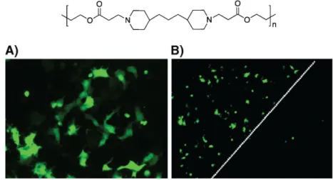
In 2005 Jewell et al. reported the use of DNA-containing multilayer films that gradually release DNA in physiological conditions. The film was built up using pDNA coding green fluorescent protein and Polymer 1 (Figure 15) on the quartz slides precoated with (PEI/PSS)n precursor layers (69). Polymer 1 is positively charged at pH=5 and belongs to the class of hydrolytically degradable (at neutral pH) synthetic polymers, (poly(β-amino ester)s. After the incubation of COS-7 cells with the LbL-coated samples during 48h, the majority of transfected cells were located directly under the LbL-coated substrate, which therefore provides the spatial control over the DNA delivery (Figure 15). It was suggested that the film released the condensed pDNA and therefore enhanced the transfection efficiency. Later study confirmed the presence of small aggregates from 100-600 nm in PBS used to incubate (Polymer1/DNA)n film (70).
Figure 15. Structure of Polymer 1 and fluorescent microscopy images of
transfected COS-7 cells located under the film-coated substrate (A) and on the edge of a film-coated slide (indicated by white dotted line) (69).
43
Recently, it was demonstrated that these films can be used to deliver pDNA across the skin (71). The insertion of the micrometer-scale needles (~750 μm long) coated with (Polymer1/fluorescently labeled DNA) film into the porcine cadaver skin over 2-h period allowed the deposition of the DNA along the needle track to the depth of 500-600 μm (Figure 16).
Figure 16. Representative fluorescence microscopy images (A) and companion
histology images (B) of porcine cadaver skin after insertion and removal the microneedles coated with polymer 1/fluorescently labeled DNA). Scale bars 400 μm (71).
The LbL assembly allows to construct the films that release DNA under particular conditions.
It was shown that the application of the reduction potential to the (Polymer1/DNA)n-coated electrode generated a rapid release of DNA over the periods of 1 to 2 min (72). The applied potential provokes the increase in the solution pH near the electrode surface resulting in the hydrolytic polymer degradation. DNA released under these conditions remained active and promoted the gene expression in COS-7 cells.
Chen and co-authors applied the cationic poly(amidoamine) polymer (Figure 17) containing di-sulfide bonds for the construction of LbL film containing DNA (73). While stable in PBS buffer, this film disassembled in the presence of reducing agent dithiothreitol (DDT). The rates of film erosion correlated with the
44
concentration of DDT. The highest film disassembly rate (30 min) was achieved in the 10 mM DDT solution, whereas the reduced DDT concentration (1 mM) generated the film erosion during 9 days.
Figure 17. Structure of cationic poly(amidoamine) polymer used in (73).
Blacklock and colleagues applied the reducible hyperbranched poly(amino amine) (Figure 18) and DNA encoding the secreted alkaline phosphate (SEAP) and GFP for film construction on the flexible stainless steel substrate (74). The release of pDNA was thought to proceed via the contact with reducing environment of plasma membrane. In vivo this film showed higher transfection efficiency compared to that of lPEI/DNA-based polyplexes. The in vivo efficiency of the films was evaluated by implanting the coated substrate and measuring the SEAP level in the blood circulation of rats. The higher blood level of SEAP was registered on the day 5 after the implantation followed by sharp SEAP level decline due to the formation of collagen fibrous capsules near the substrate that created a barrier for sufficient distribution of SEAP in the blood. Later, it was reported that the transfection activity of this kind of film depends on the film rigidity and roughness and can be controlled by the salt concentration of polymer solution used for LbL assembly (75). The transfection efficiency was found significantly higher for the cells grown on the rigid film.
45
Figure 18. Structure of the reducible hyperbranched poly(amino amine) used in
(74).
1.4. Quartz crystal microbalance/Cell studies
The variety of cellular functions and properties that depend on the cytoskeleton rearrangements such as adhesion, spreading, mitosis, apoptosis…are often studied by the methods of flow cytometry or microscopy (76). Both methods require the additional steps of the cell labeling or staining, which limit their application for in situ characterization of the cell properties.
Quartz crystal microbalance is a technology applied for studying the changes in mass and viscoelastic properties near the surface of the sensor via the changes of the resonance frequency and energy dissipation of oscillating quartz crystal (see section 2.4, Materials and Methods). It is therefore commonly used for monitoring the formation and the properties of the thin films of polymers, proteins, lipids, DNA…at the surface of the sensor (77).
Aside from the information about the deposition of the thin films, quartz crystal microbalance with enhanced dissipation (QCM-D) is capable of providing the in situ characterization of living cells. Since cytoskeleton framework determines the cell mechanical properties, it is therefore possible to follow its rearrangements by measuring the changes in the QCM-D sensor frequency and energy dissipation.
In this section, several examples of the application of QCM-D for characterization of living cell properties are described.
46
One of the pioneering works of studying the cell adhesion on the surface of QCM-D sensor was performed by Fredriksson and co-workers (78). These studies were carried out using either the native gold polystyrene-coated QCM-D sensor (hydrophobic surface) or those treated by ozone under UV irradiation (hydrophilic surface). The experiments were performed in the serum-free medium with two cell lines: Monkey kidney epithelial cells and Chinese hamster ovary epithelial cells. It was shown that the cell adhesion resulted in the decrease in the resonance frequency corresponding to deposition of the effective mass on the electrode surface, while the energy dissipation increased due to the viscoelastic energy losses. The authors demonstrated that the QCM-D response strongly depends on the cell type adhered on the same type surface and on the type of the surface under otherwise identical experimental conditions.
Later, Nimeri and collegues demonstrated that the QCM-D technique can be used to monitor the specific cell adhesion onto the sensors surfaces covered with immunoglobulin G (IgG), human serum albumin (HSA) or fibrinogen (79). Although the attachment of neutrophils onto the sensors surface resulted in the frequency decrease and increase in the energy dissipation, the shape and the magnitude of the QCM-D response were particular for each protein-covered surface. The better cell spreading onto the surface coated with IgG generates higher decrease in the resonance frequency compared to the frequency shift for cells exposed to the HSA- or fibrinogen-coated surface.
Lord and co-workers demonstrated that QCM-D is capable of detecting the differences in the cell adhesion, which are not detectable by fluorescence microscopy (80). In this work, the adhesion of NIH3T3 fibroblasts to tantalum or oxidized polystyrene covered QCM-D sensors coated with serum proteins such as bovine serum albumin (BSA), human fibronectin or newborn calf serum proteins was studied in the serum-free medium over the period of either 2 or 4h. The QCM-D responses detected for the cells seeded at non-adhesive albumin covered surface were similar for both kinds of sensors. The fluorescence microscopy studies of the cells adhered to the sensor surfaces covered with