HAL Id: tel-01512355
https://tel.archives-ouvertes.fr/tel-01512355
Submitted on 22 Apr 2017HAL is a multi-disciplinary open access archive for the deposit and dissemination of sci-entific research documents, whether they are pub-lished or not. The documents may come from teaching and research institutions in France or abroad, or from public or private research centers.
L’archive ouverte pluridisciplinaire HAL, est destinée au dépôt et à la diffusion de documents scientifiques de niveau recherche, publiés ou non, émanant des établissements d’enseignement et de recherche français ou étrangers, des laboratoires publics ou privés.
Modulating the activity of NADPH oxidase by oxidative
stress participants ; lipids and nanoparticles A cell-free
system study
Rawand Masoud
To cite this version:
Rawand Masoud. Modulating the activity of NADPH oxidase by oxidative stress participants ; lipids and nanoparticles A cell-free system study. Radiochemistry. Université Paris Saclay (COmUE), 2016. English. �NNT : 2016SACLS028�. �tel-01512355�
NNT : 2016SACLS028
T
HESE DE
D
OCTORAT
DE
L
’U
NIVERSITE
P
ARIS
-S
ACLAY
PREPAREE A
L
’U
NIVERSITE
P
ARIS
-S
UD
ÉCOLE DOCTORALE N°571
Sciences chimiques : molécules, matériaux, instrumentation et biosystèmes
Spécialité de doctorat : Chimie
Par
Mme Rawand Masoud
Modulating the activity of NADPH oxidase by oxidative stress participants; lipids
and nanoparticles A cell-free system study.
Thèse présentée et soutenue à Orsay, le 16 février 2016 :
Composition du Jury :
Mme S. Lacombe Professeure, Université Paris-Sud Présidente du Jury M. F. Fieschi Professeur, Université Joseph Fourier Rapporteur Mme. P.M .Dang Chargée de Recherche, CNRS Rapporteur M. M. Ostuni Professeure, Université Paris Diderot Examinateur
Mme. C. Houee-Levin Professeur, Université Paris-Sud Co-directeur de thèse Mme. T. Bizouarn Chargée de Recherche, CNRS Co-directeur de thèse
Titre : Modulation de l’activité de la NADPH oxydase par des participants au stress oxidatif, les nanoparticules et le cholesterol
Mots clés : NADPH oxydase, stress oxydatif, cholestérol lipides nanoparticules Résumé : La NADPH oxydase de phagocyte est un
complexe enzymatique impliqué dans la défense immunitaire contre les pathogènes. Elle est constituée du flavocytochrome b558 membranaire (Cyt b558), composé
de deux sous-unités (gp91phox et p22phox) et de quatre sous-unités cytosoliques, p47phox, p67phox, p40phox, et Rac. Sa fonction est de produire les anions superoxyde (O2•−)
qui sont transformés en d'autres espèces réactives de l'oxygène (ROS) qui vont détruire les agents pathogènes mais aussi dans certains situations pathologiques attaquer les lipides, les protéines et l’ADN environnants. Après activation du phagocyte, les sous-unités cytosoliques subissent des modifications post-traductionnelles et transloquent vers la membrane pour former avec le Cyt b558 le complexe NADPH oxydase
activé. Le rôle délétère des ROS dans les maladies inflammatoires est connu depuis longtemps. Le but de ma thèse a été d’étudier l’influence de molécules exogènes qui induisent une augmentation du stress oxydatif, sur l’activité de la NADPH oxydase.
Dans ce travail, nous avons étudié le fonctionnement de la NADPH oxydase dans un système in vitro dans lequel l’enzyme était activée par la présence d'acide arachidonique (AA). J’ai étudié l'influence de deux types de molécules: une classe de lipides et des nanoparticules (NPs). Pour simplifier le système, nous avons remplacé l’ensemble des sous-unités cytosoliques par une protéine unique appelé trimère qui correspond à une fusion des trois protéines cytosoliques p47phox, p67phox et Rac. Nous avons montré que le trimère est fonctionnellement comparable aux sous-unités cytosoliques séparées. La vitesse de production de O2•−, sa dépendances en
fonction de la concentration en AA et et sa sensibilité aux radicaux libres sont comparables lorsque le trimère ou les sous-unités séparées sont utilisés.
J’ai étudié les conséquences de la présence du cholestérol et de ses formes oxydées sur la production de O2•− par la NADPH oxydase.
Nos résultats montrent clairement que le cholestérol et les oxystérols ne sont pas des activateurs efficaces de la NADPH oxydase. L’addition d’une quantité physiologique de cholestérol déclenche une faible production d’anions superoxyde. L’addition de cholestérol à des concentrations du même ordre de grandeur pendant le processus d'assemblage (en présence de AA), a un rôle inhibiteur sur la production d’O2•−. Le cholestérol ajouté agit sur les composantes,
cytosoliques et membranaires, conduisant à un assemblage imparfait. En conclusion, le cholestérol déjà présent dans la membrane des neutrophiles est optimal pour le fonctionnement de la NADPH oxydase.
Il était intéressant de vérifier l'influence des nanoparticules de dioxyde de titane (TiO2) et de platine
(Pt) sur le comportement de la NADPH oxydase sachant que l'internalisation cellulaire de ces NPs a pour effet d’activer les neutrophiles et les macrophages et contribue à une sur-production de ROS. En l’absence d'activateur mais en présence de NPs de TiO2 ou Pt,
aucune production de O2•− n’était détectée indiquant que
les NPs de TiO2 et Pt sont incapables d'activer le
complexe par eux-mêmes aussi bien dans le système acellulaire que dans les neutrophiles. Cependant, une fois la NADPH oxydase activée (par AA), les NPs de TiO2 entrainent une augmentation de la production des
O2•− jusqu’à 40% en comparaison aux conditions où elles
sont absentes. Cet effet est dépendent de leur concentration. Par contre, les NPs de Pt n’ont aucun effet sur l’activité de la NADPH oxydase aussi bien in
vitro que dans les neutrophiles. En conclusion,
l'hyperactivation de la NADPH oxydase et l'augmentation subséquente de la production des ROS induites par les NPs de TiO2 pourraient participer au
développement du stress oxydatif ce qui ne serait pas le cas de Pt NPs étant donnée leur absence d’effet.
Title : Modulating the activity of NADPH oxidase by oxidative stress participants, nanoparticles and cholesterol Keywords : NADPH oxidase, lipids oxidative stress, cholesterol, nanoparticles
Abstract: NADPH oxidase from phagocytes is a multi-subunit enzyme complex involved in the innate defense of organisms against pathogens. It is composed of the membrane-bound flavocytochrome b558 (Cyt b558), comprising two subunits (gp91phox, and p22phox)
and four cytosolic components, p47phox, p67phox, p40phox, and Rac. Its function is to produce superoxide anions that are transformed subsequently into other reactive oxygen species (ROS) and will destroy the pathogens but also can damage in certain pathological situations lipids, proteins and DNA. Upon phagocyte activation, the cytosolic subunits undergo posttranslational modifications and migrate to the membrane bound Cyt b558 to constitute the activated
NADPH oxidase complex. The damaging role of oxidative stress induced by ROS in cardiovascular diseases has been known for some decades. The aim of my thesis was to study the influence on NADPH oxidase activity, of molecules coming from food and industrial products and known to be involved in increase of oxidative stress.
In this work, we studied the NADPH oxidase functioning in an in vitro system in which the components of the enzyme are mixed and activated by the introduction of an amphiphile the arachidonic acid (AA). During my PhD, I have studied the influence of two types of oxidative stress participants: lipids and nanoparticles (NPs). For simplicity, we have replaced the cytosolic subunits by a single protein called trimera, which is a fused construction of three cytosolic proteins p47phox, p67phox and Rac. We have shown that trimera is functionally comparable with the separated cytosolic subunits. The rates of production of O2•−, the dependences of the activity in function of AA
concentration and temperature, the presence of two states in the activation process and the sensitivity of NADPH oxidase to free radicals were comparable when either trimera or separated subunits were used.
I investigated the consequences of the addition of cholesterol on NADPH oxidase, on the production of ROS. Our results clearly show that cholesterol and oxysterols are not efficient activators of NADPH oxidase. Concentrations of cholesterol similar to what found in neutrophiles trigger a low superoxide production. Addition of cholesterol during the assembly process (in presence of AA) at similar or higher concentrations, has an inhibitory effect on the production of O2•−. Added cholesterol acts on both
cytosolic and membrane components, leading to imperfect assembly and decreasing the affinity of cytosolic subunits to the membrane ones. In conclusion, we showed that the cholesterol already present in the phagocyte membrane is optimal for the function NADPH oxidase.
It was of interest to check the influence of titanium dioxide (TiO2) and platinum (Pt) NPs on NADPH
oxidase especially that cellular internalization of NPs was shown to activate neutrophils and contribute to O2•−
overproduction via NADPH oxidase. In the absence of activators and presence of TiO2 or Pt NPs, no
production of O2•− could be detected in in vitro system
as well as in neutrophils indicating that TiO2 and Pt NPs
were unable to activate by themselves the complex. However once the NADPH oxidase was activated by AA, TiO2 NPs increased the rate of O2•− production by
up to 40%, this effect being dependent on their concentration. Differently, Pt NPs had no effect both on in vitro system as well as on neutrophils. In conclusion, the hyper-activation of NADPH oxidase and the subsequent increase in ROS production by TiO2 NPs
could participate to oxidative stress development while the absence of Pt NPs effect suggest that they do not induce inflammation status via this complex.
“
As far as you dream goes, the
earth will get bigger
”
ACKNOWLEDGEMENTS
I would like to express my gratitude to many people who supported and helped me to bring this research project to fruition.
First and foremost I offer my sincerest gratitude to Pr. Mehran MOSTAFAVI and Pr. Philippe MAITRE for welcoming me in the laboratory and for their venerable suggestions.
A very special thanks goes out to Pr. Chantal HOUEE-LEVIN who gave me the opportunity to work on this subject. She is the one professor who truly made a difference in my life. It was under her tutelage that I developed myself. I am not sure that many students are given the opportunity to develop their own individuality and self-sufficiency by being allowed to work with such independence such as I had. She provided me with aspiring guidance, unlimited support and friendly advice. I doubt that I will ever be able to convey my appreciation fully, but I owe her my eternal gratitude.
I think that no word can express the gratitude I have to my supervisor Dr. Tania BIZOUARN, without her, I would not have been able to achieve my PhD. I am sincerely grateful for her understanding, patience and availability throughout these years. She supported me not only by providing a research assistantship, but also emotionally through the rough road to finish this thesis. I could not have imagined having a better supervisor and mentor for my PhD study.
I would like to express my deepest gratitude to Pr. Frank FIESCHI and Dr. My-chan DANG for accepting to be the rapporteurs of my thesis and for their careful reading and valuable comments on my work. I also thank Pr. Sandrine LACOMBE and
Dr. Mariano OSTUNIfor having agreed to judge this work and to participate in the jury.
I would like to record my deep sense of gratitude to Dr. Laura BACIOU and Dr. Florence LEDERER for the help, encouragement and valuable scientific discussion. My sincere thanks go to Dr. Hynd REMITA for her continuous support and valuable advices throughout my PhD.
I gratefully thank Dr. Sergio MARCO for the work collaboration we had at Institut Curie which was very fruitful for shaping up my ideas and research. I also benefited by outstanding works from Dr. Sylvain TREPOUT’s help with his particular skill in handling precisely the transmission electron microscope. My thanks to Dr. Frank WEIN and Dr. Matthieu REFREGIERS for their help in achieving CD experiments in Soleil synchrotron.
I must acknowledge Abdel Karim EL OMAR for his moral support, he was a true friend ever since. Karim is an amazing person in too many ways. I must also thank Daniela SALADO for the great cooperation and nice times we had together.
I also extend my thanks for all the members of the laboratory, the professors, the researchers and particularly: Marie ERARD, Delphine ONIDAS, Sophie BERNAD, Fabienne MEROLA, Cecile SICARD, Emilie BRUN, Filippo RUSCONI and Valerie DERRIEN for creating a pleasant working atmosphere. I wish also to record my thanks to Paul MACHILLOT who has been an inspiration on how to make experiments perfect and for sharing with me his remarkable knowledge. And I also thank Yasmina BOUSMAH who has been a source of love and energy ever since. Additionally, I express my warm thanks to all the administrative members
especially Marie-François LECANU and Severine BOURGUIGNON for their indispensable help dealing with travel funds, administration.
In my daily work I have been blessed with a friendly and cheerful group of friends with whom I have shared good times particularly Myriam MOUSSAOUI, Guilda KARIMI, Cornelia ZIEGLER, Xavier SERFATY.
Where would I be without my family? My parents and especially my mother deserve special thanks for their inseparable support, love and prayers. Even if they are far away, it was with their help and under their watchful eye that I gained so much drive and an ability to tackle challenges head on. Words fail me to express my appreciation to my husband Mohammed ABUOLBA whose unwavering love, quiet patience, encouragement and persistent confidence in me, has taken the load off my shoulder. He was always there cheering me up and stood by me through the good and bad times. This accomplishment would not have been possible without him. I want to especially thank my sisters Dalia and Lina and my brother Mohammed who provided me with advice at times of critical need and consistently supported me despite the thousands of miles that separate us.
Finally, and most importantly, I must thank my daughter Louna who was born during my PhD, her smile was the biggest support during the hard times.
I would like to thank everybody who contributed to my success. Thank you all for supporting me during these years in both private life and in work.
SUMMARY
Introduction ... 18
I. NADPH oxidase history ... 19
II. NADPH oxidase family ... 21
1. Structure and Function ... 21
2. Tissue distribution and physiological roles ... 22
III. Reactive oxygen species production ... 23
IV. Phagocyte NADPH oxidase ... 26
1. Composition of the NADPH oxidase complex ... 28
2. Structure and functions of the membrane subunits ... 28
i. gp91phox ... 28
ii. p22phox protein ... 30
3. Structure and functions of cytosolic subunits ... 31
i. p47phox protein ... 31
ii. p67phox protein ... 33
iii. p40phox protein ... 34
iv. Rac ... 35
v. Trimera ... 37
V. Activation of NADPH oxidase complex ... 38
1. Physiological activation ... 38
2. Non physiological activation ... 40
VI. Diseases related to the malfunction of the NADPH oxidase ... 41
VII. Membrane lipids ... 42
2. Glycolipids ... 44
3. Sphingolipids ... 45
4. Sterols ... 46
VIII. Lipid rafts ... 48
IX. NADPH oxidase and lipids ... 49
X. Nanoparticles ... 51
1. Generalities ... 51
2. Titanium dioxide nanoparticles ... 53
3. Platinum nanoparticles ... 54
Objectives ... 56
Materials & Methods ... 58
I. Preparation of proteins ... 59
1. Preparation of membrane fractions ... 59
2. Expression and purification of cytosolic proteins ... 60
i. Culture of bacteria expressing cytosolic proteins in E.coli ... 60
ii. Extraction of proteins ... 61
iii. Purification of p47phox ... 62
iv. Purification of p67phox and Rac1Q61L ... 64
v. Purification of trimera ... 65
II. Proteins analysis ... 66
1. Determination of proteins concentration ... 66
2. Evaluation of protein purification by SDS-PAGE ... 67
III. Functional study of NADPH oxidase proteins ... 68
1. Measurement of superoxide anion production rate ... 68
IV. Neutrophil cell activity measurement ... 70
V. Structural study of NADPH oxidase proteins ... 71
1. Intrinsic fluorescence Assays ... 71
2. Circular dichroism ... 71
VI. Cholesterol measurements ... 72
1. Quantification of intrinsic cholesterol in neutrophil membrane fractions ... 72
2. Depletion of cholesterol ... 75
VII. Quantification of H2O2 produced upon NADPH oxidase activation ... 75
VIII. Nanoparticles characterization ... 76
1. Dynamic light scattering measurement ... 76
2. Transmission electron microscopy ... 77
IX. Gamma irradiations ... 77
Results & Discussion ... 80
CHAPTER 1: CHARACTERIZATION OF TRIMERA ... 81
I. Introduction ... 82
II. Characterization and assessing the validity of the trimera as a replacement of the cytosolic proteins ... 82
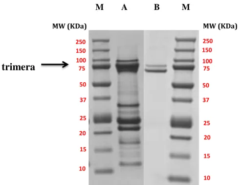
1. Purification of trimera ... 83
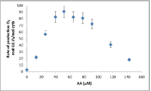
2. Optimization of AA activation with the trimera ... 84
3. Structural effects of AA ... 86
i. Effect of AA on soluble and membrane proteins ... 86
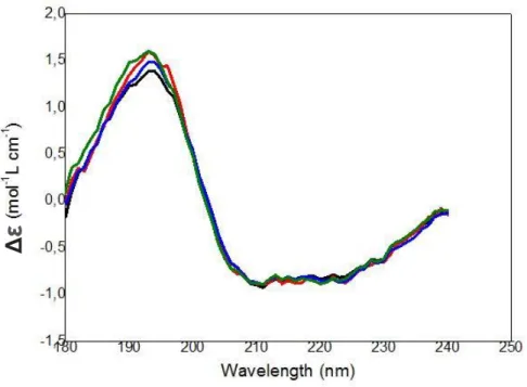
ii. Effect of AA on the secondary structure of the trimera ... 87
iii. Effect of AA on the tryptophan intrinsic fluorescence of the trimera ... 88
4. Kinetic parameters of superoxide anion production in the presence of trimera . 89 5. Assembly of NADPH oxidase in the presence of trimera ... 90
6. Quantification of H2O2 produced upon NADPH oxidase activation ... 91
7. Effect of H2O2 addition during the assembly phase ... 92
8. Sensitivity of cytosolic proteins to free radicals ... 93
9. Influence of temperature on the activity of NADPH oxidase ... 94
III. Discussion: trimera instead of separated proteins ... 97
1. On the role of AA with trimera ... 97
2. On the structural effect of AA on trimera ... 98
3. On the comparison between separated proteins and trimera ... 99
CHAPTER 2: EFFECT OF CHOLESTEROL ON NADPH OXIDASE ... 101
I. Introduction ... 102
II. Intrinsic cholesterol concentration and effect of cholesterol depletion by Methyl-β-cyclodextrin on superoxide production rate ... 102
III. Cholesterol as a Nox-activating molecule? ... 103
IV. Superoxide production in the presence of AA and added cholesterol ... 105
1. The addition of cholesterol inhibits NADPH oxidase activity ... 105
2. Effect of cholesterol on AA activation profile ... 109
3. Modification of kinetic parameters in the presence of added cholesterol ... 110
4. Effect of cholesterol addition during the assembly phase ... 112
V. Structural effects of added cholesterol ... 113
1. Effect of cholesterol on water-soluble and membrane proteins ... 113
2. Effect of cholesterol on the tryptophan intrinsic fluorescence of the trimera .... 115
3. Effect of cholesterol on the secondary structure of the trimera ... 116
VI. Discussion on the effect of lipids on NADPH oxidase ... 117
1. On the required cholesterol presence in neutrophils ... 118
3. On the conformation of the cytosolic partner ... 120
CHAPTER 3: EFFECT OF TITANUM DIOXIDE NANOPARTICLES ON
NADPH OXIDASE... 121
I. Introduction ... 122
II. TiO2 NPs size characterization... 123
III. Structural effects... 125
1. Tryptophan fluorescence of the trimera in the presence of TiO2 NPs ... 125
2. Effect of TiO2 NPs on the secondary structure of the trimera... 127
IV. Effects on the functionality ... 129
1. Effects of TiO2 NPs on the NADPH oxidase activity ... 129
2. Effect of TiO2 NPs addition at different sequences of cell free system assay ... 132
3. TiO2 NPs effect on neutrophil cells ... 133
V. Discussion on the effect of TiO2 NPs ... 134
CHAPTER 4: EFFECT OF PLATINUM NANOPARTICLES ON NADPH
OXIDASE ... 137
I. Introduction ... 138
II. Synthesis and characterization of platinum nanoparticles ... 138
III. Interaction of Pt NPs with the trimera determined by fluorescence emission... 140
IV. Effects on the functionality of NADPH oxidase ... 141
1. Pt PEG NPs as an activating molecule? ... 141
2. NADPH oxidase activation by AA in the presence of Pt-NPs ... 142
3. Effect of Pt PEG NPs on the AA-dependent activation profile ... 144
4. Pt PEG NPs effect on neutrophil cells ... 144
5. Influence of gamma irradiation on the functioning of the cell free system ... 145
Conclusions & Perspectives ... 149
References ... 152
List of publications ... 181
LIST OF FIGURES
Figure 1: Scheme of NADPH oxidase family members present in human complexed with
their regulatory proteins ... 21
Figure 2: Representative structure of the core region of NADPH oxidase enzymes ... 22
Figure 3: ROS production by a cascade of reactions initiated by NOX enzymes ... 25
Figure 4: The steps of phagocytosis and the action of NADPH oxidase ... 27
Figure 5: Electron transfer mechanism model catalyzed by flavocytochrome b558 ... 30
Figure 6: Scheme of cytochrome b558 ... 31
Figure 7: p47phox organization ... 32
Figure 8: Model of p47phox activation ... 33
Figure 9: p67phox organization ... 34
Figure 10: p40phox organization ... 35
Figure 11: Model of Rac activation ... 36
Figure 12: Trimera organization ... 37
Figure 13: Assembly of the phagocyte NADPH oxidase ... 40
Figure 14: Lipid bilayer with embedded lipid and protein molecules ... 43
Figure 15: Example of the structure of a phospholipid, the phosphatidylcholine ... 44
Figure 16: Structure of Glycolipid ... 45
Figure 17: Structure of sphingolipids ... 45
Figure 18: Structure of main sterols ... 46
Figure 19: Structures and sources of selected oxysterols ... 48
Figure 20: Scheme of lipid rafts ... 49
Figure 21: Absorption difference spectrum of Cyt b558. ... 60
Figure 22: Separation of proteins of various charges by ion exchange chromatography ... 63
Figure 23: Principle of proteins separation by affinity chromatography ... 64
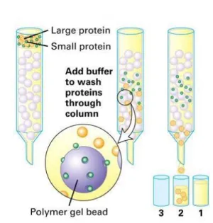
Figure 24: Separation of proteins of various sizes on a gel filtration column ... 66
Figure 25: Schematic representation of the migration of SDS PAGE ... 67
Figure 26: Example of kinetics of superoxide anion production ... 69
Figure 27: Michaelis–Menten curve for an enzyme reaction showing the relation between the protein concentration and reaction rate. ... 70
Figure 28: Schematic representation of Circular Dichroism spectrophotometer ... 72
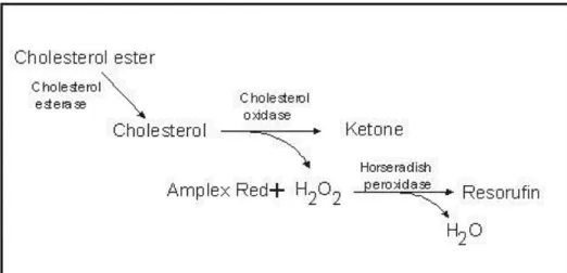
Figure 29: Enzymatic Reaction for Cholesterol Quantitation. ... 73
Figure 30: Normalized absorption and fluorescence emission spectra of resorufin ... 73
Figure 31: Detection of cholesterol using the Amplex® Red reagent–based assay ... 74
Figure 32: Structural representations of α, β and γ cyclodextrin ... 75
Figure 33: Radioactive decay of cobalt-60 ... 78
Figure 34: SDS–PAGE of trimera solutions after Ni column and gel filtration chromatography. ... 83
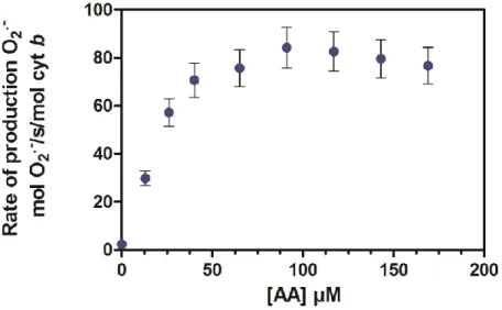
Figure 35: Superoxide anion production in function of AA concentrations for separate proteins ... 84
Figure 36: Arachidonic acid dependence of NADPH oxidase activity with the trimera. ... 85
Figure 37: Effect of AA activation of membrane fraction and trimera. ... 86
Figure 38: SR-CD spectra of trimera in the presence of increasing amounts of AA ... 87
Figure 39: Fluorescence of trimera treated with AA. ... 88
Figure 40: NADPH oxidase activity dependence on trimera concentration ... 89
Figure 41: Time course of the superoxide anion production catalyzed by the NADPH oxidase complex. ... 91
Figure 42: Effect of H2O2 on NADPH-oxidase activity. ... 93
Figure 43: Activity measurement in presence of irradiated p47phox or trimera ... 94
Figure 44: Dependence of NADPH oxidase activity as a function of temperature ... 95
Figure 45: Arrhenius plot of NADPH oxidase activity ... 96
Figure 46: Dependence of NADPH oxidase activity as a function of cholesterol concentration in the absence of arachidonic acid. ... 103
Figure 47: Dependence of NADPH oxidase activity as a function of oxysterols concentration in the absence of arachidonic acid. ... 104
Figure 48: NADPH-oxidase activity inhibition by cholesterol ... 106
Figure 49: NADPH-oxidase activity inhibition by cholesterol in the presence of separated cytosolic proteins. ... 107
Figure 50: NADPH-oxidase activity inhibition by cholesterol at 37 °C ... 108
Figure 51: NADPH-oxidase activity inhibition by addition of cholesterol to neutrophil membrane fractions from hypercholesteremic donor ... 109
Figure 52: Effect of cholesterol on the AA-dependent activation profile. ... 110 Figure 53: Effect of cholesterol on the trimera dependence NADPH oxidase activity ... 111 Figure 54: Effect of the addition time of cholesterol on NADPH-oxidase activity. ... 112 Figure 55: Effect of cholesterol on AA activation of membrane fraction and trimera. ... 114 Figure 56: Fluorescence of trimera treated with cholesterol or/and AA. ... 116 Figure 57: Effect of cholesterol on the SRCD spectra of trimera. ... 117 Figure 58: TEM images of TiO2 NPs alone and with proteins ... 124
Figure 59: Fluorescence spectrum of trimera in the presence and absence of TiO2 NPs. ... 125
Figure 60: Enlargement of the fluorescence spectrum in the region 360-500 nm. ... 126 Figure 61: Variation of the emission fluorescence intensity of the trimera-TiO2 NP
suspensions ... 126 Figure 62: Fluorescence spectrum of Trp in the presence and absence of TiO2 NPs. ... 127
Figure 63: SRCD spectra of trimera alone and with either TiO2 NPs or AA ... 128
Figure 64: Kinetics of superoxide anion production in presence of TiO2 ... 129
Figure 65: Dependence of NOX activity as a function of TiO2 NPs concentration ... 130
Figure 66: Dependence of NADPH oxidase activity as a function of TiO2 NPs concentrations
in the absence of arachidonic acid. ... 131 Figure 67: Effect of TiO2 NPs on the AA-dependent activation profile. ... 132
Figure 68: Effect of TiO2 NPs as a function of its sequence of addition in the cell free
system. ... 133 Figure 69: Characterization of platinum nanoparticles by transmission electron microscope ... 139 Figure 70: Size distribution histogram generated using TEM for (A) Pt PEG NPs, (B) naked Pt NPs. ... 140 Figure 71: Fluorescence emission spectra of the trimera-Pt PEG NPs solutions ... 141 Figure 72: Dependence of NADPH oxidase activity as a function of Pt PEG NPs
concentrations in the absence of arachidonic acid. ... 142 Figure 73: Dependence of Nox activity as a function of Pt NPs concentration ... 143 Figure 74: Effect of Pt PEG NPs on the AA-dependent activation profile ... 144 Figure 75: Influence of Gamma irradiation of Nox proteins and Pt PEG NPs on NADPH oxidase activity. ... 146
LIST OF TABLES
Table 1: Major reactions initiated by NOX enzymes ... 25 Table 2: Plasma membrane lipid composition by weight percent of mammalian red blood cells ... 43 Table 3: Kinetics parameters for cytosolic proteins using Michaelis-Menten equation in presence of AA ... 90 Table 4: kinetic parameters of the fit by a two inhibitory sites equation. ... 106 Table 5: Values of the parameters of the fit by Michaelis-Menten equation are shown ... 111 Table 6: Analysis of the SRCD spectra of trimera alone, with AA and with TiO2 NPs, using
BestSel software. ... 128 Table 7: Rates of superoxide anion production in neutrophil cells ... 134 Table 8: Rates of superoxide anion production in neutrophil cells ... 145
LIST OF ABBREVIATIONS
AA: arachidonic acid AIR: auto-inhibitory region Amp: ampicillin
AuNPs: gold nanoparticles BCA: bicinchoninic acid BSA: bovine serum albumin CD: circular dichroism cDNA: complementary DNA
CGD: chronic granulomatous disease Chl: chloramphenicol
C-ter: C-terminal CDs: Cyclodextrins Cyt b558: cytochrome b558
Cyt c: cytochrome c
DLS: Dynamic light scattering DNase: deoxyribonuclease DSB: double-strand breaks
DTT: dithiothreitol DUOX: dual oxidase
E. coli: Escherichia coli
EDTA: ethylenediaminetetraacetic acid FAD: flavin adenine dinucleotide
fMLP: formylmethionine leucyl-phenylalanine GEF: GDP/GTP exchange factor
GDP: guanosine diphosphate GTP: guanosine triphosphate Gy: Gray
HDL: High-density lipoprotein
HEPS: 4-(2-hydroxyethyl)-1-piperazineethanesulfonic acid IPTG: Isopropylthiogalatoside
Kd: dissociation constant KM: Michaelis constant
kDa: Kilodalton LB: Luria Bertoni
LDL: low-density lipoprotein LiDS: lithium dodecyl sulfate LR: lipid raft
MβCD: methyl-β-cyclodextrin MPO: myeloperoxidase MF: Membrane Fraction
NADP: nicotinamide adenine dinucleotide phosphate oxidized form NADPH: nicotinamide adenine dinucleotide phosphate reduced form NCF: neutrophil cytosolic factor
NOX: NADPH oxidase NOS: NO synthase NOXA1: Nox activator 1 NOXO1: Nox organisator1 NPs: nanoparticles
N-ter: N-terminal O2 •−: superoxide anion
PA: phosphatidic acid
PAGE: polyacrylamide gel electrophoresis PBS: Phosphate buffer saline
PB1: phox bem1 domain phox: phagocyte oxidase Pt: platinum
PI: Phosphoinositides
PI(3)P: phosphatidylinositol 3-phosphate
PI(3,4)P2: phosphatidylinositol 3,4-disphosphate PIP: phosphatidylinositol
PMA: phorbol 12-myristate 13-acetate PMN: polymorphonuclear neutrophils PMSF: phenylmethylsulfonyl fluoride
PRR: proline rich region PX: phagocyte oxidase domain ROS: reactive oxygen species RNOS: reactive nitrogen species RhoGDI: GDP dissociation Inhibitor rpm: rotation per minute
SEM: standard error of the mean SH3: src homology 3 domain SDS: sodium dodecyl sulfate
SDS-PAGE: sodium dodecyl sulfate polyacrylamide gel electrophoresis SOD: superoxide dismutase
SP Sepharose: sulfopropyl sepharose
SRCD: synchrotron radiation circular dichroism TB: terrific broth
TEM: transmission electron microscopy TiO2: Titanium dioxide
TPR: tetratricopeptide repeat
Tris: tris (hydroxymethyl) aminomethane VLDL: very low-density lipoprotein
Introduction
“Research is creating new knowledge.”
I. NADPH oxidase history
The discovery of NADPH oxidase is strongly linked to that of the oxidative burst. In the early thirties, the correlation between phagocytosis, a process used by many organisms to destroy pathogens, and oxygen metabolism, was made by Baldrige and Gerard, who found that phagocytosis, was accompanied by a strong increase of the oxygen consumption [1]. It was found that functioning of phagocytes is accompanied by an important consumption of oxygen, called the respiratory burst, although the NADPH oxidase was not yet discovered.
The respiratory burst, i.e. this increase of oxygen consumption, was initially attributed to the mitochondrial respiration. In 1959, Sbarra and Karnovsky [2] described that the phagocyte respiratory burst was an energy requiring process that depended on glucose metabolism. Shortly after, in 1961, Iyer et al. [3] demonstrated that the phagocyte respiratory burst leads to the production of hydrogen peroxide. There was at that point a major controversy over whether the main substrate for the enzyme system was NADPH or NADH. In 1964, Rossi and Zatti [4] correctly suggested that an NADPH oxidase was responsible for the respiratory burst. Then, in 1965-78 NADPH oxidase was discovered in professional immune cells called phagocytes such as neutrophils, eosinophils, monocytes and macrophages [5-7]. In 1968, Fridovich and McCord discovered the enzymatic activity of copper, zinc superoxide dismutase (SOD) to protect organisms and made the hypothesis of the formation and the toxic effects of superoxide anion [8]. Later in 1973, Babior et al. [9] demonstrated that the initial product of the respiratory burst oxidase was superoxide anion and not hydrogen peroxide.
A second important topic of studies that resulted in the discovery of the phagocyte NADPH oxidase came from clinical research. In 1957, Berendes et al. [10] identified a new and relatively rare syndrome in young boys who suffered from recurrent pyogenic infections. Quie et al. [11] reported that phagocytes of chronic granulomatous disease (CGD) patients have diminished bactericidal capacity, although many phagocyte functions, such as chemotaxis, phagocytosis, and degranulation, were shown to be intact. In 1967, it was demonstrated that the respiratory burst was absent in the phagocytes of CGD patients [12, 13].
Finally, in 1975, further characterization of the reactive oxygen species (ROS) production by phagocytes showed that NADPH oxidase: 1) produced superoxide anions and its downstream
metabolite hydrogen peroxide, 2) was insensitive to cyanide, distinguishing it from mitochondria complexes and myeloperoxidase (MPO), 3) was present in phagocytes from MPO-deficient patients, but absent in those of CGD patients; and 4) was selective for NADPH over NADH by a factor of 100 [5].
The precise identification of the proteins responsible for ROS generation in phagocytes was the next challenge. A breakthrough happened in 1978, when Segal, Jones, and colleagues [7] characterized cytochrome b558 (Cyt b558), which was absent in the leukocytes of many CGD
patients. In the late 1980s, the gene coding for the catalytic subunit of the phagocyte NADPH oxidase, gp91phox (also called NOX2), was cloned by Royer-Pokora et al. [14] and Teahan et al. [15].
However, it was rapidly revealed that NOX2 was not the only constituent of the phagocyte enzyme. In 1987, the protein p22phox was discovered as the transmembrane subunit linked to NOX2 [16-18]. The development in 1984-85 of a cell-free system permitted the activation of the phagocyte NADPH oxidase using cytosol and membrane fractions [19, 20]. This system gave important tools to discover the cytosolic subunits p47phox and p67phox [21, 22] and to identify the roles of Rac1 and Rac2 [23, 24]. In 1993, Wientjes et al. [25] described a fourth cytosolic subunit, p40phox.
In parallel with the progress toward understanding the phagocyte NADPH oxidase, during the 90s, the improvement in sensitivity of ROS detection methods allowed their identification in small amounts in different cell types and tissues other than phagocytic cells (epithelial, muscle, endothelial, neuronal ...) [26, 27]. In 1999, the first homolog of NOX2 was identified, NOX1 [28, 29].
Afterwards, six homologs of the phagocyte NADPH oxidase were identified in mammals based on sequence homology with gp91phox (NOX2) [30-34]. They are listed in two subfamilies: NOX 1-5 (for NADPH oxidase) and DUOX 1-2 (for Dual Oxidase) (Figure 1) [35]. Together with the phagocyte NADPH oxidase, these homologs are now known as the NOX family [36]. In addition, new regulatory subunits called NOXO1 (NOX organizer 1) and NOXA1 (NOX activator 1) homologues of p47phox and p67phox were identified [37, 38].
II. NADPH oxidase family
1.
Structure and Function
The NOX family is composed of proteins that share the capacity to transfer electrons across biological membranes from NADPH to dioxygen. The final product of the electron transfer reaction is either superoxide anion (NOX 1-2-3-5) or hydrogen peroxide (NOX4- DUOX1-2) [35, 39]. The biological function of NOX enzymes is therefore the generation of ROS [40] (Figure 1).
Figure 1: Scheme of NADPH oxidase family members present in human complexed with their regulatory proteins
(According to Drummond 2011) [41].
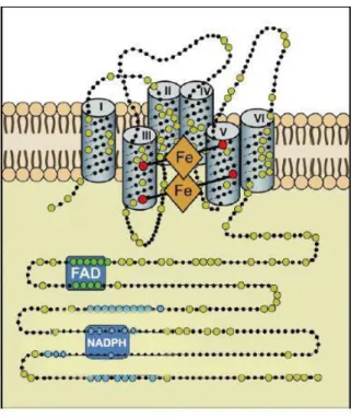
All Nox family members share a core structure consisting of: 1) six transmembrane helices, 2) NADPH and FAD binding sites at the C- terminus, and 3) four heme-binding histidines, two in the third and two in the fifth transmembrane helix (Figure 2) [42, 43]. Certain family members have additional structures. NOX5 has two polybasic domains with a domain rich in serines and a binding site for the calmodulin in the C terminal part and, in the N-terminal part, four EF-hand domains required for the protein activation by calcium [34, 44, 45]. DUOX1 and DUOX2 have two EF-hand domains followed by a seventh transmembrane helix and peroxidase homology
domain, which may allow the transformation of superoxide anion into hydrogen peroxide [33, 46].
All NOX family members contain six transmembrane domains with four histidine residues (red dots) involved in the coordination of two hemes (orange) in addition to FAD and NADPH binding domains at the C-terminus (Modified from Bedard and Krause 2007) [40].
2.
Tissue distribution and physiological roles
NOX family members have been found nearly in all superior organisms such as fungi, plants and mammals [42]. In humans, many cell types express NOX family members [26]. NOX2, the phagocyte NADPH oxidase, is the most widely distributed among the NOX isoforms. It is present in a large number of tissues, such as small intestine, colon, spleen, pancreas, ovary, placenta, prostate, and testis [47]. It is expressed in non-phagocytic cells, including neurons, cardiomyocytes, skeletal muscle myocytes, hepatocytes, endothelial cells, and hematopoietic stem cells [40]. NOX2 is a clearly established host defense enzyme [48], but it is also involved in signaling functions [49]. NOX1 is most highly expressed in the colon; however, it is also expressed in other cell types, including vascular smooth muscle cells, endothelial cells, uterus, placenta, prostate and osteoclasts [40]. NOX1 was suggested to play a role in host defense and in
blood pressure regulation [26]. NOX3 is mainly expressed in the inner ear, where it is involved in otoconia morphogenesis. Low level of NOX3 can be detected in kidney, liver and fetal tissues [50, 51]. NOX4, is strongly expressed in kidney but it is also found in vascular cells, endothelial cells, smooth muscle cells, hematopoietic stem cell, fibroblasts, keratinocytes, melanoma cells, and neurons and osteoclasts [31, 39]. NOX4 is probably involved in the oxygen sensing in the kidney cortex. NOX5, a Ca2+ activated enzyme is predominantly expressed in lymphoid tissues and testis, where it might be involved in signaling processes [44, 45]. DUOX1 is expressed in the thyroid and in respiratory epithelia while DUOX2 is found basically in the thyroid and in gastrointestinal glandular epithelia [52]. DUOX2 is clearly involved in thyroid hormone synthesis, but possibly also in epithelial host defense [33, 48, 53, 54]. The role of DUOX1 remains unclear.
III. Reactive oxygen species production
ROS are derivatives of oxygen which are very unstable and react very easily with other molecules; they can free radicals or not free radicals. A free radical is any chemical species possessing one or more unpaired electrons. Free radicals of importance in living organisms include superoxide (O2•−), hydroxyl (OH•), nitric oxide (NO•), peroxyl (RO2
·
). Hypochlorous acid (HOCl), peroxynitrite (ONOO−), hydrogen peroxide (H2O2), singlet oxygen (1O2) and ozone (O3)
are not free radicals but can easily lead to free radical reactions in living organism. [55].ROS are produced in vivo from various endogenous sources such as the mitochondrial electron transport chain [56], xanthine oxidase, cytochrome P450, peroxisome, NOX and DUOX enzymes [57]. ROS are also produced by exogenous sources such as tobacco, smoke, pollutants, drugs and ionizing radiation [58].
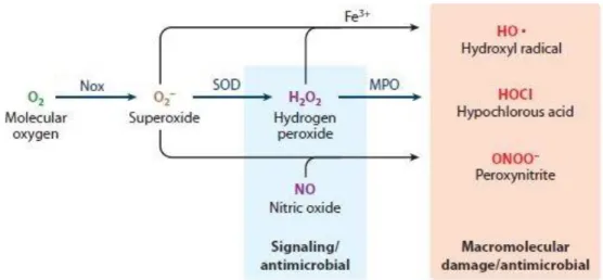
NADPH oxidase is the major source of non-mitochondrial cellular ROS in many cells including renal cells, and vascular tissues. NOX-dependent ROS production can be depicted generally as coming from a cascade of reactions that starts with the production of superoxide anion [27] (Figure 3, table 1). NOX enzymes reduce dioxygen to form superoxide anion as their sole enzymatic function (equation 1) [59]. Although superoxide is the species produced by NOX enzymes, it seems doubtful that superoxide itself is directly implicated in microbial killing. O2•− is
not highly reactive and since it is a charged molecule, it does not diffuse across the membrane. It has been known that superoxide is not reactive enough towards proteins, DNA or lipids, to be an
essential player on its own [60-62]. O2•−is the conjugate base of the perhydroxyl radical (HO2•)
with an acid dissociation constant (pKa) of 4.8, thus superoxide will be present in appreciable concentrations when the pH is above 4.8 [62]. Some reports have proposed that in nonpolar environment the reactivity of superoxide is boosted and at low pH, HO2•, the more reactive
protonated form of superoxide is abundant and could participate to the bactericidal destruction [63]. Therefore, under relevant physiological situations, such as the low pH in the phagosome and the nonpolar environment in the vicinity of cell membrane, superoxide itself could potentially be a direct player in microbial killing in addition to its role of being a substrate for the formation of secondarily derived ROS. O2•− rapidly dismutates to H2O2 either spontaneously, or catalyzed by
superoxide dismutase (SOD) (equation 2) [3, 64]. H2O2 is relatively stable and has a bactericidal
capacity. In fact, H2O2 is less reactive thus it can easilydiffuse through the membrane and react in
a different area from the area where it was produced. Other elements in the cascade of ROS generation include the iron-catalyzed Fenton reaction leading to the generation of the most powerful oxidizing agent (OH•) (equation 3) [65, 66], the myeloperoxidase (MPO) catalyze formation of potent oxidant (HOCl) from H2O2 and Cl- (equation 4) [67], and the reaction of O2•−
with NO• to form ONOO− (equation 5) [68], a powerful oxidant capable of fragmenting the DNA and oxidizing lipids and proteins. In addition to the high level of ROS production by the phagocyte NADPH oxidase for the defense mechanisms against microbes [69], lower levels of ROS production have been found in non-phagocytic cells. The discovery of NOX family led to the concept that ROS are generated in these cells with distinctive cellular functions. It is now well-established that NADPH oxidases participate in important cellular processes including signal transduction, cell proliferation, differentiation and development [70, 71].
Figure 3: ROS production by a cascade of reactions initiated by NOX enzymes (Modified from Lambeth 2014)
Table 1: Major reactions initiated by NOX enzymes Reactions Equations NADPH + 2O2 → NADP+ + H+ + 2 O2•- 1 O2•- + HO2• + H+ → O2 + H2O2 2 Fe+2 + H2O2 → Fe+3 + OH• + OH– 3 H2O2 + Cl- → HOCl + OH– 4 • NO + O2•– → ONOO– 5
Despite the important role of ROS in the immune defence, they can have harmful effects. ROS are considered as toxic molecules due to their high reactivity to many biological macromolecules which may lead to DNA damage, oxidations of polyunsaturated fatty acids and amino acids, and oxidative inactivation of specific enzymes which can contribute to the pathology of human diseases [71]. Thus, their production must be tightly controlled.
There are protection systems, enzymatic and non-enzymatic, that allow limitation of ROS concentration and their effects. The steady state of ROS is controlled by endogenous enzymatic regulatory systems. Intracellular levels of O2•− are controlled by different types of SOD: the
cytosolic CuSOD, ZnSOD and mitochondrial MnSOD. SOD accelerate the dismutation rate of O2•− to H2O2 by about four orders of magnitude [72] (equation2). Catalase catalyzes the reduction
of H2O2 to H2O and O2 and is located in the peroxisomes of most mammalian cells [73].
However, it is likely that the major H2O2 removing enzyme in mammalian cells are peroxidases,
some of them possess a selenium active site [74]. There are also antioxidant molecules which protect the organisms by interacting with ROS. They can have anti-catalytic effect by interfering with the enzymes producing ROS or with the signalization cascade implicated in their production. Some of these molecules are produced by the body, it is the case of uric acid and Glutathione [75], Some of the antioxidants are taken from nutrients in food such as tocopherol (vitamin E), ascorbic acid (Vitamin C), β-Carotene and vitamin A. Those molecules act generally as scavengers but they are more and more studied for their anti-catalytic capacities. Oxidative stress is the term referring to imbalance between generation of ROS and the activity of the antioxidant defenses. Finally, the balance between ROS and antioxidants must be optimal as both extremes are damaging [76].
IV. Phagocyte NADPH oxidase
The human polymorphonuclear neutrophil (PMN) play an essential role in innate immunity intervening in the defense of the host against pathogens (bacteria, fungi, and virus) [77]. This cell will acquire during its maturation in the marrow, azurophilic granules (primary), specific granules (secondary), granules rich in gelatin (tertiary) and secretory vesicles that contain antimicrobial peptides and proteolytic enzymes. After maturation, the PMN remain 0-5 days in the marrow then pass into the bloodstream where its half-life is typically few hours. Under the influence of various stimuli, the PMN who are at rest in the circulating blood, will adhere to the vascular wall, slipping through endothelial cells then migrate through the tissues to the site of infection following a gradient of chemoattractant substances. At this level, adhesion to the target followed by its ingestion result in the formation of the phagosome and will engage the PN in bactericidal response (Figure 4). The PMN use in cooperative way two systems to destroy the infectious agent: the first system which is independent of the oxygen, involves the release, in the phagosome, anti-microbial peptides and lytic enzymes originally contained in the granules. The second system is dependent of oxygen and involves massive and rapid production of superoxide anions by NOX2 (Figure 4).
All the experiments present in my thesis have been performed with NOX2 from neutrophils. As said in paragraph I, NOX2 was first described in neutrophils and macrophages and is known as the “phagocyte NADPH oxidase”. In these cells, NOX2 is localized in both intracellular and plasma membranes [78, 79]. In resting neutrophils, most of the NOX2 is localized in intracellular compartments, the granules (60-70% in secondary granules, 20-25% in tertiary granules, 5-20% in membrane) [80].
During phagocytosis of invading microbes, the phagocyte NADPH oxidase becomes activated and generates superoxide anion, a precursor of microbicidal ROS.
Upon phagocyte stimulation, NOX2 is incorporated to the membrane as the granules fuse with the phagosomal or the plasma membrane [78, 81]. This fusion is one of the key events for the microbicidal activity of NOX2. However, NOX2 may also be activated within the granules without a fusion with surface membranes. In this case it is harmful for the neutrophil itself [82, 83]. Neutrophils produce large amounts of O2•−, estimated between approximately 1 and 4 M in
the phagosome. However, the steady state concentration has been estimated to be in the µM range because dismutation to H2O2 is very rapid (equation 2) [84].
1.
Composition of the NADPH oxidase complex
NADPH oxidase is a multi-subunit enzyme complex composed of the membrane-bound flavocytochrome b558 (Cyt b558), comprising two subunits (NOX2 also known as gp91phox, and
p22phox) and four cytosolic components. The cytosolic components include p47phox, p67phox, p40phox, and a small GTPase Rac1 or Rac2 [85, 86].
NADPH oxidase activity is tightly regulated spatially and temporally, therefore the enzyme exists in two forms, active and inactive, which differ in localization of the subunits in the cell as well as by their structure. In resting phagocytes, the components of the complex exist as separated entities but upon cell activation, the cytosolic subunits undergo posttranslational modifications and migrate to the membrane bound Cyt b558 to constitute the activated NADPH
oxidase complex [42]. Actually this process involves a complicated set of protein-protein and protein-lipid interactions that will be discussed later.
2.
Structure and functions of the membrane subunits
The membrane part of the NADPH oxidase is a transmembrane heterodimer, the flavocytochrome b558 that constitutes the catalytic core of the enzyme. It consist of gp91phox and
p22phox [6, 17] where both subunits are essential for the stable expression of a functional cytochrome [87, 88].
i. gp91phox
The gp91phox subunit nowadays called NOX2 is a glycoprotein expressed at high levels in phagocytic cells. It is encoded by the CYBB gene located on the chromosome X [89]. It consists of 570 amino acids. The N-terminal (residues 1-290) forms six transmembrane α-helices, responsible for its anchoring in the plasma membrane, its interaction with p22phox and the electron transfer through the membrane from FAD to oxygen thus enabling the production of superoxide anions on the external side of the plasma membrane. This membrane part harbors the two non-identical haem groups of the NADPH oxidase via two histidine pairs [90-92]. The C-terminal cytoplasmic domain (residues 290-570) contains the binding sites for NADPH and FAD and is therefore responsible for the first steps of the electron transfer [93]. NADPH, the electron donor is mainly derived from the pentose phosphate pathway by the oxidation of glucose 6-phosphate [94].
The gp91phox is synthesized as a precursor of 65 kDa partially glycosylated in the endoplasmic reticulum. It is further processed in the Golgi apparatus into a highly glycosylated protein of 91 kDa [87]. This maturation requires the incorporation of both hemes in the precursor 65 kDa and the presence of p22phox protein.
Hence gp91phox contains all cofactors needed for the electron transfer reaction which could take place in four steps: two electrons and one proton are transferred from NADPH to FAD in this model. The first electron is transferred from reduced FAD to the first haem, followed by its transfer to the second haem up to O2 to form O2•−. In the same way, the second electron is
transferred from FAD to the first haem then to the second haem and afterward to oxygen forming superoxide [95-97]. However, the precise mechanism of the electron transfer is not yet known. One can propose the scheme described Figure 5. One can notice that the third and sixth steps in this electron transfer scheme are energetically unfavorable, since the inner haem has a higher midpoint redox potential than the outer haem. That may explain that the presence of oxygen is necessary for rapid electron flow through the NADPH oxidase, as the absence of a terminal electron acceptor will cause electrons to accumulate on the inner haems [98].
There is a need of a production of pure NOX proteins in high amount to identify theses electrons transfer steps and determine the structure of the protein. Two ways are tempted, in our group the production of recombinant proteins in yeast and in Fieschi’s group in the production of bacterial homologs.
Figure 5: Electron transfer mechanism model catalyzed by flavocytochrome b558
In this model, the electron transfer is carried out in seven oxido-reduction steps with a transfer of two electrons from NADPH to the FAD (1) then the transfer of a first electron via the two haems (2, 3, 4) to a first oxygen and finally the transfer of the second electron from FAD through the haems to a second oxygen (5, 6, 7) (Modified from Cross and Segal 2004) [98].
ii. p22phox protein
p22phox is a membrane protein, which closely associates with gp91phox in a 1:1 ratio [79]. It contains 195 amino acids encoded by the gene CYBA. According to the model proposed, p22phox could contain two transmembrane helices [85, 99]. The C-terminal cytoplasmic portion appears to have a proline-rich region (PRR) that contains a consensus Pro-Xaa-Xaa-Pro motif. This motif is known to be a target of the SH3 (Src homology 3) domains of p47phox [100-102] and this interaction appears to be critical for the activation of NADPH oxidase since a mutation of proline 156 on p22phox blocks the binding of the p47phox protein resulting in a lack of activity NADPH oxidase [103].
p22phox does not possess a catalytic role but remains indispensable. in vivo, p22phox has two major functions: 1) a binding to NOX proteins, that results in enzyme stabilization, and 2) a binding to organizer subunits p47phox or NOXO1 [104]. p22phox has also been shown to interact with NOX1 [105, 106], NOX3 [107], and NOX4 [108] [39]. The significance of the p22phox subunit for the phagocyte NADPH oxidase was demonstrated with the identification of CGD patients with mutations in p22phox [109].
Figure 6: Scheme of cytochrome b558
The transmembrane helices of gp91phox and p22phox are indicated. Glycosylation sites are shown by cyan dots and regions that are expected to interact with p67phox in the active state in red. The FAD and NADPH binding sites in gp91phox are indicated in cyan and pink respectively. The position of the PXXP motif in the cytoplasmic region of p22phox that interacts with p47phox is shown in grey (modified from Groemping 2005).
3.
Structure and functions of cytosolic subunits
i. p47phox protein Description
p47phox is a cytosolic protein encoded by the NCF1 (Neutrophile Cytosolic Factor 1) gene located on chromosome 7 in humans. This protein is composed of 390 amino acids with a molecular weight of 44.7 kDa. p47phox is the protein chiefly responsible for transporting other cytosolic proteins to the Cyt b558 to form the active oxidase [110]. In vitro, it has been found not
to be absolutely indispensable for oxidase activity because at sufficiently high concentrations of p67phox and Rac, superoxide production takes place in the absence of p47phox [111, 112].
However, it is essential in the neutrophil since patients whose neutrophils are deficient in p47phox have CGD [113, 114]. p47phox possesses several distinct functional domains that can either mediate intramolecular interactions or interact with other subunits. Starting from the amino acid N-terminal, p47phox consists of a phox homology (PX) domain that interact with the membrane phospholipids [115], a tandem SH3 domains that interact in the active form with the proline rich region in the C-terminal of p22phox [116], an autoinhibitory region (AIR) that prevents these previous interactions until the protein is phosphorylated [117, 118] and finally a proline rich region (PRR) in the carboxy terminus that interacts with the SH3 domain of p67phox and SH3 domain of p40phox [119, 120]. This interaction is essential for the translocation of p67phox to the Cyt b558 (Figure 7).
Figure 7: p47phox organization
The arrows represent the different intramolecular and intermolecular interactions that have been identified. The arrow in dotted lines represents interaction that is still under investigations. Activation of p47phox
In the absence of stimulation, p47phox is maintained in a self-inhibited conformation in the
neutrophils. Within the PX domain of p47phox, the PXXP motif (R70IIPHLPAP78) interacts with
its SH3 domain [121]. This intramolecular interaction masks the phosphoinositide binding sites of the PX domain of p47phox in resting neutrophils, therefore preventing its membrane association (Figure 8).
Upon neutrophil activation, phosphorylation of several serine residues including S303, S304, S359, and S370 by kinases proteins mediates a conformational change in p47phox [122], unfolding the protein [110]. Phosphorylated p47phox was suggested to increase the binding of p67phox to cytochrome b558 by acting as an adapter, bringing p67 phox into proximity with
cytochrome b558 [123-125]. The exposition of the SH3 domains of p47phox will eventually interact
stably with PRR of p22phox [126]. At the same time, the PX domain interacts with specific membrane phospholipids (phosphatidylinositol 3,4-disphosphate (PI(3,4)P2) and phosphatidic acid (PA)) [110]. This opening of p47phox is linked to an increase in hydrophilicity caused by the negative charges provided by the phosphates bound in the serine residues [122]. p47phox is described as organizer subunit of NADPH oxidase complex [127].
Figure 8: Model of p47phox activation
Both the resting and activated configurations of p47phox are shown (according to Marcoux et al, 2010) [127].
ii. p67phox protein
p67phox is a cytosolic protein encoded by the NCF2 (Neutrophil Cytosolic Factor 2) gene located on chromosome 1 in humans. This protein is composed of 526 amino acids with a molecular weight of 59.8 kDa [128, 129]. Unlike p47phox, p67phox is absolutely required for oxidase activity in cell free system [112]. Additionally, several CGD patients with functionally significant mutations of p67phox have been found [130]. All p67phox mutants responsible for CGD
are localized in the half N-terminal part (1-210) [131].
The N- terminal part of p67phox contains a domain of four tetratricopeptide repeat (TPR) motif that is responsible for mediating its interaction with Rac [132, 133] and an activation domain (amino acids 199–210) that has been shown to be essential for O2 •− production in a
flavocytochrome and thereby contributes in the regulation of electron transfer [136]. That is why p67phox is considered as the activator subunit. This domain is followed by a PRR which might interact with the SH3 domain of p47phox and by two SH3 domains that are separated by a PB1 (Phox and Bem1) domain which forms a heterodimer with the PB1 domain of p40phox [137] (Figure 9). The last SH3 domain has been shown to be important for p67phox - p47phox dimerization [105]. Fieschi’s group suggests that intramolecular interactions within p67phox may be present [138].
Figure 9: p67phox organization
Although the phosphorylation of p47phox contributes remarkably to the interaction changes, the functional consequences of the phosphorylation of p67phox are less obvious. Distinct sites for ERK2 and p38MAPK-dependent phosphorylation have been found during the activation of neutrophil [139, 140], including the threonine 233 [141, 142] and some serines but the positions of phosphorylated serine are not well determined [143] [140]. Recently, Dang et al. suggested that p67phox undergoes a continual cycle of phosphorylation/dephosphorylation in resting cells and p67phox phosphorylation would be controlled by MEK1/2 and tyrosine kinase [144].
iii. p40phox protein
p40phox is a cytosolic protein encoded by the NCF4 gene located in chromosome 22 in humans, with a molecular weight of 39 kDa. Its sequence contains 336 amino acids. The organization of p40phox domains are described in Figure 10. It consists of a PX domain, a SH3 domain and a PB1 domain. p40phox interacts with p67phox via its PB1 domain [145] while its SH3 domain has been proposed to interact with the PRR of p47phox [146, 147]. However, the affinity between the two proteins is very low (Kd = 5 µM) [120], thus this interaction is very weak in
SH3 domain can also associate with PRR in p22phox [150]. The PX domain of p40phox binds particularly to phosphatidylinositol 3-phosphate (PI(3)P), which accumulates in phagosomal membranes, and could thus facilitate oxidase assembly at this site [151, 152]. A conformation for p40phox at rest was proposed by Ueyama et al. [153]. According to their model, the intramolecular interaction of the PX domain with the PB1 one prevents binding to PI(3)P, thus this interaction must be disrupted to allow binding of p40phox to the membrane (Figure 10). The study of the p40phox deficient CGD patient found by Dinauer group goes in favor of a role of this protein for an assembly of the NADPH oxidase at the phagosome [154].
Compared to the other cytosolic proteins, the overall role of p40phox in oxidase regulation is still controversial, it has been proposed as both activator and inhibitor [155-158]. In fact, after phosphorylation of this protein, it could play a role during the complex activation [119, 159, 160]. It is probably implicated in the stabilization of p47phox /p67phox complex in the cytosol and facilitates its recruitment to the membrane during the activation of NADPH oxidase [161]. It was also reported by Dang et al. that phosphorylated p40phox may induce conformational change that permits p67 phox to interact fully with cytochrome b558 [125]. Like the previous cytosolic proteins,
p40phox undergoes phosphorylation upon oxidase stimulation. These events are produced in correlation with superoxide production [159].
Figure 10: p40phox organization iv. Rac
Rac protein is a small G-protein of about 21 kDa, composed of 192 amino acids. It exists in two isoforms in phagocytic cells (Rac2 in human neutrophils; Rac1 in human monocytes) and
both share 92% sequence homology [162]. Rac belongs to the Rho-family of small GTPases, which regulate a large variety of signaling pathways. When the neutrophil is at resting state, Rac is in an inactive form, containing a GDP in its binding site and associated to RhoGDI inhibitor (GDP dissociation inhibitor) in 1:1 stoichiometry [162-164]. The conversion between the active and inactive states is tightly controlled by GEFs (guanine-nucleotide-exchange factors) which replace GDP by GTP and allow Rac to be free from RhoGDI inhibitor and to migrate to the membrane. It has been shown that this migration occurs independently of p47phox and p67phox [165] (Figure 11).
Rac protein has an N-terminal domain (RAS effector domain (aa 20-40)) essential for its interaction with the TPR domain of p67phox and for NADPH oxidase activity [133, 166-168]. This interaction takes place when Rac is in the form linked to GTP [132, 169]. Rac undergoes geranylgeranylation at the polybasic region of its C-terminus, which facilitates its association with membranes. Although it has been suggested that Rac solely acts to place the activation domain of p67phox on Cyt b558 for regulation of electron transfer and increase the affinity of
p67phox to NOX2 [125, 170], there is also notable evidence that Rac itself interacts directly with the Cyt b558 to activate the initial electron transfer step from NADPH to FAD [165, 171-173]. In
vivo and in vitro, Rac1 and Rac2 are required for the activation of NADPH oxidase [174-177].
To study the NADPH oxidase activity in vitro in a reconstituted system, we used a mutant of Rac, Rac1Q61L, in which the residue Glutamine 61 is substituted by a leucine. The Q61L mutation leads to a constitutively active form of Rac. A study of this mutant showed that Rac1 is more stable and has a greater affinity for p67phox and the complex than the native Rac1 [178].