HAL Id: hal-01904131
https://hal.archives-ouvertes.fr/hal-01904131
Submitted on 24 Oct 2018
HAL is a multi-disciplinary open access archive for the deposit and dissemination of sci-entific research documents, whether they are pub-lished or not. The documents may come from teaching and research institutions in France or abroad, or from public or private research centers.
L’archive ouverte pluridisciplinaire HAL, est destinée au dépôt et à la diffusion de documents scientifiques de niveau recherche, publiés ou non, émanant des établissements d’enseignement et de recherche français ou étrangers, des laboratoires publics ou privés.
Spotlight on the NLRP3 inflammasome pathway
Marine Groslambert, Bénédicte Py
To cite this version:
Marine Groslambert, Bénédicte Py. Spotlight on the NLRP3 inflammasome pathway. Journal of In-flammation Research, Dove Medical Press, 2018, 11, pp.359-374. �10.2147/jir.s141220�. �hal-01904131�
Journal of Inflammation Research
Dove
press
R e v I e w
open access to scientific and medical research Open Access Full Text Article
Spotlight on the NLRP3 inflammasome pathway
Marine Groslambert1–5 Bénédicte F Py1–5
1Centre International de Recherche
en Infectiologie (CIRI), Université de Lyon, Lyon, France; 2INSeRM,
U1111, Lyon, France; 3ecole Normale
Supérieure de Lyon, Lyon, France;
4Centre International de Recherche
en Infectiologie, Université Lyon 1, Lyon, France; 5CNRS, UMR5308, Lyon,
France
Abstract: Inflammation is triggered by a repertoire of receptors detecting infections and
dam-ages. Some of these receptors directly bind microbial ligands, while others recognize endogenous molecules exposed under stress conditions, including infections. Most of these receptors can be engaged by a relatively limited number of stimuli. Differently, NLRP3 acts as a broad sensor of cell homeostasis rupture and can be activated downstream of a plethora of stimuli. NLRP3 then assembles a multiprotein platform resulting in caspase-1 activation, which controls, by direct cleavage, the maturation of cytosolic pro-cytokines including pro-interleukin-1β. In addition, caspase-1 processes cytosolic gasdermin-D and unleashes its pore-forming N-terminal domain, leading to the release of mature cytosolic cytokines and alarmins, as well as pyroptotic cell lysis. Accumulating evidences of the aggravating role of NLRP3-mediated inflammation in various highly prevalent human conditions, including diabetes, neurodegenerative and cardiovascular diseases, raises a huge clinical interest. Nevertheless, the molecular mechanism governing NLRP3 activation remains insufficiently understood. In line with the detrimental consequences of NLRP3 activation illustrated by the aforementioned pathologies, this process is tightly regu-lated. In this review, we address the current understanding of the control of NLRP3 activity which can be divided into two coordinated processes referred to as priming and activation. In particular, we detail the emerging role of NLRP3 post-translational modifications critical in inflammasome assembly regulation.
Keywords: inflammation, NLRP3, inflammasome, post-translational modifications
Introduction
Inflammation is usually triggered in response to infections and/or tissue damages. It constitutes a protective response, critical for the recruitment of immune cells mediating pathogen clearance and tissue repair at the affected site.1 Pathogens and damages are
sensed by tissue resident cells, including innate immune cells, through a repertoire of germline-encoded dedicated receptors known as pattern recognition receptors (PRRs). These receptors are activated by pathogen-associated molecular patterns (PAMPs), typically shared throughout large kindred of microbes but absent from the host organ-ism, and by host-derived cellular stress signals, markers of tissue and cell homeostasis disruption, known as damage-associated molecular patterns (DAMPs).2,3
Many inflammatory cytokines such as interleukin-6 (IL-6), tumor necrosis factor (TNF), and interferon-β (IFN-β) are regulated at the transcriptional level and secreted through the endoplasmic reticulum (ER)/Golgi pathway. Others, including IL-1β and IL-18, are produced as cytosolic precursors and their secretion is tightly controlled by
Correspondence: Bénédicte F Py CIRI, Université de Lyon, 21 Avenue Tony Garnier, 69007 Lyon, France Tel +33 4 26 73 13 52
email benedicte.py@inserm.fr
Journal name: Journal of Inflammation Research Article Designation: Review
Year: 2018 Volume: 11
Running head verso: Groslambert and Py
Running head recto: Spotlight on the NLRP3 inflammasome pathway DOI: http://dx.doi.org/10.2147/JIR.S141220
Journal of Inflammation Research downloaded from https://www.dovepress.com/ by 193.54.110.33 on 25-Oct-2018
For personal use only.
This article was published in the following Dove Press journal: Journal of Inflammation Research
Dovepress
Groslambert and Py
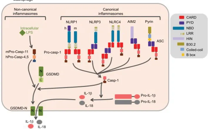
inflammatory caspases, namely caspase-1 and -11 in mice, and caspase-1, -4, and -5 in humans. These caspases are activated within cytosolic multimolecular complexes named inflammasomes (Figure 1). Canonical inflammasomes form activation platforms for caspase-1. Their assembly depends on some dedicated cytosolic PRRs from the nucleotide-binding domain leucin-rich repeat (NLR) family including NLR and pyrin domain containing receptor 1 (NLRP1), NLRP3, and NLR and caspase recruitment domain containing receptor 4 (NLRC4); the AIM2-like receptors (ALR) family including absent in melanoma 2 (AIM2); or the tripartite motif (TRIM) family including pyrin. Activation of these PRRs triggers either their oligomerization (as for NLRC4 or AIM2)4,5 or
confor-mational changes of pre-formed oligomers (as for NLRP3).6
The PRR oligomer then recruits the adaptor protein ASC (apoptosis-associated speck-like protein containing a caspase recruitment domain [CARD]) through pyrin domain (PYD)– PYD interactions and constitutes a nucleation platform for fila-ments of ASC. The surface of the filament in turn enables the
nucleation of filaments of the effector pro-caspase-1 through CARD–CARD interactions.7 Inflammasomes form specks,
also referred to as pyroptosomes, in the micron range in size and unique per cell, that can be observed by light microscopy.8
Pyroptosome assembly constitutes an all-or-nothing response at the single cell level. Noteworthy, NLRC4 which possesses a CARD domain can either directly recruit pro-caspase-1 or trigger the formation of ASC filaments, serving as an ampli-fication mechanism required for cytokine maturation but dispensable for pyroptosis.9 The inflammasomes that control
the activation of caspase-11 in mice and caspase-4 and -5 in human in response to cytosolic lipopolysaccharide (LPS) are referred to as non-canonical. Their composition remains poorly understood, and the lipid A moiety of LPS has been proposed to directly interact with the CARD of caspase-11, -4, and -5 which then undergo oligomerization, resulting in the activation of the caspases.10 Therefore, non-canonical inflammasome
assembly does not depend on dedicated PRRs, and caspase-11, -4, and -5 act as receptors themselves.
Figure 1 Canonical and non-canonical inflammasomes.
Notes: Canonical inflammasomes assemble downstream of cytosolic PRRs including NLRP1, NLRP3, NLRC4, AIM2, and pyrin and control the activation of caspase-1. The ASC adaptor forms multimeric filaments and amplifies the response. The non-canonical inflammasomes are activated by cytosolic LPS and result in caspase-4 and -5 (homologs of mouse caspase-11) activation. All these inflammatory caspases cleave GSDMD which then forms cytolytic pores leading to pyroptosis. In addition, caspase-1 controls the maturation of cytosolic pro-cytokines.
Abbreviations: PRRs, pattern recognition receptors; ASC, apoptosis-associated speck-like protein containing a CARD domain; LPS, lipopolysaccharide; GSDMD, gasdermin-D; h, human; m, mouse.
Non-canonical
inflammasomes inflammasomesCanonical
NLRP1
h m
NLRP3 NLRC4 AIM2 CARDPYD
NBD LRR HIN B30.2 Coiled-coil B box Pyrin ASC Pro-casp-1 GSDMD Casp-1 Pro-IL-1 Pro-IL-18 GSDMD-N IL-1 IL-1 IL-18 IL-18 N C N N mPro-Casp-11 hPro-Casp-4,5 Intracellular LPS Macrophage
Journal of Inflammation Research downloaded from https://www.dovepress.com/ by 193.54.110.33 on 25-Oct-2018
Dovepress Spotlight on the NLRP3 inflammasome pathway
The caspase-1 protease controls maturation of pro-IL-1β and pro-IL-18, produced as pro-cytokines in the cytosol by direct cleavage of the pro-forms.11 In addition, all
inflamma-tory caspases trigger a pro-inflammainflamma-tory form of cell death named pyroptosis (from Greek roots pyro, relating to fire or fever, and ptosis, relating to falling). This process is mediated by the cleavage of the cytoplasmic gasdermin-D (GSDMD) protein, which leads to the release of its N-terminal domain that oligomerizes as pores in the plasma membrane.12–14
GSDMD pores and cell lysis mediate the release of mature IL-1β and IL-18 together with IL-1a and many other alarm-ins, as well as the ASC speck. Extracellular ASC specks enter neighbor cells by phagocytosis and propagate the inflam-matory signal.15,16 Noteworthy, caspase-11, -4, and -5 do
not directly cleave pro-IL-1β and pro-IL-18. Non-canonical inflammasome controls cytokine maturation through sub-lytic plasma membrane permeabilization by GSDMD pores that activate the NLRP3 inflammasome.12,17
NLRP3, also known as cryopyrin, CIAS1, or NALP3, was initially described for its genetic association with forms of hereditary autoinflammation known as familial cold autoinflam-mation syndrome (FCAS), Muckle–Wells syndrome (MWS), and neonatal-onset multisystem inflammatory disease/chronic infantile neurologic cutaneous articular syndrome (NOMID/ CINCA), in the order of increasing severity.18,19 These diseases
were then collectively named cryopyrin-associated periodic syndromes (CAPS). Since then, NLRP3-mediated inflam-mation has been additionally recognized to fuel a number of multifactorial diseases including gouty arthritis, atherosclerosis, type 2 diabetes, and non-alcoholic steatohepatitis, as well as several neurodegenerative diseases, including amyotrophic lateral sclerosis, Alzheimer’s and Parkinson’s diseases, and other aging-related diseases.20–23 NLRP3 has also been involved
in the response against diverse pathogens including ameba, protozoans including Plasmodium, fungi including Candida
albicans and Saccharomyces cerevisiae, bacteria including Staphylococcus aureus and Listeria monocytogenes, and viruses
including influenza, Sendai, and adenoviruses.24 On contrary to
the abovementioned conditions, NLRP3-dependent inflamma-tion is mostly protective against infecinflamma-tions, although the impact of Nlrp3 deficiency is usually limited, probably due to some redundancies among PRRs. NLRP3 is a 115 kDa cytosolic protein composed of three domains: a C-terminal leucin-rich repeat (LRR) with poorly characterized regulatory functions, a central oligomerization domain NACHT or nucleotide binding oligomerization domain (NOD) with an ATPase activity, and a N-terminal pyrin domain (PYD) that recruits ASC.18 NLRP3
is activated by numerous and highly diverse PAMPs/DAMPs
as, for example, hyperglycemia, ceramides, fatty acids, protein aggregates (β-amyloid, islet amyloid polypeptide), crystals (cholesterol, sodium urate, asbestos, silica, calcium pyrophos-phate, alum), extracellular ATP, hyaluronic acid, pore-forming bacterial toxins including nigericin, bacterial and viral nucleic acids, and hemozoin.25–27 The great diversity in the structures of
these activators excludes the fact that NLRP3 acts as their direct receptor. Instead, it is now well accepted that NLRP3 indirectly senses cytosolic stress. In this review, we describe the recent advances in our understanding of signaling pathways control-ling NLRP3 activation, with a particular focus on the newly discovered key role of NLRP3 post-translational modifications in inflammasome assembly.
Signaling pathways controlling
NLRP3 inflammasome assembly
Regulation of the NLRP3 inflammasome has been most extensively investigated in macrophages. In these cells, NLRP3 inflammasome assembly results from a two-step process referred to as priming and activation (Figure 2). The priming signal usually corresponds to cytokines or DAMPs/ PAMPs recognized by receptors that transcriptionally control the expression of numerous genes. The activation signal can be provided by various cellular stresses and is transcription independent.
NLRP3 inflammasome priming
Consistent with the high diversity of the conditions leading to NLRP3 inflammasome assembly, a large number of recep-tors, including PRRs as well as cytokine and anaphylatoxin receptors, control NLRP3 priming. Among the PRRs, Toll-like receptors (TLRs), nucleotide-binding oligomerization domain 2 (NOD2), and receptor for advanced glycation endproducts (RAGE) can prime NLRP3 for further activa-tion.28–32 The immune system itself can autonomously prime
NLRP3 through the anaphylatoxin C3a and C5a receptors and cytokines IL-1 and TNF receptors.33–36 In addition,
exces-sive reactive oxygen species (ROS) production also leads to NLRP3 priming.
Upon TLR binding by their agonists, several pathways resulting in NLRP3 priming are successively activated over time (Figure 3). The first pathway that has been described relies on transcription activation downstream of these receptors and de novo protein synthesis.35 This pathway is
referred to as transcriptional priming or late priming, since it requires several hours (>3 h) before NLRP3 is licensed for further activation. Transcriptional priming depends on myeloid differentiation primary response 88 (MyD88), TIR
Journal of Inflammation Research downloaded from https://www.dovepress.com/ by 193.54.110.33 on 25-Oct-2018
Dovepress
Groslambert and Py
domain-containing adapter-inducing interferon-β (TRIF), Fas-associated protein with death domain (FADD), and caspase-8, as well as ROS, which activate nuclear factor-κB (NF-κB).37–39 Indeed, while NLRP3 expression is low and
the major caspase-1 substrate pro-IL-1β is not expressed in resting macrophages, both are highly transcriptionally upregulated downstream of TLR activation.35 In addition,
experimental overexpression of NLRP3 alleviates priming requirement for further NLRP3 activation, suggesting that priming mechanism may rely on this upregulation of NLRP3 expression level.35,39,40 Expressions of ASC, pro-caspase-1,
and other substrates, including IL-18, are constitutive and are not affected upon priming in cultured macrophages.
More recently, accumulative evidence highlighted a transcriptional-independent priming mechanism. Indeed, TLR agonists, but not IL-1β or TNF cytokines, license very quickly and transiently NLRP3 for further activation inde-pendently of protein synthesis.36,41–44 These reports imply
that NLRP3 basal expression level may be sufficient for the inflammasome assembly and that transcriptional priming may rely on the expression of other factors that remain to be identified. In addition, these observations suggest that several priming mechanisms may exist, and that early after the activa-tion of TLRs, priming may rely solely on post-translaactiva-tional modifications. This non-transcriptional priming is effective 10–30 min after TLR4 and TLR2 binding and depends on
Figure 2 Control of NLRP3 inflammasome assembly.
Notes: Inflammasome formation requires coordinated NLRP3 priming and activation. PAMPs/DAMPs and cytokines prime NLRP3 through rapid post-translational modifications (non-transcriptional priming) and then NF-κB-dependent transcriptional upregulation of NLRP3 and putative other factors (transcriptional priming). Activation signals are provided by various stress signals including change in plasma membrane permeability, lysosome rupture, and metabolic stress. Downstream signaling pathways rely on cytosolic K+ decrease. Increase in cytosolic Ca2+ and decrease in cAMP are also often observed. NLRP3 activation results in inflammasome assembly and caspase-1
activation.
Abbreviations: PAMPs, pathogen-associated molecular patterns; DAMPs, damage-associated molecular patterns; TLR, Toll-like receptor; TNFR, tumor necrosis factor receptor; MSU, monosodium urate; ROS, reactive oxygen species; GSDMD, gasdermin-D; ASC, apoptosis-associated speck-like protein containing a CARD domain.
Complement membrane attack complex Lysosome rupture K+ Ca2+ cAMP Phagolysosome Mitochondrial stress Mitochondria Activated NLRP3 Activation Primed NLRP3 Non-transcriptional priming ROS NLRP3 Other factors? Nucleus Nlrp3, Pro-IL-1b... Transcriptional priming NF- B pathway ROS ASC Pro-caspase-1 Caspase-1 Inflammasome GSDMD GSDMD-N Pyroptosis Cytokines/alarmins secretion IL-18 IL-1 Pro-IL-1 Pro-IL-18 N N N C Crystals (silica, MSU, asbestos,
cholesterol...) Membrane permeability ATP P2X7R PAMPs/ DAMPs TLRs IL-1 TNF Complement receptor Anaphylatoxins TNFR IL-1R Nigericin
Journal of Inflammation Research downloaded from https://www.dovepress.com/ by 193.54.110.33 on 25-Oct-2018
Dovepress Spotlight on the NLRP3 inflammasome pathway
the MyD88/IL-1 receptor-associated kinase 1/4 (IRAK1/4) pathway. Notably, IRAK1, which is required for this process and associates with NLRP3–ASC complexes, is degraded after 30 min providing a possible mechanism for the transient nature of this priming.44 Non-transcriptional priming is also
effective 30 min–1 h after TLR4 and TLR3 binding and then depends on the activation of the TRIF/IRAK1/FADD/cas-pase-8/receptor-interacting protein 1 (RIP1) pathway.37,42–45
These pathways are referred to as early and intermediate priming, respectively, and both rely on mitochondrial ROS
(mtROS), which can even bypass the requirement for TLR agonists.42 In addition, a third non-transcriptional priming
pathway has been described in macrophages deficient for caspase-8 activity. This priming pathway would bypass the requirement for NLRP3 activation and rely on FADD and mixed lineage kinase domain like (MLKL) pseudokinase.38
However, ongoing necroptosis in this setting is likely to provide NLRP3 activation signal.37,45,46
Finally, prolonged exposure (>12 h) of macrophages to TLR4 agonists, but not TNF or TLR2/1 agonists, renders
Figure 3 NLRP3 priming.
Notes: Several pathways successively control NLRP3 priming following TLR activation. The early and intermediate non-transcriptional pathways are independent of de novo protein synthesis and rely on post-translational modifications. They are, respectively, engaged by the MyD88 and the TRIF adaptors. Prolonged TLR or cytokine receptor activation prime NLRP3 through NF-κB-dependent transcriptional upregulation of NLRP3 and putative other factors. Both MyD88- and TRIF-dependent signaling drives this late transcriptional priming. All priming pathways depend on mitochondrial ROS.
Abbreviations: TLR, Toll-like receptor; ROS, reactive oxygen species; LPS, lipopolysaccharide.
Non-transcriptional Transcriptional Early 10–30 min Extracellular milieu Pam3CSK4 LPS TLR4 LPS TLR4
Polyl:C Pam3CSK4 LPS Polyl:C
TLR3 TRIF TLR4 TLR2 TLR3 TLR2 MyD88 IRAK4 primed NLRP3 unprimed NLRP3 IRAK1 TRIF MyD88 MtROS NF- B IRAK4 TRIF IRAK1 RIP1 FADD Other factors? Nucleus Nlrp3, Pro-IL-1b Pro-IL-1 Caspase-8 MtROS Intermediate 30 min–1 h Late>3 h Cytosol
Journal of Inflammation Research downloaded from https://www.dovepress.com/ by 193.54.110.33 on 25-Oct-2018
Dovepress
Groslambert and Py
NLRP3 refractory to further activation signals. The require-ment for coordinately received priming and activation signals may physiologically limit inappropriate response, and this desensitization process may therefore protect the organ-ism against detrimental inflammation. Several mechanorgan-isms support NLRP3 desensitization. LPS triggers IFN-I secre-tion which induces, in an autocrine/paracrine manner, the expression of inducible nitric oxide synthase (iNOS) and TRIM30.47,48 iNOS inhibits NLRP3 through the production
of endogenous NO,47 while TRIM30 inhibits NLRP3 by
reducing the production of ROS.48 In addition, IFN-I also
represses the expression of the inflammasome substrates pro-IL-1a/β through the secretion of IL-10.49 Moreover,
LPS-inducible A20 dampens pro-IL-1β maturation by cas-pase-1 (detailed in the “Post-translational modifications of ASC, caspase-1, and IL-1β in the regulation of the inflam-masome” section).50 Finally, the induction of Sequestosome
1 (SQSTM1, alternatively named p62) and degradation of damaged mitochondria that emit inflammasome-activating signals also participate in this desensitization.51 On the
con-trary, prolonged exposure to TNF does not make NLRP3 refractory to further activation, and may thus participate in NLRP3 priming involved in chronic inflammatory diseases, such as CAPS and Alzheimer’s disease. Indeed, β-amyloid fibrils as well as inflammation associated with aging induce the release of TNF.33,36,52,53
In summary, successive signaling pathways activated downstream of PRRs, cytokine receptors, and other priming receptors control one or several of the following events: 1) the early or intermediate switch in NLRP3 by post-translational modifications to a form competent for further activation; 2) the late transcriptional upregulation of NLRP3, inflamma-some substrates including pro-IL-1β, and probably other key regulator(s); and 3) upon their prolonged activation, the inhi-bition of subsequent inflammasome activation by targeting NLRP3, inflammasome substrate pro-IL-1β, or key elements in NLRP3 activation pathways such as ROS or mitochondria. The impact and the relevance of these successive signaling pathways in infectious or auto-inflammatory contexts in vivo remain poorly understood.
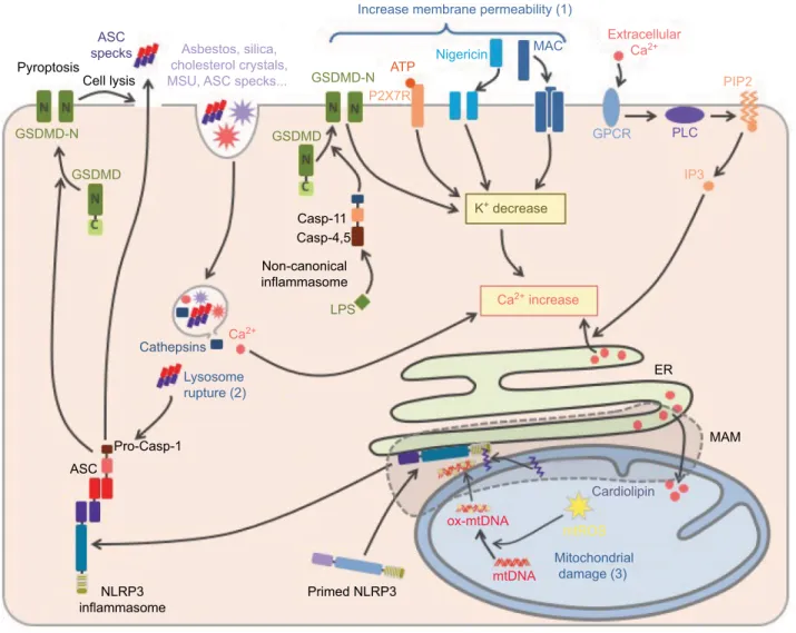
NLRP3 inflammasome activation
Priming licenses NLRP3 to subsequent activation by highly diverse agents and conditions. These activation signals can be classified according to the type of cellular stress that they cause (Figure 4). Agents leading to changes in the plasma membrane permeability to ions form the first category. It com-prises, for example, pore-forming bacterial toxins including
nigericin,54 as well as the membrane attack complex
assem-bled upon the activation of the complement system.55,56 As
already mentioned, permeabilization of plasma membrane by GSDMD pores is critical in NLRP3 activation downstream of non-canonical inflammasomes.13,17 Finally, extracellular ATP,
as released by necrotic neighboring cells, activates NLRP3 through the plasma membrane P2X7 receptor and the subse-quent opening of ionic pores.57 Crystals constitute a second
class of NLRP3 activation signals including asbestos, silica, cholesterol crystals, β-amyloid, and liposomes.58–61 They are
phagocytized but resistant to degradation, and their accumula-tion ultimately leads to the rupture of phagolysosomes and the consequent release of cathepsins and Ca2+ in the cytosol, and
both may account for inflammasome activation.58,62 Finally,
as mentioned before, particulates formed by inflammasome specks released in the extracellular milieu by pyroptotic cells may play a key role in inflammation amplification.15,16 Their
phagocytosis by neighboring macrophages leads to NLRP3 activation by lysosomal disruption similar to other particulate signals, and in addition triggers direct recruitment and activa-tion of cellular caspase-1 by these ASC specks. In addiactiva-tion, extracellular ASC speck has been recently shown to recruit β-amyloid and seed aggregates, spreading β-amyloid pathol-ogy in Alzheimer’s disease.63 Agents and conditions triggering
mitochondrial dysfunction constitute the third class of NLRP3 activators, including metabolic stress, inhibitors of respiratory chain complexes, and blockade of mitophagy.64 Finally, other
NLRP3 activation signals cannot be included in any of these classes as, for example, ultraviolet radiations and activation of G-protein-coupled receptors by the extracellular Ca2+.62,65–67
These highly diverse activation signals seem to engage several pathways to activate NLRP3.68
Although a unifying model for NLRP3 activation still remains controversial, common key signaling features observed downstream of several activators recently emerged including in particular changes in cytosolic ionic concen-tration and mitochondrial damage. A decrease in cytosolic K+ is observed upon exposure to extracellular ATP,
pore-forming toxins, and particulates.69 K+ efflux precedes
mito-chondrial dysfunctions, and correlates with inflammasome activation upon experimental up- or down-modulations of extracellular K+ concentrations, suggesting a key role of
reduction in cytosolic K+ levels in the upstream control of
NLRP3 activation. The mechanism of this control remains unknown, although it has been suggested that low cytosolic K+ concentration might participate directly or indirectly in
the conformational changes of NLRP3 oligomers associated with inflammasome formation.6 Rise in cytosolic Ca2+ is also
Journal of Inflammation Research downloaded from https://www.dovepress.com/ by 193.54.110.33 on 25-Oct-2018
Dovepress Spotlight on the NLRP3 inflammasome pathway
associated with inflammasome activation following numer-ous stimuli.62 High extracellular K+ reduces cytosolic Ca2+
rise, suggesting that K+ efflux may promote Ca2+ influx to
activate NLRP3, although its significance remains debated since the increase in cytosolic Ca2+ is not sufficient to trigger
NLRP3 activation and its role may also be restricted to K+
efflux counterbalancing.69 Several ionic channels including
transient receptor potential, inositol tri-phosphate receptor, and ryanodine receptor have been reported to mediate Ca2+
fluxes associated to NLRP3 activation depending on the inflammatory stimuli and contexts.6,56,61,62,65 In addition,
dis-ruption of lysosomes following particulate phagocytosis may lead to the release of lysosomal Ca2+ into the cytosol and may
participate in the rise of cytosolic Ca2+ levels.62 Increased Ca2+
concentration favors ASC recruitment to NLRP3.65 Lastly,
mitochondrial damage constitutes another shared feature of NLRP3 activation pathways. This mitochondrial dysfunction can be caused after mitochondrial Ca2+ overload following
rise in cytosolic Ca2+ concentration.62 The events following
mitochondrial damage such as loss of membrane potential (∆Ψm), production of ROS, release of mitochondrial DNA (mtDNA) in the cytosol, and exposure of cardiolipin at the
Figure 4 NLRP3 activation.
Notes: NLRP3 activation is triggered by various stress signals including: 1) increase in plasma membrane permeability caused by bacterial toxins, activation of P2X7R by ATP release from necrotic cells, complement membrane attack complex (MAC) or GSDMD pore formed downstream of the activation of the non-canonical inflammasome by cytosolic LPS, 2) lysosome rupture caused by phagocytosis of particulates including crystals (asbestos, silica, monosodium urate [MSU], cholesterol crystals, etc) as well as ASC specks released from neighboring cells, and 3) mitochondrial damages. NLRP3 activation is highly dependent on K+ efflux. In addition, increase in membrane permeability,
lysosome rupture, and activation of G-protein coupled receptor (GPCR) lead to a rise in cytosolic Ca2+. Subsequent Ca2+ overload induces mitochondrial damage, then
cardiolipin exposure and oxidized mtDNA participate in NLRP3 activation at mitochondria-associated eR membranes (MAMs), resulting in the inflammasome assembly and caspase-1 activation. In addition, exogenous ASC specks released in the cytosol following lysosome rupture can directly activate caspase-1 and therefore propagate rapidly the inflammatory signal to neighboring cells.
Abbreviations: TLR, Toll-like receptor; GSDMD, gasdermin-D; LPS, lipopolysaccharide; ASC, apoptosis-associated speck-like protein containing a CARD domain; mtDNA, mitochondrial DNA; eR, endoplasmic reticulum.
ASC
specks Asbestos, silica, cholesterol crystals, MSU, ASC specks... Pyroptosis GSDMD-N GSDMD GSDMD GSDMD-N ATP Nigericin MAC Extracellular Ca2+ PIP2 IP3 K+ decrease PLC GPCR Ca2+ increase ER Casp-11 Casp-4,5 Cathepsins Pro-Casp-1 Primed NLRP3 ox-mtDNA
mtDNA Mitochondrialdamage (3) Cardiolipin mtROS NLRP3 inflammasome ASC Ca2+ Lysosome rupture (2) LPS Non-canonical inflammasome MAM Increase membrane permeability (1)
P2X7R Cell lysis
Journal of Inflammation Research downloaded from https://www.dovepress.com/ by 193.54.110.33 on 25-Oct-2018
Dovepress
Groslambert and Py
outer membrane of mitochondria have all been proposed to mediate NLRP3 activation. In particular, mtDNA and cardiolipin could be direct ligands of NLRP3.70–72
Accord-ingly, inhibition of mitophagy promotes NLRP3 activation through the accumulation of defective mitochondria.72
NLRP3 inflammasome assembly is similarly controlled in most of other innate immune cells, including murine mono-cytes, mast cells, and neutrophils.73–75 Noteworthy, in human
monocytes, strong LPS priming signals have been reported to be sufficient to trigger NLRP3 inflammasome activation in some conditions, although activation signals remain necessary for potentiation of weak priming signals.73,76–78 These differential
requirements may be due to ATP secretion by human monocytes upon priming. Released ATP may then act as an activation signal in an autocrine/paracrine manner.76,77 More recently, it has been
proposed that in human monocytes, an alternative inflamma-some pathway may be triggered without the requirement of activation signals.73 This species-specific alternative pathway
is independent of K+ efflux and leads to a low and gradual
IL-1β response without pyroptosome formation and therefore significantly differs from the all-or-nothing speck response trig-gered by canonical activation stimuli. This alternative pathway depends on TRIF, RIPK1, FADD, and caspase-8 signaling pathways as evidenced in BLaER1 post-mitotic monocytes transdifferentiated from immortalized B cells.
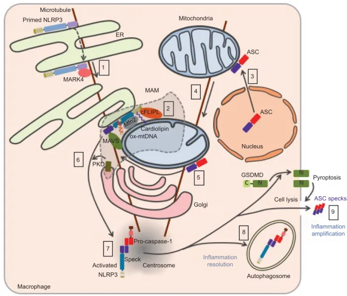
NLRP3 trafficking toward
inflammasome assembly
The trafficking of NLRP3 seems to be tightly regulated upon both priming and activation processes and appears to be criti-cal for inflammasome assembly (Figure 5). Nevertheless, the current model remains controversial due to possible artifacts of overexpression approaches, concerns on the specificity of anti-NLRP3 antibodies raising doubts on endogenous stain-ing, and extensive use of artificial cell lines. NLRP3 is both cytosolic and associated with the ER in THP1 and resting bone marrow-derived macrophages (BMDMs).64,79 Upon
NLRP3 activation, NLRP3 is recruited to mitochondria-associated ER membranes (MAMs).64,71,80 This recruitment
is independent of ASC and therefore probably precedes NLRP3 interaction with its adaptor.80,81 Consistently, NLRP3
recruitment to the mitochondria has been recently reported to occur during priming.82 Consistent with the inhibition of
NLRP3 pathway by colchicine or nocodazole,79 NLRP3
co-migrates with LPS-induced microtubule-affinity regulating kinase 4 (MARK4) along microtubules to reach mitochondria, suggesting a role of microtubules in NLRP3 recruitment to the MAMs.83 MARK4 kinase domain interacts with NLRP3
PYD and NACHT domains, and its expression is required for NLRP3 localization to mitochondria. Other interactors of NLRP3 have been described to be required for – or to facili-tate – NLRP3 relocation and activation including cardiolipin, normally located in the inner mitochondrial membrane, that interacts with NLRP3 LRR domain,71 mitochondrial antiviral
signaling (MAVS) that interacts with NLRP3 PYD,80 cellular
FLICE-inhibitory protein long form (cFLIPL) upregulated by TLR signaling and interacting with NLRP3 LRR domain.84,85
Noteworthy, the role of MAVS remains controversial and has been differentially shown by others to be restricted to NLRP3 control downstream of RNA virus infections, and implicated in the priming of NLRP3 and ASC stabilization.86,87 In
addi-tion, ∆Ψm-dependent binding of NLRP3 with mitofusin-2 (Mfn 2) may potentiate its interaction with MAVS, upon RNA virus infections, but the role of mitofusin-2 in NLRP3 recruitment to the mitochondria has not been investigated.88
Finally, small heterodimer partner (SHP) that co-translocates to the mitochondria upon NLRP3 activation and interacts with its PYD domain acts as a negative regulator. Interestingly, SHP deficiency abolishes NLRP3 recruitment to the mito-chondria suggesting a not yet characterized role in NLRP3 trafficking.81 Other inflammasome components have also
been shown to relocate upon priming or activation. While caspase-1 is cytosolic in basal condition, it is recruited to the mitochondria upon priming probably through its interaction with externalized cardiolipin.82 In basal condition, ASC has
been reported to be mostly in the nucleus but is also found in the cytoplasm where it associates with mitochondria.79,89 Upon
NLRP3 activation, its mitochondrial recruitment increases.82
In addition, mitochondria then relocate in a microtubule net-work-dependent manner to approach the ER at the perinuclear region where acetylated tubulin accumulates.79 Inhibition of
a-tubulin deacetylase sirtuin 2 (SIRT2) caused by mitochon-drial damage and subsequent decrease in NAD+ may induce
this process.79 NLRP3 undergoes self-oligomerization and
interacts with ASC in the MAMs before being released in the cytosol.81,90 Clustering of mitochondria around the Golgi and
phosphorylation of NLRP3 by Golgi-associated protein kinase D (PKD, detailed hereunder) control this release.90 NLRP3
and ASC then colocalize in one cytosolic perinuclear speck of 1–6 µm.89,91 This speck has recently been described to localize
at the centrosome, suggesting a role of microtubule network in speck formation and providing a mechanistic explanation for the formation of a single speck per cell.83 Consistently,
the centrosomal protein NIMA-related protein kinase 7 (NEK7) associates with the inflammasome complex.92–94 On
the contrary, a previous study reported the speck to be distinct
Journal of Inflammation Research downloaded from https://www.dovepress.com/ by 193.54.110.33 on 25-Oct-2018
Dovepress Spotlight on the NLRP3 inflammasome pathway
from the centrosome.95 Finally, inflammasome targeting to
autophagosome plays a role in the control or resolving of the response.96,97 If the inflammasome is not degraded by
autopha-gosome, the abovementioned release of specks participates in the amplification of the inflammatory response.15,16
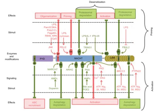
To conclude, the assembly of the NLRP3 inflammasome in macrophages is a tightly controlled event that requires coordinated priming and activation signals. Signaling path-ways and molecular mechanisms involved are not yet fully understood. Notably, accumulative studies recently highlight the role of NLRP3 post-translational modifications in both processes, that is, priming and activation (Figure 6; Table 1).
NLRP3 post-translational
modifications as a regulation
mechanism of inflammasome
assembly
NLRP3 post-translational modifications in
inflammasome priming
As already mentioned, NLRP3 priming can occur indepen-dently of new protein synthesis, and therefore relies primarily on post-translational modifications in addition to the extra need of transcriptional gene upregulation in some condi-tions.42 Phosphorylation of S198 (corresponding to S194 in Figure 5 Current model of NLRP3 subcellular localization during its activation process.
Notes: Primed NLRP3 is both cytosolic and associated with endoplasmic reticulum (eR). Upon activation, NLRP3 is recruited to mitochondria-associated eR membranes (MAMs) by co-migration with MARK4 along microtubules (1). NLRP3 is stabilized in the MAMs through its interaction with cardiolipin, MAvS, cFLIPL, Mfn 2, and oxidized mtDNA at the surface of the mitochondria (2). Upon NLRP3 activation, ASC relocates from the nucleus to the mitochondria network (3), which approaches the eR in a microtubule-dependent manner (4). NLRP3 associates with ASC in the MAMs (5). Phosphorylation of NLRP3 by Golgi-associated PKD leads to the release of NLRP3/ASC complex from the MAMs (6) and assembly of a single speck at the centrosome (7). Autophagy dampens inflammation by targeting the inflammasome (8), while release of ASC speck from pyroptotic cells propagates the inflammatory signal to neighboring cells (9).
Abbreviations: MAvS, mitochondrial antiviral signaling; mtDNA, mitochondrial DNA; ASC, apoptosis-associated speck-like protein containing a CARD domain; PKD, protein kinase D; mtDNA, mitochondrial DNA; GSDMD, gasdermin-D.
Activated Macrophage 7 Primed NLRP3 Microtubule 1 MARK4 6 PKD MAVS Mfn2 cFLIPL 2 4 5 8 Golgi Inflammation resolution Pro-caspase-1 Speck Centrosome Autophagosome NLRP3 3 ASC MAM Cardiolipin ASC Nucleus GSDMD Cell lysis Pyroptosis 9 Inflammation amplification ASC specks ox-mtDNA ER Mitochondria
Journal of Inflammation Research downloaded from https://www.dovepress.com/ by 193.54.110.33 on 25-Oct-2018
Dovepress
Groslambert and Py
mouse NLRP3) by JNK1 has been recently proposed as a universal feature of NLRP3 priming, required for both tran-scriptional-dependent and -independent priming pathways, and downstream of both MyD88 and TRIF signaling.98 S198
phosphorylation kinetics correlates with NLRP3 priming by these different pathways. S198 phosphorylation leads to NLRP3 deubiquitination and promotes its oligomerization. S198N polymorphism has been identified in CAPS patients.99
This observation is surprising since CAPS is typically caused by gain of function mutations in NLRP3. Nevertheless, this patient bore another well characterized CAPS mutation Y570C that was probably causing over-activation of NLRP3 without the need of S198 phosphorylation. These results are consistent with earlier findings that NLRP3 is constitutively ubiquitinated and that non-transcriptional priming induced by short LPS exposure leads to NLRP3 deubiquitination in a mtROS-dependent manner.42 More recently and in opposite
to its constitutive ubiquitination model, NLRP3 has been reported to be very transiently ubiquitinated with K63 chains following LPS priming.100 This ubiquitination depends on the
E3 ligase Pellino 2, whose deficiency significantly impairs NLRP3 response in cultured BMDMs, as well as in several
mouse models. Most E3 ubiquitin ligases and deubiquitinases controlling NLRP3 priming remain to be identified. Notewor-thy, chaperones such as suppressor of the G2 allele of skp1 (SGT1) and heat shock protein 90 (HSP90), which have E3 ligase activity, associate with NLRP3 in resting conditions.101
In addition, the increase in NLRP3 level upon transcriptional priming may in part rely on stabilization of NLRP3 through post-translational regulation. Indeed, NLRP3 ubiquitination on K691 (K689 in mouse) by skp-cullin-F box L2 (SCFFBXL2)
E3 ligase targets NLRP3 to proteasomal degradation.102,103
Transcriptional priming leads to the expression of the SCF F-BXO3 E3 ligase that destabilizes SCFFBXL2 via ubiquitination
and degradation, resulting in increased NLRP3 half-life. Other NLRP3 modifications have been reported upon LPS priming including S-glutathionylation, but their relevance in the priming process remains to be established.104
NLRP3 post-translational modifications in
inflammasome activation
Accumulative studies point to the critical role of NLRP3 post-translational modifications in NLRP3 activation step. We first described that NLRP3 inflammasome assembly requires
Figure 6 Post-translational modifications of NLRP3 involved in its priming, activation, and desensitization.
Notes: Stimuli, enzymes, modifications, and targeted residues that promote or inhibit inflammasome assembly are represented in pink and green, respectively.
Abbreviations: LPS, lipopolysaccharide; ASC, apoptosis-associated speck-like protein containing a CARD domain; Nig, nigericin; Sil, silica; MSU, monosodium urate; PYD, pyrin domain. Effects Desensitization LPS FBXL2 TRIM31 MARCH7 AMPc Dopamin Autophagy
degradation Activation degradationAutophagy ASC recruitment ARIH2 PKA AMPc Bile acid PGE2 Priming Activation Oligomerization Priming Proteasomal degradation LPS/IL-1 PELI2 JNK LPS, Pam3CSK4, Poly(I:C), Flagellin, R848, ARN CpG LPS, zymosan Ub Ub Ub Ub Ub NACHT NO Ub Ub BRCC3 PKD DAG ATP, Nig,
Sil, MSU ATP, Nig,Sil, MSU ATP, Nig,Sil, MSU Ca2+ PTPN22 LRR S295 S198 S5 K691 Y861 IFN-I/II Activation Proteasomal degradation Stimuli Enzymes and modifications Signaling PYD P P P P Stimuli Effects
Journal of Inflammation Research downloaded from https://www.dovepress.com/ by 193.54.110.33 on 25-Oct-2018
Dovepress Spotlight on the NLRP3 inflammasome pathway
NLRP3 deubiquitination by the BRCA1/BRCA2-containing complex subunit 3 (BRCC3) deubiquitinase downstream of all tested activation signals.105 Therefore, this
deubiq-uitination can be considered as a key common regulatory mechanism of inflammasome activation. It does not depend on mtROS, and therefore signaling differs from deubiquiti-nation associated with NLRP3 priming.42 NACHT and LRR
domains of NLRP3 are independently ubiquitinated, and both K48 and K63 ubiquitin chains associate with NLRP3.105 The
K63-specific deubiquitinase BRCC3 specifically targets the NLRP3 LRR domain, but the precise ubiquitinated residue and the structural consequences of this deubiquitination remain unknown. BRCC3 inhibition promotes NLRP3 ubiq-uitination and blocks inflammasome assembly but does not target NLRP3 for degradation.105 Consistently, the ARIH2
ubiquitin ligase has recently been identified in the speck.106
While ARIH2 promotes NLRP3 ubiquitination with K63 and K48 chains and inhibits the inflammasome activation, it does not reduce NLRP3 stability. In addition, NLRP3 acti-vation is also downregulated by degradative ubiquitination. The inflammasome inhibitor dopamine induces an increase in cAMP level through dopamine receptor D1 (DRD1).107
cAMP then binds to NLRP3 and triggers the ubiquitination of NLRP3 NACHT and LRR domains with K48 ubiquitin
chains by the E3 ubiquitin ligase membrane associated ring-CH-type finger 7 (MARCH7), targeting NLRP3 to autophagy-mediated degradation.107
Phosphorylation of NLRP3 also controls its activation process. Phosphorylation of S295 (S291 in mouse NLRP3) in the NACHT domain has recently emerged as a key regula-tory point in NLRP3 activation process. Initially, bile acids binding Takeda G-protein-coupled receptor 5 (TGR5) and eicosanoid prostaglandin E2 (PGE2) binding E prostanoid 4 receptor (EP4) have been shown to inhibit NLRP3 through production of cAMP, activation of protein kinase A (PKA), phosphorylation of NLRP3 by PKA on S295 leading to its inactivation by non-degradative ubiquitination.108,109 On the
opposite, S295 phosphorylation by PKDs has recently been reported to be required for NLRP3 activation.90 Indeed,
in response to inflammasome activators, Golgi-associated diacyl-glycerol (DAG) recruits PKDs while mitochondria cluster to the Golgi. Then PKDs phosphorylate MAM-associated NLRP3 on S295, leading to its release from the MAMs and inflammasome assembly in the cytosol. PKDs are dispensable for mitochondrial clustering to the Golgi and act downstream of mitochondrial damage. Reconciling all data, both NLRP3 phospho-null and phospho-mimetic mutants are inactive, suggesting that S295 phosphorylation needs to be
Table 1 Post-translational modifications of NLRP3 inflammasome
Protein Process Effect Modification Domain (residue human/mouse)
Regulatory enzyme
References
NLRP3 Priming Activatory Phosphorylation NACHT (S198/S194) JNK1 Song et al98
Activatory Deubiquitination ? ? Juliana et al42
Activatory Ubiquitination ? PeLI2 Humphries et al100
Inhibitory Ubiquitination LRR (K691/K689) SCFFBXL2 Han et al102
? S-glutathionylation ? ? Guglielmoet al104
Activation Activatory Deubiquitination LRR BRCC3 Pyet al105
Inhibitory Ubiquitination NACHT ARIH2 Kawashima et al106
Inhibitory Ubiquitination NACHT and LRR MARCH7 Yan et al107
Inhibitory Phosphorylation NACHT (S295/S291) PKA Guoet al, Mortimer et al108,109
Activatory Phosphorylation NACHT (S295/S291) PKD Zhanget al90
Activatory Dephosphorylation LRR (Y861/Y859) PTPN22 Spalingeret al112
? Activatory Dephosphorylation PYD (S5/S3) PP2A Stutzet al110
Desensitization Inhibitory Ubiquitination ? TRIM31 Song et al113
Inhibitory S-nitrosylation LRR ? Hernandez-Cuellaret al47
ASC Priming Activatory Ubiquitination CARD (K174/K172) TRAF3 Guanet al87
? Phosphorylation PYD ? Stehlik et al118
Activation Activatory Deubiquitination ? USP50 Leeet al117
Activatory Phosphorylation CARD (Y146/Y144) SYK/JNK Haraet al119
? Activatory Ubiquitination ? LUBAC Rodgerset al116
Resolving Inhibitory Ubiquitination ? ? Shiet al96
Caspase-1 Activation Activatory Ubiquitination CARD cIAP-2 Labbéet al120
? Inhibitory S-glutathionylation p10 subunit (C362 and C397) ? Meissneret al121
Pro-IL-1β Desensitization Inhibitory Deubiquitination K133 A20 Duonget al50
Note: ? indicates unknown.
Abbreviations: ASC, apoptosis-associated speck-like protein containing a CARD domain; LRR, leucin-rich repeat; PYD, pyrin domain; CARD, caspase recruitment domain.
Journal of Inflammation Research downloaded from https://www.dovepress.com/ by 193.54.110.33 on 25-Oct-2018
Dovepress
Groslambert and Py
timely and locally regulated. Indeed, S295 phosphorylation initially prevents NLRP3 recruitment to MAMs necessary for its activation, while S295 phosphorylation later by PKDs allows its release from the MAMs to form cytosolic specks.90
In addition, inflammasome assembly also requires the dephosphorylation of S5 (S3 in mouse NLRP3) in the PYD of NLRP3.110 Indeed, phosphorylation of S5 prevents ASC
recruitment by electrostatic repulsion. Protein phosphatase 2A (PP2A) could be involved in this dephosphorylation.110,111
However, it remains unclear whether S5 dephosphorylation occurs during the priming or the activation step. Noteworthy, the critical role of kinase NEK7 in NLRP3 inflammasome assembly has recently been discovered by several indepen-dent studies.92–94 NEK7 interacts with NLRP3 LRR domain
and takes part in the inflammasome complex. However, its catalytic activity is dispensable for the inflammasome activity and therefore does not regulate inflammasome assembly by direct phosphorylation of NLRP3 or other components of the complex but rather by scaffolding, subcellular relocation, or other functions that remain to be elucidated. Finally, the phos-phorylation on Y861 (Y859 in mouse) in the LRR domain dampens the inflammasome activity by targeting NLRP3 to autophagosomes.97,112 Phosphorylated NLRP3 interacts with
SQSTM1 in an ASC-dependent manner, suggesting that this downregulation may occur later in the process after inflam-masome assembly.97 Upon activation signals, protein tyrosine
phosphatase non-receptor type 22 (PTPN22) is recruited to NLRP3 in an ASC-dependent manner and sustains IL-1β secretion by dephosphorylating Y861.112 NLRP3
dephos-phorylation in a model of dextran sulfate sodium-induced colitis requires PTPN22 and correlates with caspase-1, IL-1β, and IL-18 maturation as well as granulocyte recruit-ment, suggesting that NLRP3 Y861 phosphorylation may play a significant role in inflammatory diseases. Noteworthy, PTPN22 is genetically associated with several inflammatory disorders linked to inflammasome dysregulation.
NLRP3 post-translational modifications
involved in its desensitization upon
prolonged priming
As mentioned previously, prolonged TLR4-mediated prim-ing in the absence of timely activation causes NLRP3 to be refractory to late activation stimuli. This desensitization probably participates in maintaining NLRP3 under tight control preventing inappropriate inflammasome-mediated inflammation and relies partly on NLRP3 post-translational modifications. Indeed, the E3 ubiquitin ligase TRIM31, which is induced after LPS and IL-1β, prolonged priming triggers
K48 ubiquitination of NLRP3 and leads to its proteasomal degradation.113 In addition, as already mentioned, Y861
phosphorylation targets the inflammasome to autophago-somes preventing NLRP3 activation.97,112 Therefore, this
mechanism may support desensitization upon prolonged priming. Last but not least, independent studies evidenced the key role of iNOS in this desensitization of NLRP3.47,114
Prolonged exposure to LPS triggers IFN-I secretion, which in turn induces iNOS expression in an autocrine/paracrine manner. iNOS inhibits NLRP3 at least in part through its direct modifications. Indeed, the subsequent production of NO leads to the S-nitrosylation of the cysteine-rich LRR domain of NLRP3, a modification that prevents ASC oligo-mer formation.47 Notably, IFN-β inhibits NLRP3 through
the same mechanism.115
Post-translational modifications of
ASC, caspase-1, and IL-1b in the
regulation of the inflammasome
The NLRP3 inflammasome activity is also regulated by post-translational modifications of its other components including ASC, caspase-1, and its substrate IL-1β. ASC linear ubiquiti-nation by heme-oxidized IRP2 ubiquitin ligase 1L (HOIL-1L) in the linear ubiquitin chain assembly complex (LUBAC) is required for its recruitment to NLRP3, inflammasome assem-bly, and activation.116 However, whether ASC linear
ubiquiti-nation occurs downstream of priming or activation signaling has not yet been investigated. Besides, ubiquitination of ASC at K174 residue with K63 chains by tumor necrosis factor receptor-associated factor 3 (TRAF3) downstream of MAVS stabilizes ASC and supports inflammasome assem-bly upon RNA virus infections.87 Interestingly, ASC is also
ubiquitinated with K63 chains upon LPS priming, and later deubiquitinated by ubiquitin specific peptidase 50 (USP50) upon NLRP3 activation, suggesting a conserved regulation of speck formation by dynamic ASC K63 ubiquitination.117
Finally, ASC ubiquitination with K63 ubiquitin chain also participates in the resolution of the response by targeting the inflammasome to degradation through the autophagy machinery.96 Phosphorylation of ASC also likely plays a role
in inflammasome regulation. A previous report shows that ectopically expressed ASC PYD domain can be phosphory-lated in response to TNF treatment.118 More recently, ASC
has been shown to be phosphorylated upon inflammasome activation stimuli through an unknown pathway dependent on both spleen tyrosine kinase (SYK) and c-Jun N-terminal kinase (JNK), which are required for ASC oligomerization and speck formation but not for ASC recruitment to NLRP3.
Journal of Inflammation Research downloaded from https://www.dovepress.com/ by 193.54.110.33 on 25-Oct-2018
Dovepress Spotlight on the NLRP3 inflammasome pathway
Among several other phosphorylated residues, Y146 in ASC CARD domain (Y144 in mouse ASC) seems to be the critical target of this pathway.119 The inflammasome effector
caspase-1 has been reported to be ubiquitinated in its CARD domain with K63 ubiquitin chains by cellular inhibitor of apoptosis 2 (cIAP-2) upon NLRP3 activation signals.120
Since cIAP-1/2 and TRAF2-containing complex, which associates with caspase-1, is required for IL-1β secretion, this non-degradative ubiquitination may promote inflammasome activity but the mechanism remains unknown. In addition, caspase-1 is also unexpectedly directly inhibited by some ROS. Indeed, accumulation of intracellular superoxide, as observed in superoxide dismutase 1 (SOD-1) deficiency, leads to caspase-1 S-glutathionylation on C362 and C397 and its inhibition.121 Finally, post-translational modifications of
the caspase-1 substrate pro-IL-1β also impact inflammasome activity. Indeed, pro-IL-1β associates with K63 ubiquitin chains on K133 and unanchored K48 chains in a complex pre-formed upon LPS transcriptional priming and containing RIPK1/3, pro-caspase-1/8, and A20.50 While A20 reduces
pro-IL-1β K63 ubiquitination and maturation, on the contrary RIPK3 increases pro-IL-1β association with ubiquitin chains and promotes its spontaneous NLRP3-dependent maturation upon A20 deficiency. Therefore, pro-IL-1β ubiquitination supports its caspase-1-mediated proteolytic processing. It may be interesting in future studies to address ubiquitina-tion of other caspase-1 substrates including pro-IL-18 and GSDMD.
Conclusion
While inflammation is important for organism protection, its dysregulation is highly detrimental as tragically exemplified by a number of inflammatory diseases. Accordingly, NLRP3 inflammasome assembly is strictly regulated by the require-ment of coordinated priming and activation signals. These signals are highly diverse, and despite almost 20 years of research, our understanding of the signaling pathways and the molecular mechanisms controlling NLRP3 remains largely incomplete. Post-translational modifications of NLRP3 and other inflammasome components have recently emerged as an important mechanism in the control of its assembly. In the near future, ongoing characterization of the molecular consequences of these modifications will fill some of these gaps. In addition, identification of the enzymes and their co-factors controlling these modifications may lead to the identification of novel druggable targets for specific anti-inflammatory therapies.
Acknowledgments
This work was supported in part by grants to BFP from the Agence Nationale pour la Recherche (ANR-13-JSV3-0002-01) and the European Research Council (ERC-2013-CoG_616986). We thank Pablo Palazon-Riquelme, Danish Patoli, and Amaury Rey for comments on the manuscript.
Disclosure
The authors report no conflicts of interest in this work.
References
1. Medzhitov R. Origin and physiological roles of inflammation. Nature. 2008;454(7203):428–435.
2. Medzhitov R, Janeway CA. Innate immunity: the virtues of a nonclonal system of recognition. Cell. 1997;91(3):295–298.
3. Matzinger P. Tolerance, danger, and the extended family. Annu Rev
Immunol. 1994;12:991–1045.
4. Hu Z, Zhou Q, Zhang C, et al. Structural and biochemical basis for induced self-propagation of NLRC4. Science. 2015;350(6259):399–404. 5. Fernandes-Alnemri T, Yu J-W, Datta P, Wu J, Alnemri ES. AIM2
activates the inflammasome and cell death in response to cytoplasmic DNA. Nature. 2009;458(7237):509–513.
6. Compan V, Baroja-Mazo A, López-Castejón G, et al. Cell volume regulation modulates NLRP3 inflammasome activation. Immunity. 2012;37(3):487–500.
7. Lu A, Magupalli VG, Ruan J, et al. Unified polymerization mecha-nism for the assembly of ASC-dependent inflammasomes. Cell. 2014;156(6):1193–1206.
8. Masumoto J, Taniguchi S, Ayukawa K, et al. ASC, a novel 22-kDa protein, aggregates during apoptosis of human promyelocytic leukemia HL-60 cells. J Biol Chem. 1999;274(48):33835–33838.
9. Dick MS, Sborgi L, Rühl S, Hiller S, Broz P. ASC filament formation serves as a signal amplification mechanism for inflammasomes. Nat
Commun. 2016;7:11929.
10. Shi J, Zhao Y, Wang Y, et al. Inflammatory caspases are innate immune receptors for intracellular LPS. Nature. 2014;514(7521):187–192. 11. Schroder K, Tschopp J. The inflammasomes. Cell. 2010;140(6):821–832. 12. Kayagaki N, Stowe IB, Lee BL, et al. Caspase-11 cleaves gas-dermin D for non-canonical inflammasome signalling. Nature. 2015;526(7575):666–671.
13. Aglietti RA, Estevez A, Gupta A, et al. GsdmD p30 elicited by cas-pase-11 during pyroptosis forms pores in membranes. Proc Natl Acad
Sci U S A. 2016;113(28):7858–7863.
14. Bergsbaken T, Fink SL, Cookson BT. Pyroptosis: host cell death and inflammation. Nat Rev Microbiol. 2009;7(2):99–109.
15. Baroja-Mazo A, Martín-Sánchez F, Gomez AI, et al. The NLRP3 inflammasome is released as a particulate danger signal that amplifies the inflammatory response. Nat Immunol. 2014;15(8):738–748. 16. Franklin BS, Bossaller L, De Nardo D, et al. The adaptor ASC has
extracellular and “prionoid” activities that propagate inflammation.
Nat Immunol. 2014;15(8):727–737.
17. Rühl S, Broz P. Caspase-11 activates a canonical NLRP3 inflammasome by promoting K+ efflux. Eur J Immunol. 2015;45(10):2927–2936. 18. Hoffman DHM. Familial cold autoinflammatory syndrome. J Allergy
Clin Immunol. 2001;108(4):615–620.
19. Shinkai K, Mccalmont TH, Leslie KS. Cryopyrin-associated periodic syndromes and autoinflammation. Clin Exp Dermatol. 2008;33(1):1–9. 20. Martinon F, Pétrilli V, Mayor A, Tardivel A, Tschopp J. Gout-associ-ated uric acid crystals activate the NALP3 inflammasome. Nature. 2006;440(7081):237–241.
Journal of Inflammation Research downloaded from https://www.dovepress.com/ by 193.54.110.33 on 25-Oct-2018
Dovepress
Groslambert and Py
21. Vandanmagsar B, Youm Y-H, Ravussin A, et al. The NALP3/NLRP3 inflammasome instigates obesity-induced autoinflammation and insulin resistance. Nat Med. 2011;17(2):179–188.
22. Youm Y-H, Grant RW, McCabe LR, et al. Canonical Nlrp3 inflamma-some links systemic low grade inflammation to functional decline in aging. Cell Metab. 2013;18(4):519–532.
23. Heneka MT, Kummer MP, Latz E. Innate immune activation in neu-rodegenerative disease. Nat Rev Immunol. 2014;14(7):463–477. 24. Franchi L, Muñoz-Planillo R, Núñez G. Sensing and reacting to
microbes via the inflammasomes. Nat Immunol. 2012;13(4):325–332. 25. Jo E-K, Kim JK, Shin D-M, Sasakawa C. Molecular mechanisms regulating NLRP3 inflammasome activation. Cell Mol Immunol. 2016;13(2):148–159.
26. Ratsimandresy RA, Dorfleutner A, Stehlik C. An update on PYRIN domain-containing pattern recognition receptors: from immunity to pathology. Front Immunol. 2013;4:440.
27. Elliott EI, Sutterwala FS. Monocytes take their own path to IL-1β.
Immunity. 2016;44(4):713–715.
28. Alexiou P, Chatzopoulou M, Pegklidou K, Demopoulos VJ. RAGE: a multi-ligand receptor unveiling novel insights in health and disease.
Curr Med Chem. 2010;17(21):2232–2252.
29. Xiang M, Shi X, Li Y, et al. Hemorrhagic shock activation of NLRP3 inflammasome in lung endothelial cells. J Immunol. 2011;187(9):4809–4817.
30. Fritz JH, Ferrero RL, Philpott DJ, Girardin SE. Nod-like pro-teins in immunity, inflammation and disease. Nat Immunol. 2006;7(12):1250–1257.
31. Meylan E, Tschopp J, Karin M. Intracellular pattern recognition recep-tors in the host response. Nature. 2006;442(7098):39–44.
32. Mitchell JA, Paul-Clark MJ, Clarke GW, McMaster SK, Cartwright N. Critical role of toll-like receptors and nucleotide oligomerisa-tion domain in the regulaoligomerisa-tion of health and disease. J Endocrinol. 2007;193(3):323–330.
33. Franchi L, Eigenbrod T, Núñez G. Cutting edge: TNF-alpha mediates sensitization to ATP and silica via the NLRP3 inflammasome in the absence of microbial stimulation. J Immunol. 2009;183(2):792–796. 34. Sutterwala FS, Haasken S, Cassel SL. Mechanism of NLRP3
inflam-masome activation. Ann N Y Acad Sci. 2014;1319:82–95.
35. Bauernfeind FG, Horvath G, Stutz A, et al. Cutting edge: NF-kappaB activating pattern recognition and cytokine receptors license NLRP3 inflammasome activation by regulating NLRP3 expression. J Immunol. 2009;183(2):787–791.
36. Bauernfeind F, Niepmann S, Knolle PA, Hornung V. Aging-associated TNF production primes inflammasome activation and NLRP3-related metabolic disturbances. J Immunol Baltim Md 1950. 2016;197(7):2900–2908.
37. Gurung P, Anand PK, Malireddi RKS, et al. FADD and caspase-8 mediate priming and activation of the canonical and noncanonical Nlrp3 inflammasomes. J Immunol. 2014;192(4):1835–1846. 38. Zhang X, Fan C, Zhang H, et al. MLKL and FADD are critical for
suppressing progressive lymphoproliferative disease and activating the NLRP3 inflammasome. Cell Rep. 2016;16(12):3247–3259. 39. Bauernfeind F, Bartok E, Rieger A, Franchi L, Núñez G, Hornung V.
Cutting edge: reactive oxygen species inhibitors block priming, but not activation, of the NLRP3 inflammasome. J Immunol Baltim Md
1950. 2011;187(2):613–617.
40. Juliana C, Fernandes-Alnemri T, Wu J, et al. Anti-inflammatory compounds parthenolide and Bay 11-7082 are direct inhibitors of the inflammasome. J Biol Chem. 2010;285(13):9792–9802.
41. Schroder K, Sagulenko V, Zamoshnikova A, et al. Acute lipopolysaccha-ride priming boosts inflammasome activation independently of inflam-masome sensor induction. Immunobiology. 2012;217(12):1325–1329. 42. Juliana C, Fernandes-Alnemri T, Kang S, Farias A, Qin F, Alnemri ES. Non-transcriptional priming and deubiquitination regulate NLRP3 inflammasome activation. J Biol Chem. 2012;287(43):36617–36622.
43. Fernandes-Alnemri T, Kang S, Anderson C, Sagara J, Fitzgerald KA, Alnemri ES. Cutting edge: TLR signaling licenses IRAK1 for rapid activation of the NLRP3 inflammasome. J Immunol. 2013;191(8): 3995–3999.
44. Lin K-M, Hu W, Troutman TD, et al. IRAK-1 bypasses priming and directly links TLRs to rapid NLRP3 inflammasome activation. Proc
Natl Acad Sci U S A. 2014;111(2):775–780.
45. Kang S, Fernandes-Alnemri T, Rogers C, et al. Caspase-8 scaffold-ing function and MLKL regulate NLRP3 inflammasome activation downstream of TLR3. Nat Commun. 2015;6:7515.
46. Kang T-B, Yang S-H, Toth B, Kovalenko A, Wallach D. Caspase-8 blocks kinase RIPK3-mediated activation of the NLRP3 inflamma-some. Immunity. 2013;38(1):27–40.
47. Hernandez-Cuellar E, Tsuchiya K, Hara H, et al. Cutting edge: nitric oxide inhibits the NLRP3 inflammasome. J Immunol. 2012;189(11):5113–5117.
48. Hu Y, Mao K, Zeng Y, et al. Tripartite-motif protein 30 negatively regulates NLRP3 inflammasome activation by modulating reactive oxygen species production. J Immunol Baltim Md 1950. 2010;185(12): 7699–7705.
49. Guarda G, Braun M, Staehli F, et al. Type I interferon inhibits interleukin-1 production and inflammasome activation. Immunity. 2011;34(2):213–223.
50. Duong BH, Onizawa M, Oses-Prieto JA, et al. A20 restricts ubiquitina-tion of pro-interleukin-1β protein complexes and suppresses NLRP3 inflammasome activity. Immunity. 2015;42(1):55–67.
51. Zhong Z, Umemura A, Sanchez-Lopez E, et al. NF-κB restricts inflammasome activation via elimination of damaged mitochondria.
Cell. 2016;164(5):896–910.
52. McGeough MD, Wree A, Inzaugarat ME, et al. TNF regulates transcrip-tion of NLRP3 inflammasome components and inflammatory mol-ecules in cryopyrinopathies. J Clin Invest. 2017;127(12):4488–4497. 53. Casal C, Serratosa J, Tusell JM. Relationship between beta-AP peptide aggregation and microglial activation. Brain Res. 2002;928(1–2): 76–84.
54. Cookson BT, Brennan MA. Pro-inflammatory programmed cell death.
Trends Microbiol. 2001;9(3):113–114.
55. Laudisi F, Spreafico R, Evrard M, et al. Cutting edge: the NLRP3 inflammasome links complement-mediated inflammation and IL-1β release. J Immunol. 2013;191(3):1006–1010.
56. Triantafilou K, Hughes TR, Triantafilou M, Morgan BP. The complement membrane attack complex triggers intracellular Ca2+ fluxes leading to NLRP3 inflammasome activation. J Cell Sci. 2013;126(13):2903–2913.
57. Solle M, Labasi J, Perregaux DG, et al. Altered cytokine production in mice lacking P2X(7) receptors. J Biol Chem. 2001;276(1):125–132. 58. Hornung V, Bauernfeind F, Halle A, et al. Silica crystals and alumi-num salts mediate NALP-3 inflammasome activation via phagosomal destabilization. Nat Immunol. 2008;9(8):847–856.
59. Duewell P, Kono H, Rayner KJ, et al. NLRP3 inflammasomes are required for atherogenesis and activated by cholesterol crystals. Nature. 2010;464(7293):1357–1361.
60. Halle A, Hornung V, Petzold GC, et al. The NALP3 inflammasome is involved in the innate immune response to amyloid-beta. Nat Immunol. 2008;9(8):857–865.
61. Zhong Z, Zhai Y, Liang S, et al. TRPM2 links oxidative stress to NLRP3 inflammasome activation. Nat Commun. 2013;4:1611. 62. Murakami T, Ockinger J, Yu J, et al. Critical role for calcium
mobiliza-tion in activamobiliza-tion of the NLRP3 inflammasome. Proc Natl Acad Sci U
S A. 2012;109(28):11282–11287.
63. Venegas C, Kumar S, Franklin BS, et al. Microglia-derived ASC specks cross-seed amyloid-β in Alzheimer’s disease. Nature. 2017;552(7685):355–361.
64. Zhou R, Yazdi AS, Menu P, Tschopp J. A role for mitochondria in NLRP3 inflammasome activation. Nature. 2011;469(7329):221–225.
Journal of Inflammation Research downloaded from https://www.dovepress.com/ by 193.54.110.33 on 25-Oct-2018