HAL Id: tel-01132704
https://tel.archives-ouvertes.fr/tel-01132704
Submitted on 17 Mar 2015
HAL is a multi-disciplinary open access
archive for the deposit and dissemination of sci-entific research documents, whether they are pub-lished or not. The documents may come from teaching and research institutions in France or abroad, or from public or private research centers.
L’archive ouverte pluridisciplinaire HAL, est destinée au dépôt et à la diffusion de documents scientifiques de niveau recherche, publiés ou non, émanant des établissements d’enseignement et de recherche français ou étrangers, des laboratoires publics ou privés.
Plasmodium, the causative parasite of malaria
Partho Sen
To cite this version:
Partho Sen. Integrated modelling of lipid metabolism in Plasmodium, the causative parasite of malaria. Parasitology. Université Montpellier II - Sciences et Techniques du Languedoc, 2013. En-glish. �NNT : 2013MON20153�. �tel-01132704�
L'UNIVERSITE MONTPELLIER 2
Sciences Chimiques et Biologiques pour la Santé
Dynamique des Interactions Membranaires Normales et
Pathologiques, (DIMNP-UMR 5235)
Prof. Jean-Pierre MAZAT
Institut de Biochimie et Génétique Cellulaires, CNRS,Université Bordeaux Segalen, France
Rapporteur
Prof. Vassily HATZIMANIKATIS
Ecole polytechnique fédérale de Lausanne & Institut des sciences et ingénierie chimiques, Laussane,Suisse.
Rapporteur
Dr. Cyrille BOTTE
Laboratoire Adaptation et Pathogenie des Microorganismes, CNRS, Université. Grenoblel,France
Examinateur
Dr. Henri VIAL
DR INSERM, DIMNP UMR 5235, CNRS, Université Montpellier 1 & 2, Montpellier,France
Président du jury
Prof. Ovidiu RADULESCU
DIMNP UMR 5235, CNRS, Université Montpellier2 Montpellier, France.
Directeur de thèse
Modélisation intégrée du métabolisme des lipides chez
Plasmodium, parasite causal du paludisme
Integrated modelling of lipid metabolism in Plasmodium,
the causative parasite of malaria.
SCIENCES ET TECHNIQUES DU LANGUEDOC
THESEpour obtenir le grade de
DOCTEUR DE L'UNIVERSITE MONTPELLIER 2
Spécialité
:Biologie Santé
Ecole Doctorale: CBS2 - Sciences Chimiques et Biologiques pour la Santé
Titre:
Modélisation intégrée du métabolisme des lipides chez Plasmodium,
parasite causal du paludisme
Integrated modelling of lipid metabolism in Plasmodium, the causative
parasite of malaria.
présentée et soutenue publiquement par
Partho SEN
le 17 Décember 2013
JURY
Prof. Jean-Pierre MAZAT
Institut de Biochimie et Génétique Cellulaires, CNRS,Université Bordeaux Segalen, France
Rapporteur
Prof. Vassily HATZIMANIKATIS
Ecole polytechnique fédérale de Lausanne & Institut des sciences et ingénierie chimiques, Laussane, Suisse.
Rapporteur
Dr. Cyrille BOTTE
Laboratoire Adaptation et Pathogenie des
Microorganismes, CNRS, Université. Grenoblel, France
Examinateur
Dr. Henri VIAL
DR INSERM, DIMNP UMR 5235, CNRS, Université Montpellier 1 & 2, Montpellier,France
Président du jury
Prof. Ovidiu RADULESCU
DIMNP UMR 5235, CNRS, Université Montpellier2 Montpellier, France.
The period approaching towards end of Ph.D is mingled with emotions. To extend this feelings, I would like to thank all those who made this thesis possible. First and foremost, my research supervisor, Prof.Ovidiu Radulescu who has been extremely encouraging and supportive. His early realization of my research inclination gave me a platform to perform interdisciplinary science in an effective enviornment. The basic dicussions and motivations helped me to orient my thinking to cope up with this fascinating disciple. His constructive criticism helped me to attain the highest possible level of perfection. He had always granted the freedom and encouragement to work independently. These helped me to develop as an independent thinker and a confident researcher. All in all, it was a great experience to work with him and have him as my supervisor.
I am deeply grateful to Dr.Henri Vial, for his support and guidance throughout my PhD thesis. His tremendous knowledge in biology, guided me in troubleshooting conceptual issues. He is always motivating and inspiring. Thanks for providing time from your busy schedule and discussing with me as or when needed. With him I got a chance to meet with other fellow biologist, which enhanced my vision on various aspects in parasitology.
I acknowledge the referees of the jury, Prof.Jean-Pierre Mazat, Prof.Vassily Hatzi-manikatis, Dr.Cyrille Botte, who devoted their valuable time to examine the thesis.
I am also thankful to Prof.Catherine Braun-Breton, director of the lab DIMN-P-UMR-5235, for providing a cordial atmosphere in the lab. Prof.Andrea Parmeggiani for his kind help and useful discussions. Dr.Laurence Berry for useful suggestions. Christine Bousquet for all the administrative work.
Friends form an important part of life, and my friends have always been great companions to me. Thanks ! Vincent Noel for helping me out as or when required. Olivier Dauloudet for cheering me up, with coffee breaks. I pay a similar consideration to my fian´cee-Palki, who remained non-demanding and understood the time crunch, I was going through.
Le paludisme est responsable de la mort de pr`es d’un million de personnes chaque ann´ee. Cette maladie est caus´ee par Plasmodium, parasite protozoaire appartenant `a la famille des Apicomplexes. Dans cette th`ese, nous avons d´evelopp´e des approches de biologie des syst`emes pour l’´etude du m´etabolisme des phospholipides (PL) et de sa r´egulation chez Plasmodium. Ces voies m´etaboliques sont d’une importance pri-mordiale pour la survie du parasite. Lors du d´eveloppement intra-´erythrocytaire, les esp`eces de Plasmodium exploitent un nombre important de voies de synth`ese phos-pholipidique, qui sont rarement trouv´ees ensemble dans un seul organisme : (i) la voie ancestrale CDP-diacylglyc´erol des procaryotes ii) les voies eucaryotes de novo dite de Kennedy, impliquant CDP- choline ou CDP-´ethanolamine (iii) de plus, P. falci-parum et P. knowlesi utilisent une voie transversale impliquant s´erine d´ecarboxylase et phospho-´ethanolamine-methyltransf´erase (SDPM), caract´eristique des plantes, et source additionnelle de phosphatidyl-´ethanolamine (PE) et de phosphatidyl-choline (PC). Pour comprendre la dynamique d’acquisition et le m´etabolisme des phospholipi-des chez Plasmodium, nous avons construit un mod`ele cin´etique quantitatif bas´e sur phospholipi-des donn´ees de flux m´etaboliques (fluxomique) obtenues chez P. knowlesi. La dynamique in vitro d’incorporation de phospholipides r´ev`ele plusieurs voies de synth`ese. Nous avons construit un r´eseau m´etabolique d´etaill´e et nous avons identifi´e les valeurs de ses param`etres cin´etiques (taux maximaux et constantes de Michaelis). Afin d’effectuer une recherche globale dans l’espace des param`etres, nous avons con¸cu une m´ethode hybride d’optimisation, discr`ete et continue. Des param`etres discrets ont ´et´e utilis´es pour ´echantillonner le cˆone des flux admissibles, alors que les constantes de Michaelis et les taux maximaux ont ´et´e obtenus par la minimisation locale d’une fonction ob-jectif, du type moindres carr´es. Cette m´ethode nous a ´egalement permis de pr´edire la r´epartition des flux au sein du r´eseau, pour diff´erentes concentrations des pr´ecurseurs m´etaboliques. Cette analyse quantitative a ´egalement ´et´e utilis´ee pour comprendre les liens de compl´ementarit´e entre les diff´erentes voies. La principale source de PC est la voie de synth`ese de novo (Kennedy). Des exp´eriences de knock-out in silico ont montr´e l’importance comparable des voies phospho´ethanolamine-N-m´ethyltransf´erase
(PMT) et phosphatidyl´ethanolamine-N-m´ethyltransf´erase (PEMT) pour la synth´ese de PC. Les valeurs des flux indiquent que la plus grande partie de PE d´eriv´ee de la s´erine est form´ee suite `a une d´ecarboxylation de s´erine, alors que la synth`ese de PS parait surtout impliquer des r´eactions d’´echange de base. L’analyse des flux de la voie CDP-choline montre que la r´eaction dentr´ee de la choline dans le parasite et la r´eaction cytidylyltransf´erase de la phosphocholine ont, dans cet ordre, les influences les plus importantes sur le flux de cette voie, mais ne permet pas de distinguer une r´eaction comme l’unique ´etape limitante. Ayant comme objectif la compr´ehension de la r´egulation de l’expression g´enique chez P. falciparum et son influence sur le fonction-nement m´etabolique, nous avons aussi effectu´e une ´etude bioinformatique int´egrative des donn´ees du transcriptome et du m´etabolome pour les principales enzymes im-pliqu´ees dans le m´etabolisme PL. L’´etude de la d´ependance temporelle des variables m´etaboliques et transcriptomiques au cours du cycle intra-´erythrocytaire, a mis en ´evidence deux modes d’activation des voies PL. Les voies Kennedy sont activ´ees pen-dant la phase schizogonique et au d´ebut de la phase anneau, alors que les voies SDPM et d’´echange de bases sont activ´ees vers la fin de la phase anneau et lors de la phase trophozo¨ıte.
Malaria is responsible of the death of up to one million people each year. This disease is caused by Plasmodium, a protozoan parasite. In this thesis we have de-veloped and applied Systems Biology approaches to the study of phospholipid (PL) metabolism and its regulation in Plasmodium. These pathways are of primary impor-tance for the survival of the parasite. At the blood stage, Plasmodium species display a bewildering number of PL synthetic pathways that are rarely found together in a sin-gle organism (i) the ancestral prokaryotic CDP-diacylglycerol dependent pathway (ii) the eukaryotic type de novo CDP-choline and CDP-ethanolamine (Kennedy) pathways (iii) Plasmodium falciparum and Plasmodium knowlesi exhibits additional reactions that bridge some of these routes. A plant-like pathway that relies on serine to pro-vide additional PC and PE, is named the serine decarboxylase-phosphoethanolamine-methyltransferase (SDPM) pathway. To understand the dynamics of PL acquisition and metabolism in Plasmodium we have used fluxomic data to build a quantitative kinetic model. In vitro incorporation dynamics of phospholipids unravels multiple synthetic pathways. A detailed metabolic network with values of the kinetic param-eters (maximum rates and Michaelis constants) has been built. In order to obtain a global search in the parameter space, we have designed a hybrid (discrete and con-tinuous), optimisation method. Discrete parameters were used to sample the cone of admissible fluxes, whereas the continuous Michaelis and maximum rates constants were obtained by local minimization of an objective function. The model was used to predict the distribution of fluxes within the network of various metabolic precur-sors. The quantitative analysis was used to understand eventual links between dif-ferent pathways. The major source of phosphatidylcholine (PC) is the CDP-choline Kennedy pathway. In silico knock-out experiments showed comparable importance of phosphoethanolamine-N-methyltransferase (PMT) and phosphatidylethanolamine-N-methyltransferase (PEMT) for PC synthesis. The flux values indicate that, major part of serine derived phosphatidylethanolamine (PE) is formed via serine decarboxy-lation, whereas the phosphatidylserine (PS) is mainly predominated by base-exchange reactions. Metabolic control analysis of CDP-choline pathway shows that the
carrier-mediated choline entry into the parasite and the phosphocholine cytidylyltransferase reaction have the largest control coefficients in this pathway, but does not distinguish a reaction as an unique rate-limiting step.
With a vision to understand regulation of gene expression in P.falciparum and its influence on the metabolite expression, we have performed an integrative study of the transcriptome and metabolome data for the main enzymes involved in PL metabolism. The study of the correlated time dependence of metabolic and transcriptomic vari-ables during the intraerythrocytic cycle showed that there are two modes of activation of PL pathways. Kennedy pathways are activated during schizogony and early ring stages, whereas SDPM and base exchange pathways are activated during late ring and tropozoite stages.
Contents
Acknowledgements i R´esum´e en Fran¸cais ii R´esum´e en Anglais iv List of Tables x List of Figures xi Abbreviations 1 1 Introduction to Malaria 1 1.1 History of malaria . . . 21.2 Plasmodium, the causative organism . . . 4
1.2.1 Lifecycle of Plasmodium falciparum . . . 5
1.2.1.1 The Asexual stages . . . 6
1.2.1.2 The Sexual stages . . . 11
1.3 Symptoms and pathology . . . 12
1.4 Global malarial control strategy . . . 14
1.4.1 Vector control . . . 14
1.4.2 Malarial Vaccine . . . 15
1.4.3 Chemotherapy . . . 16
2 Glycerophospholipid metabolism in Plasmodium 20
2.1 Introduction to Glycerophopholipids . . . 21
2.1.1 Phospholipid (PL) acquisition in Prokaryotes and Eukaryotes . . 23
2.1.1.1 PL acquisition in Prokaryotes . . . 23
2.1.1.2 PL acquisition in Eukaryotes . . . 26
2.1.1.3 PL acquisition in Plasmodium species . . . 28
2.1.2 PL biogenesis in Plasmodium . . . 32
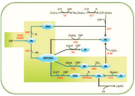
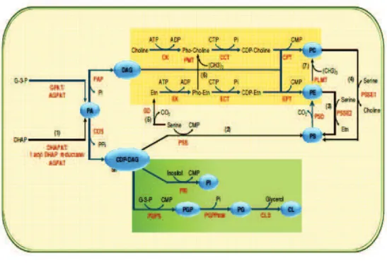
2.1.2.1 Biochemical scheme of glycerophospholipid metabolism in Plasmodium . . . 33
2.1.2.2 Different pathways involve in PL metabolism . . . 34
2.1.2.3 Enzymes involved in PL metabolic pathways . . . 37
2.1.3 Pharmacology of choline uptake . . . 41
2.1.3.1 Albitiazolium (bis-thiazolium compound), an antimalar-ial drug candidate designed by Vantimalar-ial and group . . . 43
2.2 Regulation of PL metabolic pathways . . . 46
2.3 Future scope . . . 49
3 Kinetic modelling of Phospholipid synthesis in P.knowlesi 51 3.1 Introduction and Background . . . 52
3.2 The Model description . . . 53
3.3 The hybrid method of optimization. . . 54
3.4 Flux Balance Analysis. . . 60
3.5 Results and Discussion . . . 61
3.5.1 Model simplification . . . 62
3.5.2 Training the PL model . . . 63
3.5.3 Model predictions and analysis . . . 66
3.5.4 Source of PC production in absence of CDP-choline pathway. . . 69
3.5.5 Rate-limiting steps for PC synthesis . . . 73
4 Insights of Plasmodium omics from High-throughput analysis 78
4.1 An overview of Plasmodium genome . . . 79
4.2 Functional genomics in Plasmodium . . . 80
4.2.1 Genome diversity and genetic polymorphism . . . 81
4.2.2 Epigenetics and epigenomics of P. falciparum . . . 83
4.2.3 Comparative genomics of P. falciparum isolates . . . 84
4.2.4 Transcriptomics . . . 85
4.2.5 Proteomics . . . 86
4.2.6 Regulation of gene expression in P. falciparum . . . 89
4.2.6.1 Mechanism of transcriptional and post-transcriptional regulation . . . 90
4.2.6.2 Mechanism of translational and post-translational reg-ulation . . . 91
4.2.7 Metabolomics . . . 93
4.2.8 Fluxomics . . . 95
4.3 Methodology for identification, quantification and integration of OMICS 95 4.3.1 Processing and quantification of transcripts or gene expression. . . 96
4.3.1.1 DNA-Microarray . . . 96
4.3.1.2 Next-Generation sequencing (NGS) . . . 98
4.3.1.3 qPCR . . . 103
4.3.2 Processing and quantification of protein expression . . . 104
4.3.3 Quantification and analysis of metabolic response. . . 107
4.3.4 Quantification of Fluxes in the Metabolic network. . . 110
4.3.4.1 Modelling approaches to metabolism of P. falciparum . 111 4.3.5 Conclusion and future perspectives for functional genomics of Plasmodium . . . 112
5 Integrating transcriptome, proteome and metabolome in blood stages of P falciparum 115 5.1 Introduction and background . . . 116
5.2 A model for translation control during ring to trophozoite transition. . 119
5.2.1 Dynamical equations relating transcriptome and proteome . . . 119
5.2.2 Ranking transcriptome and proteome according to phase . . . . 120
5.3 The experimental study of the relation between transcriptome and metabolome125 5.3.1 Materials and Methods . . . 125
5.3.2 Processing and quantification of metabolomics data . . . 125
5.3.3 Processing and quantification of qPCR data . . . 127
5.3.4 Results and Discussions . . . 127
5.3.4.1 Metabolomics of Host erythrocyte/RBC . . . 127
5.3.5 Regulation of LP genes and metabolites along cell-cycle . . . 130
5.3.5.1 Regulation of CDP-choline and CDP-ethanolmaine path-way . . . 131
5.3.5.2 Regulation of serine and base-exchange reactions . . . 137
5.3.5.3 Regulation of LP genes and metabolites in Serine de-carboxylation pathway . . . 137 5.4 Conclusion . . . 138 6 General Conclusion 140 Bibliography 145 Article published/submitted 174 Appendix A 175 Appendix B 183
List of Tables
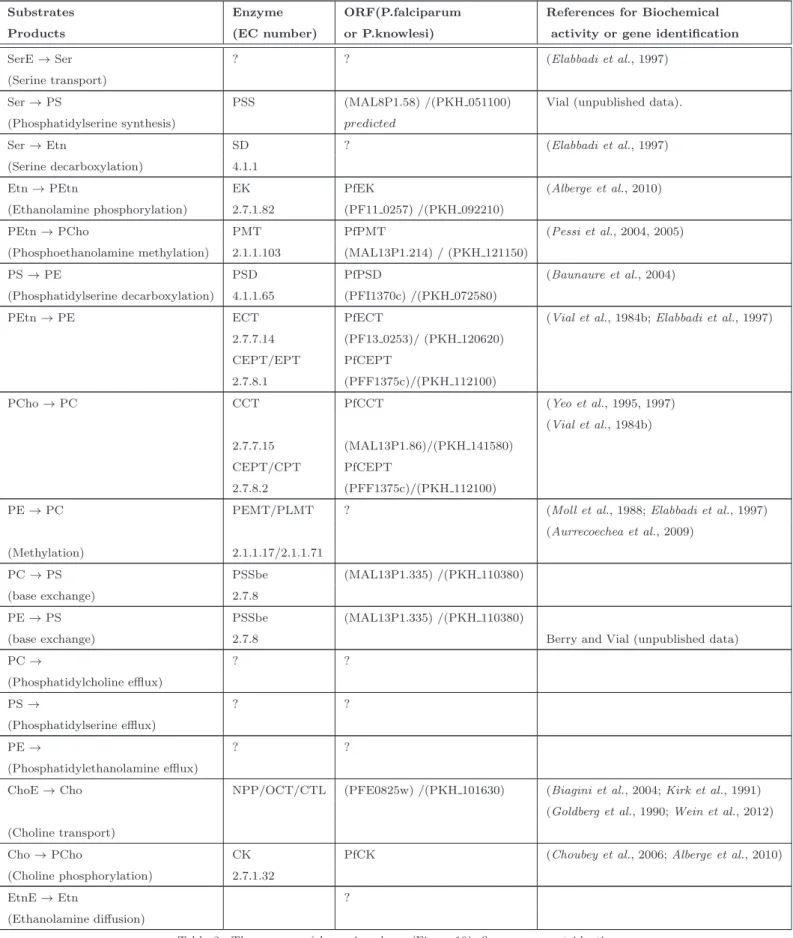
1 Abbreviation List . . . xiv 2 List of enzymes involved in PL metabolism and their corresponding ORFs . 40 3 List of the Km and Vm of some of the enzymes involved in phopholipid
metabolism of P.falciparum . . . 49 4 Parameter values for the ODE (PL) Model. . . 66 5 Transcript counts in during development stage of P. falciparum . . . . 86 6 Peptide spectral count during the Intra-developement cycle (IDC) of P.
falciparum . . . 88 7 RNAseq evidences in P. falciparum, source:PlasmoDB . . . 100 8 Evidences of post-infection quantitative proteomics in P. falciparum . . 105 9 Factors for volume corrections that corresponds to T1...T7, seven different
time-points. Volume of Plasmodial cell varies from 3−40 femto liters. . . 127 10 List of enzymes and ORF indicated in P. falciparum . . . 130
1 An illustration of global malaria burden . . . 3
2 Figure of P.falciparum with major organelles . . . 4
3 Several asexual and sexual stages of development of P.falciparum . . . 6
4 The sporozoite journey to the hepatocyte and subsequent liver stage development. . . 8
5 Structure of glycerophospholipids . . . 22
6 Scheme of PL metabolism in Prokaryotes . . . 24
7 Scheme of PL metabolism in Eukaryotes . . . 26
8 Composition of phospholipids in P.falciparum infected erythrocytes . . 29
9 FAS II systems in Plasmodium . . . 31
10 Schematic overview of PL metabolism in Plasmodium . . . 33
11 Kennedy pathway in P.knowlesi . . . 34
12 SDPM pathway in Plasmodium . . . 35
13 Base-exchanges in Plasmodium . . . 36
14 Scheme of choline transport in P.knowlesi and P.falciparum . . . 41
15 Scheme of Albitiazolium entry in P.falciparum . . . 45
16 Discretization of the cone of admissible fluxes . . . 57
17 Inversion of the Michaelis-Menten relation. . . 58
18 Flowchart illustrating the hybrid optimisation method . . . 58
19 Schematic overview of P.knowlesi reactions in structural phospholipid biosynthesis as demonstrated by experimental work . . . 63
20 Fit between the steady state concentration of serine and choline
incor-porated metabolites with the extracellular serine and choline . . . 64
21 Distribution of fluxes in the network with four different concentrations of SerE, 0 − 100 µM . . . 68
22 In silico knock-out of PMT (R5) . . . 71
23 In silico knock-out of PEMT/PLMT (R9) . . . 72
24 Sensitivity coefficients matrix . . . 75
25 A schematic representation of contribution of Functional genomics . . 81
26 Genetic diversity within Gene Ontology (GO) classified on functional categories . . . 82
27 Functional profiles of the expressed proteins. Proteins identified in each stage are plotted as a function of its functional class. . . 89
28 Schematic overview of process of translation in Plasmodium . . . 90
29 Schematic overview and the scope of identification, quantification and integration of Plasmodium ’OMICS’ . . . 96
30 Illustration of DNA Microarray, a general protocol . . . 97
31 Translating ribosome bound mRNA . . . 100
32 Step by step flow of quantification of NGS data . . . 102
33 Illustration of identification and quantification of metabolites . . . 118
34 Transcriptome and proteome data for 121 oscillatory genes during intra-erythrocytic stage of P. falciparum . . . 121
35 Volume of the parasite during intra-erythrocitary stage of P. falciparum as a function of time . . . 123
36 Transition in expression of genes and proteins . . . 124
37 Transmission X-ray tomography of asexual stages of P. falciparum . . . 126
38 Biochemical scheme showing glycerophospholipid metabolism in P. fal-ciparum . . . 129
40 The fold change in expression of PL genes and metabolites obtained by qPCR and LC/MS−MS along the 48 hours cell cycle in P.falciparum 3D7 . . . 132 41 Regulation of genes and metabolites of CDP-choline pathway along the
cell cycle . . . 132 42 Regulation of genes and metabolites of CDP-ethanolamine pathway along
the cell cycle . . . 133 43 Regulation of serine . . . 137 44 Dynamic ranges of substrate concentrations. . . 178 45 Parameter profiles and confidence intervals for a uncorrelated,
multi-plicative perturbation scheme . . . 179 46 Parameter profiles and confidence intervals for a correlated,
Abbreviations Names Abbreviations Names
ACT Artemisinin-based combination therapy PA Phosphatidic acid
ADP Adenosine bisphosphate PAP PA phosphatase
AGPAT 1-acyl-G-3-P acyltransferase PC Phosphatidylcholine
ATP Adenosine triphosphate PCS PC synthase
CCT CTP:phosphocholine cytidylyltransferase PCho/P-Cho/CholP Phosphocholine
CDP Cytidine bisphosphate PE Phosphatidylethanolamine
CDP-Cho CDP-choline PMT Phosphoethanolamine
-N-methyltransferase CDP-DAG CDP-diacylglycerol PEtn/P-Etn/EthaP Phosphoethanolamine
CDP-Etn CDP-ethanolamine PEMT PE N-methyltransferase
CDS CDP-DAG synthase PG Phosphatidylglycerol
CEPT Choline/ethanolamine-phosphotransferase PGPS PGP synthase
Cho Choline PGP Phosphatidylglycerolphosphate
CK Choline kinase PGPPase PGP phosphatase
CL Cardiolipin PI Phosphatidylinositol
PIS PI synthase
CLS Cardiolipin synthase PL(s) Phospholipid(s)
CPT Cholinephosphotransferase PLMT PL N-methyltransferase
CTP Cytidine triphosphate PS Phosphatidylserine
DAG Diacylglycerol PSD PS decarboxylase
DHAP Dihydroxyacetone phosphate PSS PS synthase
DHAPAT DHAP acyltransferase PPM Parasite’s plasma membrane
Etn Ethanolamine PV Parasitophorous vacuole
EK Ethanolamine kinase PVM
Parasitophorus-vacuolar-membrane ECT CTP:phosphoethanolamine cytidylyltransferase Ser Serine
EPT Ethanolaminephosphotransferase SDPM Serine decarboxylase - phosphoethanolamine-methyltransferase pathway
FA Fatty acid SD Serine decarboxylase
FAS II Type II Fatty-acid biosynthesis Km Affinity constant
FBA Flux Balance Analysis Vm Maximum velocity/rate
FV Food vacuoles Glu Glucose GPAT G-3-P acyltransferase G-3-P Glycerol-3-phosphate HT Hexose transporter Ino Inositol IS Inositol synthase IDC Intra developmental cycle ITN Insecticide treated net IRS Insecticide resistant spray IRBC Infected Red Blood Cells NPP New Permeation Pathway(s) OCT Organic-Cation Transporter ODE Ordinary Differential Equations
The chapter gives an overview of malaria, its origin and pathology. It discusses about life cycle of causative organism (Plasmodium) in human host and vector causing malaria. It also highlights the controls and measures undertaken to combat human malaria. The later part focus on mechanisms of various anitmalarials that has been used for chemotherapy.
1.1
History of malaria
C.Laveran, a French army surgeon stationed in Constantine, Algeria, was the first to notice parasites in the blood of a patient suffering from malaria. This occurred on 6th of November 1880. For his discovery, Laveran was awarded the Nobel Prize in 1907. On August 20th, 1897, Ronald Ross, a British officer in the Indian Medical Service, was the first to demonstrate that malaria parasites could be transmitted from infected patients to mosquitoes. In further work with bird malaria, Ross showed that mosquitoes could transmit malaria parasites from bird to bird. This necessitated a sporogonic cycle (the time interval during which the parasite developed in the mosquito). Thus, the problem of malaria transmission was solved. For his discovery, he was awarded the Nobel Prize in 1902.
The history of malaria predates humanity, as this ancient disease evolved before humans. The ancestors of malaria parasites were about half a billion years old. The pathology and symptoms of malaria was first described in China about 5000 years ago. Then it was reported in Egypt (3500 − 4000 years ago). The enlarge spleens of Egyptians mummies are known to caused by malarial infections. The disease reached India about 3000 years ago, northern Europe between 500 − 1000 years ago.
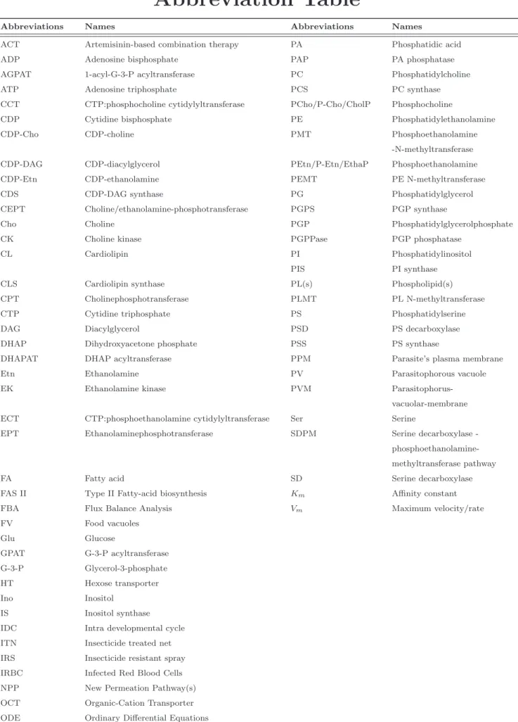
Global distribution of malaria
According to World Malaria Report (WHO, 2010) there were about 219 million cases of malaria (with an uncertainty range of 154−289 million) and an estimated 6.6 million deaths (with an uncertainty range of 4.9 − 8.36 million). Most deaths occur among
Figure 1: An illustration of malaria transmission, death occured by region in accordance with WHO,World Malaria Report,2010, (Courtesy: Jenny Ridley)
children living in Africa where a child dies every minute from malaria. Country level burden estimates (available for 2010) shows that 80% of malaria deaths occur in 14 countries. Together, the Democratic Republic of the Congo and Nigeria account for over 40%.
In 2011, 99 countries and territories had ongoing malaria transmission. Specific population risk groups include : −
• Young children in stable transmission areas who have not yet developed protective immunity against the most severe forms of the disease.
• Non-immune pregnant women as malaria causes high rates of miscarriage and can lead to maternal death.
• Semi-immune pregnant women in areas of high transmission. Malaria can result in miscarriage and low birth weight, especially during first and second pregnan-cies.
• Semi-immune HIV-infected pregnant women in stable transmission areas, during all pregnancies. Women with malaria infection of the placenta also have a higher
risk of passing HIV infection to their newborns. • People with HIV/AIDS (Cohen et al., 2005).
• International travelers from non-endemic areas because they lack immunity. • Immigrants from endemic areas and their children living in non-endemic areas
and returning to their home countries to visit friends and relatives are similarly at risk because of waning or absent immunity.
Transmission of Malaria
Malaria is transmitted exclusively through the bites of Anopheles mosquitoes. The intensity of transmission depends on factors related to the parasite, the vector, the human host, and the environment. Transmission also depends on climatic conditions that may affect the number and survival of mosquitoes, such as rainfall patterns, tem-perature and humidity.
1.2
Plasmodium
, the causative organism
Figure 2: Diagram of P.falciparum with major organelles including the rhoptries, micronemes and polar rings near the apical end.
Malaria is caused by a protozoan parasite called Plasmodium, a genus of Apicom-plexan parasite. It was described in 1885 by Ettore Marchiafava and Angelo Celli. Currently over 200 species of this genus are recognized and newspecies continue to be described (Siddall and Barta, 1992; Chavatte et al., 2007; Perkins, 2008). Plasmodium is entirely an obligate intracellular parasite that grow and replicate only within the host cells. Four species of Plasmodium , P.falciparum , P. vivax , P. ovale and P. malariae commonly infect humans, and a fifth, P.knowlesi , has recently been identi-fied as being responsible for a significant number of human cases in South-East Asia (Cox-Singh et al., 2008; Kantele and Jokiranta, 2011).
1.2.1
Lifecycle of Plasmodium falciparum
Plasmodium falciparum is the causative agent of malaria. It causes most severe forms of human infectious diseases. The closest known relative of P.falciparum is reichenowi (Rich et al., 2009). The life cycle of the P. falciparum, malaria parasite is complex and comprises two hosts: an intermediate vertebrate host (human) and a definitive invertebrate host (female Anopheles mosquito) where the asexual and sexual stages of the parasite occur respectively.
All these stages have their own unique shapes and structures and protein comple-ments. The survival and development of the parasite is made possible by more than 5,000 genes and their specialized proteins which helps the parasite to invade and grow within multiple cell types and to evade host immune responses (Greenwood et al., 2008; Florens et al., 2002).
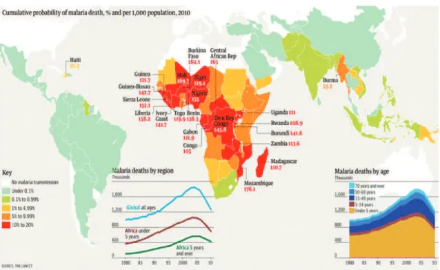
Figure 3: Several asexual and sexual stages of development of P.falciparum such as the sporozoites (Gr. Sporos = seeds; the infectious form injected by the mosquito), merozoites (Gr. Meros = piece; the stage invading the erythrocytes), trophozoites (Gr. Trophes = nourishment; the form multiplying in erythrocytes), schizonts are round to oval inclusions that contain the deeply staining merozoites and gametocytes (sexual stages).
1.2.1.1 The Asexual stages
Man is the intermediate host for malaria, wherein asexual phase of the life cycle occurs.This stage is further divided into two sub phases a) Pre-erythrocytic phase b) Erythrocytic phase. The sporozoites inoculated by the infested mosquito initiate this phase of cycle in the liver, and in the latter part it continues within the red blood cells.
a) Pre-erythrocytic phase
When the infected Anopheles mosquito takes a blood meal on mammalian host, tens to hundred invasive sporozoites are introduced into the skin (Jin et al., 2007). Following the intradermal deposition, some sporozoites are destroyed by the local macrophages, some enter the lymphatics, while others find a blood vessel (Yamauchi et al., 2007; Vaughan et al., 2008; Silvie et al., 2008). The sporozoites that enter a lymphatic vessel reach the draining lymph node wherein some of the sporozoites partially develop into exoerythrocytic stages (Vaughan et al., 2008). The parasite might prime the T cells to mount a protective immune response (Good and Doolan, 2010). The sporozoites that enters a blood vessel reach the liver within a few hours.
It has been shown that the sporozoites travel by a continuous sequence of stick and slip motility, using the thrombospondin related anonymous protein (TRAP) family and an actin-myosin motor (Baum et al., 2006; Yamauchi et al., 2007; M¨unter et al., 2009). The sporozoites then negotiate through the liver sinusoids, and migrate into a few hepatocytes, where they multiply and grow within parasitophorous vacuoles. Each sporozoite develop into a schizont containing 10, 000 − 30, 000 merozoites (or more in case of P.falciparum) (Amino et al., 2006; Jones et al., 2006; Kebaier et al., 2009).
The growth and development of the parasite in the liver cells is facilitated by a favor-able environment created by the circumsporozoite protein of the parasite (Prudˆencio et al., 2006; Singh et al., 2007). The entire pre-erythrocytic phase lasts about 5−16 days depending on the parasite species. It averages between 5 − 6 days for P.falciparum, 8 days for P.vivax, 9 days for P.ovale, 13 days for P.malariae and 8 − 9 days for P.knowlesi. The pre-erythrocytic phase remains a silent phase, with little pathology and no symptoms, as only a few hepatocytes are affected (Vaughan et al., 2008).
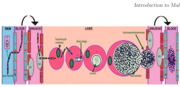
Figure 4: Courtesy: (Vaughan et al., 2008). The figure illustrates the sporozoite journey to the hepatocyte and subsequent liver stage development(Parasite/Host Interactions). The infectious sporo-zoite is deposited into the skin and subsequently enters the bloodstream through a capillary endothelial cell (CE). A number of sporozoites also enter draining lymph nodes and can partially develop within the lymphoid endothelium (LE). Once in the liver sinusoid, sporozoites glide along the fenestrated endothelia (SE) and cross the sinusoidal cell barrier by traversing a resident Kupffer cell (KC). The sporozoite then traverses a number of hepatocytes before invading a hepatocyte with the formation of a parasitophorous vacuole membrane (PVM). Massive replication and growth lead to the formation of erythrocyte- infectious merozoites that enter the sinusoid packaged in extrusomes/merosomes and are subsequently released in the pulmonary bloodstream. The parasite and host proteins known to be involved in the individual steps of this cascade are listed under the appropriate location, and a timeline for the whole process for rodent parasites and human parasites (postinfection, PI) is depicted at the base of the figure.
b) Erythrocytic phase
The merozoites released from the liver recognize, attach, and enter the red blood cells (RBCs) (see Figures. 3 & 4) by multiple receptor-ligand interactions in as little as 60 seconds. This quick disappearance from the circulation into the red cells minimizes the exposure of the antigens on the surface of the parasite, thereby protecting these parasite forms from the host immune response (Cowman and Crabb, 2006; Greenwood et al., 2008).
The invasion of the merozoites into the red cells is facilitated by molecular interac-tions between distinct ligands on the merozoite and host receptors on the erythrocyte membrane. P.vivax invades only ”Duffy” blood group-positive red cells, using the Duffy-binding protein and the reticulocyte homology protein, found mostly on the reticulocytes. The more virulent P.falciparum uses several different receptor families and alternate invasion pathways that are highly redundant.Varieties of Duffy binding– like (DBL) homologous proteins and the reticulocyte binding-like homologous proteins of P.falciparum recognize different RBC receptors other than the Duffy blood group or the reticulocyte receptors. Such redundancy is helped by the fact that P.falciparum has four Duffy binding-like erythrocyte-binding protein genes, in comparison to only one gene in the DBL-EBP family as in the case of P.vivax, allowing P.falciparum to invade any red cell (Mayer et al., 2009; Weatherall et al., 2002).
The process of attachment, invasion, and establishment of the merozoite into the red cell is made possible by the specialized apical secretory organelles of the merozoite, called the micronemes, rhoptries, and dense granules. The initial interaction between the parasite and the red cell stimulates a rapid wave of deformation across the red cell membrane, leading to the formation of a stable parasite-host cell junction. Following this, the parasite pushes its way through the erythrocyte bilayer with the help of the actin-myosin motor, proteins of the thrombospondin-related anonymous protein family (TRAP) and aldolase. It thus creates a parasitophorous vacuole to seal itself from the host-cell cytoplasm, thus creating a hospitable environment for its development within the red cell. At this stage, the parasite appears as an intracellular ring (Cowman and Crabb, 2006; Haldar and Mohandas, 2007).
Within the red cells, the parasite numbers expand rapidly with a sustained cycling of the parasite population. Even though the red cells provide some immunological advantage to the growing parasite, the lack of standard biosynthetic pathways and intracellular organelles in the red cells tend to create obstacles for the fast growing intracellular parasites. These impediments are overcome by the growing ring stages by several mechanisms (a) restriction of the nutrient to the abundant hemoglobin, (b) dramatic expansion of the surface area through the formation of a tubovesicular
network, and (c) export of waste and range of remodeling and virulence factors into the red cell (Silvie et al., 2008).Hemoglobin from the red cell, the principal nutrient for the growing parasite, is ingested into a food vacuole and degraded. The amino acids thus made available are utilized for protein biosynthesis and the remaining toxic heme is detoxified by heme polymerase and sequestrated as hemozoin (malaria pigment). The parasite depends on anaerobic glycolysis for energy, utilizing enzymes such as pLDH, plasmodium aldolase etc. As the parasite grows and multiplies within the red cell, the membrane permeability and cytosolic composition of the host cell is modified (Lew et al., 2003; Kirk, 2004). These new permeation pathways induced by the parasite in the host cell membrane help not only in the uptake of solutes from the extracellular medium but also in the disposal of metabolic wastes, and in the origin and maintenance of electrochemical ion.
The erythrocytic cycle occurs every 24 hours in case of P.knowlesi, 48 hours in cases of P.falciparum, P.vivax and P.ovale and 72 hours in case of P.malariae.During each cycle, each merozoite grows and divides within the vacuole into 8 − 32 (average 10) new merozoites, through the stages of ring, trophozoite, and schizont. At the end of the cycle, the infected red cells rupture, releasing the new merozoites that in turn infect more RBCs. With sunbridled growth, the parasite numbers can rise rapidly to levels as high as 1013 per host (Greenwood et al., 2008). A small proportion of asexual parasites
do not undergo schizogony but differentiate into the sexual stage gametocytes. These male or female gametocytes are extracellular and nonpathogenic and help in transmis-sion of the infection to others through the female anopheline mosquitoes, wherein they continue the sexual phase of the parasite’s life cycle. Gametocytes of P.vivax develop soon after the release of merozoites from the liver, whereas in case of P.falciparum, the gametocytes develop much later with peak densities of the sexual stages typically occurring 1 week after peak asexual stage densities (Pukrittayakamee et al., 2008).
1.2.1.2 The Sexual stages
Mosquitoes are the definitive hosts for the malaria parasites, wherein the sexual phase of the parasite’s life cycle occurs. The sexual phase results in the development of innumerable infecting forms of the parasite within the mosquito that induce disease in the human host following their injection with the mosquito bite (see Figure.3).
Gametocytes are the only erythrocytic forms of the parasite that are capable of infecting Anopheles mosquito vectors (Talman et al., 2004). When the female Anophe-les draws a blood meal from an individual infected with malaria, the male and female gametocytes of the parasite find their way into the gut of the mosquito. The molec-ular and cellmolec-ular changes in the gametocytes help the parasite to quickly adjust to the insect host from the warm-blooded human host and then to initiate the sporo-gonic cycle. The female gametocyte develops into a single female gamete, the male gametocyte undergoes exflagellation that gives rise to 8 thread-shaped motile male ga-metes (Rawlings et al., 1992). In the mosquito midgut, the male gamete fuses with the female gamete resulting in a diploid zygote (see Figure.3). Within 20 − 24 hours following the blood meal,non-motile zygote undergoes morphological changes and de-velops into a diploid/tetraploid motile ookinete (Alano, 2007). Growth and division of each oocyst produces thousands of active haploid forms called sporozoites. After the sporogonic phase of 8 − 15 days, the oocyst bursts and releases sporozoites into the body cavity of the mosquito, from where they travel to and invade the mosquito salivary glands. When the mosquito thus loaded with sporozoites takes another blood meal, the sporozoites get injected from its salivary glands into the human bloodstream, causing malaria infection in the human host. The sporozoites along with the saliva are now ready to be injected into the vertebrate host during the next blood meal of the mosquito (Alano, 2007). It has been found that the infected mosquito and the parasite mutually benefit each other and thereby promote transmission of the infection. The Plasmodium-infected mosquitoes have a better survival and show an increased rate of blood feeding, particularly from an infected host (Barillas-Mury, 2007; Hill, 2006; Ferguson and Read, 2004).
1.3
Symptoms and pathology
Malaria typically begins after 8 − 25 days of infection. However,symptoms may occur later in those who have taken antimalarial medications as prevention (Nadjm and Behrens, 2012). Initial manifestations of the disease common to all malaria species are flu-like symptoms, (Bartoloni and Zammarchi, 2012) and can resemble other conditions such as septicemia, gastroenteritis, and viral diseases (Nadjm and Behrens, 2012).
The clinical symptoms of malaria are related to the development of asexual par-asites in the blood. The disease can be broadly classified into two types, a) mild or uncomplicated malaria and b) severe or complicated malaria. The symptoms of mild or uncomplicated malaria are nonspecific, such as fever, shivering, chills, headache, musculoskeletal and abdominal pain, fatigue, vomiting and/or diarrhoea etc. These symptoms present a clinical picture that resembles the symptoms of many other child-hood infectious diseases. These non-specific signs of malaria are believed to be caused by the release of a malarial toxin which induces macrophages to secrete tumour necrosis factor α (TNF α) and interleukin-1 (IL-1), common mediators induced by Plasmodium species. Although the nature of malarial toxin is controversial, it is generally agreed that it is released at the time of schizont rupture (Miller et al., 2002). Uncomplicated malaria is terminated either by host immunity or by drug treatment.
The classic symptom of malaria is paroxysm, a cyclical occurrence of sudden cold-ness followed by rigor and then fever and sweating, occurring every two days (ter-tian fever) in P.vivax and P.ovale infections,and every three days (quartan fever) for P.malariae.P.falciparum infection can cause recurrent fever every 36 − 48 hours or a less pronounced and almost continuous fever.
Symptoms of P.falciparium malaria arise 9 − 30 days after infection (Bartoloni and Zammarchi, 2012).Individuals with cerebral malaria frequently exhibit neurological symptoms, including abnormal posturing, nystagmus, conjugate gaze palsy (failure of the eyes to turn together in the same direction), opisthotonus, and seizures (Bartoloni and Zammarchi, 2012).
P.falciparum malaria indicate a complex syndrome, established by host and para-site factors. The main virulence phenotypes are related to cytoadherence, rosetting and antigenic variation. Cytoadherence or adhesion to the endothelium has an important role in the pathogenicity of the disease causing occlusion of small vessels and contribut-ing to the failure of many organs. Rosettcontribut-ing signifies the formation of rosettes due to adhesion of uninfected erythrocytes with erythrocytes infected with mature forms of the parasite. In P.falciparum malaria, rosetting seems to increase microvascular ob-struction of the blood flow and can hide the infected cell thereby protecting it from phagocytosis (Rowe et al., 2009). In P.falciparum, various surface antigens mediate adhesion to several receptors of the host endothelium, preventing the infected ery-throcytes from passing through the spleen, where they would be destroyed. In doing so however, the erythrocyte surface proteins make the parasite ”visible” to the host immune system and thus the parasite needs to vary the proteins to avoid destruction (Scherf et al., 2008). It is important to understand the role of host receptors and par-asite ligands involved in the development of different clinical syndromes for developing new control methods to reduce the mortality rates of this disease.
Pathological disorders of P.falciparum malaria have shown severe complications. The obstruction of cerebral venules and capillaries with erythrocytes containing mature trophozoites and schizonts causes convulsions and coma. Acidosis and hypoglycemia are the most common metabolic complications in severe malaria. Acute pulmonary edema is also a common fatal complication, presenting interstitial edema with swollen endothelial cells and monocytes narrowing the capillary lumen. Acute renal failure is another important complication in severe malaria and is defined by an increase in the serum creatinine or an increase in blood urea (Greenwood et al., 2008).
Some of the major pathological disorder caused by malaria are listed below : − • Anaemia is the inevitable consequence of the infection of red cells with parasites,
but other alterations of the infected red cell may lead to cell rigidity and to changes in cytoadherent properties.
• Sequestration of P.falciparum in the brain leads to cerebral malaria, the most dramatic complication of falciparum malaria.
• Enlargement of the spleen is the most constant clinical sign of malaria infections, but in some individuals this may lead to hyperreactive malarial splenomegaly. • As in most infection, transient glomerulonephritis may occur in acute forms of
the disease. Immune complex deposition may lead to fatal forms of ”quartan malarial nephropathies”.
• The intervillous spaces of the placenta offer a very favorable environment for parasite development which may lead to placental malaria, with its consequences on foetal growth.
1.4
Global malarial control strategy
Control,elimination and research are the 3 key startegies implemented to combat the disease. Control indicates controlling the disease in malaria-endemic countries. This makes a substantial impact on their malaria burden by controlling it with existing tools. Secondly, by reducing all locally-acquired infections within a country to zero will bring the world closer to the ambitious goal of global eradication. Together, malaria control and elimination efforts will require international research activities. This will help to discover new tools to combat malaria.
With a vision to eradicate the malaria; vector control,vaccination and chemotherapy are in prime focus amongst implemented strategies.
1.4.1
Vector control
Vector control is an important part of the global malaria control strategy. The idea behind vector control is to reduce the levels of mortality and morbidity by reducing transmission of the disease. Basically, there are many efforts focused on preventing man-vector contact by large-scale implementation of (i) Indoor Residual Spraying (IRS) which can be achieved by applying long-lasting chemical insecticides on the walls and the roofs of the houses and (ii) Insecticide Treated Nets (ITNs) which can be efficient at places where a large proportion of human-biting by local vectors takes place after
people have gone to sleep. In community-wide trials in several African settings, ITNs have been shown to reduce mortality rate by about 20%. Other measures includes larviciding, this is only used for vectors which tend to breed in permanent or semi-permanent water bodies that can be identified and treated.
1.4.2
Malarial Vaccine
Recombinant proteins, synthetic peptides, DNA vaccines, inactivated whole para-sites, and vaccines (comprising mixtures of a large variety of potential antigens) are continously evaluated againist malaria.
Vaccine that are categorized specifically on the different stages of the parasite are as
follows:-a). Pre-erythrocytic Vaccine: It aims to protect against the early stage of malaria infection. The first malaria vaccination trial against pre-erythrocytic stages was made with radiation attenuated sporozoites. Initial studies have shown that in humans, vaccination with radiation attenuated sporozoites provided about 90% protection when subsequently challenged by bite of infected mosquitoes (Van-derberg, 2009). Even though a high degree of protection could be achieved using irradiated sporozoites, it presents some challenges in extracting and purifying sporozoites from mosquito salivary glands. Nevertheless, there are some liver stage subunit vaccines in clinical trials which are based on immunogenic compo-nents of sporozoites or liver stage parasites. The most promising vaccine currently in phase III clinical trial is RTS,S which is directed against the CSP protein in the liver stage. Recent data from phase III clinical trials showed that RTS,S pro-vided 50% reduction in incidence of malaria in young children (Agnandji et al., 2011).
b). Erythrocytic stage vaccine: It targets the malaria parasite at its most de-structive strategy rapid replication of the organism in human red blood cells. Vaccines targeting erythrocytic stages include antigens like PfMSP-1, PfMSP3 are in phase II clinical trials (Pierce and Miller, 2009).
par-asite by inducing antibodies that prevent the parpar-asite from maturing in the mosquito after it takes a blood meal from a vaccinated person. Most extensive studies have focused on sexual stagespecific antigens; Pfs48/45, Pfs230, Pfs25, Pfs28 of P.falciparum and orthologues in other Plasmodium species (Sutherland, 2009).
Although a safe and effective malaria vaccine would be the easiest way to control malaria, but even after decades of research, that vaccine is still elusive. The complex life cycle of the parasite involving human and vector mosquitoes as well as its allelic diversity, antigenic variations and difficulties in generating high levels of durable im-munity continue to make the development and implementation of an effective vaccine problematic.
1.4.3
Chemotherapy
Successful malaria control depends greatly on treatment with efficacious anti-malar-ial drugs. To adequately treat malaria, drugs must be fast acting, highly potent against asexual blood stage infections, minimally toxic and affordable to residents of endemic regions. Correct use of such an antimalarial drug will not only shorten the duration of malaria illness but also reduce the incidence of complications and the risk of death. The state of the art knowledge on currently existing and developing antimalarial drugs has been reviewed in several recent articles (N Burrows et al., 2011). A major problem leading to a decline in the efficacy of currently existing antimalarials is the growing emergence of drug resistance. Resistance to antimalarial drugs normally arises as a re-sult of spontaneously-occurring mutations that affect the structure and activity of the drug target in the malaria parasite or affect the access of the drug to the target. Recent progress in understanding the mechanisms of parasite’s resistance to antimalarials has been described by (Mita et al., 2009; Sridaran et al., 2010).
1.4.3.1 Pharmacology of antimalarials
Choloroquine which is a 4−Aminoquinolines derivative binds with toxic heme (released during haemoglobin catabolism in erythrocytic cycle) to prevent its crys-tallization to hemozoin. This allows heme concentration to rise and kill the parasite (Bray et al., 1999). Chloroquine resistance has been associated to point mutation in Chloroquine Resistance Transporter (PfCRT), an integral membrane protein localized to the parasite’s internal digestive vacuole. These mutations result in a marked reduc-tion in the accumulareduc-tion of chloroquine by the parasite (Martin et al., 2009; Sidhu et al., 2002).
Quinine, an aryl amino alcohol, is a naturally occurring compound obtained from the bark of American cinchona tree. It was the first antimalarial and its use dates back to 17th century. It is accumulated in the digestive vacuole of the erythrocytic parasites. It is less effective than chloroquine and has a narrow therapeutic range.Resistance to quinine has been developing in Southeast Asia (White, 1992). However, the short term efficacy of quinine plus antibiotics, notably quinine-clindamycin regimens, has been reported in the treatment of severe malaria in Africa (Winstanley, 2001).
Mefloquine, an amino alcohol, is also thought to work in much the same way as quinine. It is well absorbed from the gut and elimination is very slow (half-life ranging from 15−33 days). However, it is used only for uncomplicated malaria and is relatively expensive. Furthermore, in recent years, resistance to mefloquine has been reported in some parts of Southeast Asia (especially the Thai borders with Burma and Cambodia). Mefloquine can cause severe idiosyncratic adverse reactions, however these are rare. In contrast, dosedependent symptomatic reactions, most commonly gastrointestinal upset and dizziness are common. The most serious of these include psychoses, seizures, and acute encephalopathy (Croft and Black, 1999).
Halofantrine also seems to have a mechanism of action similar to that of chloro-quine. Its parenteral formulation is under development. Like mefloquine, it is an expensive drug and is unaffordable for general use throughout tropical Africa. It is incompletely absorbed from the gut (Amponsaa-Karikari et al., 2009).
Primaquine is a synthetic 8-Aminoquinoline compound and is mainly used to eradicate liver hypnozoites of P.vivax and P.ovale to prevent late relapses of malaria (Baird and Rieckmann, 2003). It is normally started when the course of chloroquine has been completed, and the patient is recovering. Primaquine is well absorbed from the gut after oral administration but has a short half-life (half-life 56 h) and needs to be administered daily. It may cause mild gastrointestinal adverse effects. One major problem of primaquine is that it can cause more serious toxicity in patients with glucose-6-phosphate dehydrogenase deficiency. Tafenoquine (WR238605) is a new synthetic analogue of primaquine which is currently in phase IIb clinical trials. It has a larger therapeutic index than primaquine and much slower elimination (half-life 14 days) (Brueckner et al., 1998).
Combination of antimalarials to overcome resistance
Pyrimethamine, a diamino pyrimidine, was always used in combination with sul-fadoxine (Fansidar). The potential benefits of pyrimethamine combinations included synergistic effects resulting in improved efficacy and reduced exposure time of para-site populations (biomass) to drugs thus reducing occurrence of drugresistance muta-tions. Sulfadoxine-pyrimethamine combination was used only in uncomplicated cases of P.falciparum malaria because it produced clinical improvement too slowly in severe disease. Pyrimethamine inhibits the dihydrofolate reductase enzyme thereby blocking the synthesis of purines which are essential for DNA synthesis. Unfortunately, as a result of drug pressure, resistance to sulfadoxine + pyrimethamine combination has been reported which limits its use in many areas. The resistance is correlated to mu-tations in dihydrofolate reductase (DHFR) and dihydropteroate synthase (DHPS), the respective targets of pyrimethanime and sulfadoxine (Sridaran et al., 2010).
Artemisinin is sesquiterpene lactone peroxide that is extracted from the Chinese herb Artemisia annua that have been used since the late 1970s. They reduce the para-site load substantially earlier than other antimalarial drugs. Additionally, they can kill Plasmodium gametes and thus lower transmission rates. Their mechanism of action is unclear. Although highly effective, artemisinins are recommended for use in
combi-nation with the more long-lasting quinolines or antifolates due to their short halflife in vivo in order to ensure complete elimination of residual parasites. Hence, the ra-tionale behind artemisinin-based combinations is that the rapid action of artemisinin compounds reduces parasite load, thereby leaving fewer parasites for the long-lasting combination drug to tackle and thus minimizing the chances of selecting drug-resis-tant organisms. Artemisinin derivatives commonly used include: (a) artemether, (b) artesunate and (c) dihydroartemisinin (Adjuik et al., 2004). The choice of the ACT is based on the efficacy of the combination drug in the country or area of intended use. In many African nations, ACTs in combination with vector control methods (like IRS and ITNs) have resulted in dramatic decrease in malaria associated morbidity and mortality (Barnes et al., 2005; White, 1992).
Albitiazolium, a new choline analogue
Our lab has identified a range of compounds that interfere with a ”new” target, that is parasite phospholipid metabolism. One such compound which is a choline analog, albitiazolium that blocks the entry of choline into the parasite thereby disrupting phos-phatidylcholine biosynthesis in the parasite and eventually inhibiting parasite growth in vitro as well as in vivo. Albitiazolium is accumulated inside the infected red blood cells. Its oral bioavailability is low and so it is now being investigated for parenteral formulation to treat severe and uncomplicated malaria (Nicolas et al., 2005; Vial et al., 2004). In an effort to seek for potential chemotherapeutic targets, our lab has been involved in unravelling the intricacies of phospholipid biosynthetic machinery. A detail report on albitiazolium could be found in chapter.2.
Chapter 2
Glycerophospholipid metabolism in
Plasmodium
Lipid metabolism in Plasmodium has been covered in previous reports from a gen-eral perspective (Holz Jr, 1977; Sherman and Greenan, 1984; Vial et al., 1989a, 1992; Vial H, 1998; Vial and Mamoun, 2005) and via specific topics such as fatty acids (FAs) (Mazumdar and Striepen, 2007; Tarun et al., 2009), neutral lipids (Coppens and Vielemeyer, 2005), phospholipids (PLs) (Vial et al., 2003; Vial and Mamoun, 2005), sphingolipids (Haldar, 1996; Vial and Mamoun, 2005; Pankova-Kholmyansky and Flescher, 2006), phosphoinositide signalling (Vial et al., 2003), glycosylphos-phatidylinositol (GPI) (Channe Gowda, 2002; Boutlis et al., 2005; Debierre-Grockiego and Schwarz, 2010), galactolipids (Mar´echal et al., 2002), lipid rafts (Murphy et al., 2006) and haemozoin formation within lipids (Egan, 2008; Pisciotta and Sullivan, 2008; Hoang et al., 2010). A detailed scheme of lipid metabolic pathways could be found in, ”the Malaria Parasite Metabolic Pathways database” (Ginsburg, 2006).
The chapter discusses about glycerophospholipid acquisition, metabolism and reg-ulation in Plasmodium species (mainly in P.falciparum, P.knowlesi). It also focus on different metabolic pathways involved in the synthesis of these lipids and their roles in prokaryotes and other eukaryotes.
2.1
Introduction to Glycerophopholipids
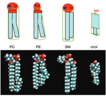
The term ’glycerophospholipid’ signifies any derivative of sn-glycero-3-phosphoric acid that contains at least one O-acyl, or O-alkyl, or O-alk-1’-enyl residue attached to the glycerol moiety and a polar head made of a nitrogenous base, a glycerol or an inositol unit. The alcohol here is glycerol, to which two fatty acids and a phos-phoric acid are attached as esters. This basic structure is a phosphatidate which is an important intermediate for the synthesis of many phosphoglycerides. Examples of some glycerophospholipids and their structures as compared to cholesterol is shown in Figure.5.
Figure 5: The structure of the major membrane phospholipids. The more or less cylindrical glyc-erophospholipid phosphatidylcholine (PC) carries a zwitterionic phosphocholine headgroup on a glycerol with two fatty acyl chains (diacylglycerol), usually one unsaturated (bent). Phosphatidylethanolamine (PE) has a small headgroup and a conical shape and creates a stress in the bilayer: the PE-containing monolayer has a tendency to adopt a negative curvature. The phosphosphingolipid sphingomyelin (SM) tends to order membranes via its straight chains and its high affinity for the flat ring structure of cholesterol (chol). Courtesy (Fahy et al., 2005).
Major roles of phospholipids
Phospholipids (PLs) are the most abundant class of biological lipids. They are found widely from prokaryotes to multicellular eukaryotes. Despite of similarity of their structures they have distinct roles both in constituting the membrane structure and in biological process. Phosphatidylcholine (PC) (also known as lecithin) is the most abundant phospholipid in animal and plant tissues. PC biosynthesis is required for nor-mal secretion of very low density lipoproteins (VLDL) by liver (Li and Vance, 2008). Phosphatidylethanolamine (PE) (also known cephalin) is an abundant phospholipid in microbial, plant, and animal cells. PE plays a critical role during cell division by mediating co-ordinated movements between the contractile ring and the plasma mem-brane that are required for proper progression of cytokinesis (Emoto et al., 2005). PE also serves as a precursor of the ethanolamine moiety of glycosylphosphatidylinositol
anchors that are required for attachment of proteins on the cell surface (Menon and Stevens, 1992). Phosphatidylserine (PS) is involved in biological processes including apoptosis, blood coagulation, and activation of protein kinase C (signal transduction). PS expression on cell surface acts as a signal by which apoptotic cells are recognized and phagocytosed by macrophages (Fadok et al., 2001). A class of phospholipids with a high rate of metabolism is the phosphatidylinositols (PI) which have varying degrees of phosphorylation in the polar head group myo-inositol. The metabolic conversion of phosphatidylinositols to diacylglycerols and inositol phosphates is important in regula-tion of vital cellular funcregula-tions such as differentiaregula-tion, proliferaregula-tion, and apoptosis, and in anchoring proteins via a glycosyl-bridge to the plasma membrane. Phosphatidyl-glycerol (PG) has important functions in bacterial membranes, chloroplasts and lung surfactants. It is a metabolic intermediate in the biosynthesis of cardiolipin.
2.1.1
Phospholipid (PL) acquisition in Prokaryotes and
Eu-karyotes
Phospholipid biosynthesis is a vital facet of cellullar physiology that begins with the synthesis of the fatty acids which are grafted on lysophosphatidic acid by fatty acid synthetase type I (FAS I) or fatty acid synthetase type II (FAS II). Then, it assembles with phosphatidic acid (PA), a primary PL biosynthezied.
2.1.1.1 PL acquisition in Prokaryotes
The bacterial glycerol-phosphate acyltransferases utilize the complete fatty acid chains to form the first membrane phospholipid and thus plays a critical role in the regulation of membrane biogenesis (Zhang and Rock, 2008). Phospholipids in bacteria comprise about 10% of the dry weight of the cell, and each mole of lipid requires the FAS II (an assembly of series of distinct discrete protein) and about 32 mole of ATP for its synthesis. The enzymes of fatty acid synthesis are cytosolic, while those of membrane lipid synthesis are mainly integral inner membrane proteins (Heath and Rock, 2004).
Figure 6: The scheme represents a model for prokaryotic phospholipid synthesis (from thesis of Dechamps, 2009). The pathways (in green shaded region) and the reactions (marked with arrows within this box) occurs in all prokaryotes. The lipid precursors and products are shown in ovals. The reactions/pathways outside this region are present in less than 10% of all prokaryotes
In most of bacteria (for example : E.coli), phosphatidylethanolamine (PE) forms major bulk (75%) of PLs with phosphatidylglycerol (PG) (15 − 20%), cardiolipin (CL), phosphatidylserine (PS) and phosphatidic acid (PA) comprises (5 − 10%) (Kanfer and Kennedy, 1964; Heath and Rock, 2004).
Cytidine diphosphate diacylglycerol (CDP-DAG) which comprise 0.05% (Heath and Rock, 2004) of total PLs bulk, is high energy lipid intermediate which is synthesized from PA and cytidine triphosphate (CTP) by the action of the enzyme CDP-DAG synthase (CDS)(EC 2.7.7.41).
CDP-DAG dependent pathways are source of the major PLs like PE,PG, PS and CL (Kanfer and Kennedy, 1964). PS is synthesized from CDP-DAG and L-serine by PS synthase (PSS). PS is then decarboxylated by PS decarboxylase (PSD) to PE (Kanfer and Kennedy, 1964). Phosphatidylglycerol (PG) is synthesized in two steps. In the first step, phosphatidylglycerolphosphate (PGP) is formed from CDP-DAG and G-3-P
by the action of the enzyme phosphatidylglycerolphosphate synthase (PGPS). In the second step, PGP is dephosphorylated by the enzyme PGP phosphatase (PGPPase), thereby leading to the formation of PG (Kanfer and Kennedy, 1964).
Phosphatidic acid (PA), is formed by the acylation of glycerol-3-phosphate (G-3-P) in two enzymatic steps. In the first step, PA is acylated to 1-acyl-G-3-P (or lyso PA) by the enzyme G-3-P acyltransferase (GPAT) and in the second step,1 acyl-G-3-P is acylated by a distinct enzyme, 1-acyl-G-3-P acyltransferase (AGPAT) to form PA (Zhang and Rock, 2008).
Phosphatidylinositol (PI) is absent in majority of prokaryotes with a few exceptions like Pseudomonas syringae, E.coli, Treponema pallidum and Mycobacterium smegmatis. The synthesis of PI in these organisms is catalyzed by PI synthase (PIS) with inositol and CDP-DAG as substrates (Salman et al., 1999).
Apart from these PLs, phosphatidylcholine (PC) is also present in some bacteria that are photosynthetic or in close association with eukaryotes (Sohlenkamp et al., 2003). Synthesis of PC in such prokaryotes occurs via three pathways: (a) CDP-DAG dependent pathway in which an enzyme called PC synthase (PCS) condenses CD-P-DAG and choline to PC (L´opez-Lara and Geiger, 2001), (b) PE methylation pathway in which PE undergoes methylation to form PC by PL N-methyltransferase (PLMT) enzyme (this pathway is also present in yeast and mammalian liver) and (c) CDP– choline pathway or the de novo Kennedy pathway which is also a major pathway for PC synthesis in mammals (Sohlenkamp et al., 2003).
Transport of exogenous long chain fatty acids are utilized (example: E.coli ) in two ways. Firstly, they can be incorporated into the membrane phospholipids by the acyltransferase system. Secondly, they can be used as the sole carbon source for growth, and are in fact an important source of energy (Clark and Cronan, 1996). Degradation of fatty acids proceeds via an inducible set of enzymes that catalyze the pathway of β-oxidation (Clark and Cronan, 1996).
Figure 7: Scheme represents a model for eukaryotic phospholipid synthesis This diagram represents a model for eukaryotic phospholipid synthesis (from thesis of Dechamps, 2009).The Kennedy and the CDP-DAG dependent pathways (in shaded yellow and dark green box respectively,) commonly occur in all eukaryotes. The lipid precursors and products are shown in ovals .The reactions/ pathways outside this shaded box with arrows are present only in yeast and mammals (1), yeast and plants (2), mammals and plants (3, 4), Plasmodium and plants (5, 6), yeast and mammalian liver cells (7).
2.1.1.2 PL acquisition in Eukaryotes
PL synthesis in eukaryotes is more complex and diversified. The major PLs in eukary-otes are PC (40 − 55%) and PE (20 − 40%) followed by smaller quantities of PI and PS (Vance and Steenbergen, 2005). PLs are known to be synthesized by two pathways in eukaryotes i) CDP-DAG dependent pathways, ii) the de novo Kennedy pathways.
In yeast, CDP-DAG dependent pathways of PS synthesis are the major source of the most abundant phospholipids like PC and PE, where as de novo Kennedy path-ways are the auxiliary pathpath-ways for the synthesis of PC and PE. In mammalian cell, CDP-DAG dependent PS synthesis is not present and the bulk of PE or PC are more biosynthesized.
In most eukaryotes, PA is synthesized from G-3-P by two successive acylation reac-tions in a quite similar way as seen in prokaryotes. In addition, in yeast and mammals, PA can also be synthesized from dihydroxyacetone phosphate (DHAP) which is the product of dehydrogenation of G-3-P by the enzyme G-3-P dehydrogenase. PA then en-ters PL biosynthetic pathways in two forms: CDP-DAG and DAG. Like in prokaryotes, CDP-DAG, precursor of CDP-DAG dependent pathways, arises as a result of transfer of PA to CTP catalyzed by enzyme CDS (Carter and Kennedy, 1966). Alternatively, DAG , a precursor for the Kennedy pathways, is synthesized by dephosphosphorylation of PA by PA phosphatase (PAP) (Carman and Han, 2009), which is then condensed and activated by CDP-Cho and CDP-Etn to produce PE and PC respectively.
PS, PI and PG are synthesized by CDP-DAG dependent pathways in a very similar way to the prokaryotic pathways. As in prokaryotes, PS in yeast and plants is also synthesized by transfer of phosphatidyl group from CDP-DAG to serine by the enzyme PSS (Yamashita and Nikawa, 1997).
In plants and mammalian cells, PS can also be synthesized by mammalian base-ex-change mechanism. In yeast and mammalian mitochondria, PE is synthesized by de-carboxylation of PS mediated by the enzyme PSD. PI is synthesized by transfer of phosphatidyl group from CDP-DAG to inositol in the presence of enzyme PI synthase (PIS) (Cooke, 2009). PG is synthesized in two steps, PGP is formed by G-3-P and CDP-DAG mediated by enzyme PGPS, thereafter PGP is dephosphorylated by the enzyme PGPase giving rise to PG.
In higher eukaryotes, PC and PE biosynthesis primarily takes place by the de novo routes, that is, CDP-choline and CDP-ethanolamine routes respectively (Kennedy and Weiss, 1956) while in yeast these pathways appear to be dispensable as long as PC and PE are supplied by CDP-DAG dependent pathways.
In plants along with Kennedy pathways, PE and PC are also synthesized by transver-sal pathway or serine decarboxylase phosphoethanolamine methylation (SDPM) path-way. In this pathway, serine is decarboxylated to form ethanolamine by an enzyme called serine decarboxylase (SD). This ethanolamine then enters the CDP-ethanolamine
pathway eventually leading to the formation of PE. On the other hand, phospho-ethanolamine so formed as a result of de novo CDP-phospho-ethanolamine pathway can be converted into phosphocholine by a unique enzyme, phosphoethanolamine N-methyl-transferase (PMT) which then enters the de novo CDP-choline pathway to form PC (Bolognese and McGraw, 2000). In mammals, the de novo Kennedy pathways are the major source of PC and PE. PS is synthesized only by base exchange mechanism wherein PS is formed at the expense of pre-existing phospholipids such as PC and PE and is mediated by the enzymes PS synthase I (PSSE1) and PS synthase II (PSSE2) re-spectively. While PSSE1 catalyzes the exchange of phospholipid head groups between serine and PC, PSSE2 catalyzes the exchange of phospholipid head groups between serine and PE (Vance, 2008).
In some other cases as in yeast, mammalian liver cells, PC can also be synthesized by triple methylation of PE. This reaction is S-adenosylmethionine (SAM) dependent which acts as a methyl group donor. In yeast, this reaction is mediated by the two classes of phospholipid N-methyltransferases (PLMT) while in mammalian liver, it is mediated by a single enzyme, phosphatidylethanolamine-N-methyltransferase (PEMT) (Bell and Coleman, 1980).
2.1.1.3 PL acquisition in Plasmodium species
Upon infection, the parasite multiplies within red blood cells leading to a production of 10-20 merozoites every 48 hours which rapidly invade further erythrocytes. This rate of production demands high metabolic activity in the parasite, particularly through in-creased requirement for novel membrane formation.Glycerophospholipids (PL) are the main Plasmodium membrane constituents, with a preponderance of PC and PE and with an increase in phosphatidylinositol (PI) involved in signaling. Phospholipids play a crucial role in the development of intracellular malaria parasite. Infected erythro-cytes show a marked increase in PL content enhanced by approximately six-fold at the trophozoite stage. PC and PE together represent 75 − 85% of the parasite total PL composition after infection (Vial et al., 1992).