HAL Id: tel-03139847
https://tel.archives-ouvertes.fr/tel-03139847
Submitted on 12 Feb 2021HAL is a multi-disciplinary open access archive for the deposit and dissemination of sci-entific research documents, whether they are pub-lished or not. The documents may come from teaching and research institutions in France or abroad, or from public or private research centers.
L’archive ouverte pluridisciplinaire HAL, est destinée au dépôt et à la diffusion de documents scientifiques de niveau recherche, publiés ou non, émanant des établissements d’enseignement et de recherche français ou étrangers, des laboratoires publics ou privés.
fumigatus hydrophobins
Borja Rodriguez de Francisco
To cite this version:
Borja Rodriguez de Francisco. Self-assembly into functional amyloids of Aspergillus fumigatus hy-drophobins. Cell Behavior [q-bio.CB]. Sorbonne Université, 2019. English. �NNT : 2019SORUS332�. �tel-03139847�
Sorbonne Université
Institut Pasteur
Ecole Doctorale Complexité du vivant
Biological NMR Technological PlatformSelf-assembly into functional amyloids of Aspergillus
fumigatus hydrophobins
Par Borja Rodríguez de Francisco
Thèse de Doctorat de Biochimie et Biologie Structurale
Dirigée par Dr. Iñaki Guijarro
Présentée et soutenue publiquement le 13 décembre 2019
Devant un jury composé de :Bertrand Friguet Président
Cristina Sizun Rapporteur
André Matagne Rapporteur
Olivier Lequin Examinateur
Thierry Fontaine Examinateur
i
Preface
This dissertation is submitted to the Ecole Doctorale Complexité du Vivant accredited by the Sorbonne University.
The work presented here has been carried out at the Institut Pasteur (CNRS UMR 3528), within the Biological NMR Technological Platform under the supervision of Dr. Iñaki Guijarro. I was granted with a 3 years doctoral fellowship from the Institut Pasteur University International PhD Program (PPU). This program has received funding from the European Union's Horizon 2020 research and innovation program under the Marie Sklodowska-Curie grant agreement No. 665807. The last three months I was funded by the French National Research Agency (ANR FUNHYDRO ANR-16- CE110020-01) awarded to Dr. I. Guijarro. The work was funded by the ANR (ANR-16- sCE110020-01) and the 800 MHz NMR spectrometer used in this work was partially funded by the Région Ile de France (SESAME 2014 NMRCHR grant no 4014526).
ii
Acknowledgments
This doctoral dissertation would not have been possible without the contribution of several people to whom I would like to express my appreciation to:
First of all, I would like to express my sincere gratitude to my supervisor Dr. Iñaki Guijarro for his continuous support during my PhD. His guidance and constant feed-back, not only professionally but also personally, made it possible for me to work on a challenging topic that was of great interest to me.
I am also very grateful to Dr. Isabel Valsecchi for always being concerned and providing me with her assistance through my project.
I would like to thank the Thesis Advisory Committee Members for their valuable assistance, insightful comments and encouragements.
I very much appreciated the Structural Bioinformatics Unit and Biological NMR platform for always willing to help me. Many thanks to Dr. Muriel Delepierre and Dr. Michael Nilges for their generous support working in their Units. Furthermore, I would like to thank Dr. Bruno Vitorge for his support and technical help, Dr. Aracelys López Castilla for her valuable comments, Dr. Nadia Izadi-Pruneyre who profoundly believes in my abilities, and all the office members, Dr. Sebastian Brier, Dr. Florence Cordier, Dr. Catherine Simenel, Dr. Benjamin Bardiaux and all students in the group who have been a big support during this time.
I would like to recognize the help that I received from Dr. Gérard Péhau-Arnaudet for his assistance during the electron microscopy experiments.
Also, I sincerely acknowledge my collaborators Dr. Antoine Loquet, Dr. Vincent Duprès, Dr. Frank Lafont, Dr. Vishu Aimanianda Dr. Anne Beauvais and Dr. Jean-Paul Latgé for their constructive suggestions. Sven van Teeffelen for allowing me to use their instruments. Also, I would like to extend my sincere thanks to Dr. Margaret Sunde and Dr. Chi L.L. Pham for their invaluable contributions and experience to our collaborative work.
Besides, I would like to thank my friends in Institute Pasteur, for always making me feel very welcome. To the PPU family, it was a pleasure coming to work every day with you there. I would also like to extend my deepest gratitude to the STAPA association and members, my personal growth would not have been possible without the support of this fantastic team, and to every friend that has been close during these years in good and bad times.
iii
Finally, I am indebted to all my family and friends, for their unconditional love and concern and for putting your faith on me and urging me to do better. To my mum, for her advice, love and care. To my dad, for his support in every decision I made and being a model for me. To my sister, for always being concerned, and to all my friends in Madrid, Grajal de Campos and abroad, for having incented me to achieved my goals.
I have no valuable words to express my thanks. I am extremely grateful to every person that made my PhD stage one of the best periods of my personal and professional career.
iv
Résumé
Les hydrophobines sont des protéines fongiques qui s’assemblent aux interfaces hydrophobes/hydrophiles ou air-eau (IHH) pour former des fibres amyloïdes fonctionnelles qui s’organisent en couches dont le caractère amphiphile détermine leurs fonctions. Les spores du champignon pathogène opportuniste Aspergillus fumigatus sont couvertes d’une couche de bâtonnets constitués de fibres amyloïdes formés par l’hydrophobine RodA. Ce revêtement hydrophobe facilite la dispersion des spores et les rend inertes vis à vis du système immunitaire humain. Deux proches homologues de RodA, RodB et RodC, sont aussi présents dans la paroi cellulaire des spores. Nous avons réalisé une étude comparative de l’auto-assemblage de ces trois protéines avec un accent particulier sur RodC. Nous avons montré que RodA-C nécessitent d’une interface IHH pour s’assembler en fibres et mis en évidence l’importance de la nature de l’interface dans la morphologie de leurs assemblages. Nous avons observé une auto-inhibition de la fibrillation avec la concentration et montré que celle-ci est due à la saturation de l’interface air-eau. En étudiant l’effet de mutations ponctuelles sur les cinétiques de fibrillation de RodC, nous avons établi des similitudes et des différences par rapport aux régions importantes pour la formation de fibres pour RodC et RodA, étudiée au préalable. La transition du monomère à l’état amyloïde est accompagnée d’une perte de régions désordonnées et un gain de feuillets intermoléculaires en accord avec les analyses par mutagénèse dirigée, qui indiquent que des résidus hydrophobes dans des régions flexibles du monomère sont impliqués dans le squelette -croisé des fibres.
Mots clés: amyloïdes fonctionnels, hydrophobine, bâtonnets, Aspergillus fumigatus, assemblage de protéines, biophysique.
v
Abstract
Hydrophobins are fungal proteins that spontaneously self-assemble at hydrophobic/hydrophilic or air/water interfaces to form functional amyloids that associate laterally into layers. The amphiphilic character of these layers is at the origin of the hydrophobin biological roles. The spores of the airborne fungal pathogen Aspergillus fumigatus are covered by an amyloid layer with rodlet morphology made up by the RodA hydrophobin. This hydrophobic coat facilitates air-dispersal of the spores and renders these inert relative to the human immune system. Two close homologs of RodA, named RodB and RodC, are also present in the spore cell wall. We have performed a comparative study on the self-assembly of the three proteins with particular emphasis on RodC. We have shown that RodA-C require an interface to form amyloids and revealed the importance of the nature of the interface in determining the morphology of hydrophobin assemblies. We have observed that the fibrillation of RodA-C is auto-inhibited (slower at higher concentrations) and shown that this phenomenon can be explained by saturation of the air-water interface. The analysis of the effect of single point mutations on the fibrillation kinetics of RodC revealed the regions that are important for fiber formation, which showed differences and similarities relative to the previously studied RodA. The transition from monomers to amyloids is accompanied by a loss of unordered regions and a gain in intermolecular β-sheets, in agreement with the mutational analyses of RodA and RodC that indicated that hydrophobic residues in flexible loops are involved in the cross-β core of the fibers.
Keywords: functional amyloid, hydrophobin, rodlet, Aspergillus fumigatus, protein assembly, biophysics.
vi
Table of contents
Preface
... iAcknowledgments
... iiRésumé
... ivAbstract
... vTable of contents
... viIndex of figures
... ixIndex of tables
... xiiAbbreviations
... xiiiIntroduction
... 151.1 Overview ... 15
1.2 The Fungal Kingdom ... 16
1.3 Fungal diseases in humans ... 17
1.4 Aspergillosis ... 18
1.5 Aspergillus fumigatus ... 19
1.5.1 Biology ... 20
1.5.2 Fungal Cell Wall ... 21
1.5.3 Aspergillus fumigatus Cell Wall ... 22
vii
1.7 Amyloid fibers... 25
1.7.1 Functional and non-functional amyloids ... 29
1.7.2 How does the amyloid state form? ... 31
1.8 Hydrophobins ... 34
1.8.1 General aspects ... 34
1.8.2 Biological functions ... 39
1.8.3 Applications ... 40
1.8.4 Aspergillus fumigatus hydrophobins... 40
1.9 Project description ... 45
Materials & Methods
... 472.1 Proteins and peptides studied ... 47
2.2 Plasmid design... 48
2.3 Expression and purification ... 49
2.4 Solution nuclear magnetic resonance spectroscopy ... 50
2.4.1 Assignment principle ... 50
2.4.2 Assignment of NMR backbone chemical shifts of RodC ... 54
2.4.3 Secondary structure calculation ... 54
2.5 Amyloid formation followed by ThT fluorescence... 55
2.5.1 Effect of RodC point mutations on the fibrillation process ... 56
2.5.2 Effects of cell wall components on amyloid formation ... 57
viii
2.7 Fourier-transform infrared spectroscopy ... 58
2.8 Transmission electron microscopy ... 60
2.9 Atomic force microscopy ... 60
Results & Discussion
... 623.1 Chapter 1: Manuscript. Probing structural changes during self-assembly of surface-active hydrophobin proteins that form functional amyloids in fungi ... 62
3.1.1 Introduction ... 62
3.1.2 Framework and objective ... 62
Manuscript ... 62
3.2 Chapter 2: Hydrophobin self-assembly... 90
3.2.1 Introduction ... 90
3.2.2 A. fumigatus Class I hydrophobins form bona fide amyloids ... 90
3.2.3 Solution and ssNMR ... 94
3.2.4 Self-assembly kinetics of Rod hydrophobins ... 99
3.2.5 Effects of major fungal cell wall components on RodC self-assembly ... 124
Conclusions & perspectives
... 128ix
Index of figures
Fig. 1. Aspergillus morphology. ... 19
Fig. 2. A. fumigatus life cycle. ... 20
Fig. 3. Image of different fungal cell walls (extracted from Gow, Latgé, & Munro, 2017). .. 22
Fig. 4. A. fumigatus conidia. ... 24
Fig. 5. Amyloid fiber features. ... 26
Fig. 6. Examples of high-resolution structures of amyloid fibers from the PDB database. .... 27
Fig. 7. Amyloid morphology and structure diagram. ... 28
Fig. 8. Examples of stacked β-strands. ... 29
Fig. 9. Comparison of the secondary structure of the globular (native) and amyloid forms of an all- protein. ... 31
Fig. 10. Protein aggregation landscapes. Adapted from Landreh et al., 2016... 32
Fig. 11. Amyloid fibril formation kinetics plot. ... 32
Fig. 12. Microscopic processes underlying amyloid formation. ... 33
Fig. 13. Hydrophobin pattern of cysteines. ... 35
Fig. 14. Alignment of Class I and Class II of some commonly studied hydrophobins. ... 35
Fig. 15.Rodlets images seeing under AFM. ... 36
Fig. 16. Class I and Class II hydrophobins cartoon and surface representations showing the structure and electrostatic potential, respectively. ... 37
Fig. 17. Proposed simple model of in vitro Class I hydrophobin aggregation. ... 38
Fig. 18. RodA-C sequence and hydropathy profiles comparison. ... 41
Fig. 19. Alignment of A. fumigatus hydrophobins. ... 42
Fig. 20. RodA solution structure... 44
Fig. 21. RodC sequence. ... 48
x
Fig. 23. HSQC experiment. ... 51
Fig. 24. Complementary pairs of triple resonance experiments for backbone assignment. .... 52
Fig. 25. Example of contiguous spin systems assignment with a set of two complementary sequences. ... 53
Fig. 26. Amyloid formation followed by ThT fluorescence ... 56
Fig. 27. RodC sequence with the single point mutations. ... 57
Fig. 28. Fourier-transform infrared spectroscopy example spectrum. ... 59
Fig. 29. AFM of RodA-C self-assemblies. ... 91
Fig. 30. Negative stain TEM of RodA-C self-assemblies. ... 92
Fig. 31. RodA-C XR fiber diffraction ... 93
Fig. 32. 1H -15N HSQC spectra of RodA (25 °C), RodB (25 °C) and a SOFAST experiment for RodC (45 °C). ... 95
Fig. 33. RCI as a function of residue number. ... 96
Fig. 34. Carbon CP-MAS (left) and INEPT (right) experiments of RodA, RodB and RodC. 97 Fig. 35. Carbon-carbon PDSD spectra of RodA-C amyloids. ... 98
Fig. 36. RodA-C amyloid formation monitored by ThT fluorescence kinetics. ... 100
Fig. 37. Lag phase time and half-time of amyloid fibril formation. ... 100
Fig. 38. TEM of RodA- and RodC-monomer samples. ... 102
Fig. 39 . Influence of an AWI on RodA-C fiber formation. ... 103
Fig. 40. RodC fiber formation kinetics at different protein concentrations. ... 104
Fig. 41. RodC fiber formation kinetics experiments at 40 °C. ... 107
Fig. 42. RodA-C fibrillation kinetics under native and reducing conditions... 108
Fig. 43. Concentration dependence of RodC fibrillation kinetics under reducing conditions. ... 109
xi
Fig. 45. Comparison of the kinetics of reduced RodC in the presence or absence of an
AWI... 111
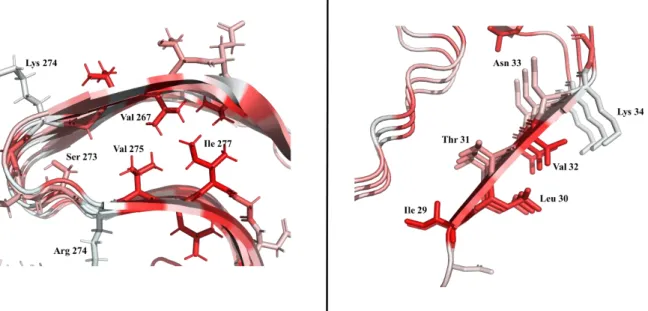
Fig. 46. RodC single-point mutations’ position... 112
Fig. 47. RodC single-point mutants’ purity and foldedness. ... 114
Fig. 48. RodC single-point mutant self-assembly kinetics – Comparison. ... 115
Fig. 49. RodC single-point mutant self-assembly kinetics – Individual kinetic traces ... 117
Fig. 50. RodC single-point mutant self-assembly kinetics – Lag phase. ... 118
Fig. 51. RodC single-point mutant self-assembly kinetics – Growth phase half-time. ... 118
Fig. 52. RodC single-point mutants’ kinetics – Lag phase and secondary structure. ... 120
Fig. 53. RodC quadruple point mutants’ SOFAST-15N HMQC experiments. ... 121
Fig. 54. RodC quadruple-point mutant self-assembly kinetics... 122
Fig. 55. RodC-derived peptides seen by TEM. ... 123
Fig. 56. Effect of different major fungal cell wall components on the lag phase of RodC fibrillation kinetics. ... 126
Fig. 57. RodC fibrillation kinetics in the presence of increasing concentrations of melanin. ... 127
xii
Index of tables
Table 1. Most common fungal diseases ... 18
Table 2. Examples of functional amyloids that can be found in nature. ... 31
Table 3. Rod hydrophobins. ... 47
xiii
Abbreviations
AFM Atomic force microscopy
AIDS Acquired Immunodeficiency Syndrome
ATR Attenuated total reflection
AWI Air-water interface
CP-MAS Cross polarization – magic angle spinning experiment DSS Sodium salt of 4,4-dimethyl-4-silapentane-1-sulfonic acid
DTT Dithiothreitol
ECM Extracellular matrix
FT-IR Fourier-transform infrared spectroscopy HHI Hydrophilic:hydrophobic interface HOPG Highly Ordered Pyrolytic Graphite
HMQC Heteronuclear multiple quantum correlation HSQC Heteronuclear Single Quantum Coherence
IA Invasive pulmonary aspergillosis
INEPT Insensitive nuclei enhanced by polarization transfer IPTG Isopropyl β-D-1-thiogalactopyranoside
LB Luria-Bertani broth
MCW Major cell wall
NMR: Nuclear magnetic resonance
PAMPs Pathogen-associated molecular patterns PDSD Proton-driven spin diffusion
RCI Random coil index
SOFAST band-selective optimized flip-angle short-transient experiment SRCD Synchrotron radiation circular dichroism
TEM Transmission electron microscopy
TFA Trifluoroacetic acid
ThT Thioflavin T
xiv
WT Wild-type
15
Introduction
1.1 Overview
Amyloids are defined as proteinaceous fibers that share a skeleton with the so-called cross-β structure. Since their first description by Virchow in 1854 (Sipe and Cohen, 2000) on abnormal brain tissues, amyloid fibers have been linked to disease. Indeed, over 60 pathologies associated to proteins that irreversibly misfold into an amyloid form have been described (Chiti & Dobson, 2017). However, in the late 90s, a number of proteins that are not associated to any disease, were shown to form amyloids under appropriate (Guijarro et al., 1998; Chiti et al., 1999), indicating that the amyloid structure was not restricted to some disease-related proteins. These findings ultimately lead to the generic amyloid hypothesis (Dobson, 1999; Stefani and Dobson, 2003) and to the recognition that protein sequences have in general evolved to avoid this self-propagating fold. Concomitantly, however, in the 90s, proteins that adopt an amyloid structure to perform their function were discovered (Fowler et al., 2005; Maji et al., 2009; Otzen and Riek, 2019). Nowadays, it is clear that many proteins have evolved to fold into amyloids to perform their biological function.
My PhD work was devoted to characterize the assembly of one of the first examples of functional amyloids, namely fungal proteins from the hydrophobin family, and in particular hydrophobins produced by the opportunistic pathogen A. fumigatus.
In this introduction, I will first present general aspects of the fungal kingdom, such as its diversity, ecological and biotechnological importance, as well as the impact of fungi in public health, particularly focusing on aspergillosis. Then, I will address, in more detail, the biology and pathobiology of A. fumigatus and describe the cell wall organelle, which is of outmost importance for pathogen-host interactions. The outer layer of the cell wall of A. fumigatus spores consists of amyloid fibers formed by a hydrophobin protein called RodA. This layer is at the forefront of the interactions of the spores with their environment.
I will then describe hydrophobins, small fungal proteins unique to filamentous fungi. These proteins show remarkable physicochemical properties upon which rely their biological functions and possible biotechnological applications. Hydrophobins spontaneously auto-associate at air-water (AWI) or hydrophobic-hydrophilic interfaces (HHI) to form amphipathic layers. I will expose what it is known about the monomeric soluble form, the mechanism of
16
auto-association and the structure of the assemblies. Finally, I will focus on A. fumigatus hydrophobins and introduce the objectives and questions that we posed during this work on their mechanism of self-assembly and structure.
1.2 The Fungal Kingdom
The Fungal Kingdom is one of the largest and more diverse group of organisms on Earth. Last estimates suggest that there are between 2.2 and 3.8 million species, and only the 8% is named (Hawksworth and Lücking, 2017). Fungi appeared on Earth more than 890 million years ago and have colonized almost every part of the globe. Fungi can indeed live in aquatic or terrestrial environments and play important roles in diverse ecosystems. These organisms have evolved into three major ecological roles: saprobes, mutualistic symbionts and parasites. As saprobes, fungi are the main decomposers of the complex components of plant debris, e.g. cellulose and lignin, and some animal debris, e.g. chitin and keratin, thus being important players in the carbon and nitrogen cycles. Fungi are also very important in mutualistic symbioses with plants (mycorrhizae) and algae (lichens) where fungi exchange nitrogen, phosphorus and minerals for carbohydrates or aminoacids from the photobiont. Finally, fungi are important pathogens with an important ecological function clearing unfit individuals and favoring evolution (Kendrick, 2001; Harrison, 2005).
In addition, fungi have many applications. Since ancient times, humans have made wine or beer with yeasts, leavened bread or collected edible mushrooms. Nowadays, fungi are a very important source of antibiotics and enzymes used in biotechnology. To give but one example, the antibiotic penicillin, produced by Penicillium spp, discovered by Alexander Fleming almost one hundred years ago, has saved hundreds millions of lives (Gaynes, 2017).
However, fungi can also be harmful to other living organisms. Fungi can be plant pathogens, for example, Magnaporthe grisea causes the most destructive disease on rice worldwide, while
Fusarium graminearum infects all cereal species and Ustilago maydis is the major corn parasite
(Dean et al., 2012). Animals can also suffer from fungal infections; Batrachochytrium
dendrobatidis, that causes chytridiomycosis in amphibians, is responsible of a mass amphibian
extinction and decline worldwide (Scheele et al., 2019). In humans, recent estimates indicate that every year fungi affect more than a billion people, of which 11.5 million suffer from life-threating infections and more than 1.5 million die (Tudela and Denning, 2017). Together, the three major fungal pathogens, namely Candida albicans, Cryptococcus neoformans and A.
17
fumigatus, kill as many people as the major infectious disease (tuberculosis, 1.5-1.7 million)
and more than HIV (770 000) or malaria (430 000; WHO, Global Health Observatory, 2018 data).
1.3 Fungal diseases in humans
There is a broad range of fungi (> 600) infecting humans causing diseases that can be from mild to lethal. Mycosis can affect the skin, nails, hair, the eyes, mucosas, lungs, and can develop into systemic diseases. Severe fungal infections are due to opportunistic pathogens that infect immunosuppressed people and are most of the time associated to other diseases or medical conditions such as asthma, leukemia, organ transplants, AIDS or immunosuppressive corticosteroid therapies. Despite the number of deaths and the increase in severe mycosis, fungal infections remain neglected by public health authorities (Bongomin et al., 2017). The table below summarizes the most important fungal infections (Table 1).
Most common fungal diseases
Fungal nail infections Caused by many different environmental species of fungi (yeasts or molds). Easy to cure with common antimycotics.
Dermatophytosis Fungal infection of the skin caused by Trichophyton, Microsporum or
Epidermophyton spp. Very common and not very dangerous for the patient. Easy to
cure with common antimycotics.
Vaginal candidiasis Caused by the yeast Candida that normally lives inside the body but can cause an opportunistic vaginal infection. In the United States, it is the second most common type of vaginal infection after bacterial vaginosis.
Fungal diseases that affect people who live in or travel to certain areas
Blastomycosis Caused by Blastomyces spp. Originated in North America. Symptoms are very similar to those of flu. Most people will need treatment with antifungal medication. Coccidioidomycosis or
Valley fever
Caused by Coccidioides spp. Endemic in North America. People who get sick will get better on their own within weeks to months but some people will need antifungal medication.
Cryptococcosis by
Cryptococcus gattii
Happens in tropical and sub-tropical regions and can produce a severe lung infection that will need antifungal medication treatment.
Histoplasmosis Histoplasma is the infectious agent and it is worldwide, very severe and will need
18
Paracoccidioidomycosis Usually happens in rural areas with the presence of Paracoccidioides spp. It can be treated with azole antifungals.
Fungal diseases that affect people with weakened immune systems
Aspergillosis Worldwide mycosis caused by Aspergillus spp. People with weakened immune system or lung diseases are at a higher risk of developing fatal health problems. Invasive candidiasis Serious infection caused by Candida spp. that affects the blood, heart, brain, eyes,
bones, or other parts of the body.
Cryptococcosis Strong disease provoked by Cryptococcus neoformans. It affects the lungs or the central nervous system and can lead to cryptococcal meningitis.
Mucormycosis Caused by different mucormycetes, mainly affects people with weakened immune systems. It is not a life-threatening disease but needs antifungal medication.
Pneumocystis pneumonia Caused by the fungus Pneumocystis jirovecii. It was very important in the late 1980s when around 75% of people living with AIDS developed this type of pneumonia. Sporotrichosis Caused by the fungus Sporothrix spp. It can be cutaneous, pulmonary or
disseminated. These infections are not life-threatening but must be treated with prescription of antifungal medication for several months.
Table 1. Most common fungal diseases
Adapted from the Center for Disease Control and Prevention (CDC) (accessed June 4, 2019) and (Fausto, Rodrigues and Coelho, 2019).
1.4 Aspergillosis
Aspergillosis are diseases caused by species belonging to the genus Aspergillus. This genus comprises more than 300 species but only 20 of them are pathogenic to humans. Among them,
A. fumigatus is the most important species, it causes approximately 90% of the aspergillosis
cases (Latgé, 1999; Margalit and Kavanagh, 2015), followed by A. flavus, A. niger and A.
terreus (Cadena, Thompson and Patterson, 2016).
A. fumigatus is a major airborne fungal pathogen that infects the lungs. The spectrum of
aspergillosis is very broad and comprises several clinical entities: allergic bronchopulmonary aspergillosis, chronic pulmonary aspergillosis syndromes and invasive pulmonary aspergillosis (IA) (Cadena, Thompson and Patterson, 2016).
Recent estimates indicate that more than 250 000 people die every year from of invasive aspergillosis and that 5 million people suffer from allergic bronchopulmonary aspergillosis
19
(Bongomin et al., 2017). However, the difficulties in diagnosis suggest that these numbers could be under estimated by a factor of two. In the case of invasive aspergillosis, the mortality ratio varies between 30 and 95%. It is related to the degree of immunosuppression, the geographic region, socioeconomic conditions and cultural habits (Brown et al., 2012; Krappmann, 2016). In the absence of treatment with antifungal drugs, the mortality rate can be 100%.
1.5 Aspergillus fumigatus
Aspergillus spp. are filamentous fungi that belong to the phylum Ascomycota, characterized by
the production of sexual spores inside closed compartments called asci. Aspergillus spp., like many other filamentous fungi spend most, if not all of their life, in an asexual form and reproduce through asexual spores called conidia held on aerial structures called conidiophores. The name Aspergillus was coined in 1729 by Pier Antonio Micheli to describe the asexual form of the conidiophores that resembled an aspergillum (Fig. 1), a liturgical device used to sprinkle holy water.
Fig. 1. Aspergillus morphology.
Left: Aspergillum (source Wikipedia). Middle, Aspergillus sp. (source: CDC’s webpage) and right, Aspergillus sp. morphological diagram (adapted from Pinterest). Conidia are located on the conidiophore and are held by specialized cells called phialides.
The genus Aspergillus contains ~350 species, of which around 20 species are pathogenic. Among these, A. fumigatus causes approximately 90% of the aspergillosis cases (Latgé, 1999; Margalit and Kavanagh, 2015).
20
1.5.1 Biology
A. fumigatus is a thermotolerant and saprophytic fungus that lives in the soil worldwide playing
an important role in the carbon and nitrogen cycle (Willger, Grahl and Cramer, 2009). This species lives in its asexual form during the whole life cycle (Fig. 2).
This mold thrives in moist substrates where it sporulates abundantly leading to high concentration of its conidia in air (ca. 1-100 conidia/m3): it has been estimated that on average, an adult inhales more than 100 conidia per day. The conidia globular form, small size < 3 µm and hydrophobicity, allow them to efficiently disperse in the air and reach lung alveoli (> 10 µm) (Kwon-Chung and Sugui, 2013).
Fig. 2. A. fumigatus life cycle.
a) Asexual hydrophobic spores (conidia) are dispersed by the air, b) when dormant conidia find a convenient place to grow, these swallow, the metabolism is activated and conidia germinate. c) Germinating conidia. d) Development of the mycelia formed by the fungal cells (called hyphae). e) Cells emerge from the substrate to form aerial hyphae and the reproductive structures (conidiophores). f) The conidiophores contain substructures named phialides, from which the new dormant conidia emerge via evagination, closing the life cycle.
21
1.5.2 Fungal Cell Wall
In contrast with animal cells, fungal cells are enveloped by a thick cell wall that endows extraordinary properties to the hyphae and spores. The fungal cell wall is indeed an essential dynamic organelle that can change its composition and structure depending on environmental conditions and the life cycle. It is the interface between the fungus and its environment, including possible interactions with their hosts. It is an armor that protects fungi against chemical or physical stress. Its strength and rigidity allows fungi to resist to turgor pressures as high as 20 times the atmospheric pressure (Money, 2001) and penetrate solid substrates or host cells, as well as to grow and differentiate into complex shapes.
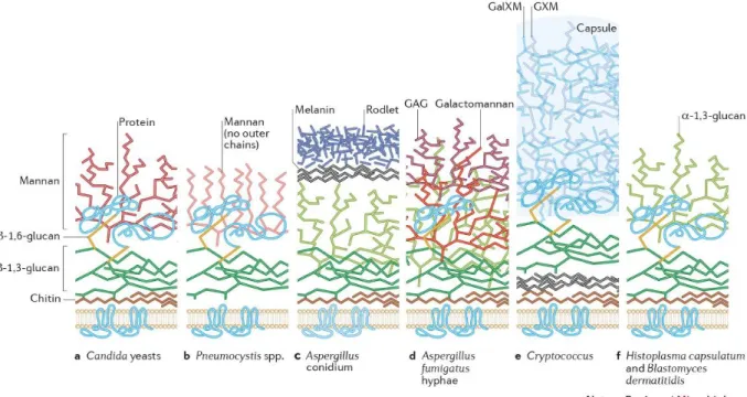
Although the fungal cell wall is a complex organelle than changes overtime and between species, most fungal cell walls have a common core of covalently branched β-(1,3)-glucan and chitin [β-(1,4) N-acetylglucosamine]. These polysaccharides form a scaffold that surrounds the cell. In many species, β-(1,6)-glucan is branched to β-(1,3)-glucan. The rest of the cell wall carbohydrate composition and the nature of the carbohydrates attached to the core can be quite different between fungal species as can be observed in Fig. 3, which shows some examples of cell wall from different fungal pathogens. (Gow, Latgé and Munro, 2017).
In addition to the different polysaccharides, that normally comprise 90% of the cell wall mass, the fungal cell wall contains enzymes specialized in carbohydrate synthesis and remodeling. Chitin and glucan synthases are embedded in membrane while the remodeling enzymes, such as glycohydrolases, glycosidases or transglycosidases are anchored to the plasma membrane through a GPI-anchor (glycosyl-phosphatidyl inositol).
22
Fig. 3. Image of different fungal cell walls (extracted from Gow, Latgé, & Munro, 2017).
1.5.3 Aspergillus fumigatus Cell Wall
The hyphal and conidial morphotypes display different structural organizations based on the same fibrillar core. Most of what is known on the composition of A. fumigatus cell wall comes from the chemical and enzymatic analysis of extracted material of wild type and mutant strains from the laboratory of J. -P. Latgé at the Institut Pasteur. The extracted material is fractionated into alkali soluble and alkali insoluble fractions. The fibrillar core is alkali-insoluble and is composed of (1,6) branched (1,3)-glucan (4% branching) cross-linked to chitin through β-(1,4) bonds. Chitosan, β-(1,3)/β-β-(1,4)-glucan and β-(1,5)-galacto-α-(1-2)- and α-(1-6)-mannan are covalently bound to branched β-(1,3)-glucan. The alkali-soluble fraction is amorphous and mainly composed of linear chains of α-(1-3)-glucan with intra-chains of α-(1-4)-glucan linked to glucose units (Latgé and Beauvais, 2014).
Relative to conidia, hyphae contain two additional polysaccharides: galactomannan (GA) and galactosaminogalactan (GAG). GA, which is used as biomarker for the detection of invasive aspergillosis, is composed of a main chain of α-mannoside and short side-chains of galactofuranose residues (Fontaine, 2017). GAG is a heteropolysaccharide composed of α-(1,4) linked galactose and N-acetylgalactosamine. Because of its importance in virulence and pathogenesis, GAG is considered nowadays as a potential novel drug target (Speth et al., 2019).
23
Although absent in wild-type conidia, it has recently been established by Valsecchi et al., 2019a that GA and GAG can appear in conidia as a result of remodeling when key-polysaccharide synthases are mutated.
Because conidia are the infectious form of the mold, they have attracted much interest for many years (Carrion et al., 2013; Margalit and Kavanagh, 2015; Croft et al., 2016; Latgé, 2017). In addition to GA and GAG, the main difference between hyphae and conidial cell walls is the presence of a layer of the black pigment melanin that is covered by an outer hydrophobic proteinaceous layer with rodlet morphology. Both the melanin and the rodlet layers disappear upon swelling and germination.
The melanin layer protects the fungus against UV radiation, reactive oxygen species and inhibits host-cell phagocytosis, cytokine production and apoptosis (Langfelder et al., 2003). A.
fumigatus melanin is of the “DHN” type, formed from 1,8-dihydroxynaphthalene (DHN),
which is synthesized from the precursors acetyl-coA or malonyl-coA. Although the intracellular synthesis pathway of DHN is known, its polymerization and oxidation to form melanin, as well as melanin structure and degradation, are still poorly understood (Eisenman and Casadevall, 2012).
The rodlet layer, which covers the conidial surface, confers hydrophobicity to the conidia, thus facilitating dispersion and rendering conidia waterproof. Besides, it protects conidia from desiccation.
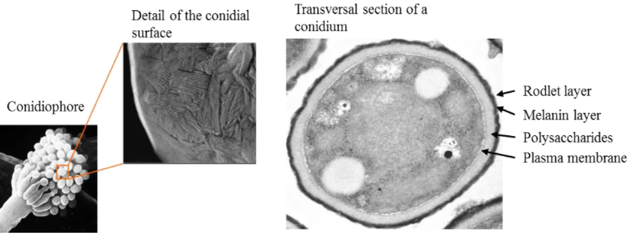
It is made up of the RodA protein (UniProtKB accession number B0Y4B10 and A. fumigatus Genome Database number AFUA_5G09580) that belongs to the hydrophobin family. This protein self-assembles into amyloids fibers with a characteristic rodlet morphology (Fig. 4). Moreover, this rodlet layer masks the pathogen-associated molecular patterns rendering the conidia inert relative to the immune system (Aimanianda et al., 2009). Indeed, while the conidia covered with RodA rodlets do not trigger any response from the innate and adaptive immune systems, conidia lacking this mask (deletion mutants of the rodA gene, germinating conidia or conidia treated with concentrated acids to extract RodA) do elicit a response.
24
Fig. 4. A. fumigatus conidia.
Left, A. fumigatus conidiophore and SEM (scanning electron micrograph) detail of the conidial surface. Right, transversal section of a conidia TEM micrograph (credits: Aspergillus Unit).
1.6 Pathobiology
The airborne conidia are the infectious form of the mold. Although the majority of conidia are expulsed mechanically by the mucociliary transport some conidia bypass the mucociliary clearance and penetrate the lung alveoli (Krappmann, 2016). Conidia attach to the extracellular matrix on the surface of the airway epithelial cells (either at bronchial or alveolar level) of the lower respiratory tract, in a process that is not very well understood. During the process, the rodlet and melanin layers hide the immunogenic cell wall polysaccharides avoiding an inflammatory response. The interaction between the conidium and the lung cells results in clearance by healthy individuals whereas in susceptible people it results in infection. In immunocompetent host, clearance of inhaled conidia is highly coordinated by the immune response that eliminates pathogens while preventing inflammatory lung damage. Intracellular swelling leading to germination, which occurs in macrophage phagosomes, is concomitant to the removal of the rodlet layer and the emergence of cell wall pathogen-associated molecular patterns (PAMPs), including β-glucans and other immunostimulatory polysaccharides and subsequent activation of Dectin-1 and other C-type lectin receptor’s signaling pathways. These signaling pathways trigger an adaptive immune response that normally ends up in fungal killing (Brown, 2006; Latgé, Beauvais and Chamilos, 2017). In contrast, in immunocompromised individuals, A. fumigatus evades the debilitated immune system, conidia swell and germinate in the macrophages’ phagosomes leading to a colonization of the lung parenchyma (Croft et
25
As mentioned before, in addition to its biological role, the RodA amyloid rodlet layer provides immune inertness to the spores of A. fumigatus. This functional amyloid coat mediates initial host-fungal interactions and protects the spores from the innate immune system. Nevertheless, the RodA coat is not a virulence factor. Although RodA reduces killing by macrophages and helps the fungus to resist against NETosis (neutrophil extracellular traps), its removal does not influence pathogenicity (Thau et al., 1994; Paris et al., 2003). Spores from other fungal species within the phylum Ascomycota are covered with an amyloid rodlet layer as well. It has been shown that the hydrophobin coat is also immunologically inert for other pathogenic (A. flavus), allergenic (Cladosporium cladosporioides), edible (P. camemberti) or toxic fungi (P.
verrucosum). Although the amyloid coat might present an advantage for pathogenic fungi
infection, it has also beneficial effects for humans by hiding the allergenic components of the cell wall and preventing allergies and asthma (Bayry et al., 2014). The immune inertness of this functional amyloids contrasts with the observation that amyloids from several disease-related proteins are immunogenic and induce inflammatory responses and the hypothesis that the cross- structure triggers the inflammatory response (Gustot et al., 2015).
1.7 Amyloid fibers
Amyloid fibers are formed by soluble proteins that self-assemble into insoluble aggregates that are resistant to disaggregation and rich in β-structure.
The name “amyloids” was coined by Virchow in 1854 to describe some deposits that appeared in brain tissue and could be stained with iodine and sulphuric acid. The staining properties of the deposits were starch-like and he inferred that these consisted of carbohydrates. Some years later, in 1859, Friedrich and Kekulé, from the high nitrogen contained in liver tissue infiltrated with amyloids, suggested that the deposits were made of proteins (Friedreich and Kekulé, 1859; Sipe, 1992) . Over a century later, Glenner and Eanes discovered that amyloid fibers exhibited an X-ray (XR) diffraction signature (Fig. 5) known as the “cross-β arrangement” (Eanes and Glenner, 1968). Today, a protein is considered to be in an amyloid state when it forms long fibers with the cross-β structure. Amyloids can be revealed using reporter dyes such as Congo red or thioflavin T (ThT) (Chiti and Dobson, 2006; Eisenberg and Jucker, 2012).
26
Fig. 5. Amyloid fiber features.
Amyloids are long and unbranched fibers when seeing by electron microscopy. When amyloids are observed with XR, a characteristic cross-β diffraction pattern with two reflections is observed, one at 10-12 Å that corresponds to the inter-sheet space while the 4.7 Å reflection corresponds to the inter-strand space in the direction of hydrogen bonding. When the fibers are aligned, the XR reflections appear as arcs (like in the diagram). For unaligned fibers, the reflections are ring-shaped.
The structure of these type of fibers is very difficult to study by well-established techniques such as XR crystallography or solution NMR spectroscopy, mostly because of their non-crystalline and insoluble character. More recently, ssNMR and cryo-EM techniques have generated substantial advances in structural information and some amyloid structures have been determined (Fig. 6) (Loquet et al., 2018; Fitzpatrick and Saibil, 2019; Röder et al., 2019). On the one hand, the main advantage of using ssNMR over cryo-EM is the capacity to work with biological assemblies that show micro heterogeneities (in otherwise homogeneous and reproducible samples) that are a big problem on EM, although recent advances in cryo-EM image recording, processing and analysis are helping to deal with this problem. On the other hand, ssNMR needs large amounts of labelled protein samples due to its low sensitivity and requires long-time for data analysis.
27
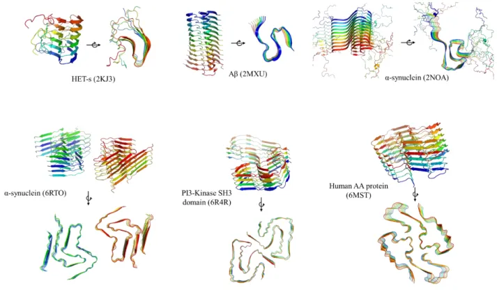
Fig. 6. Examples of high-resolution structures of amyloid fibers from the PDB database.
Top, ssNMR structures; bottom, cryo-EM structures with resolution below 4 Å.
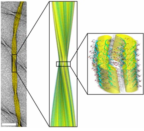
Amyloids are composed of an ordered arrangement of thousands of copies of the same peptide or protein showing a repetitive structure. Under EM, the long unbranched fibers have a diameter between 6–12 nm. Protein or long peptide amyloids show structures that are more complex than it was anticipated from the study of short peptides but are still based on very flat -sheets (Fig. 7). The low angle observed between strands usually leads to fibers with a helical rotation about their central axis showing a long pitch.
28
Fig. 7. Amyloid morphology and structure diagram.
Left, image of a fiber (11 residue peptide from transthyretin) taken using TEM. Center, ssNMR structure of the triplet fiber fitted into the cryo-EM reconstruction. Right, fiber surface and the constituent β-sheets. Adapted from Fitzpatrick et al., 2013.
In amyloids, hydrophobic residues are mainly packed within the fiber core while hydrophilic and charged residues point towards the solvent (Fig. 8, left). This arrangement is kept between the strands of all the subunits to maintain the charge balance constant and stable. The high number of backbone hydrogen bonds and of hydrophobic interactions between complementary side chains are at the origin of the high stability and strength of amyloid fibers. It has been shown that the large network of intermolecular hydrogen bonds can confer to amyloids a mechanical strength comparable to that of steel (Lonescu-Zanetti et al., 1999; Knowles et al., 2007; Mankar et al., 2011). Interactions like amide side chain hydrogen bonds observed for glutamine and asparagine residues (Nelson et al., 2005), π stacking between aromatics (Gazit, 2002) or other steric zippers also significantly contribute to the stability of amyloids (Sawaya
29
Fig. 8. Examples of stacked β-strands.
Left, HET-S (2KJ3) detail. Right, PI3-Kinase SH3 Domain (6R4R). Aminoacids are colored by hydrophobicity using PyMOL, the redder the more hydrophobic is the residue.
1.7.1 Functional and non-functional amyloids
Amyloids have been (and still are) historically related to disease. To date, there are 36 extracellular fibril proteins and 7 intracellular protein-based inclusions associated to amyloids diseases, including Alzheimer’s, type II diabetes and Parkinson’s (Chiti & Dobson, 2017). The societal impact of these generically called protein misfolding diseases is huge and their prevalence is ever increasing with the ageing of population and life style. Alzheimer’s, type II diabetes and Parkinson’s affect nowadays more than 500 million people and numbers keep increasing. Only in the US, it is estimated that the treatment of Alzheimer’s costs more than 200 US billion dollars (Dobson, 2017).
The long time interest in amyloids is due to the importance of associated diseases and was further stimulated by the demonstration by the Prusiner’s group that infectious diseases such as human (as Creutzfeld Jacob or kuru) or bovine/ovine spongiform encephalopathies (mad cow disease, scrapie) were caused and propagated by proteins with an aberrant fold, the prions (Prusiner, 1998). The believe that the amyloid fold was unique to some proteins involved in disease was first challenged by a serendipitous finding that an SH3 domain (unrelated to any disease) under acidic conditions that destabilized the native fold could form amyloids and the
30
demonstration led by the Dobson’s group that under appropriate conditions many if not all normally globular proteins could self-assemble into amyloid fibers (Guijarro et al., 1998; Chiti
et al., 1999). These findings stimulated research to understand how protein evolution had
managed to limit/avoid this highly stable and self-propagating fold. Besides chaperons and cellular mechanisms (localization, synthesis regulation and degradation), some of the solutions to avoid irreversible formation of amyloids are encoded in the sequence of proteins (for instance, folding to hide aggregation prone sequences).
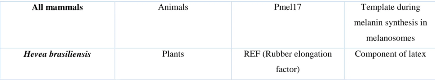
Concomitantly, proteins that adopt an amyloid fibrillar form to perform their biological function began to be discovered. Nowadays, described functional amyloids probably outnumber disease-related amyloids and new functional amyloids keep being reported. Functional amyloids have indeed been found in the three domains of life, Bacteria, Archaea and Eukaryotes, including humans. Amyloids can play important physiological roles (table 2), which can be structural, templating, storage, adhesion, biofilm and extracellular matrix formation, regulatory, etc. The role of several functional amyloids can also be to power-off the function of the soluble form of the protein.
Species Organism type Protein Function
Mycobacterium tuberculosis
Bacteria MTP Adhesion, biofilm formation and cell-cell
communication
Escherichia coli Bacteria Curli Adhesion and biofilm formation
Bacillus subtilis Bacteria TasA Colony biofilm formation
Methanosaeta thermophila
and
Methanospirillum hungatei
Archaea (methanogens) MspA (major sheath protein A) Part of cell wall (tubular
sheaths) and possibly gas vesicles
Aspergillus fumigatus Fungi RodA Surface attachment, aerial hyphae formation and surface
tension modification
Saccharomyces cerevisiae Fungi Ure2p Nitrogen catabolism (soluble protein) Mouse, fish and silk moth Animals ZP proteins (ZP1-ZP3) Formation of the
amyloid matrix around the oocyte
31
All mammals Animals Pmel17 Template during melanin synthesis in
melanosomes
Hevea brasiliensis Plants REF (Rubber elongation factor)
Component of latex
Table 2. Examples of functional amyloids that can be found in nature. Extracted from Otzen & Riek, 2019; Pham, Kwan, & Sunde, 2014.
1.7.2 How does the amyloid state form?
The amyloid state can be adopted by globular proteins with a variety of folds and secondary structure topology, by intrinsically disordered proteins (e.g. α-synuclein) or short peptides. Irrespective of the initial structure of the protein, and of whether amyloids arise from misfolding (disease related) or folding (functional) events, protein amyloids share a common cross-β structure, suggesting that some common features must exist in the process of amyloid formation. Amyloid formation implies important conformational changes, even in the case of globular all-β sheet proteins (Fig. 9).
Fig. 9. Comparison of the secondary structure of the globular (native) and amyloid forms of an all- protein.
Position of the β-strands in the fiber protein and in the natively folded of the immunoglobulin light chains (PDB 1BJM). Reproduced and modified from Radamaker et al., 2019.
The high number of hydrogen bonds involved in the cross-β structure, the inter β-sheet side chain packing, possible intra β-sheet side-chain hydrogen bonds (Q/N) or aromatic rings
π-32
stacking interactions render the amyloid state highly stable (more stable than the native globular fold or unfolded forms) and hence thermodynamically very favorable (Gazit, 2002; Baldwin et al., 2011; Buell et al., 2012). However, the energy barrier(s) to form amyloids can be very high (Fig. 10). The formation of amyloid fibers is an aggregation process that is thereby concentration dependent. It is also a nucleated phenomenon. First, for stable globular proteins, some destabilizing condition (such as pH, temperature or a destabilizing mutation) must favor unfolding or partial unfolding to permit conformational modifications to occur and allow the protein to exit the kinetically-trapped native meta-stable conformation. Then, oligomerization/conformational modification events must occur to form a nucleus that is competent to readily self-assemble into a cross-β structure. In vitro, amyloid fiber formation kinetics are usually monitored by the fluorescence of an amyloid reporter dye. The observed kinetics show a sigmoidal shape that is characteristic of nucleated phenomena (Fig. 11).
Fig. 10. Protein aggregation landscapes. Adapted from Landreh et al., 2016.
33
The fluorescence intensity reveals the appearance of the cross-β structure at the core of the fibers. The sigmoidal curve can be divided in three stages: the lag phase, the growth phase and a final plateau.
During the lag phase, soluble monomers form nuclei consisting of oligomers. The process that govern this phase is primary nucleation, in which monomers constantly interchange with forming nuclei (Fig. 12). At some point, the primary nucleation process leads to a critical concentration of nuclei from which fibers can form during the growth phase that is usually dominated by elongation and can have significant contributions from fragmentation and secondary nucleation processes. It can be an extremely rapid process in which monomers are added to the existing fibers. Fragmentation of fibers increase the number of sites at which monomers can incorporate to the fibers. Secondary nucleation is a self-catalytic process in which the surface of an existing fiber serves as a template to incoming monomers to form new fibers. This secondary nucleation mechanism is monomer concentration dependent and can dramatically accelerate fiber formation. After some time, the majority of the monomers are in the amyloid final form and speed decreases, it is the final stage, the final plateau, also called stationary phase, but still different processes such as fragmentation, elongation, secondary nucleation or exchange with monomers are happening (Arosio, Knowles and Linse, 2015; Meisl et al., 2016).
Fig. 12. Microscopic processes underlying amyloid formation.
Primary nucleation from monomers in solution (a), elongation (growth) by monomers addition to existing aggregates (b), surface catalyzed secondary nucleation from monomers on the fiber surface (c) and fragmentation (d) (adapted from Arosio et al., 2015).
Understanding the mechanism of amyloidogenesis is very important both, in the case of disease-related amyloids and of functional amyloids. In the case of amyloid-related diseases, shedding light on amyloidogenesis and the intermediates involved is actually more important than understanding the structure of the fibers, because small aggregates seem to be at the origin
34
of toxicity (Gustot et al., 2015). Also, disease-related proteins show different polymorphs with distinct physical properties that can lead to a broad spectrum of disease phenotypes (Fitzpatrick and Saibil, 2019). Moreover, understanding the auto-association process might help in finding drugs to avoid protein aggregation (Gustot et al., 2015). In the case of functional amyloids, understanding the mechanism of fibrillation and conformational plasticity of proteins to acquire their functional form is interesting per se and can provide useful information for amyloid related diseases. Perhaps, one of the main differences between proteins that aggregate into fibrils under abnormal conditions or have evolved to form amyloids, is the fiber polymorphism. Although in both cases, fiber structure is related to the influence of the environment, functional amyloids tend to show less polymorphism under controlled conditions (Tycko, 2015).
Therefore, the knowledge on how amyloid self-assemble is of vital importance to better understand amyloid-related diseases and important to understand how functional amyloids fold to perform their role in biological systems.
1.8 Hydrophobins
1.8.1 General aspects
Hydrophobins are surface-active proteins of low molecular weight (6-15 KDa) characterized by a conserved pattern of eight cysteine residues. These proteins are unique to filamentous fungi.
Hydrophobins were discovered by Wessels and co-workers in the early 1990s when studying gene expression during the development of the Schizophyllum commune fruiting body (J. Wessels et al., 1991). Later studies showed that these proteins are surface-active proteins that spontaneously self-assemble at hydrophilic/hydrophobic surfaces into amphipathic layers or films with rodlet morphology resembling those previously reported in the 1960s by Hess and co-workers on the surface of Penicillium and Aspergillus conidia (Hess, Sassen and Remsen, 1968; Hess and Stocks, 1969; J. J. G. H. J. Wessels et al., 1991; Wessels, 2000; Wösten, 2001). The proof that the hydrophobin layer covering fungal spores was hydrophobic came from the observation that a mutant of Neurospora crassa lacking the gene of the Eas hydrophobin is hydrophilic (Dempsey and Beever, 1979). The protein was named EAS, after the ‘EASy wettable’ phenotype observed for the mutant.
35
Hydrophobins show, in general, very little conservation except for the idiosyncratic pattern of eight cysteine residues that form four disulfide bridges. These cysteines are interconnected as shown below (Fig. 13).
Fig. 13. Hydrophobin pattern of cysteines.
Eight cysteine (C) residues form four disulfide bonds with the same pattern (C1–C6, C2–C5, C3–C4, C7–C8). Cysteines C2-C3 and C6-C7 are contiguous in the sequence.
Based on their sequence, hydropathy pattern, solubility and structure formed upon self-assembly, hydrophobins are divided in two major classes, the Class I and Class II. Class I hydrophobins typically display low conservation and very variable inter-cysteine spacing whereas Class II are more conserved and show less inter-cysteine-spacing variability (Fig. 14). The hydropathy profile of hydrophobins also varies between classes I and II.
Fig. 14. Alignment of Class I and Class II of some commonly studied hydrophobins. The highly variable N and C terminal parts of the sequences have been cropped. The conserved eight cysteine residues are highlighted in blue. The UniprotKB accession number and species of the aligned protein sequences are: PRI2 (Q9Y8F0) Cyclocybe
cylindracea; ABH1 (P49072) Agaricus bisporus; HCF1 (Q00367) Passalora fulva;
MPG1 (P52751) Magnaporthe oryzae; SC4 (P16934) Schizophyllum commune; EAS (Q04571) Neurospora crassa; RodA (B0Y4B10) A. fumigatus; HCF6 (Q9C2X0)
36
reesei; CFTHI (Q9UVI4) Claviceps fusiformis; HYD4 (Q6YF29) Gibberella moniliformis and CU (Q06153) Ophiostoma ulmi.
Class I hydrophobins form rodlet layers displaying the hallmarks of amyloid fibers and are extremely robust, requiring treatment with concentrated acids (e.g. net TFA, 50% HF, 50% formic acid) to induce disassembly. On the other hand, Class II hydrophobins form non-fibrillar amorphous aggregates that can be solubilized in organic solvents or detergents at high temperature and do not show amyloid characteristics (Kwan et al., 2006; Kordts et al., 2018). The morphology of the films formed by Class I and II hydrophobins also differs: while Class I hydrophobins form very ordered rodlets, Class II hydrophobins generally form amorphous films (Fig. 15).
Fig. 15.Rodlets images seeing under AFM.
AFM images of films formed on HOPG (highly oriented pyrolytic graphite) by EAS∆15 (Class
I) and NC2 (Class II) from Neurospora crassa (reproduced from Lo et al., 2014).
Based on bioinformatics, a third class (Class III) of hydrophobins with intermediate or different characteristics has been proposed. Putative hydrophobins from Class III have an intermediate hydropathy profiles and/or inter-cysteine spacings and can display more cysteine residues, lack one of them, lack secretion signal peptides or have very long N or C-terminal tails (Jensen et
al., 2010; Littlejohn, Hooley and Cox, 2012). It should be mentioned that to the best of our
knowledge, no Class III hydrophobin has been studied so far.
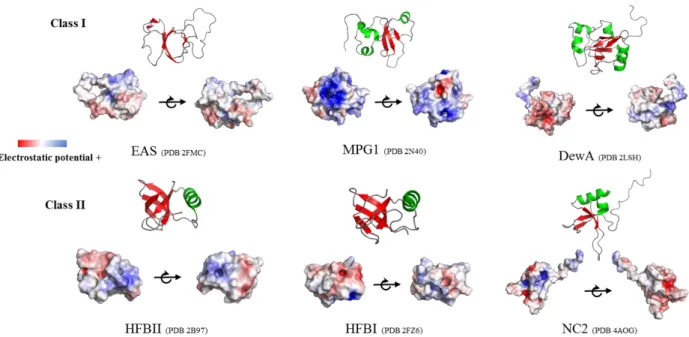
Hydrophobins are secreted by fungi as soluble proteins. Several structures of the soluble form of hydrophobins that auto-associate to form amphipathic layers have been described. The structures show a common central β-barrel or semi-β-barrel topology organized around the four disulfide bridges. Two of the disulfide bridges are located inside the sheet core linking β-strands, whereas the other two link this core to external structural elements. In Class II
37
hydrophobins, the central β-sheets generally form a β-barrel, whereas this core is a semi-β-barrel in most of Class I structures. In addition to the core, hydrophobins display flexible loops and α-helical elements (Fig. 16). The diversity of hydrophobin sequences is reflected in the differences in structure shown by hydrophobins.
Fig. 16. Class I and Class II hydrophobins cartoon and surface representations showing the structure and electrostatic potential, respectively.
EAS (2FMC) from Neurospora crassa, MPG1 (2N40) from Magnaporthe oryzae, DewA (2LSH) from Aspergillus nidulans, HFBII (2B97) from Trichoderma reesei, HFBI (2FZ6) from Trichoderma reesei and NC2 (4AOG) from Neurospora crassa. β-sheets are colored in red and α-helices in green. The PyMOL plugin APBS was used to display electrostatic potentials.
Despite their structural differences, hydrophobins show two other common features in addition to the central rigid core. First, the N-terminal region upstream cysteine C1 and the loop regions between cysteines C3-C4, C5-C6 and C7-C8 are highly flexible (Pille et al., 2015). Second, the monomers display hydrophobic and charged patches at their surface or are amphipathic (Fig. 16). This amphipathic character enables the proteins to migrate to the AIW or HHI (Fig. 17), where the monomers undergo conformational changes triggered by the physical properties of the interface and assemble into amyloid or amorphous layers. Studying the effect of surfactants (ionic, non-ionic) and alcohols on the auto-association of the hydrophobins DewA and EAS, it has been shown that surface tension is the driving force to address hydrophobins
38
to the surface and that a minimal surface tension (which can be different for each hydrophobin) is required to self-assemble at an AWI (Morris et al., 2011; Pham et al., 2016; Sunde, Pham and Kwan, 2017).
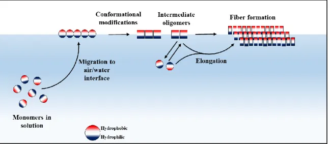
Fig. 17. Proposed simple model of in vitro Class I hydrophobin aggregation. The monomers with surface exposed hydrophobic regions reach the AWI or HHI, undergo conformational changes and associate into oligomeric nuclei that can readily convert into amyloid fibers that laterally associate into layers.
Aggregation of hydrophobins can follow a complex mechanism. For instance, the Class I hydrophobin SC3 assembles to the rodlet state via two intermediates. The first is an α-helical state that is formed upon binding to an hydrophobic solid, the second is an amorphous β-sheet state formed at the AWT, which after some hours spontaneously, acquires the rodlet morphology (de Vocht et al., 2002).
Because hydrophobin amyloids are self-propagating structures, different mechanisms have been described to regulate or limit aggregation of the monomers that could be harmful for the fungal cells. DewA, a protein found in the spores of A. nidulans, populates two conformers (with the same disulfide bridges) in solution that are amyloidogenic. While both conformers readily form amyloids at high concentrations, one of them forms off-pathway dimers thus reducing the rate of self-assembly into amyloids (Morris, Kwan and Sunde, 2013).
In silico studies with EAS have suggested that large flexible loops may inhibit aggregation in
solution because of the high cost of reducing their conformational entropy and only in the presence of an anisotropic surface providing surface tension (that can be seen as a force that counterbalances conformational entropy) the protein can self-assemble (De Simone et al.,
39
2012). Based on the relative rates of the reduced and oxidized forms of SC3, it has also been proposed that the idiosyncratic S-S bonds of hydrophobins can reduce the rate of fiber formation and that they represent a mechanism to avoid fast deleterious aggregation within cells (De Vocht et al., 2000).
Although Class I hydrophobin self-assembly can differ from one hydrophobin to another, these proteins share common characteristics. Within very variable structures, the central β-barrel or half-barrel core and the amphipathic character of the monomer allowing it to reach the HHI have been observed for all the monomeric proteins studied. Also, it has been shown for some hydrophobins that an HHI is required to trigger the conformational changes that lead to self-assembly, although some hydrophobins like SC3 or Vmh2 can form nanorods or amyloids in bulk solution, respectively (Zykwinska et al., 2014; Gravagnuolo et al., 2016).
1.8.2 Biological functions
The function of hydrophobins relies upon the physicochemical characteristics of the layers they form. One of the best studied hydrophobin is SC3, a protein produced by Schizophyllum
commune when conditions are optimum to generate aerial hyphae to form the fruiting body and
reproduce. The main role of SC3 is to cover the surface of the aerial hyphae and allow these structures to reduce the surface tension of water and breach the AWI. S. commune SC3 deletion mutants are not able to form aerial structures but addition of recombinant SC3 to the medium complements the deletion and allows the fungus to form aerial structures. These observation indicated the critical role of SC3 in the correct development of the fungus (Wösten, 2001). Similarly, hydrophobins (hypA) have been found in the “portobello mushroom”, the edible
Agaricus bisporus. Here, hydrophobins reduce wetting and help mediate resilience to
environmental stresses (Lugones et al., 1996). We can appreciate this edible layer when cleaning in the kitchen the mushroom’s cap, a very smooth semi-waterproof layer that covers it.
Hydrophobins can help on dispersion too. Indeed, the hydrophobic character of the hydrophobin coat that covers the spores facilitates air dispersal. Moreover, this coat provides resistance to desiccation and wetting, while warranting gas exchange.
Hydrophobins can play an important role in attachment, especially in the case of plant pathogens. For example, the hydrophobin CU from Ophiostoma ulmi that causes the Dutch elm disease, participates during the attachment of the fungus to the plant at the first stages of the
40
infection. Similarly, MPG1 produced by Magnaporthe grisea, the main parasite of rice cultivars is used by the fungus to attach to the leaf surface (Dean et al., 2012). Other roles have been described for hydrophobins in plant-fungi or insect-fungi interactions (reviewed in Bayry
et al., 2012).
1.8.3 Applications
Like their biological roles, the foreseen applications of hydrophobins mainly rely on their remarkable physicochemical properties. Their high surfactant activity, their capacity to cover surfaces and invert their polarity (hydrophobic into hydrophilic and vice versa), their stability and, mechanical and chemical resistance make these proteins interesting candidates for applications in biotechnology. For example, their potential as emulsifying agents, hydrophobin-based nanoparticles for hydrophobic drug delivery, in electro-chemical biosensing applications, cell immobilization, or protein purification tags, has been investigated (Bayry et al., 2012; Berger and Sallada, 2019). Recently, Winandy et al., 2019 have used DewA and HFBI to coat stones or concrete for conservation of architectural heritage. They have shown that coating with a hydrophobin layer confers an effect similar to “Gore-tex”, a waterproof material consisting of Teflon® fibers that is permeable to air and widely used to make fabrics.
The immunological inertness of hydrophobins (Aimanianda et al., 2009) coupled to their capability to form highly stable membranes make them attractive candidates for orthopedic implant coatings. In this sense, Boeuf et al., 2012 have shown that coating with DewA enhanced mesenchymal stem adhesion without interfering with their functionality and preventing bacteria cell attachment and infection. Coating of allergenic/immunogenic proteins with immunologically inert hydrophobins has also been proposed (Bayry et al., 2012). Nonetheless, large-scale applications of hydrophobins might be difficult to implement due to the production cost of recombinant proteins and might be reserved to high-added value applications and will require an optimization of the production.
1.8.4 Aspergillus fumigatus hydrophobins
In addition to RodA that coats A. fumigatus spores, facilitating dispersion, rendering the spore’s waterproof, preventing desiccation, helping the conidia to reach the lung alveoli and masking the immunogenic components of the cell wall from the immune system, A. fumigatus genome codes for 6 other hydrophobins (RodB-RodG).