ORIGINAL RESEARCH
Mirror neuron activity during contagious
yawning
—an fMRI study
Helene Haker&Wolfram Kawohl&Uwe Herwig& Wulf Rössler
Published online: 7 July 2012
# Springer Science+Business Media, LLC 2012
Abstract Yawning is contagious. However, little research has been done to elucidate the neuronal representation of this phenomenon. Our study objective was to test the hy-pothesis that the human mirror neuron system (MNS) is activated by visually perceived yawning. We used function-al magnetic resonance imaging to assess brain activity dur-ing contagious yawndur-ing (CY). Signal-dependent changes in blood oxygen levels were compared when subjects viewed videotapes of yawning faces as opposed to faces with a neutral expression. In response to yawning, subjects showed unilateral activation of their Brodmann’s area 9 (BA 9) portion of the right inferior frontal gyrus, a region of the MNS. In this way, two individuals could share physiological and associated emotional states based on perceived motor patterns. This is one component of empathy (motor empa-thy) that underlies the development of cognitive empathy. The BA 9 is reportedly active in tasks requiring mentalizing abilities. Our results emphasize the connection between the MNS and higher cognitive empathic functions, including mentalizing. We conclude that CY is based on a functional substrate of empathy.
Keywords Empathy . fMRI . Mirror neuron system . Social cognition . Imitation . Resonance . Inferior frontal gyrus Abbreviations
BA Brodmann’s Area
BOLD Blood oxygenation level-dependent CY Contagious yawning
fMRI Functional magnetic resonance imaging IFG Inferior frontal gyrus
IPL Inferior parietal lobule MNS Mirror neuron system STS Sulcus temporalis superior
Introduction
Little research has been done to elucidate an origin for the fascinating phenomenon of contagious yawning (CY) (Provine 1986). In contrast to spontaneous yawning, which is considered evolutionarily old (Vischer1959; Sepulveda and Mangiamarchi1995), CY is phylogenetically and ontogenet-ically young, and may not appear until the second year after birth (Piaget1951; Provine1989; Anderson and Meno2003). Whereas CY occurs in only a limited number of animal species besides humans, including chimpanzees (Anderson et al. 2004), macaques (Paukner and Anderson 2006), baboons (Palagi et al.2009), and dogs (Joly-Mascheroni et al. 2008), spontaneous yawning can be found in almost all vertebrates. Why does CY require such a high degree of evolutionary and developmental specialization? CY is an in-teraction between two individuals, with one person experienc-ing and sharexperienc-ing the physiological and emotional state of the other, and a mechanism for synchronizing the state of a group. This implicit link between two persons in CY is considered an easily observable sign of empathy (Lehmann1979; Provine 2005; Senju2010; Arnott et al.2009).
CY is impaired in children with autism spectrum disorder (Senju et al. 2007; Senju et al.2009), patients with PTSD (Nietlisbach et al. 2010), and those with schizophrenia (Haker and Rössler 2009) or schizotypal personality traits (Platek et al.2003). All of these conditions are accompanied by reduced empathic abilities. Currently accepted concepts of empathy state that contagion constitutes one functional component of empathy—motor empathy—and is mediated by brain areas involved in the mirror neuron system (MNS)
H. Haker (*)
:
W. Kawohl:
U. Herwig:
W. Rössler Department of General and Social Psychiatry, Psychiatric University Hospital Zurich,Militärstrasse 8, POB 1930, 8021 Zürich, Switzerland e-mail: [email protected]
(Gallese2007; Preston and de Waal2002; Leslie et al.2004; Blair2005; Decety and Lamm2006; Keysers and Gazzola 2007; Uddin et al.2007; Haker et al.2010)
The MNS is a network of visuo-motor neurons that was first discovered in a macaque in area F5 of the pre-motor cortex (Rizzolatti et al. 1996). These neurons are active when a particular action is performed or when the same action, done by another individual, is observed. Mirror neurons with similar properties have been found in the posterior parietal cortex, reciprocally connected with area F5 (Rizzolatti et al.2001). Experimental evidence suggests that an analogous action observation–execution matching system exists in humans. Studies using electroencephalog-raphy, trans-cranial magnetic stimulation, positron emission tomography, and functional magnetic resonance imaging (fMRI) have revealed a network composed of the pars opercularis of the inferior frontal gyrus (IFG), the anterior part of the inferior parietal lobule (IPL), and the superior temporal sulcus (STS) (Rizzolatti and Sinigaglia2010).
Because one’s own motor patterns can be activated while observing an individual and anticipating its effect from the same perspective as the one who is acting, the mirror mech-anism generates the basis for shared perception (Gallese 2003). In this way not only simple motor actions but also emotional states can be shared, as if by contagion, between human beings (Carr et al.2003). By applying video sequen-ces, Platek et al. (2005) have found bilateral activity in the posterior cingulate and in the precuneus of individuals ex-posed to yawning faces contrasted to laughing faces. These regions belong to a medial fronto-parietal network that mediates processes focused on internal, mental, emotional, and experiential characteristics of others or oneself (Lieberman2006). Schürmann et al. (2005) have reported that the right STS is activated when a person is stimulated by a video-taped yawning face but not one that is performing similar non-yawning mouth movements. The STS is a region of the externally oriented fronto-parietal network, which is thought to represent the main visual input to the MNS and to detect specifically socially meaningful stimuli (Iacoboni2005).
Our aim was to search for possible activation of regions associated with the MNS, as IFG (as a motor core of the human MNS), as well as the IPL and STS (Rizzolatti and Craighero2004), during visual contagion by yawning. This mechanism, as hypothesized by Cooper et al. (2008), has been found in auditory contagious yawning by Arnott et al. (2009) but, according to our knowledge, has not yet been verified in a visual paradigm.
To compare the effects of stimulations, we used video sequences that depicted yawning faces in contrast to faces showing minimal, physiological, smooth-head, -mouth, and -gaze movements by a person scanning the environ-ment without emotional mimic expression (i.e., a
non-contagious biological motion). We conducted fMRI to monitor changes in blood oxygen level-dependent (BOLD) signals. In contrast to the above-mentioned study by Platek et al. (2005), who contrasted a neutral condition against two contagious conditions, yawning and laughing, we considered our contrast to be more specific to the contagious potential of the yawning stimulus. Thus, we hypothesized that the BOLD signal would increase in regions attributed to the MNS when persons viewed yawning faces but not faces with neutral expressions.
Methods Participants
Eleven right-handed healthy adults (five male, six female; 21–55 years old, mean age 31.5) volunteered for this study. Participants gave written informed consent. The study was approved by the local ethics committee and conducted in accordance with the guidelines stipulated in the Helsinki Declaration. Participants were fitted with earplugs, and pad-ding was used to minimize involuntary head movements. Stimuli and task
The paradigm consisted of three conditions presented on videos: yawning faces, those with neutral expressions, and scrambled faces generated from each of the other video sequences as a static baseline. The video stimuli were taken from a battery of eight yawning, eight laughing, and eight neutral-expression faces used in behavioral tasks described elsewhere (Haker and Rössler2009). A set of three male and three female yawning stimuli, plus three male and three fe-male neutral stimuli were selected for this imaging paradigm. The mean age of the“actors” was 40 years (range 27–58 years, all working at the Psychiatric University Hospital of Zurich). During the stimulus recording, yawns were induced by talking about yawning and pretending to yawn, until natural yawns occurred. Those genuine yawns were then selected as stimuli. The contagiousness of the entire original battery was behav-iorally tested on 60 participants (30 male, 30 female; range 16–63 years, mean age 34). Psychologists rated contagion after reviewing the tapes of participants’ faces in response to the stimuli. Participants were instructed to relax and to imag-ine a situation in a waiting room sitting vis-à-vis another person. They were further instructed not to suppress any effect the other person’s behavior might have on them. The stimulus was rated as contagious if minimal signs of yawning—such as yawning-like visible muscular activity around the mandible or deep breaths—were detected in the participant even without fully apparent yawning. From the four male and four female yawning stimuli obtained in the original battery, the three most
contagious stimuli from both male and female were chosen for our paradigm. The yawns selected showed a mean contagion rate of 40 % (SD 3.56 %) in the behavioral testing.
For the condition of neutral expression, three male and three female stimuli were selected randomly from the original four male and four female stimuli. To obtain this condition, the actors were again asked to imagine a situation in a waiting room sitting vis-à-vis another person, scanning the face and the close environment of that other person who was producing minimal physiological, smooth-head, -mouth, and -gaze move-ments without emotional mimic expression. The fMRI para-digm comprised six individual yawning and six individual neutral videos. Each of these 12 sequences consisted of three seamless repetitions of one individual yawning or neutral expression stimulus (3×10.5 s), followed by the static baseline condition for 10.5 s (in total, 42 s per sequence). The entire paradigm spanned 12×420504 s. The video sequences were grouped into blocks of three male yawn sequences, three female yawn sequences, three male neutral sequences, and three female neutral sequences. The order of the blocks was counterbalanced across participants to exclude order effects.
The videos were presented via goggles (Resonance Technology Inc., Northridge, U.S.A.) and covered a visual angle of 30° horizontally and 22.5° vertically. Participants were aware of the goal of this stimulation—inducing conta-gion by yawning—and were instructed to view the videos with full attention, and to avoid head movements. They were requested to suppress overt yawning by keeping their mouths closed. Prior to the experiment, they were instructed and tested to suppress yawning without distracting grimaces or mouth movements. After each yawn sequence (consisting of three repetitions of one stimulus), they had to indicate whether they had felt a contagion (i.e., the tendency to yawn) by pressing a button with the right index (left button indicating“contagion”, right button indicating “no contagion”). Subjects were instructed to rate yawns as contagious if they felt compelled to yawn (but were able to suppress any overt motor reaction based on the requirement that they not make any mouth or other head movements). To avoid additional motor activation after the yawn videos compared to the neutral videos, partic-ipants had to press the right button after each neutral video. Image acquisition and analysis
Imaging was performed on a 3.0 Tesla MR system (GE Healthcare Signa Twin Speed Excite). For functional imag-ing, axial slices (covering the entire brain) were acquired with a BOLD contrast sensitive echo planar imaging (EPI) sequence (repetition time, TR02.8 s; echo time, TE032 ms; field of view, FOV0240 mm×240 mm; flip angle080°; image matrix096×96; 37 slices of 3-mm thickness).
Data were pre-processed and analyzed using Statistical Parametric Mapping (SPM2; Wellcome Department of
Cognitive Neurology, London,http://www.fil.ion.ucl.ac.uk/ spm). The first four images were discarded to allow for establishment of steady-state magnetization. For the analy-sis, all images were realigned, transformed into the stan-dardized stereotactic reference system (template provided by the Montreal Neurological Institute, MNI), and smoothed with a 9-mm full-width at half-maximum Gaussian kernel.
The stimulation paradigm was devised as a box-car mod-el, convolved with the canonical hemodynamic response function. Data were temporally high pass-filtered with a cut-off period of 128 s. Serial correlations were accounted for using an autoregressive model of the first order. Changes in regional BOLD contrast were determined by applying the general linear model to each voxel. The conditions were modeled using a canonical hemodynamic response function. Contrast maps were obtained by comparing stimuli types: neutral videos versus baseline condition, and yawning ver-sus neutral videos. A within-group voxel-wise comparison of the BOLD response was performed using t-statistics. The resulting set of voxel values for each contrast constituted a statistical parametric map of the t-statistics (SPM(T)). Activations corresponding to an uncorrected threshold of p <0.001 on the voxel level and p<0.05 on the cluster level were considered significant. Furthermore, for reporting results, we set the cluster threshold at 20 voxels. Finally, MNI coordinates of the activated voxels were converted to equivalent Talairach coordinates by applying mni2tal.m (pro-vided by Matthew Brett; http://www.mrc-cbu.cam.ac.uk/ Imaging/Common/mnispace.shtml). Those coordinates were localized with a Talairach atlas (Talairach and Tournoux1988).
Results Behavioral data
The subjects indicated a mean conscious contagion in 55 % of the six yawning video sequences (SD030, min033 %, max0100 %). Contagiousness was balanced, with no stim-ulus being significantly more contagious than another. fMRI data
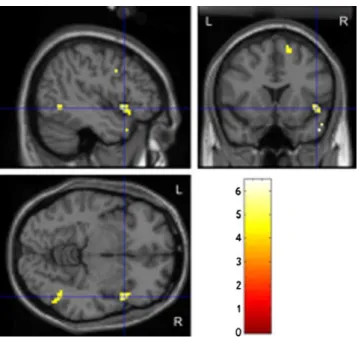
The contrast of yawn videos versus neutral videos revealed activation in the following regions: right inferior frontal gyrus (BA 9), right middle temporal gyrus, and right supe-rior frontal gyrus (Fig.1, Table1A). The contrast of yawn-ing vs. baseline showed activity in the left insula, left inferior frontal gyrus, and right middle temporal gyrus (Table 1B). The perception of neutral expression faces vs. baseline activated regions within the left and right inferior frontal gyrus (BA 45, 46, 47), left superior frontal gyrus, and right middle temporal gyrus (Table1C).
Discussion
We tested the hypothesis that the MNS is activated when persons view CY. Changes in BOLD signaling were inves-tigated in the regions attributed to the MNS while the sub-jects watched video-taped yawning faces, neutral-expression faces, and a baseline condition of static scram-bled face pictures. We found bilateral activations in regions reported to be involved in the human MNS and in face
perception (i.e., inferior frontal and middle temporal gyrus) in both dynamic-stimulation conditions (neutral expression faces and yawning faces) contrasted to the static baseline (Rizzolatti and Craighero 2004; Talairach and Tournoux 1988; Puce et al.1998). The response to faces with neutral expressions included only minimal, physiological, and smooth movements of the head, mouth, and eyes. These are motions associated with an individual who is quietly scanning the environment and whose perception activates MNS regions. This finding is in accord with that of Nahab et al. (2009), who reported MNS activation under both yawn-ing and non-yawnyawn-ing (gape and cough) conditions. The difference between our conditions of yawning and neutral faces was assumed to be the effect of contagiousness.
On the behavioral level, a contagion was indicated in approximately 55 % of the stimulations. This rate is com-parable to that described by Provine (1989) and Platek et al. (2003), whose stimuli were rated within similar settings. Because contagion is considered to be primarily an auto-matic phenomenon, with a conscious cognitive process be-ing only a secondary response, we based our evaluation on all trials rather than just those where a conscious feeling of contagion was indicated or on a comparative analysis. When we contrasted the two conditions (yawning vs. neutral), we found only right-sided activation: besides activation of the middle temporal gyrus, we found specific activation in the BA 9 portion of the right IFG and in the right superior frontal gyrus. The BA 9 is involved in higher social cogni-tive functioning such as mentalizing (Ohnishi et al. 2004). Thus, we concluded that activation of this area in our con-trast might represent the effect of contagiousness, possibly
Fig. 1 Significant activation when contrasting yawn videos with neu-tral videos (threshold T > 4.0 corresponding to voxel threshold p < 0.001, uncorrected)
Table 1 Local maxima of cerebral blood flow change for experimental contrasts (threshold p<0.001 voxel level and p<0.05, cluster level, uncorrected, with the threshold of reporting being a minimum 20 voxels; BA0 Brodmann’s Area. Talairach coordinates obtained using
Matthew Brett’s mni2tal procedurehttp://imaging.mrc-cbu.cam.ac.uk/ imaging/MniTalairach. Anatomical structures according to Talairach and Tournoux1988)
Contrast Anatomical region Talairach coordinates T-max voxel level Cluster size (voxel) x y z
A) Yawn > Neutral Right inferior frontal gyrus (BA 9) 50 7 30 6.48 28 Right middle temporal gyrus (BA 37) 42 −55 0 5.87 27 Right superior frontal gyrus (BA 6) 9 14 52 5.46 29 B) Yawn > Baseline Left insula/superior temporal gyrus (BA 13/22) −44 −14 3 9.64 326 Left inferior frontal gyrus (BA 44/9) −59 5 20 6.39 45 Right middle temporal gyrus (BA 37/39) 48 −58 1 6.12 143 Right middle temporal gyrus, sublobar 38 −51 3 5.89 27 Right middle temporal gyrus (BA 39) 46 −72 12 5.10 28 C) Neutral > Baseline Left inferior frontal gyrus (BA 45/47) −42 20 4 8.89 26 Right middle temporal gyrus (BA 21/22) 55 −47 −4 8.65 130 Right inferior frontal gyrus (BA 46) 34 37 2 7.84 28 Left superior frontal gyrus (BA 10) −18 50 20 7.84 41 Right middle temporal gyrus (BA 37) 46 −58 3 5.98 38
linking the MNS to higher cognitive functions such as cognitive empathy (Haker et al. 2010). This involvement of an area associated with higher cognitive functions, which are not developed at birth, may explain why CY is ontoge-netically seen only in later stages of a person’s development. The right hemispheric dominance for processes involving mentalizing is supported by our results and is also in accor-dance with results from neuropsychological studies on hemispheric lesions (Siegal and Varley2002). We interpret the other specific activation in the right superior frontal gyrus as representing the suppression of the urge to yawn during the experiment. Beauregard et al. (2001) reported activation of this region during a task of volitional inhibition of a comparable vegetative reaction induced by visual stim-ulation, i.e., sexual arousal. By comparison, for motor tasks such as finger movements, the temporo-parietal junction and the anterior fronto-median cortex have been identified as involved in inhibiting imitation (Brass et al. 2009). However, these movements do not elicit a vegetative urge such as yawning or sexual stimuli. Therefore, other mecha-nisms may be involved here.
The absence of MNS activation in CY has been described in previous imaging studies by Platek et al. (2005) and Schürmann et al. (2005). This might be explained because those earlier tests contrasted yawning with two other poten-tial MNS activators (Platek: laughing; Schürmann: mouth movements similar to yawning), as has already been dis-cussed by Arnott et al. (2009). The finding by the Platek group of activation in the cortical midline structures sup-ports their “empathic modeling hypothesis” of CY. This concept considers contagious yawning to be “a primitive form of empathic modeling that is subserved by substrates that are precursors to a more sophisticated and distributed system involved in conscious self-processing”, i.e., an ele-ment of cognitive empathy (Platek et al.2003). In line with Platek, we consider our evidence for BA 9 activation during CY as a bottom-up input for cognitive empathy and as a basis for such higher-level aspects of cognitive empathy, e.g., conscious self-processing or the attribution of mental states to other persons (Gallese 2007; Haker et al. 2010). Schürmann et al. (2005) have reported IFG activation in both stimulation conditions when contrasted to a baseline. Therefore, the IFG was no longer seen in the contrast between those conditions. Schürmann et al. explained this STS activation when contrasting the two conditions as evi-dence of an affinity in this region to socially meaningful cues (in this case, yawning). They conclude that“viewing another person yawn seems to circumvent the essential parts of the MNS, in line with the nature of contagious yawns as automatically released behavioral acts—rather than truly imitated motor patterns”. However, the behavioral act (i.e., the manifest yawn) did not occur during the scanning in their study either, as participants were instructed to avoid
head movements. We interpret the difference between their two conditions as the potential of the true yawn stimulus to elicit a highly stereotypical vegetative reaction based on the activation of the MNS, whereas the mere yawn-similar mouth movements lead to a comparable MNS (IFG) activa-tion that lacks this potential. After the scanning, their par-ticipants had to rate their covert tendency to yawn during the scanning. There, they indicated a greater tendency to yawn during the yawn vs. the control condition. However, their urge to imitate covertly the other mouth movements in the control condition was not reported. Based on the IFG acti-vation in the control condition, we assume that the tendency to imitate those mouth movements was also present in the control condition.
In addition to the results described here, we must also address some limitations. One might argue that the activation observed under our test conditions might have been due to participants observing mouth movements associated with yawning, such reflecting mere movement observation. However, a major function of the MNS is to copy and extract the goal of observed movements in order to behave intuitively or automatically like the person being observed. Thus, the associated activity can be interpreted as yawning-related mir-ror neuron activity because the contagious element represents yawning-associated mouth movements. With regard to the stimuli used here, we cannot deny that the yawning videos were inherently more interesting than the neutral videos. This may have influenced the level of activity observed during stimulation with yawning vs. neutral videos. However, we did not find any attention-specific differences in activation patterns under those two conditions.
Our examination was further hindered because of an essen-tial methodological issue, for which we had to ask that the subjects not perform yawning motions in order to avoid introducing any movement artifacts in the scanner. Consequently, one might argue that a motor inhibition might also have led to activation of the IFG region, particularly because both factors (motor inhibition and mirror neuron activity) may be associated with IFG activation (Rowe and Siebner2012; Bien et al.2009). However, we do not consider any possible inhibition component to be more prominent because the mirror component is essentially a presumption for the other, and the overt imitation of most mirror percep-tions in healthy adult humans is non-volitionally inhibited, leading to covert imitation (Barkley2001). Nevertheless, it is impossible to differentiate this definitely.
Another limitation may have been the task-imminent inequality between our two sets of dynamic stimuli, espe-cially that concerning the amount of biological motion. Whenever a task is designed to provide differentiated stim-uli in this way, one cannot entirely exclude the possibility that the extra activation in BA 9 under the yawning condi-tion is merely due to addicondi-tional facial mocondi-tions. Nevertheless,
BA 9 has previously been reported to be active in higher cognitive functioning (see above). The small number of participants used here (11 total) might also be regarded as a limitation because it did not allow us to perform correla-tional analyses between the activation and the contagions indicated by the participants.
Via the MNS, physiological and associated emotional states of two individuals can be shared based on perceived motor patterns (Carr et al. 2003). This so-called motor empathy or empathic resonance is one component within a multi-component model of human empathy that is adjacent to and underlies the development of cognitive and emotional empathy (Gallese2007; Preston and de Waal2002; Meltzoff and Decety 2003; Decety and Lamm 2006; Keysers and Gazzola 2007; Uddin et al. 2007; Blair 2005). Based on our results, we conclude that a connection can be demon-strated between the MNS and higher cognitive empathic functions such as mentalizing, as represented in the BA9.
In summary, we conclude that the easily observable be-havioral sign of CY is based on MNS activity and, therefore, it can be considered an expression of an individual’s em-pathic abilities. It would be interesting to study the conta-gion effect besides the behavioral level, utilizing functional imaging of patients with impairments in their empathic abilities, such as those with autism (Senju et al. 2007), psychopathy (Hagenmuller et al.2012), PTSD (Nietlisbach et al.2010), or schizophrenia (Haker and Rössler2009).
Acknowledgements We thank Mengia Dosch, Thomas Loenneker, and Ernst Martin for their support in data acquisition and analysis. Conflicts of interest The authors declare that they have no conflict of interest.
References
Anderson, J. R., & Meno, P. (2003). Psychological influences on yawning in children. Current Psychology Letters,http://cpl.revues.org/docu ment390.html. Accessed 01–12 2012.
Anderson, J. R., Myowa-Yamakoshi, M., & Matsuzawa, T. (2004). Contagious yawning in chimpanzees. Proceedings of the Biological Sciences/The Royal Society, 271(Suppl 6), S468–S470. Arnott, S. R., Singhal, A., & Goodale, M. A. (2009). An investigation of auditory contagious yawning. Cognitive, Affective, & Behavioral Neuroscience, 9(3), 335–342.
Barkley, R. (2001). The executive functions and self-regulation: an evolutionary neuropsychological perspective. Neuropsychology Review, 11(1), 1–29.
Beauregard, M., Levesque, J., & Bourgouin, P. (2001). Neural corre-lates of conscious self-regulation of emotion. Journal of Neuroscience: The Official Journal of the Society for Neuroscience, 21(18), RC165.
Bien, N., Roebroeck, A., Goebel, R., & Sack, A. T. (2009). The brain’s intention to imitate: the neurobiology of intentional versus auto-matic imitation. Cerebral Cortex, 19(10), 2338–2351.
Blair, R. J. R. (2005). Responding to the emotions of others: dissoci-ating forms of empathy through the study of typical and psychi-atric populations. Consciousness and Cognition, 14(4), 698–718. Brass, M., Ruby, P., & Spengler, S. (2009). Inhibition of imitative behaviour and social cognition. Philosophical Transactions of the Royal Society of London. Series B, Biological Sciences, 364 (1528), 2359–2367.
Carr, L., Iacoboni, M., Dubeau, M. C., Mazziotta, J. C., & Lenzi, G. L. (2003). Neural mechanisms of empathy in humans: a relay from neural systems for imitation to limbic areas. Proceedings of the National Academy of Sciences, 100(9), 5497–5502.
Cooper, N. R., Puzzo, I., & Pawley, A. D. (2008). Contagious yawn-ing: the mirror neuron system may be a candidate physiological mechanism. Medical Hypotheses, 71(6), 975–976.
Decety, J., & Lamm, C. (2006). Human empathy through the lens of social neuroscience. The Scientific World Journal, 6, 1146–1163. Gallese, V. (2003). The roots of empathy: the shared manifold hypoth-esis and the neural basis of intersubjectivity. Psychopathology, 36 (4), 171–180.
Gallese, V. (2007). Before and below ‘theory of mind’: embodied simulation and the neural correlates of social cognition. Philosophical Transactions of the Royal Society of London. Series B, Biological Sciences, 362(1480), 659–669.
Hagenmuller, F., Rössler, W., Endrass, J., Rossegger, A., & Haker, H. (2012). Impaired resonance in offenders with psychopathic traits (Empathische Resonanzfähigkeit bei Straftätern mit psychopathischen Persönlichkeitszügen). Neuropsychiatrie, 26(2). doi:10.1007/s40211-012-0015-9.
Haker, H., & Rössler, W. (2009). Empathy in schizophrenia: impaired resonance. European Archives of Psychiatry and Clinical Neuroscience, 259(6), 352–361.
Haker, H., Schimansky, J., & Rössler, W. (2010). Sociophysiology: basic processes of empathy (Soziophysiologie: Grundlegende Prozesse der Empathiefahigkeit). Neuropsychiatrie, 24(3), 151– 160.
Iacoboni, M. (2005). Neural mechanisms of imitation. Current Opinion in Neurobiology, 15(6), 632–637.
Joly-Mascheroni, R. M., Senju, A., & Shepherd, A. J. (2008). Dogs catch human yawns. Biology Letters, 4(5), 446–448.
Keysers, C., & Gazzola, V. (2007). Integrating simulation and theory of mind: from self to social cognition. Trends in Cognitive Sciences, 11(5), 194–196.
Lehmann, H. E. (1979). Yawning: a homeostatic reflex and its psycho-logical significance. Bulletin of the Menninger Clinic, 43, 123–136. Leslie, K. R., Johnson-Frey, S. H., & Grafton, S. T. (2004). Functional imaging of face and hand imitation: towards a motor theory of empathy. NeuroImage, 21(2), 601–607.
Lieberman, M. D. (2006). Social cognitive neuroscience: a review of core processes. Annual Review of Psychology, 58(1), 1. Meltzoff, A. N., & Decety, J. (2003). What imitation tells us about
social cognition: a rapprochement between developmental psy-chology and cognitive neuroscience. Philosophical Transactions of the Royal Society of London. Series B, Biological Sciences, 358 (1431), 491–500.
Nahab, F. B., Hattori, N., Saad, Z. S., & Hallett, M. (2009). Contagious yawning and the frontal lobe: an fMRI study. Human Brain Mapping, 30(5), 1744–1751.
Nietlisbach, G., Maercker, A., Rössler, W., & Haker, H. (2010). Are empathic abilities impaired in posttraumatic stress disorder? Psychological Reports, 106(3), 832–844.
Ohnishi, T., Moriguchi, Y., Matsuda, H., Mori, T., Hirakata, M., Imabayashi, E., et al. (2004). The neural network for the mirror system and mentalizing in normally developed children: an fMRI study. Neuroreport, 15(9), 1483–1487.
Palagi, E., Leone, A., Mancini, G., & Ferrari, P. F. (2009). Contagious yawning in gelada baboons as a possible expression of empathy.
Proceedings of the National Academy of Sciences of the United States of America, 106(46), 19262–19267.
Paukner, A., & Anderson, J. R. (2006). Video-induced yawning in stumptail macaques (Macaca arctoides). Biology Letters, 2(1), 36–38.
Piaget, J. (1951). Play, Dreams and Imitation in Childhood. New York: Norton.
Platek, S. M., Critton, S. R., Myers, T. E., & Gallup, G. G. (2003). Contagious yawning: the role of self-awareness and mental state attribution. Brain Research. Cognitive Brain Research, 17(2), 223–227.
Platek, S. M., Mohamed, F. B., & Gallup, G. G., Jr. (2005). Contagious yawning and the brain. Brain Research. Cognitive Brain Research, 23(2–3), 448–452.
Preston, S. D., & de Waal, F. B. (2002). Empathy: its ultimate and proximate bases. The Behavioral and Brain Sciences, 25(1), 1– 20.
Provine, R. R. (1986). Yawning as a stereotyped action pattern and releasing stimulus. Ethology, 72, 109–122.
Provine, R. R. (1989). Faces as releasers of contagious yawning: an approach to face detection using normal human subjects. Bulletin of the Psychonomic Society, 27(3), 211–214.
Provine, R. R. (2005). Yawning. American Scientist, 93(6), 532–539. Puce, A., Allison, T., Bentin, S., Gore, J. C., & McCarthy, G. (1998).
Temporal cortex activation in humans viewing eye and mouth movements. Journal of Neuroscience: The Official Journal of the Society for Neuroscience, 18(6), 2188–2199.
Rizzolatti, G., & Craighero, L. (2004). The mirror neuron system. Annual Review of Neuroscience, 27(1), 169–192.
Rizzolatti, G., & Sinigaglia, C. (2010). The functional role of the parieto-frontal mirror circuit: interpretations and misinterpreta-tions. Nature Reviews Neuroscience, 11(4), 264–274.
Rizzolatti, G., Fadiga, L., Gallese, V., & Fogassi, L. (1996). Premotor cortex and the recognition of motor actions. Brain Research. Cognitive Brain Research, 3(2), 131–141.
Rizzolatti, G., Fogassi, L., & Gallese, V. (2001). Neurophysiological mechanisms underlying the understanding and imitation of action. Nature Reviews Neuroscience, 2(9), 661–670.
Rowe, J. B., & Siebner, H. R. (2012). The motor system and its disorders. NeuroImage, 61(2), 464–477.
Schürmann, M., Hesse, M. D., Stephan, K. E., Saarela, M., Zilles, K., Hari, R., et al. (2005). Yearning to yawn: the neural basis of contagious yawning. NeuroImage, 24(4), 1260–1264.
Senju, A. (2010). Developmental and comparative perspectives of contagious yawning. Frontiers of Neurology and Neuroscience, 28, 113–119. Senju, A., Maeda, M., Kikuchi, Y., Hasegawa, T., Tojo, Y., & Osanai,
H. (2007). Absence of contagious yawning in children with au-tism spectrum disorder. Biology Letters, 3(6), 706–708. Senju, A., Kikuchi, Y., Akechi, H., Hasegawa, T., Tojo, Y., & Osanai,
H. (2009). Brief report: does eye contact induce contagious yawning in children with autism spectrum disorder? Journal of Autism and Developmental Disorders, 39(11), 1598–1602. Sepulveda, W., & Mangiamarchi, M. (1995). Fetal yawning.
Ultrasound in Obstetrics & Gynecology, 5(1), 57–59.
Siegal, M., & Varley, R. (2002). Neural systems involved in“theory of mind”. Nature Reviews Neuroscience, 3(6), 463–471.
Talairach, P., & Tournoux, J. A. (1988). Stereotactic Coplanar Atlas of Human Brain. Stuttgart: Thieme.
Uddin, L. Q., Iacoboni, M., Lange, C., & Keenan, J. P. (2007). The self and social cognition: the role of cortical midline structures and mirror neurons. Trends in Cognitive Sciences, 11(4), 153–157. Vischer, A. L. (1959). About yawning and it’s spontaneous associated
movements. Schweizerische Medizinische Wochenschrift, 89, 1356–1359.