Olfactory dysfunction in the pathophysiological continuum of dementia
Praveen Bathini
a, Emanuele Brai
b, Lavinia Alberi Auber
a,c,⁎aDepartment of Medicine, University of Fribourg, Fribourg, Switzerland
bVIB-KU Leuven Center for Brain & Disease Research, Laboratory for the Research of Neurodegenerative Diseases, Leuven, Belgium
cSwiss Integrative Center of Human Health, Fribourg, Switzerland
A R T I C L E I N F O Keywords: Olfaction Smell loss Olfactory nerve Aging Dementia Alzheimer’s disease Microbes Inflammation Olfactory diagnostics A B S T R A C T
Sensory capacities like smell, taste, hearing, vision decline with aging, but increasing evidence show that sensory dysfunctions are one of the early signs diagnosing the conversion from physiological to pathological brain state. Smell loss represents the best characterized sense in clinical practice and is considered as one of the first preclinical signs of Alzheimer’s and Parkinson’s disease, occurring a decade or more before the onset of cognitive and motor symptoms. Despite the numerous scientific reports and the adoption in clinical practice, the etiology of sensory damage as prodromal of dementia remains largely unexplored and more studies are needed to resolve the mechanisms underlying sensory network dysfunction. Although both cognitive and sensory domains are progressively affected, loss of sensory experience in early stages plays a major role in reducing the autonomy of demented people in their daily tasks or even possibly contributing to their cognitive decline. Interestingly, the chemosensory circuitry is devoid of a blood brain barrier, re-presenting a vulnerable port of entry for neurotoxic species that can spread to the brain. Furthermore, the exposure of the olfactory system to the external environment make it more susceptible to mechanical injury and trauma, which can cause degenerative neuroinflammation. In this review, we will summarize several findings about chemosensory impairment signing the conversion from healthy to pathological brain aging and we will try to connect those observations to the promising research linking environmental influences to sporadic dementia. The scientific body of knowledge will support the use of chemosensory diagnostics in the presymptomatic stages of AD and other biomarkers with the scope of finding treatment strategies before the onset of the disease.
1. Introduction
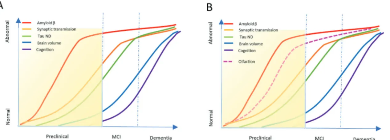
Alzheimer’s disease (AD), the most common type of dementia, is a progressive neurodegenerative pathology affecting 50 million people worldwide (World Alzheimer Reports, 2018) and no curative therapy is currently available. In the last two decades only one palliative drug, memantine, was approved, while 99.6% of the clinical trials for disease-modifying drugs have failed partially as a result of the ad-vanced staging of the patient cohorts and the reductionist focus on late phase biomarkers. Dementia has an inherent problem, its char-acteristic cognitive and executive symptoms, are typically diagnosed late in the disease progression at a time when irreversible processes have already set in (Fig. 1A). This mental disorder should be con-sidered not as one disease but as a pathophysiological continuum of sequential events starting decades before the onset of the clinical symptoms. Based on the complexity and the latency of the pathology still little is known about the processes occurring in the
asymptomatic phase. Currently, diagnosis of AD entails cognitive, psychological, functional imaging testing and confirmation by mo-lecular detection of Amyloid-β (Aβ) and tau species in cerebrospinal fluid (CSF). It is, therefore, expected that the amnestic phase of the disorder remains long underdiagnosed limiting the possibility for any reversal and restricting the eligible patient population for early therapeutics. Due to the intrinsic pathophysiological dynamic of dementia and the reductionist view of one target-one disease, we are currently lacking a proper understanding of the early mechanistic changes occurring in the pre-symptomatic stage. This is further ag-gravated by the fact that the current transgenic mouse models of AD mimic genetic AD pathophysiology but not its more frequent sporadic form (95% of the cases). The dramatic lack of reproduci-bility between rodents and humans emphasizes the need to decom-posing complex behavior and molecular events in humans to dis-tinguish between healthy and pathological aging. As such, the development of biomarkers in early stages or in preclinical AD would
⁎Corresponding author at: Swiss Integrative Center for Human Health, Fribourg, Switzerland.
E-mail address:[email protected](L.A. Auber).
http://doc.rero.ch
Published in "Ageing Research Reviews 55(): 100956, 2019"
which should be cited to refer to this work.
increase chances of interventions thereby decreasing the risk of fu-ture cognitive decline (Budson and Solomon, 2012).
The suggested framework of preclinical AD combines three main stages: 1) asymptomatic amyloidosis, 2) amyloidosis and neurode-generation and 3) development of subtle cognitive changes leading to mild cognitive impairment (Sperling et al., 2011). Interestingly, our senses (smell, hearing and vision) that allow us to communicate with the external environment, while declining naturally with aging, have been shown to be prodromal of dementia (Fig. 1B). Substantial re-search shows the presence of Amyloid-β and tau pathology in the central sensory and motor domains in AD patients with few sensory circuitries also being affected peripherally (Arnold et al., 2010;
Christen-Zaech et al., 2003;Goldstein et al., 2003;Lewis et al., 1987).
Despite the numerous clinical studies indicating that sensory dys-function precedes cognitive loss, the generalized sensory impairment in several forms of dementia, has refrained the investigation of a mechanistic relationship between sensory and cognitive deficit leaving these studies mainly observational and correlative. The nasal neuroepithelium and the eye are the only CNS appendices in direct communication with the external environment without an interlining blood brain barrier (BBB), therefore representing a susceptible port of entry for exogenous toxins to the brain. This hypothesis is supported by the accumulation of microbial species in AD brains (Itzhaki et al., 2016). Furthermore, in these areas, host-pathogen interactions gain particular importance as peripheral inflammatory responses are pro-pagated to the CNS without anatomical impediment. Noteworthy, several gene variants, including Apolipoprotein E type epsilon 4 (ApoEε4), identified as risk factors for AD, play a major role in in-flammatory responses (Zhang et al., 2011), suggesting a common mechanistic link between environmental adaptive processes and net-work dysfunction that could unfold through the sensory organs. While vision and hearing are just beginning to emerge as clinically relevant for AD, chemosensory impairment is established in AD foreplay and AD progression.
This review focuses on olfactory impairment as preclinical indicator of dementia and contains the most updated collection of clinical and preclinical studies treating olfactory processing in the context of brain aging and neurodegenerative diseases. The aim of this manuscript is to provide the foundation for associating the infectious hypothesis of AD to early chemosensory impairment (Fig. 2) and emphasize the great
potential of olfaction as prognostic non-invasive behavioral signature to classify preclinical AD.
2. Anatomy of the olfactory system in AD
2.1. Primary olfactory area 2.1.1. Nasal cavity
The nose exerts respiratory and olfactory function and is also in-volved in taste through the nasopharynx connecting the mouth. The nasal cavity is structured in highly vascularized cartilaginous turbinates covered by respiratory epithelium rostrally and olfactory epithelium (OE) dorso-posteriorly (Fig. 2). The contraction of the turbinates through the nasal valve increases the airflow pressure facilitating the passage to the lungs, while the cilia serve as a filtering system for air particulates and microbial species. The depletion of respiratory cilia enhances microbial infections, altering inhalation and smell (Cohen, 2006). Odorants reach the olfactory epithelium through the nares or the nasopharynx and get first trapped in the mucus layering this neuroe-pithelium (Pelosi, 1994). The mucus secretions produced by the inter-lining Bowman’s glands are rich in glycoproteins and contain odorant-binding proteins, growth- and immune- factors supporting the integrity and the odorant response of the olfactory sensory neurons (OSN)
(Federico et al., 1999). Furthermore, the mucus creates a barrier to
xenobiotics favoring the colonization of resident microbial species, which are involved in OE turnover and the efficiency of the odorant responses (François et al., 2016). Cholinergic and adrenergic efferents innervate the OE and regulate the secretory activity of Bowman’s gland
(Zielinski et al., 1989). As one of the first neurotransmission deficits in
AD concerns the adrenergic afferents to the secretory gland (Grudzien
et al., 2007), it is expected that mucus alteration may play out early in
disease and could explain the discrepant results of an olfactory threshold dysfunction in mild cognitive impairment (MCI) (Kareken
et al., 2001; Djordjevic et al., 2008). Nevertheless, to date no
in-vestigation of the mucus biological content has been conducted in AD. Whereas, it has been demonstrated that patients with Parkinson’s dis-ease (PD) (Friedman et al., 2008) and amyotrophic lateral sclerosis (ALS) display a change in mucus composition (Federico et al., 1999), likely affecting the functional integrity of the OE.
Fig. 1. Dynamic biomarkers’ model underlying the progressive staging of dementia. A) Graphical sketch of the typical progression of known biomarkers in progression of dementia, with the yellow area indicating the preclinical phase. B) Graphical sketch supplemented with chemosensory decline detectable in the preclinical phase of dementia. Redrawn from Alzheimer‘s and Dementia Vol 7–3, 2011.
2.1.2. Olfactory neuroepithelium
Besides the olfactory sensory neurons projecting to the brain, the OE receives afferents from the trigeminal nerve and the nervus terminalis
(Fuller and Burger, 1990). The nervous efferents and afferents of the OE
represent a sink for microbial species that can be propagated to the brain causing a neuroinflammatory response associated with the de-velopment of dementia (Fig. 3A) (Fülöp et al., 2018). Interestingly, bacterial and viral species have been found in the entorhinal cortex (EC), which is part of the olfactory circuitry. In the OE, sustentacular cells are intercalated between the OSN forming a protective and sup-portive apical monolayer with regenerative capacity. Sustentacular cells have glial-like properties and their renewal is promoted by pro-liferating globose cells progenitors (Chen et al., 2014). Head trauma or viral infection can temporarily induce the proliferation of the basal cells population (globose and horizontal basal cells) warranting reconstitu-tion of the OE after injury. Nevertheless, when the insult is protracted the chronic inflammatory reaction impairs OE basal proliferative re-sponses and the function of OSN, leading in the worst case to OE atrophy and anosmia (Choi and Goldstein, 2018). The progressive loss of olfactory function with age-related neurodegeneration has been at-tributed to the recurrent insults over a lifespan and the progressive decline in proliferative capacity of senescent stem cells, compromising OE integrity (Witt et al., 2009). Each olfactory sensory neuron expresses a specific G-protein-coupled receptor on its apical cilia, embedded in the nasal mucus layer, that traps the odorants and increases its binding to cognate olfactory receptors. Ciliary defects as result of genetic mu-tations in the Bardet-Biedl syndrome-gene family members (Kulaga
et al., 2004;Iannaccone et al., 2005) or the gene Centrosomal protein
290 (McEwen et al., 2007) cause a complete loss of smell. Moreover, long-term exposure to xenobiotics or chemicals can cause a ciliopathy contributing to the decline in smell with aging (Goncalves and
Goldstein, 2016). Furthermore, cilia have been implicated in retrograde
transport of several virus species (adenovirus, herpesvirus, poliomye-litis, influenza A and rabies) to the brain (Doty, 2008;Mori et al., 2005) and ablation of the OE or olfactory bulb (OB) in experimental monkeys prevents the entry of the poliomyelitis virus in the CNS (Brodie and
Elvidge, 1934). The access of virus into the CNS through the olfactory
route is supported by several studies in animals using intranasal in-oculation of influenza, herpes and Nipah viruses to study their life cycle and develop vaccine carriers (Bodewes et al., 2011;Plourde et al., 2012;
Schrauwen et al., 2012;El-Habashi et al., 2010;Munster et al., 2012;
Quinn et al., 2011; van Riel et al., 2015). These studies in animal
models are corroborated by increasing reports on the herpesviridae load in the brains of demented patients (Haas and Lathe, 2018;Eimer
et al., 2018;Itzhaki, 2014). Further, a damaged olfactory epithelium is
permissive to entry of bacterial species to the olfactory bulb within hours curtailing any protective inflammatory response from macro-phages of the OE and olfactory ensheathing cells of the olfactory nerve layer (ONL) (Herbert et al., 2012). Further, Amyloid-β aggregates have been reported to accumulate in the olfactory mucosa (OM) from MCI patients (Ayala-Grosso et al., 2015) (Fig. 3B), where the formation of Amyloid-β peptides may result from the exposure to microbes (Hill and
Lukiw, 2015) building an antibacterial biofilm (Spitzer et al., 2016).
These aggregation-prone peptides interfere with OE turnover, likely affecting olfactory transmission (Ayala-Grosso et al., 2015).
In humans, there are about 100–200 different G-protein-coupled olfactory receptors, which are considerably lower than the number of encoding genes (500–600). However, different odorants can activate spatially distinct olfactory receptors (ORs) and the combinatorial affi-nity of odors to their receptors allow humans to discriminate more than 10′000 odors (Purves et al., 2001a). OR transduction efficiency through c-AMP determines odor threshold sensitivity. With aging, OR expres-sion changes (Verbeurgt et al., 2014) and senescent OR have a dete-riorated c-AMP signal transduction, which may explain the age-de-pendent decline in odor threshold (Purves et al., 2001b).
2.1.3. Olfactory nerve layer
Each olfactory sensory neuron projects its axons to the olfactory bulb in bundles through the cribriform plate cavities (Fig. 3A). Occlu-sion of these foramina occurs naturally with aging and interrupts the olfactory signal transmission from the OSN to the OB affecting the sense of smell (Kalmey and Thewissen, 1998). The olfactory sensory nerve terminals project to the glomerular region of the olfactory bulb main-taining a spatial odorant map defined by the odor class specificity (Mori
et al., 1999). After axonal resection of the ONL, as in traumatic brain
injury (TBI), rewiring between the OE and the OB can be only partially re-established, since odorant map specificity is compromised altering the sense of smell in the long-term (Costanzo, 2005).
2.2. Secondary olfactory area 2.2.1. Olfactory bulb
The olfactory bulb is the rostral enlarged appendix of the olfactory tract (Fig. 3A). Imaging studies report a progressive reduction in OB Fig. 2. Anatomy and neuropathological processes of the olfactory system in Alzheimer’s disease. Gross anatomy of the nose, the olfactory nerve and the olfactory bulb, reported neuropathology, known pathogenic risk factors, genetics of the host (red) and type of deficits manifests in the early phases of AD. OSN = olfactory sensory neurons, DA = dopaminergic, OT = olfactory tract, PC = piriform cortex, AMG = amygdala, EC = entorhinal cortex, OFC = orbitofrontal cortex, PM = particulate matter with a diameter below 2.5 μm and UFPM = ultrafine particulate matter.
volume in AD patients from the MCI stage on (Ridha et al., 2008;
Thomann et al., 2009) as well in PD subjects (Mueller et al., 2005;
Hummel et al., 2010). The change in mass is associated to a gradual
tauopathy in this region (Kovacs and Cairns, 1999; Attems and
Jellinger, 2006), but a loss in cell density could not be confirmed
(Hummel et al., 2010;Gómez-Isla et al., 1997;Kril et al., 2002),
sug-gesting neuronal network breakdown as the main cause for atrophy. At the tip of the olfactory bulb, olfactory sensory neurons and the apical dendrites of mitral cells converge in a synaptic hallow zone called the glomerular region. In histological studies, this area can be localized using the olfactory marker protein filling the glomeruli or Hematoxylin-Eosin staining showing circular disposed cells. Periglomerular GA-BAergic and DAergic neurons innervate the dense synaptic regions to modulate the strength of the incoming signal and further tune the specificity of the odorant map (Lledo et al., 2004). The fate of the bulbar GABAergic interneurons in humans remains largely unaddressed
(Saiz-Sanchez et al., 2016). On the other hand, DAergic periglomerular
neurons are increased in number in AD, PD and frontotemporal de-mentia (FTD) (Mundiñano et al., 2011), suggesting a possible ex-citatory-inhibitory network dysbalance in the pathogenesis of olfactory dysfunction. This hypothesis is further supported by studies showing alteration in cholinergic (Kovacs et al., 1998; Lehéricy et al., 1993;
Ruberg et al., 1986;Rudi et al., 2014), serotonergic (Mössner et al.,
2000; Yang and Schmitt, 2001) and noradrenergic (Grudzien et al.,
2007; German et al., 1992;Weinshenker, 2008;Zarow et al., 2003)
innervations to the OB in AD, PD and FTD patients. Those corticofugal fibers from the forebrain (Horizontal limb of the diagonal band of Broca; HDB) and hindbrain nuclei (Locus coeruleus and Raphe Nuclei) typically modulate the excitability of mitral cells through inhibitory potentiation (Nai et al., 2009; Nai et al., 2010). APP/PS1 transgenic mice show locus coeruleus degeneration which aggravates odor dis-crimination and memory (Rey et al., 2012). Mechanistically, early functional corticofugal denervation depresses mitral cells excitatory tone, which interferes with synaptic transmission and signal transduc-tion efficiency. Aberrant excitatory transmission has been observed in AD rodent models (Palop et al., 2007) as well as AD patients (Palop and
Mucke, 2009;De Simone et al., 2010). In addition, our recent study
demonstrates augmented activity-dependent signaling in mitral cells of MCI patients, which then dissipates with the severity of dementia
(Bathini et al., 2018). Progressive network breakdown is supported by
other studies showing a gradual proteinopathy that evolves early on in the OB and is mainly characterized by hyperphosphorylated tau neu-rofibrils and tangles (Bathini et al., 2018;Lachén-Montes et al., 2017). The Amyloid-β pathology remains largely diffused at glomerular level
(Fig. 3C) (Mundiñano et al., 2011;Bathini et al., 2018) suggesting that
intracellular oligomerization may reflect microbial transformation and cell-to-cell transport. Indeed, trans-synaptic propagation of proteotoxic species along the olfactory nerve could accompany and mimic the trajectory of microbial species from the olfactory bulb to central limbic structures as the entorhinal cortex and hippocampus (Rey et al., 2018). In rodents, granule and periglomerular interneurons are con-tinuously regenerated also in adult life. In humans, neurogenesis in the olfactory bulb remains a matter of debate (Bergmann et al., 2012,2015) and interneurons of this region have been marginally characterized. Only a couple of reports indicate that somatostatin and secretogogin interneurons decrease in number in the olfactory bulb and tract with the onset of dementia (Saiz-Sanchez et al., 2010;Attems et al., 2012). It is unclear whether other GABAergic populations of Calbindin, Parval-bumin and Calretinin neurons are affected. However, if the vulner-ability of these populations is confirmed, the partial shunting of feed-back inhibition would result in de-synchronization of mitral cell firing and an olfactory transmission deficit to higher brain areas.
2.2.2. Olfactory tract
From the olfactory bulb, mitral cells send their long axonal pro-jection in a bundle forming the olfactory tract (OT) connecting the
olfactory bulb to higher olfactory areas (Fig. 3A). Axonal loss as in-dicated by a 40% decrease in OT cross sectional areas and 52% loss in myelinated axons has been previously reported in AD patients (Davies
et al., 1993). The alteration in structural integrity of the OT, was also
detected in our study showing an abrupt decrease in neurotic varicosity already in MCI patients (Bathini et al., 2018).
In the olfactory bulb and tract, the anterior olfactory nucleus (AON) is subdivided in pars bulbar (AONb), interpeduncular (AONi), retro-bulbar (AONr). Developmentally, those nuclei are extravagination of the olfactory cortex, AONc (pars corticalis), with layered principal and GABAergic neurons and a progressive proteinopathy characterized by abundant neurofibrillary tangles (NFTs) and small core plaques in AD
(Kovacs and Cairns, 1999;Bathini et al., 2018;Hyman et al., 1984;Ter
Laak et al., 1994). Despite not completely clarified, these intercalating
structures receive projections from mitral neurons and function as in-termediate signal integrator (Brunjes and Kenerson, 2010). The pro-nounced proteinopathy in this region suggests that its activity is sig-nificantly impaired in the progression of dementia (Fig. 3D).
2.3. Olfactory cortices 2.3.1. Olfactory tuberculum
The olfactory tract splits at a bifurcation point called the olfactory tubercle which triages the axons directed to the fornix (medial stria) or the piriform cortex, entorhinal cortex and amygdala (lateral stria)
(Fig. 3A). With aging, neurofibrillary tangles accumulate in the
olfac-tory tuberculum compromising the long-range olfacolfac-tory connectivity
(Hyman et al., 1984).
2.3.2. Piriform cortex
The majority of the lateral olfactory tract axons project to the two-layered piriform cortex (PC) (Fig. 3A), which represent a sensory-as-sociative center, where odor perception is shaped. Removal of the temporal lobe, often occurring in epileptic patients, affects odor per-ception but not odor threshold (Gottfried, 2010). Odor coding in the piriform cortex is sparse and results from the integration of excitatory-inhibitory circuitry located rostrally (Anterior Piriform cortex; APC) to caudally (Posterior Piriform cortex; PPC). This structural organization determines the spatial and temporal encoding of odors in dispersed rather than discrete ensembles (Rennaker et al., 2007). This organiza-tion allows multi-component odors to be sparsened through local feed-forward inhibition into a dominant encoding of the most abundant perceived odor (Poo and Isaacson, 2009) and explains why structurally similar odors are often perceived as different based on the discrete neuronal ensemble recruited in the PC (Gottfried, 2010). Further, a pioneering functional magnetic resonance imaging (fMRI) cross-adap-tation study, indicates that the function of the piriform cortex is spa-tially dissociated in a structure-encoding area (APC) and quality-en-coding area (PPC) (Gottfried et al., 2006). MCI patients are unable to qualitatively and categorically distinguish odors during odor identifi-cation and discrimination tasks as a result of a disorganization of quality-coding signal strength after repetitive stimuli (Li et al., 2010a). Sensory perception interference may be attributed by different patho-physiological mechanisms ongoing in this region i) the synaptic loss
(Arendt, 2009;Giannakopoulos et al., 2009), ii) the observed changes
in GABAergic population distribution (decreasing number of calretinin and somatostatin neurons in contrast to increasing parvalbumin cells) and iii) associated tau and Amyloid-β pathology evident with the pro-gression of dementia (Fig. 3E) (Saiz-Sanchez et al., 2015). Interestingly, in the Tg2576 animal model of AD, while odor memory is impaired, odor perception is spared suggesting that higher order quality dis-crimination in human may be more susceptible to early network changes independently of the proteinopathy (Xu et al., 2014). Along these lines, treatment with scopolamine in rats reduces olfactory per-ceptual memory (Wilson, 2001), suggesting that in humans early per-turbations of cholinergic innervation to the piriform cortex could result
in an odor perception deficit (Geula and Mesulam, 1996). From the PC, monosynaptic afferents reach the orbitofrontal cortex (OFC), which is involved in odor discrimination learning/recognition and multisensory integration (Gottfried, 2010). An older report described a generalized damage and tau pathology in the OFC of AD patients (Van Hoesen et al., 2000). A spatial topography study confirmed that the OFC undergoes aging-related atrophy (Bakkour et al., 2013), confirming aging as a critical factor in functional olfactory deterioration (Doty and Kamath,
2014).
2.3.3. Amygdala
The bypass lateral olfactory tract (LOT) axons further project to the amygdala (AMG) (Fig. 3A). Regional cerebral blood flow PET study in humans indicated that the AMG is recruited bilaterally at exposure of highly aversive stimuli, along with the OFC (Zald and Pardo, 1997). The activity in the AMG is associated with emotional processing of per-ceived aversiveness of odors, which is in line with the involvement of this limbic structure in negative emotional processing (Halgren, 1982). Neuropathological examination has shown a pronounced tauopathy and cell loss in the AMG with AD progression (Arriagada et al., 1992;
Scott et al., 1992;Vereecken et al., 1994). A more recent neuroimaging
study indicated that AMG atrophy is prominent in early AD and relates to the severity of the symptoms (Poulin et al., 2011). The loss in cell mass profoundly compromises olfactory nociception triggering a typical flight response and puts these individuals at risk of exposure to household toxic volatile substances as CO2 and fire.
2.3.4. Entorhinal cortex
The most posterior monosynaptic connections of the LOT end in the entorhinal cortex (EC) (Fig. 3A), organized in 6 cortical layers. Studies in guinea pigs using peristimulus responses indicate that the lateral EC is the target of olfactory inputs from the LOT and associative afferents from the piriform cortex, whereas the medial entorhinal cortex is in-nervated by hippocampal relay fibers (Biella and de Curtis, 2000). In humans this connectivity is conserved with bidirectional afferents (layer I/II) and efferents (layer II/III) from and to the OB, the PC, the AMG and hippocampus (Cleland and Linster, 2003). The EC mediates odor discrimination memory, as demonstrated by alteration in olfactory recognition in animals with aspiration lesion in this area (Stäubli et al.,
1984;Wirth et al., 1998). Further, inhibitory projections from the
en-torhinal cortex to the piriform cortex contribute to the top-down phasic inhibition and tuning of the piriform cortex resulting in fine odor dis-crimination ability of similar but not distinct odors (Chapuis et al., 2013), which is a trait affected early on in AD. This is consistent with the stereotypical, layer II (Stranahan and Mattson, 2010), tauopathy with the progression of AD, which is evident already at Braak stage II
(Braak et al., 2006;Braak and Del Tredici, 2015). Amyloid-β
deposi-tions remain scant in the human EC (Braak and Braak, 1991a) sug-gesting that other mechanisms contribute to the fine odor discrimina-tion deficit in the early stages of AD (Wilson et al., 2014). That said, olfactory discrimination and identification, which depends on the in-tegrity of the PC to EC and OB network remains an important biometric to distinguish normal from pathological brain aging (Murphy, 1999;
Bahar-Fuchs et al., 2011; Rahayel et al., 2012). Further, the
con-nectivity of the EC to the hippocampus and AMG explains why odorant exposure may trigger a vivid emotional memory, which is engraved in the subject’s history (Mouly and Sullivan, 2011). Besides the long term odor memory, psychophysical testing establishes also the presence of short-term odor memory, as smaller number of odours can be better remembered than a larger panel of odour 2–3 weeks from the first ex-posure (Schab, 1991). Functional magnetic resonance imaging (fMRI) studies indicate that epileptic patients with temporal lobe lesions show impairment in olfactory memory (Eskenazi et al., 1986). In AD, inter-ference with EC connectivity as a result of the prominent temporal tauopathy impairs odor recognition as evidenced by the investigation of olfactory identification/discrimination in the prediction of dementia
(Devanand et al., 2015).
2.4. Thalamocortical olfactory relay
Odors are processed to conscious odor discrimination and memory through bidirectional projections from the olfactory cortices to/from the thalamocortical relay consisting of the medial dorsal thalamus (MDT) and the OFC. These regions are involved in multisensory in-tegration, attention and conscious sensory perception.
2.4.1. Mediodorsal nucleus of the thalamus
The MDT receives afferents from the olfactory cortices, the olfactory tubercle and innervates the OFC, while it has feedback connections to the piriform cortex (Fig. 3A). Stimulation of the lateral olfactory tract in animals or the exposure to a variety of odors can induce electrical re-sponses in the MDT in phase with the β-oscillation of the PC supporting the reciprocal connectivity of these areas (Courtiol and Wilson, 2014). Lesions to the MDT affect odor preference in animals altering sexual behavior (Sapolsky and Eichenbaum, 1980). Similarly in humans, da-mage to the MDT as a consequence of hemorrhage or ischemia impairs hedonic judgements (Sela et al., 2009). Furthermore, MDT lesions in rats impair olfactory memory and learning (Slotnick and Risser, 1990) and reward association related to odors (Kawagoe et al., 2007) but not visual discrimination, which suggests a specialized function for the MDT in olfactory information coding (Tham et al., 2011a). Based on the role of the thalamus as the gateway of attention, MDT appears to modulate odor attention processing which allows for fine olfactory discrimination and identification of complex mixtures (Tham et al.,
2009; Zelano et al., 2011). A study addressing the role of the MDT
pathways indicated that patients with MDT lesions perform well in a general olfactory test, but show impairment in olfactory attention and olfactory naming (Tham et al., 2011b). Imaging studies demonstrated that attention to odors increases the functional connectivity of the ol-factory cortices-DMT-OFC network (Veldhuizen and Small, 2011;
Plailly et al., 2008). These two works support a role of MDT in
pre-diction error signaling where the magnitude of the MDT responses is driven by the expectation of odor stimuli (Zelano et al., 2011). Overall, the studies in animals and humans indicate that MDT executes high-order functions by converting external olfactory stimuli into conscious awareness of odorants through attention. While a variability in neuro-pathological hallmarks was observed in the MDT of AD patients with a more pronounced tauopathy as compared to senile plaques (Paskavitz
et al., 1995;Braak and Braak, 1991b), MRI analysis reveals a robust
thalamic degeneration in Alzheimer’s patients (Zarei et al., 2010;
Vasavada et al., 2017). Morphometry studies indicate that a reduction
in thalamic volume is observable already in amnestic MCI (Chételat
et al., 2005;Sorg et al., 2007;Yi et al., 2016) and the grade of thalamic
shrinkage correlates with the cognitive status (Pedro et al., 2012), suggesting thalamic atrophy as a diagnostic biomarker for conversion from MCI to AD. Furthermore, hypometabolism in the medial thalamus is associated to early decline in executive functions (Reinvang et al., 2012) revealing the relevant connectivity of this region to the frontal cortices. The volumetric changes in the olfactory projection areas support that thalamic relays dysfunction also contributes in the olfac-tory discrimination and olfacolfac-tory memory deficit observed in the pre-clinical phase of AD.
2.4.2. Orbitofrontal cortex
The OFC is characterized by 4 broad giri, with reciprocal connec-tions with the olfactory tubercle, the MDT and all primary olfactory areas, including the piriform cortex, amygdala and entorhinal cortex, in the absence of an obligatory thalamic relay (Fig. 3A). Non-overlapping regions of the OFC receive sensory input from gustatory and visual centers, as well as information about visceral states, providing a neural substrate for associative learning and cross-modal integration. In non-human primates, the posterior OFC has been identified as the site of
olfactory perception (Gottfried, 2006). On the other hand, in humans there is a spatial dissociation between the caudal aspect of the OFC, associated to low-level olfactory processing such as passive smelling and odor detection and its rostral portion devoted to higher order ol-factory computation related to associative learning, and odor recogni-tion memory (Gottfried and Zald, 2005). A further topological distinc-tion across the medial lateral axis has been observed in accordance to the pleasantness of the odor with the medial portion of the OFC en-coding hedonic odor stimuli and the lateral OFC, unpleasant ones
(Gottfried et al., 2002;Anderson et al., 2003;Rolls et al., 2003). The
valence-dependent medio-lateral pattern of the OFC is conserved for other sensory modalities (Gottfried, 2006; Small et al., 2001;
OöDoherty et al., 2003). The OFC is the primary sensory neo-cortical
area, participating in a wide variety of complex olfactory functions related to multimodal integration, reward processing, olfactory con-sciousness and goal-directed learning and behavior (Gottfried, 2006;
Schoenbaum and Eichenbaum, 1995). The OFC has unique role in
sensory convergence of odor/taste and odor/vision and semantic cor-respondence between odors and tastes or between odors and pictures determined also by the anatomical topology (Gottfried, 2006). Orbito-frontal lesions following post-traumatic injury are accompanied by ol-factory perception/identification deficits (Jones-Gotman and Zatorre,
1988; Potter and Butters, 1980) underlining the role of the OFC in
conscious olfactory experience and multimodal sensory-semantic
integration. A more recent work studied an anosmic subject with OFC lesion after TBI using brain imaging, autonomic recordings and olfac-tory psychophysics and demonstrated that despite the “blind smell” responses registered in the OFC, the subject was completely lacking conscious olfactory perception (Li et al., 2010b). In AD patients, the OFC is characterized by an abundant tauopathy with NFT in layer III and V containing projection neurons to olfactory cortices (Van Hoesen
et al., 2000). The NFT burden correlates with agitation in AD patients
(Tekin et al., 2001). More recently, a paper using positron emission
tomography (PET) with 11C-pyridinyl-butadienyl-benzothiazole 3 (11C-PBB3) and 11C-Pittsburgh compound-B (11C-PiB), tracers for tau and Aβ respectively, indicated that elevated tau accumulation in the OFC positively correlates with OFC atrophy and apathy scale scores in the patients, while Aβ did not show any significant interaction (Gordon
et al., 2018). The sparse Amyloid-β pathology in the OFC is confirmed
in Tg2576 mice at 16 months (Wesson et al., 2010). Topography cross-sectional studies indicate that incipient AD patients show a 20–30% grey matter loss in the OFC, which becomes less significant with the progression of the disease when temporal atrophy is dominant (Frisoni
et al., 2009). This may explain the early sensory deficit observed in the
prodromal phase of AD, which is later overruled by the cognitive phenotype at the MCI and moderate stage of AD. While the OFC has been widely studied in FTD for its role on executive function, this area remains less explored in AD and further investigation is required to Fig. 3. Olfactory circuitry with typical hallmarks of AD. A) Magnified drawing of the olfactory network superimposed to a half brain hemisphere shows: the peripheral olfactory neuroepithelium in which olfactory sensory neurons (red) are embedded; the olfactory nerve layer projecting to the glomeruli (pink) where synaptic connection with mitral cells (red) are established; mitral cell axons forming the olfactory tract project and bifurcate their monosynaptic terminal either to the fornix or to the olfactory cortices (piriform cortex, entorhinal cortex, amygdala; black). The olfactory cortices are connected viareciprocal connections (dotted blue double arrowed lines) with second order structures (blue), the medial dorsal thalamus and the orbitofrontal cortex; the forebrain nucleus of the Horizontal limb of the diagonal band of Broca and the hindbrain nuclei of the Locus coeruleus and Raphe nucleus send their corticofugal projections to the olfactory bulb. B) Aβ42 aggregates in the olfactory neuroepithelium. C) Aβ42 positive diffused plaques in the glomerular region. D) Tauopathy (pTau) in the olfactory tract. E) Core plaques Aβ42 positive in the entorhinal cortex. OE = olfactory epithelium, ONL = olfactory nerve layer, CP = cribriform plate, OT = olfactory tract, OB = olfactory bulb, GL = glomeruli, AMG = amygdala, EC = entorhinal cortex, PC = piriform cortex, HDB= Horizontal limb of the diagonal band of Broca, MDT = medial dorsal thalamus, OFC = orbitofrontal cortex.
understand the involvement of the OFC in the early olfactory pheno-type.
2.5. Host-pathogen interactions in the olfactory route
2.5.1. The olfactory vector hypothesis in neurodegenerative diseases
Based on the well documented early olfactory deficit in a variety of neurodegenerative diseases and the accessibility of the olfactory system to the external environment and exogenous species, an olfactory vector hypothesis has been put forward since more than two decades. Environmental factors such as bacteria, viruses, toxins can enter through the airways and disseminate to the brain in the absence of a BBB and also through the subarachnoid space which expands into the nasal cavity. Microbial species, such as Herpes viridae (Itzhaki et al.,
1997; Jamieson et al., 1991), Spirochete (Miklossy, 2008a; Miklossy
et al., 1994), and Chlamydiaspecies (Balin et al., 1998), have been found
in the olfactory nerve and connected limbic structures suggesting that those species infecting the respiratory tract can enter the brain through the nasal cavity, triggering neuroinflammation and inducing the pro-duction and seeding of Amyloid-β species for microbicidal purposes
(Eimer et al., 2018;Miklossy, 2016). A continuously expanding
litera-ture shows that viruses and bacteria inoculated through the nose in rodents can upregulate Aβ processing/deposition (Eimer et al., 2018;
Wozniak et al., 2007;Moir et al., 2018;Wozniak et al., 2009;Yount
et al., 2006; Little et al., 2004, 2014) through upregulation of
β-se-cretase and nicastrin (a protein component of the γ-seβ-se-cretase complex)
(Wozniak et al., 2011) and/or via interfering with Aβ intracellular
trafficking and clearance (Cheng et al., 2011;Shipley et al., 2005). In AD patients, the diffused Aβ pathology in the glomerular region pos-sibly results from microbial infection spreading inter-neuronally through the ONL to the glomeruli (Bathini et al., 2018). Amyloid-β peptides have agglutinating properties forming a microbicidal trap likely and limiting the pathogen’s dissemination ability (Spitzer et al.,
2016; Kumar et al., 2016). On the other hand, a recent contrasting
study has advanced that fibrillar Aβ covering the viral corona increase the infectivity of the pathogen to the brain (Ezzat et al., 2019). While these latter results await confirmation, the overall consensus is that fibrillary Aβ represents the first innate response to viral and bacterial infection exerting a double sword effect in protection from microbes at the expense of neuronal damage. In addition, microbes in the brain can induce pro-inflammatory reactions with the release of interleukins (IL-1β, IL-6, IL-8) and tumor necrosis factor-α (TNF-α) (Lokensgard et al., 2001), which further contribute to neural network breakdown also supported by the prominent and progressive tau pathology with AD.
2.5.2. Herpesviridae
In contrast to the punctual host-invasions of bacterial species, Herpesviridae infections are life long and represent one of the most common infectious diseases, with 90% of the adult population positive for HHV-6 antigens and 60% for HSV-1 and HSV-2. The latter reside in the body in a latent phase with herpetic outbreaks in 25–40% of the cases suggesting intraindividual immunological variability. After the first colonization, HSV-1 remains in a latent form in the sensory ganglia, with outburst in condition of stress or immune repression
(Itzhaki and Wozniak, 2008). Herpesviridae infections are associated
with the production of Amyloid-β species that mediate antimicrobial activity through the binding of viral glycoproteins expressed on the surface of infected neurons in vitro (Eimer et al., 2018). This innate immune response explains how viral-induced β-amyloidosis exacerbate the neurotoxicity following infection. Interestingly, ApoEε4 is posi-tively associated with HSV-1 infections and cold sores in humans
(Itzhaki et al., 1997) and ApoEε4 carriers have increased cerebral load
of HSV-1 (Burgos et al., 2006) with a greater risk for females than males mice (Guzman-Sanchez et al., 2012). These data, in addition to the evidence that ApoEε4 accelerate early seeding of Aβ (Liu et al., 2017), support a functional interaction between ApoEε4 and the innate
immune reaction to viral infections. Microbial traces in the brain of elderly AD patients emphasize that these species are kept under im-munological control until senescence when HSV-1 reactivation from latency is accompanied by upregulation of neuroinflammatory (toll-like receptor-4, interferon α/β, and p-IRF3) and early neurodegenerative markers (phospho-tau and TauC3) (Martin et al., 2014). A recent study integrating genomic, transcriptomic, proteomic and histopathological data indicated a functional interaction between the presence of HSV-1, HSV-2, HHV-6A and HHV-7 in the prefrontal cortex and hippocampal regions and the incidence of dementia (Jamieson et al., 1991;Readhead
et al., 2018). Previously HHV-6A and HHV-6B have been shown to have
a tropism for astrocytes in vitro with lytic and nonlytic activity for the two viruses respectively (Donati et al., 2005). When marmoset were intranasally inoculated with both strains, traces of HHV-6B, but not HHV-6A were observed in saliva, blood and in the frontal cortex/ol-factory bulb and hippocampus, causing local neuroinflammation and experimental autoimmune encephalomyelitis (Leibovitch et al., 2018). From autopsy brains, HHV-6 shows preferential tropism for the bulb/ tract and both HHV-6B and HHV-6A are generally found in the nasal mucus and saliva from healthy individuals, patients with multiple sclerosis and anosmic subjects (Harberts et al., 2011), independently of the neurological condition. To validate the olfactory route hypothesis of CNS invasion, the same group has confirmed the astrogliosis tropism by infecting with HHV-6A and HHV-6B primary human olfactory en-sheathing cells (OEC) typically surrounding the ONL. HHV-6A but not HHV6-B could productively infect the OEC and cause an inflammatory response. Despite the discrepancy of findings between the animal and human specimen, the presence of HHV-6A or HHV-6B in the olfactory system is well documented, whereas it remains to be established whe-ther one or both strains are causative for olfactory-mediated patho-genesis of AD.
2.5.3. Chlamydia pneumoniae
Among the airways bacterial species, Chlamydiapneumoniae (C.
Pneumoniae), has been found in 90% of AD brains while only 5% of the
control brains were tested positive. C. Pneumoniae attaches to the nasal epithelium and neuroepithelium and can spread to the brain through the OB, where bacterial traces were found (Balin et al., 1998). To confirm the intranasal spread of the airborne C. Pneumoniae to the brain, a study showed the presence of C. Pneumoniae antigens along with Amyloid-β plaques and astrogliosis in the brain of Balb/c mice, 6 months after intranasal infection (Little et al., 2004,2014). As for HSV-1, the load of C. Pneumoniae in the Alzheimer’s brain varies with APOE genotype with ApoEε4 carriers showing higher copy numbers of C.
Pneumoniae as compared to subjects lacking the risk variant (Gérard
et al., 2005). Chlamydial LPS and other membrane proteins induce the
secretion of proinflammatory cytokines (IL-6 and MCP-1) and reactive oxygen species from astrocytes with neurotoxic activity (Boelen et al.,
2007).
2.5.4. Spirochete
Spirochetes, such as Borrelia Burgdorferi (B. burgdorferi) and
Treponema denticola, cause latent or persistent infection throughout life
(Miklossy, 2008b). With characteristic neurotropism, they can spread to
the brain through the blood, lymphatic system and peripheral as well as central nerves including the olfactory tract (Mann et al., 1988;
Miklossy, 2011). A meta-analysis has indicated an odd ratio between
100–300% of identifying one or more species of spirochetes in AD brains or CSF as compared to controls (Miklossy, 2011). B. Burgdorferi was cultivated from postmortem brain of AD patients (MacDonald and
Miranda, 1987) and DNA traces were detected both in brains, blood
serum and CSF of AD patients (D’Aniello et al., 1992;Miklossy et al.,
1996;Miklossy and Martins, 2008;Miklossy et al., 2004). B. Burgdorferi
antigens were associated to neuritic plaques, which are typically more abundant in the olfactory tract, and appear to trigger amyloidogenesis and neuroinflammation (Miklossy, 2008a). In Lyme disease, B.
burgdorferi frequently co-infects with other pathogens, e.g. C. Pneumo-niae (Nicolson, 2008) and Herpes viruses (Gylfe et al., 2002). Spir-ochetes and their surface lipoproteins activate Toll-like Receptor (TLR) signaling through CD14 in macrophages and brain microglia (Sellati
et al., 1998) triggering an innate immunity reaction, inducing the
bacteriolysis and removal of affected cells but also causing the break-down of neuronal network integrity.
2.5.5. Other bacterial species
A recent report linking dysbiosis in bacterial taxa Moraxella & Staphylococcus in PD patients and reduced olfactory function further supports the olfactory vector hypothesis in neurodegenerative dementia
(Koskinen et al., 2018). One alternative possibility, which might
ex-plain the presence of resident microorganisms could be related to the maternal transmission during pre- and post- natal life (Dominguez-Bello
et al., 2010). A more recent article, has found that nasal microbial
signatures can differentiate between normosmic and hyposmic in-dividuals showing that the microbiome composition of the nasal cavity has a profound effect on olfactory functions (Koskinen et al., 2018). Furthermore, the olfactory route was also successfully employed to deliver the bacteriophage M13, encoding an antibody for Aβ, in the OB and hippocampus (Frenkel and Solomon, 2002), confirming the olfac-tory vector hypothesis.
The bacterial and viral infection are presumably recurrent over a lifetime, however studies indicate that the viral infectious burden cor-relates better with cognitive decline than the bacterial burden alone
(Strandberg et al., 2003). Furthermore, viral and bacterial interactions
have been extensively reported in the upper respiratory tract triggering a synergistic action for neuroinflammatory responses and brain colo-nization (AATM et al., 2013). The clinical relevance of these findings is further corroborated by recent studies showing that antiviral therapy can reduce Aβ and phospho-tau accumulation (Wozniak et al., 2011;
Wozniak and Itzhaki, 2013) and that antiherpetic medications reduce
the risk of dementia in patients with HSV infections by 90.8% (Tzeng
et al., 2018). That said, careful work from the Itzhaki group indicated
that microbial species can be also found in aging healthy individuals
(Jamieson et al., 1991), suggesting that pathogen-host interactions are
instrumental for the deleterious effects of HSV-1 infection. A more re-cent GWAS study has found several variants associated with increased herpesviridae infections, however the investigation used a top down multiscale approach to find viral network driver in different existing study cohorts, without parallelization (Readhead et al., 2018). The existing data suggest that microbial pathogens may enter through the nasal route to colonize the brain in slow asymptomatic phase of de-mentia. Nevertheless, the colonization of the olfactory neuroepithelium by microorganisms appears to be instrumental for the development and the function of the olfactory circuit as demonstrated by the altered ki-netics of odorant responses and odorant signal transduction in germ free mice (François et al., 2016). Other infectious agent besides the respiratory pathogens have been implicated in the etiology of AD and a recent study has demonstrated that the periodontal pathogen, Por-phyromonas Gingivalis (P. Gingivalis), progressively accumulates in the brain of AD patients and gingipains inhibitor treatment in a mouse model of AD reduces the spread of P. Gingivalis and the associated Amyloid-β pathology (Dominy et al., 2019), supporting the use of an-tibiotics in the prevention of the disease. Finally, fungal infections from Candida species are disseminated across various brain regions in AD patients, including the entorhinal cortex (Pisa et al., 2015a, b). The presence of more than one fungal species has also been observed in neuritic varicosities, called corpora amylacea, in the insular cortex of demented subjects (Pisa et al., 2018). These large extracellular vesicles, likely used as a disposal system by brain cells, are of synaptic origin and are disseminated in the olfactory tract in the older brains (Bathini et al., 2018). While yet to be demonstrated, the presence of fungal, bacterial or viral species (unpublished report) in corpora amylacea may support the polymicrobial invasion of the brain through the olfactory tract.
According to the host-pathogen etiology of AD, the reported gut mi-crobiota dysbiosis (Vogt et al., 2017) could also trigger peripheral in-flammatory processes immunocompromising the host and priming it to secondary infections through the airways. Another body of work in-dicates that on top of microbial antigens, also reactive endogenous molecules released in response to the pathogens (i.e. Amyloid-β) bind to TLRs on microglia perpetuating the neuroinflammatory response
(Heneka et al., 2015). In countertendency to the emerging vector
hy-pothesis, a provocative discussion in the emerging topic session the Alzheimer’s Association International Conference in 2019 has put for-ward that late stage infections in immunosuppressed dying demented patient could represent a major confounder to the presence of microbes in the brains as compared to elderly control, who die mainly of cardiac arrest. Considering this last very pertinent argument against the in-fectious etiology of AD, and the wealth of data supporting it, more studies are needed to assess which and how microbial communities nesting the brain contribute to the disease progression. Additional en-deavors comparable to the recent P. gingivalis report along with inter-ventional studies are needed to identify the causal relationship between respiratory pathogens and the genetic-environmental-immunological reservoir of the host determining the conversion from physiological to pathological brain aging.
2.5.6. Antimicrobial function of proteofibrils
Recent exciting data in animals and in vitro models of AD indicate that bacteria, viruses and fungi trigger the release of amyloid-species from the host cells, forming an antimicrobial trap (Eimer et al., 2018;
Kumar et al., 2016;Soscia et al., 2010). The insoluble Amyloid-β
pep-tides form a fibrillary net that confines the pathogens but also trigger neuroinflammation. There are about 100 naturally occurring anti-microbial peptides in humans (LAMP, 2019). In addition, another paper described that α-synuclein, which is highly expressed in the olfactory neurons, represents the first line of defense to the invasion of reoviruses inoculated through the nose (Tomlinson et al., 2017). While Amyloid-β is prevalently diffused and intracellular in the olfactory bulb, the abundance of α-synuclein aggregates throughout the olfactory nerve supports innate immune mechanisms against the entry of pathogens to connected limbic areas.
2.5.7. Air pollutants
Air pollution consists of a complex mixture of particulate matter (PM), gases (e.g. ground-level ozone, carbon monoxide, sulfur oxides, nitrogen oxides), organic compounds (e.g. polycyclic aromatic hydro-carbons and endotoxins) and metals (e.g. vanadium, nickel, and man-ganese) (Akimoto, 2003). Besides the lungs and the blood stream, pollutants present in outdoor and indoor air can enter the brain through the olfactory neuroepithelium (Doty, 2008). Fine particles below 2.5 μm of diameter (PM2.5) and ultrafine particles (UFPM) of less than 0.1 μm of diameter are most common in the urban environment and can cross the mucosal barrier of the lungs and nose gaining access to the peripheral circulation and the brain (Simkhovich et al., 2008). Subjects chronically exposed to air particulates, display OB hyperplasia accom-panied by nanoparticles accumulation in neurons and Aβ aggregates
(Calderón-Garcidueñas et al., 2010). Feral dogs exposed to high
con-centration of urban pollution show accumulation of nickel and vana-dium traces along the olfactory circuit from the olfactory mucosa to the frontal cortex (Calderon-Garciduenas et al., 2002, 2003). Similarly, heavy metals such as lead, manganese and thallium, which have been associated with an early onset of progressive neurodegenerative disease as AD, PD and ALS, are inhaled through the airways and accumulate in the brain interfering with cellular metabolism, neuronal transmission and inducing cell death (Chen et al., 2016). Studies in humans and animal models confirm that exposure to air pollutants increases the levels of proinflammatory cytokines (Interleukin1-β, IL1-β; cyclo-oxy-genase 2, COX2), deposition of proteotoxic species (Amyloid-β and α-synuclein), BBB leakage (Calderón-Garcidueñas et al., 2008a),
increased microglia activation, infiltrating monocytes as well as pre-frontal cortex lesions (Calderón-Garcidueñas et al., 2008b). Young hu-mans and dogs appear particularly vulnerable to the air pollutants causing a life-long inflammatory state and carrying a considerable risk for the development of AD and PD with aging. On the other hand, paradoxically, smoking has been inversely correlated with the devel-opment of olfactory dysfunctions and cognitive impairment (Hoffman
et al., 2016). Finally, exposure to pesticides and herbicides increases the
risk of developing AD (Richardson et al., 2014) and PD (Parrón et al., 2011), without significantly affecting olfactory functions (Steenland
et al., 2000).
2.5.8. Head trauma and pathogen entry
Head trauma represents a significant risk for the development of dementia (Gottlieb, 2000). Even low grade head concussion, mild TBI, can cause hyposmia or in the worst case anosmia in the long term as a result of reduced olfactory bulb volume, lesions in the OFC (Han et al.,
2018;Proskynitopoulos et al., 2016), olfactory nerve denervation and/
or degeneration of the olfactory neuroepithelium (Holbrook et al., 2005). The mechanical shearing of the olfactory structures damages their integrity and causes a sustained inflammatory response that may deplete the innate immune reservoir making it more susceptible to pathogen entry and invasion to the brain. In line with the role of ApoE in regulating the efficiency of the immune response, ApoEε4 carriers have poorer long-term outcome after TBI as compared to non-carriers
(Ponsford et al., 2011;Nathoo et al., 2003). Even chronic rhinosinusitis
(CRS) causes a protracted inflammatory damage (Lane et al., 2010) to the respiratory and olfactory mucosa resulting in chronic hyposmia in 30%–70% of the cases and a potential neuropathological spread to the brain. Hyposmia after head trauma may represent a first sentinel for subsequent conversion to dementia (Hüttenbrink et al., 2013).
2.6. Olfactory gateway of peripheral to central inflammatory responses
The absence of a BBB in the olfactory circuitry makes it susceptible for spreading immune reactions from the periphery to the brain. With the protracted exposure to xenobiotics the olfactory neuroepithelium releases Amyloid-β peptides, forming a bactericidal trapping biofilm on one side but also affecting OSN integrity triggering local immune pro-cess and potentially stimulating the infiltration of innate immune cells to the brain. The local immune reaction in the nasal epithelium in re-sponse to microbes was demonstrated by intranasal administration of the viral mimicking molecule Polyinosinic:polycytidylic acid (PolyI:C), which causes a transient invasion of neutrophils, T-cells, monocytes and macrophages, tissue atrophy at 3 days and regeneration at 9 days post-injection (Kanaya et al., 2014). Interestingly, the inflammatory reaction to xenobiotics contributes to the neurogenesis of basal progenitors stem cells through the macrophages-mediated release of TNF-α and nuclear factor kappa-light-chain-enhancer of activated B cells (NFk-B) (Borders
et al., 2007; Chen et al., 2017). As for other organs the regenerative
capacity of the olfactory neuroepithelium decays with aging both in humans and rodents (Child et al., 2018;Holbrook et al., 2011;Kondo
et al., 2010), limiting the neurogenesis-promoting action of the innate
immunity, while maintaining the inflammatory effect. This scenario is common in patients with chronic rhinosinusitis with hyposmia or transient anosmia, showing structurally and functionally altered neu-roepithelium (Doty and Mishra, 2001). In CRS mouse model, producing a persistent release of TNF-α, the integrity of the olfactory epithelium is compromised with evident neuronal loss (Farbman et al., 1999;Suzuki
and Farbman, 2000). A more recent report, showed that nasal
admin-istration of LPS causes rostral-caudal alterations following the olfactory circuitry trajectory: i) a rapid local increase in IL-1β within 3 days, ii) infiltration of macrophages to the OM with concomitant loss of OSN, iii) degeneration of olfactory sensory axons, astrogliosis in the OB’s glo-meruli, iv) reduction in the dopaminergic periglomerular neurons, v) a decrease in synapses on mitral/tufted cell dendrites and vi) a decline
interneurons of the granular layer (Hasegawa-Ishii et al., 2017). The authors did not explore changes in the olfactory cortical areas but infer alterations in these regions based on the synaptic loss of mitral/tufted neurons, likely affecting their long-range transmission. In humans, CRS has been identified as a putative risk factor for the development of AD
(Yasue et al., 2015). In a population based case-control study of more
than 17′000 subjects, prior incidence of CRS has been shown to increase the risk of dementia by 44% as compared to controls (Chung et al., 2015). Another retrospective study shows that reported allergic rhinitis is positively associated with PD, whereas the use of non-steroidal anti-inflammatory drugs (NSAID) is negatively linked to PD onset. The study suffers from the referral bias from the sampled population and the lack of phenotype classification but provides an additional piece of evidence that chronic inflammation in response to infection may underlie the development of PD (Bower et al., 2006). Taken the emergent role of ApoE (Zhang et al., 2011; Gonzalez et al., 2017), clusterin (CLU)
(Falgarone and Chiocchia, 2009;Hong et al., 2016), triggering receptor
expressed on myeloid cells 2 (TREM2) in innate immunity (Fahrenhold
et al., 2018;Jay et al., 2015;Shi and Holtzman, 2018), and their
con-vergence as risk variants for sporadic AD, it is likely that an altered inflammatory response to exogenous substances in the airways is transferred through the olfactory neuroepithelium to the connected olfactory circuitry. This is supported by the increased infections in-cidence of viruses in ApoEε4 carriers (HSV-1, HIV1, Hepatitis C, He-patitis B) (Itzhaki et al., 1997;Burgos et al., 2006;Finch and Morgan,
2007; Burt et al., 2008; Hishiki et al., 2010; Yin et al., 2010) and
bacteria (C. Pneumoniae, Gram negative bacteria) (de Bont et al., 1999;
Van Oosten et al., 2001; Kattan et al., 2008). Along the same lines,
subjects with at least one ApoEε4 allele display early olfactory identi-fication and olfactory memory deficits and have a higher risk to convert to AD (Murphy et al., 2009;Calhoun-Haney and Murphy, 2005;Gilbert
and Murphy, 2004;Bacon et al., 1998). ApoE suppresses inflammation
through its binding to the very low density lipoprotein receptor (VLDLR) and apolipoprotein receptor 2 (ApoER2) receptors in macro-phages (Baitsch et al., 2011) and by inhibiting the complement cascade
(Yin et al., 2019). As macrophages influence the regeneration of the
olfactory epithelium after mechanical injury or infection, it is possible that the effect of ApoE on the turnover of the olfactory epithelium
(Nathan et al., 2007,2005;Hussain et al., 2013) and OSN transduction
(Wetter and Murphy, 2001;Covington et al., 1999) is directly mediated
by the immunogenic phenotype of macrophages. Accordingly, anosmia in MCI and AD patients is concurrent to the frequency of ApoEε4 allele and the signs of inflammation in the OM (Ayala-Grosso et al., 2015). Despite this evidence, more studies are needed to address whether the olfactory deficit observed in ApoEε4 results from an early inflammatory imbalance anticipating an olfactory neuropathological spread. In ad-ditional support to the transfer of peripheral immune cells centrally, a more permeable/disrupted BBB or a dystrophic neuroepithelium as a result of aging, chronic inflammation or TBI allows the infiltration of monocytes, T- and B-cells to the brain (Prinz and Priller, 2017;Cross
et al., 2013). A chronic low inflammatory grade, as with airways
in-fections, can create the premises for the xenobiotic spread triggering the production of anti-microbial amyloids and an abnormal neuroin-flammatory response underlying the progressive pathogenesis of AD
(Mori et al., 2005;Gillet et al., 2015).
2.7. Olfactory biomarkers in AD
Olfaction can be tested routinely using inexpensive self-adminis-trable odor threshold, odor identification/discrimination and odor memory tests. Few studies with different olfactory tests assessing the relationship between olfactory function and its association with cog-nitive decline are listed inTable 1.
Odor threshold is considered a purely olfactory activity indicator, depending on the integrity of the olfactory neuroepithelium and OSN transduction. Changes in the odor threshold are usually detected using
a T&T olfactometer and olfactory perception threshold test kits like Olfactolab No.11 (Saiki et al., 1994;Fortier et al., 1991). The test re-quires repetitive trials with defined intervals between exposures to the odors.
Olfactory identification pertains the detection of odors based on an historic repertoire of previously smelled odors, it engages both per-ipheral and central olfactory areas. Olfactory discrimination requires both memory of previous odors as well as intact executive verbal functions allowing to differentiate between 2 or more odorants, it in-volves mainly olfactory cortical areas.
Finally, olfactory memory is one of the most engrained formed of memory and entails the recall of a situation, image, name based on the smelled odor. It depends on the transmission of the olfactory signal to the hippocampus and the retrieval of context-dependent memories. In other studies using psychophysical tests, olfactory event-related po-tentials and functional imaging, olfactory impairment served as a dif-ferentiating factor to detect early AD (Peters et al., 2003;Förster et al.,
2010).
Olfactory alteration in late phases of dementia is characterized by a marked increase in olfactory threshold levels (Doty et al., 1987) and a decreased smell recognition profile in AD patients (Knupfer and
Spiegel, 1986). Olfactory identification tests are the most commonly
employed and olfactory identification deficits in elderly individuals can diagnose the conversion to dementia in a 3 years period (Devanand
et al., 2008). As chemosensory impairment is not limited to olfaction
alone, in a multiple chemosensory task, Koss et al. studied both olfac-tory identification and taste detection in patients with mild symptoms of AD. In contrast to unaltered odor detection and taste detection thresholds, subjects with mild AD scored worse in olfactory identifi-cation task following trigeminal stimulation, suggesting that the che-mosensory impairment in AD is central rather than peripheral (Koss
et al., 1988). PET studies showed that olfactory and visual
dis-crimination deteriorate with severity of dementia. In the olfactory discrimination task, AD patients show severe impairment and that the differences between mild and moderated AD are very subtle but in the visual discrimination task subjects with mild AD symptoms performed better than the moderate AD ones (Elisabeth, 1986). In another multi-sensory study researchers demonstrated increased olfactory threshold levels in early phases of dementia without any change in their taste threshold (Murphy et al., 1990). Overall, these evidence indicate that chemosensation is ideally suited for early detection and monitoring of AD onset and progression.
3. Conclusions
Chemosensory functions decline with aging, therefore affecting the abilities of elderly people and impacting on their quality of life. Smell loss is further aggravated in dementia and in some cases precedes the onset of cognitive decline. Chemosensory deficit is an overarching condition, lacking specificity for one type of dementia, and as such its diagnostic power has been underestimated. However, sensory bio-metrics represent an opportunity to study common mechanisms in neurodegenerative diseases both at network as well as molecular level. In light of the recent studies corroborating the viral hypothesis of de-mentia, and the causal link between infection and amyloidogenesis, the sensory peripheral organs devoid of blood brain barrier, such as the olfactory circuitry, assume particular clinical relevance for a neuroin-flammatory spreading that can underlie the slow and progressive rostral to caudal neurodegeneration in AD. On the other hand, there is an in-creasing interest in exploiting the accessibility of the olfactory nerve for biomarker discovery and there is a concrete potential for using the combination of multiple sensory modalities to improve the early diag-nostic of AD. This review has attempted to collect the most up to date literature and we realize that while the functional anatomy of the chemosensory circuit is well characterized, host-pathogenic interac-tions are just beginning to emerge. If host-pathogen mechanisms of
Table 1 Olfactory tests for smell loss in e valuation of cognitive decline. Test Type Response mode Parameters People participated in the study D iagnosis for A D R eference University o f P ennsylvania Smell Identification T est (UPSIT) Doty et al., 1984 Scratch a nd sniff microencapsulated o dorant strips (40 items) four option words depicting scent objects Identification 148 MCI (converters and non-converters) 6 3 matched controls Low U PSIT scores indicate risk of conversion to AD (p < 0 .0001) Devanand e t a l., 2008 Sniffin` Sticks Test (SST) ; Hummel e t a l., 1997 12 or 16 smell identification pen-like dispenser d evice four option words depicting scent objects
Threshold, identification, discrimination
174 MCI [150 amnestic (aMCI), and 2 4 non-amnestic (naMCI)] 262 AD 292 Healthy controls SST with M ontreal Cognitive assessment scores increased the diagnostic accuracy for A D prediction even in MCI subgroups Quarmley et al., 2016 Brief Smell Identification T est (B-SIT) Doty et al., 1996 12-item version o f U PSIT Four response alternative words Identification 147 MCI 100 AD 63 Healthy elderly controls The test categorizes well the AD stages (p < 0 .001) and B -SIT score for MCI (9.56 ± 2 .21), AD converters (7.95 ± 2 .67) and non-converters (10.12 ± 1 .70) upon follow up detected early A D Tabert e t a l., 2005 Scandinavian odor Identification Test (SOIT) Nordin et al., 1998 Odors culturally validated to Scandinavian people Four alternatives (written words) Identification Follow up study in 1087 subjects. P atients with impaired episodic memory (n = 110) of which ApoE status ε4+ve (45); Normal g roup (n = 977) with ε4+ve carriers (279) Decreased olfactory score was a ssociated with a decline in e pisodic m emory a s well a s A poE ε4 status Olofsson e t a l., 2016 San D iego Odor Identification Test (SDOIT) Murphy e t a l., 1994 Common n atural odours in opaque jars Option with pictures Identification Longitudinal study with 1920 cognitively normal p articipants Deficit in olfactory identification predicts the 5 y ear incidence of cognitive impairment; o dds ratio (3.72) 95% Confidence Interval = 2 .31,5.99) Kobayashi, 2005 ; Schubert e t a l., 2008