Drug sensitivity of single cancer cells is
predicted by changes in mass accumulation rate
The MIT Faculty has made this article openly available. Please share how this access benefits you. Your story matters.Citation Stevens, Mark M; Maire, Cecile L; Chou, Nigel; Murakami, Mark A; Knoff, David S; Kikuchi, Yuki; Kimmerling, Robert J, et al. “Drug Sensitivity of Single Cancer Cells Is Predicted by Changes in Mass Accumulation Rate.” Nature Biotechnology 34, no. 11 (October 2016): 1161–1167.
As Published http://dx.doi.org/10.1038/nbt.3697
Publisher Nature Publishing Group
Version Author's final manuscript
Citable link http://hdl.handle.net/1721.1/108662
Terms of Use Creative Commons Attribution-Noncommercial-Share Alike
Drug sensitivity of single cancer cells is predicted by changes in mass accumulation
rate
Mark M. Stevens1,2,*, Cecile L. Maire6,*, Nigel Chou1,3,*, Mark A. Murakami6, *, David Knoff 6, Yuki Kikuchi1,7, Robert J. Kimmerling1,3, Huiyun Liu6, Samer Haidar6, Nicholas L. Calistri1, Nathan Cermak1,4, Selim Olcum1, Nicolas Cordero6, Ahmed Idbaih8,9,10,11,12, Patrick Y. Wen6, David M. Weinstock 6,13,†, Keith L. Ligon6,14,15,†, Scott R. Manalis1,3,5,†
AFFILIATIONS: 1Koch Institute for Integrative Cancer Research, Massachusetts Institute of Technology, Cambridge, MA, 2Department of Biology, Massachusetts Institute of Technology, Cambridge, MA, 3Department of Biological Engineering, Massachusetts Institute of Technology, Cambridge, MA, 4Department of
Computational and Systems Biology, Massachusetts Institute of Technology, Cambridge, MA, 5Department of Mechanical Engineering, Massachusetts Institute of Technology, Cambridge, MA, 6Department of Medical Oncology, Dana-Farber Cancer Institute, Harvard Medical School, Boston, MA, 7Hitachi High-Technologies Corporation, Ibaraki-ken, Japan, 8AP-HP, Groupe Hospitalier Pitié-Salpêtrière, Service de Neurologie 2-Mazarin, Paris, France, 9Sorbonne Universités, Paris, France, 10Inserm, Paris, France, 11CNRS, Paris, France, 12Institut du Cerveau et de la Moelle epiniere, Paris, France, 13Broad Institute, Cambridge, MA, 14Department of Pathology, Children’s Hospital Boston, Boston, MA, 15Department of Pathology, Brigham and Women’s Hospital, Boston, MA
* these authors contributed equally to this work † corresponding authors
Assays that can determine the response of tumor cells to cancer therapeutics could greatly aid the selection of drug regimens for individual patients. However, no functional assays are currently implemented
clinically, and predictive genetic biomarkers are available for only a small fraction of cancer therapies. Here we demonstrate that the single-cell mass accumulation rate (MAR), profiled over many hours with a suspended microchannel resonator, accurately defines the drug sensitivity or resistance of glioblastoma multiforme (GBM) and B-cell acute lymphocytic leukemia (B-ALL) cells. MAR reveals heterogeneity in drug sensitivity not only between different tumors but also within individual tumors and tumor-derived cell lines. MAR measurement predicts drug response using samples as small as 25 µL of peripheral blood while maintaining cell viability and compatibility with downstream characterization. MAR measurement is a promising approach for directly assaying single-cell therapeutic responses and for identifying cellular subpopulations with phenotypic resistance within heterogeneous tumors.
The choice of drug regimens for individual cancer patients has historically been based on treatment responses observed in large studies across heterogeneous populations. The shortcomings of this approach have motivated a broad effort to personalize treatment decisions for each patient based on the presence or absence of genetic, epigenetic or other biomarkers within an individual tumor 1, 2. Although population-based studies have been successful in some instances (e.g., in lung cancers with mutations of EGFR or rearrangements involving ALK), the vast majority of cancer therapeutics have no known markers for susceptibility or resistance 3. Even when marker-based predictions can be made, they do not guarantee patient response, as many are the result of correlations from population-based studies 4, 5.
The second major shortcoming of nearly all available biomarkers is that they are derived from analyses of bulk tumor populations, and thus do not predict the emergence of resistant subpopulations. More sensitive approaches of genetic characterization, like single-cell sequencing, are becoming increasingly common as research platforms but are not yet amenable to a clinical setting 6, 7. These approaches also suffer from the same shortcoming as bulk
assays, i.e., the lack of predictive genetic or transcriptional markers.
In contrast to most biomarkers, functional assays can provide phenotype-driven predictors of therapeutic response that represent the integrated output of multi-parameter datasets, including genetic, epigenetic, environmental and other variables that determine response. Detection of clinical functional response in a patient following treatment initiation is currently measured indirectly by imaging (e.g. bulk tumor volume) or more rarely by direct
measurement of tumor burden (e.g. peripheral blast counts). However, these assessments are delayed in time (ranging from days to months) and clinical indicators are only useful for making post-hoc treatment decisions. In the ideal scenario, therapeutic functional assays would be used to guide selection of treatment that would induce response and thereby avoid problematic side effects from inefficacious therapies.
Although functional assays are essential clinical tools in assessing the antibiotic susceptibility of microbes, no such approaches have been widely adopted for patients with cancer 8, 9. Existing platforms to measure cancer cell growth, such as ATP-based assays (CellTiter-Glo), require extended time in culture and a large number of tumor cells 10. This precludes their use for the large majority of patients, who have limited amounts of cancer tissue available. Furthermore, these bulk approaches are ill-suited for characterizing therapeutic susceptibility of subpopulations that exist within heterogeneous tumors 10.
An ideal functional assay for predicting therapeutic response in patients with cancer would have the following characteristics: 1) accurately measure responses to both single drugs and drugs in combination, 2) require minimal sample input, 3) avoid artifacts that result from long-term, in vitro culture, 4) quantify therapeutic response at the single-cell level, 5) return results within a timeframe conducive to therapeutic decision making, and 6) maintain cell viability to allow for downstream functional and molecular interrogations.
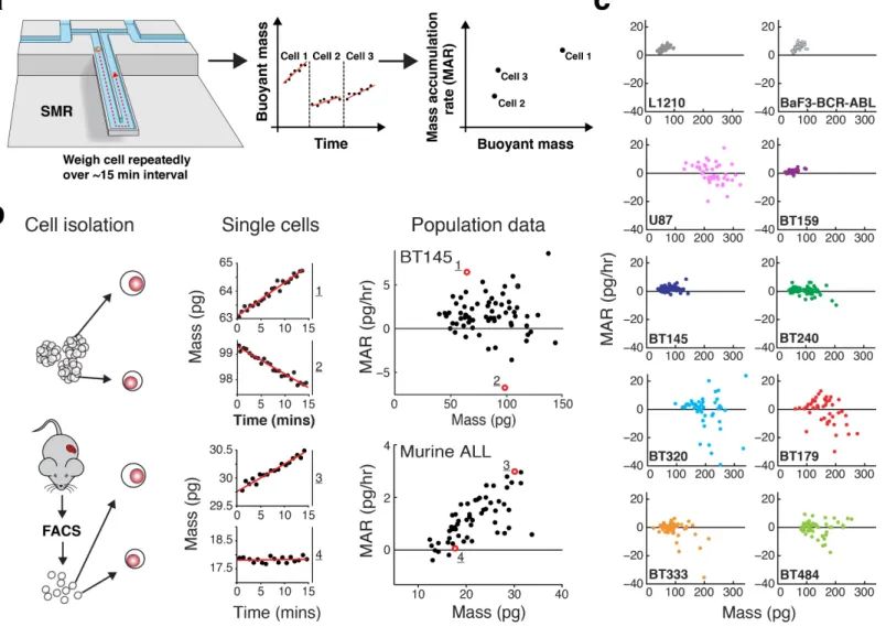
We have developed an approach for functionally assessing the therapeutic sensitivity of single cancer cells by weighing each cell repeatedly over a 15-minute period in a suspended microchannel resonator (SMR) (Fig. 1a) 11-13, either in the presence or absence of cancer therapeutics. Resonator-based approaches have been used to measure an array of cellular physical properties14, and, in one preliminary study, response to therapeutics 15. Following the incubation of tumor cells with drug, the SMR can detect changes in the growth of single cells to predict therapeutic response without the need for extended culture. To validate this approach, we applied the SMR to traditional cancer cell lines, patient-derived cell lines (PDCLs) and primary leukemia cells.
RESULTS
Mass accumulation rate (MAR) measurement
The SMR is a cantilever-based microfluidic mass sensor that measures the buoyant mass (referred to hereafter simply as mass) of live single cells with a resolution near 50 fg, which is highly precise given that the average buoyant mass of a hematopoietic cell is ~75 pg 11. Cells are measured in suspension while under culture
conditions, with controlled media temperature and CO2 concentration to maintain cell viability and growth13. A series of mass measurements is made on an individual cell every ~30 seconds for ~15 minutes, allowing for determination of the mass accumulation rate (MAR), which is defined as the change in mass over time (Fig. 1a)12. In addition to the MAR we also use the absolute single-cell mass as a biomarker, which is determined for each cell during the MAR measurement. By performing the MAR measurement on multiple cells from the same population, the SMR reveals heterogeneity in mass and MAR across the population, rather than an average of the tumor bulk. The degree to which mass and MAR behave as independent biomarkers varies depending on conditions and cell type. Although linear discriminate analysis (LDA) maximizes the predictive capability of these two biomarkers, we have used a simplified metric of MAR normalized by mass for most of the studies in this paper.
Single-cell MARs reveal tumor growth heterogeneity
In order to better characterize the platform’s performance, we applied this method to two cancer cell types known to be viable and proliferate in suspended cell culture: GBM and acute leukemias. First, we analyzed a fast growing GBM-PDCL (BT145) which grows as free-floating “stem-like” cells and tumorspheres, as well as primary
leukemia cells isolated directly from mice with genetically-engineered, BCR-ABL-expressing acute lymphoblastic leukemia (BCR-ABL ALL). Consistent with our previous findings 12, the SMR was able to quantify MAR of single cells over ~15 minutes intervals with high signal-to-noise ratios in both tumor types (Fig. 1b). During this time, the cells acquired less than a few picograms of biomass. This is equivalent to an increase in cell diameter on the order of only 10 nm.
To determine whether cell proliferation potential could be maintained after passage through the SMR, single BT145 GBM cells were isolated after MAR measurement and then assayed for their ability to form tumorspheres. Overall, 14 (35.9%) of 39 single cells formed tumorspheres compared to 217 (45.2%) of 480 single cells isolated directly from a bulk culture (p=0.26; Supplementary Fig. 1). Thus, MAR measurement had no statistically significant effect on viability or stem-like cell phenotype.
The heterogeneity of single-cell growth across cell lines or patient models has not been well characterized. In BT145 cells, MAR measurements enabled delineation of different growth populations, identifying cells of both large and small mass with positive, zero and negative MAR (Fig. 1b). GBM tumors are known to harbor an exceptionally diverse admixture of growing, senescent, quiescent, and dying cells 16, 17. Examination of BT145 by immunohistochemistry also supported maintenance of these heterogeneous properties (Supplementary Fig. 2). Thus, heterogeneity of MAR and mass within this line seems to parallel the known growth heterogeneity and morphological diversity of cells seen in primary GBMs. In contrast, almost all primary BCR-ABL ALL cells had positive MARs that monotonically increased with cell mass (Fig. 1b).
To determine the range of growth diversity that might exist across patients in a single tumor type, we measured MARs of single cells from seven different GBM-PDCLs with diverse genotypes (Supplementary Table 1) over three successive passages (Supplementary Fig. 3)18. MAR measurements demonstrated significant inter- and intra-PDCL heterogeneity across the seven intra-PDCLs with single-cell diversity in both MAR and mass. These findings were in stark contrast with the homogeneity of MARs across conventional hematopoietic cell lines, such as the L1210 leukemia cell line and the murine lymphoblastoid BaF3 cell line engineered to express BCR-ABL (BaF3 BCR-ABL) (Fig. 1c). Such homogeneity did not appear related to the advanced age and passage of these lines as the conventional GBM cell line U87 exhibited significant MAR heterogeneity. Some lines like BT159 and BT145 had more homogenous patterns of cell proliferation, whereas BT320 and BT179 exhibited the most heterogeneous phenotypes, findings supported by immunohistochemistry analysis (Supplementary Fig. 2). The extent of growth heterogeneity within individual GBM-PDCLs was maintained across consecutive passages (Supplementary Fig 3), suggesting that intra-tumoral diversity in growth is an inherent property of GBM PDCLs even during in vitro propagation.
MAR predicts cell line sensitivity to targeted therapy
To test the ability of MAR measurements to predict drug susceptibility in an established cell line, we utilized BaF3 cells engineered to express either wild-type BCR-ABL or the imatinib-resistant mutant BCR-ABL T315I19, 20. Treatment of BaF3-BCR-ABL cells with the tyrosine kinase inhibitor (TKI) imatinib at a therapeutically
achievable concentration of 1 µM for only 2-4 hrs significantly decreased MAR without altering the distribution of mass (Fig. 2). With longer durations of exposure to imatinib, the reduction in MAR became more pronounced (Fig. 2b) and cell mass was reduced (Fig. 2c). When the same conditions were applied to BaF3-BCR-ABL T315I cells, no significant change was observed in MAR or mass distributions (Fig. 2). However, exposure of these cells to the third-generation TKI ponatinib, which retains activity against BCR-ABL T315I, recapitulated the same reduction in MAR observed upon imatinib treatment of BaF3-BCR-ABL cells (Fig. 2b)20. Thus, MAR can distinguish therapeutic susceptibility from resistance within single BaF3 cells after only a few hours of drug exposure.
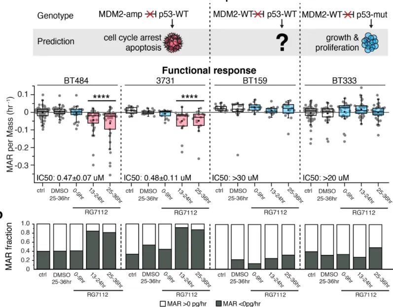
Next, we tested the ability of MAR measurements to predict therapeutic susceptibility when applied to GBM- PDCLs which have heterogeneous and complex MAR profiles and include cycling (non-G0) as well as non- cycling cells (G0) (Figure 1, Supplementary Fig. 2)16, 17. GBM is highly resistant to most therapeutics, but we previously reported sensitivity of some GBM-PDCLs to targeted MDM2 inhibition 21. In bulk tumors and cultures, MDM2 inhibitors are known to induce response in cells which have MDM2 amplification and wild-type TP53, whereas cells with mutant or deleted TP53 are completely resistant to MDM2 inhibitors. However, cells with wild-type MDM2 and wild-wild-type TP53 have unpredictable sensitivity to MDM2 inhibitors in GBM and other tumor cell types 22. We utilized the MDM2 inhibitor RG7112, a compound being evaluated in clinical trials, to test how single-cell MAR response of five GBM-PDCLs across this spectrum of MDM2 and TP53 mutational backgrounds might empirically correspond with genetics and CTG bulk sensitivity measures 23. BT484 and 3731 cells, which are MDM2 amplified and TP53 wild-type, had primarily negative MARs after incubation with 1 µM RG7112 for 13-24 hrs (Fig. 3, Supplementary Fig. 4). With increasing time, the number of cells with negative MAR further increased until 25-36 hrs, when >80% of cells were losing mass in both PDCLs (Fig. 3, Supplementary Fig. 4). In comparison, BT333 cells, which are MDM2 wild-type and TP53 mutant, had no significant change in MAR compared to a DMSO-treated control over 36 hrs of RG7112 exposure (Fig. 3, Supplementary Fig. 4).
Contrasting the predictable sensitivity and resistance of the aforementioned lines, the response of MDM2-
wildtype, TP53-wildtype cells is not clearly correlated to MDM2/TP53 genotype. For example, targeted and whole exome sequencing of BT159, which are MDM2 and TP53 wild-type, identified no mutations or copy number alterations in TP53, TP63, TP73, CDKN2A, MDM4, mitochondrial apoptosis mediators or other p53- related genes that could mediate MDM2 inhibitor resistance. However, BT159 exhibited no evidence of single- cell response by 36 hrs using MAR measurements, similar to the complete resistance exhibited by TP53-mutant BT333 cells (Fig. 3, Supplementary Fig. 4). MAR measurements for all lines was consistent with the response in viability as measured by CTG after 72 hours of RG7112 exposure (Supplementary Fig. 5).
MAR predicts primary cell sensitivity to targeted therapy
Next, we asked whether MAR measurements could effectively predict therapeutic response in primary tumor cells measured immediately after isolation from the in vivo setting. We harvested transgenic murine ALLs that express BCR-ABL or BCR-ABL T315I from the spleen of mice and flow-sorted to purify leukemia cells (Supplementary Fig. 6) 24. Single-cell MAR data was collected after 10-20 hrs of treatment with 1 µM imatinib or 100 nM
ponatinib. Across three independent biological replicates, we observed a significant reduction in average MAR for leukemias expressing BCR-ABL upon treatment with imatinib or ponatinib, as well as for leukemias expressing BCR-ABL T315I upon treatment with ponatinib (Fig. 4a). By contrast, imatinib had no effect on leukemias expressing BCR-ABL T315I (Fig. 4a). We confirmed that BCR-ABL T315I leukemias are truly resistant to the imatinib analog nilotinib in situ by treatment of mice engrafted with these leukemias (Supplementary Fig. 7). In contrast to the marked effect of imatinib on MAR of wild-type BCR-ABL leukemia cells, exposure to imatinib in
vitro for 24 hrs had no effect on the viability of leukemias expressing wild-type BCR-ABL in bulk culture, as
determined by flow cytometry for Annexin V and DAPI (Supplementary Fig. 6a). Thus, the effect on MAR precedes these more standard metrics of drug response.
In order to gauge how robustly MAR measurements can predict primary ALL single-cell drug sensitivity, we generated a receiver-operating characteristic (ROC) after performing linear discriminate analysis (LDA) on each replicate’s dataset. So far we have used the single metric of MAR per mass, however we also considered MAR and mass as independently variable biomarkers. LDA projects the populations of the two-dimensional MAR versus mass data onto a single axis that provides the best ability to distinguish two populations, and then defines the ideal threshold for this classification (Fig. 4b). Subsequent ROC curve analysis is performed and its area under the curve (AUC) is a metric of the ability to properly identify a single cell’s classification as sensitive or resistant to therapy 25. A random classifier has an AUC equal to 0.5, and a perfect classifier has an AUC of 1. The average AUC of non-selective conditions (DMSO-treated compared to imatinib-treated T315I-mutant leukemia) was 0.57,
consistent with the expectation that resistant cells are indistinguishable from untreated cells (Fig. 4a,c). Under selective conditions, the ROC curves for MAR versus mass showed excellent resolution of sensitive and resistant populations, with an average AUC of 0.85 (Fig. 4a,c). ROC curves using mass or MAR as single parameters had significant power to classify single cells, but the single parameters were less consistent between replicates and on average less accurate than using both parameters for classification (Fig. 4c, Supplementary Fig. 8).
MAR predicts sensitivity of circulating leukemia cells
Although GBM PDCLs and murine spleens provide access to an unlimited number of tumor cells, we wanted to measure MARs from small samples, simulating the limited tissue available with patient biopsies. To this end, we isolated tumor cells from the peripheral blood of mice by cheek bleed, which resulted in only 25 µL of total
volume and does not compromise mouse survival. We performed these bleeds when circulating disease was as low as 4% of circulating mononuclear cells. This approach typically provided on the order of 103 total tumor cells for measurement following purification by flow sorting. In order to measure samples of low cell count and volume, we implemented a next-generation SMR array device that greatly simplifies fluidic handling, increasing throughput by 20-fold and enables the use of low-volume samples 26.
Single-cell MAR data was then collected on both cheek-bleed samples (~25 µL) and cardiac bleeds (~500 µL) exposed to either DMSO or 100 nM ponatinib for 14-20 hrs in vitro. Classification of single-cell drug response was similar using cheek-bleeds (AUC = 0.85) compared with measurements from splenocytes (AUC = 0.85) or cardiac bleeds (AUC = 0.80) (Fig. 4d, Supplementary Fig. 9). The ability of MAR measurements to assay drug sensitivity of single cells isolated from very small amounts of blood makes it feasible to longitudinally screen for phenotypic resistance within individual patients through iterative sampling.
Patient cells reduce MAR when treated ex vivo or in vivo
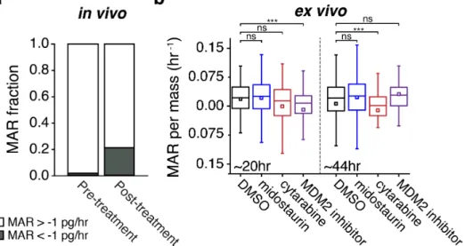
samples. First, we assayed ficolled peripheral blood samples from a patient with relapsed acute myeloid leukemia (AML). A sample obtained while the patient was not receiving treatment was composed largely of slightly positive MARs (Fig. 5a, Supplementary Fig. 10). After the patient had received 48 hrs of treatment with an experimental MDM2 inhibitor, a second peripheral blood sample was taken. This demonstrated a broader distribution of MARs compared to the pretreatment sample. Furthermore, a large population of cells with negative MARs of less than -1 pg/hr appeared, indicating a shift in population MAR dynamics in vivo among the leukemia present in the
peripheral blood (Fig. 5a, Supplementary Fig. 10). This shift towards negative MARs following treatment is consistent with our observations from ex vivo treatment of susceptible GBM patient-derived cell lines (Fig. 3) and murine primary cells (Fig. 4).
Finally, we performed ex vivo treatment of a patient sample analogous to the approach applied to primary murine samples. Bone marrow leukemia cells from a patient presenting with newly diagnosed FLT3-mutated positive AML with MDS-related changes was treated in media with a range of therapeutics, including DMSO, 1 µM cytarabine (cytotoxic chemotherapy), 100 nM midostaurin (FLT3 and multikinase inhibitor), and an experimental MDM2 inhibitor (Fig. 5b). These cultures were incubated, and MARs were measured on three next- generation SMR array devices in parallel during 10-hour windows that centered on 20 and 44 hrs. Cells in midostaurin showed no significant change in their distribution of MAR as compared to a DMSO control, consistent with the limited activity of single-agent midostaurin in FLT3-mutated AML27. In comparison, cytarabine did result in a reduction in MAR that was highly significant (p=0.00015) at 48 hrs as compared to the control. Finally, cells treated with the experimental MDM2 inhibitor showed reduced MAR at 24 hrs, which rebounded by 48 hrs, potentially indicating a brief period of p53 target induction followed by the rapid induction of adaptive resistance.
DISCUSSION
Here we have presented a functional assay for assessing single cancer cell therapeutic sensitivity based on measurements of MAR and mass of individual cells. We validated the predictive power of combining MAR and
mass measurements by confirming susceptibility or resistance of genetically-defined cell lines, human GBM-PDCLs, and primary murine ALLs in response to targeted therapeutics. Additionally, by measuring mass
accumulation rates of single cells across a population and establishing a growth profile for different GBM-PDCLs and cell lines, we have shown for the heterogeneity of single-cell growth both within and across these populations. A substantial body of additional experiments will need to be performed to prove utility of MAR measurements for clinical decision making. However, our initial data with primary patient leukemia samples, treated either ex vivo or
in vivo, showed responses that were consistent with our previously assayed in vivo models.
MAR measurement within the SMR is not a terminal assay, as cells are kept viable throughout the measurement and thereby remain compatible with downstream analyses. Thus, a key advantage of MAR measurements is that cells can be studied downstream of the SMR using other single-cell assays, as we demonstrated by quantifying the tumorsphere-forming potential of GBM-PDCL cells after passage through the device (Supplementary Fig. 1). This ability will ultimately allow for correlations between single-cell changes in MAR, other functional outcomes and non-functional biomarkers (e.g. genetics, gene expression, chromatin modifications). Further studies are needed to assess the effects of passage through the SMR on aspects of tumor cell biology, including changes in the
transcriptome, genome, and proteome. Previous studies have found that cellular and genomic properties of single cells can be measured using techniques such as RNA-sequencing and are well- preserved following exposure to microfluidic environments28.
Recent studies have used other functional approaches to predict therapeutic sensitivity of individual cancers.For example, Crystal et al. applied standard proliferation-based assays to quantify therapeutic susceptibilities of bulk cultures of PDCLs 10. The results from their ex vivo screening predicted the response of in vivo xenografts to combination therapies. However, cell-to-cell heterogeneity is not captured by population approaches, and the utility of this strategy for clinical decision making is limited by the months of prolonged culture required for PDCL creation.
Montero et al. recently reported a functional approach called ‘dynamic BH3 profiling’, in which therapeutics are applied to cell lines or clinical tumor isolates, and then the percentage of cells that undergo mitochondrial outer membrane permeabilization (MOMP) is measured after introduction of a pro-apoptotic BH3 peptide29. Dynamic BH3 profiling robustly predicted which patients would respond to a given therapy across multiple cancer types. However, this approach requires cell permeabilization, which complicates the application of both downstream assays and phenotype validation, and does not clearly distinguish subsets of cells with phenotypic heterogeneity.
MAR measurement addresses many of these limitations by assessing therapeutic susceptibility in single, live cells without the need for PDCL generation, but is subject to its own set of constraints. Most notably, in vitro culture is still necessary for a length of time adequate to elicit a growth response to applied therapeutics. In BaF3 cells this occurred within 2-4 hrs, but GBM cells required longer culture to appreciably change MAR in the presence of MDM2 inhibition. Thus, the MAR measurement can reduce, but does not completely eliminate, the time within in
vitro culture. Additionally, the SMR currently requires cells to be in a single-cell suspension or small clumps for
mass and MAR measurement. Future studies will explore the utility in solid tumor systems, where the extent of dissociation required may perturb cellular viability and/or response. MAR measurements also initially suffered from low throughput. However, single-cell mass and MAR can now be obtained with a throughput exceeding 60 cells/hr/device without sacrificing precision26.
Perhaps the most important shortcoming of our approach, and the vast majority of functional assays, is a potential bias towards assessing only cell intrinsic drug susceptibility. Microenvironmental interactions are known to influence in vivo drug response, but cellular ‘memory’ of these interactions may degrade during the course of ex
vivo treatment9
. There have been recent advancements in this arena involving implantable devices, but these approaches are currently only compatible with solid tumors, and require tumors of a minimum size that are easily accessible30
should explore whether alternative culture conditions can help address the contribution of cell extrinsic factors under controlled conditions; e.g., tumor cells could be maintained in vitro in the presence of both drug and co-culture with stromal and/or immune cells prior to measurement. Alternatively, patient drug response in situ could be monitored. For example, patient tumor cells could be analyzed with MAR measurements immediately before and hours to days following treatment to help inform pharmacokinetics and pharmacodynamics. In fact, we applied this approach to a patient with AML (Figure 5) to assess overall feasibility for clinical scenarios that could be explored in the future.
Significant future work needs to be performed to define the utility of mass and MAR as biomarkers fortreatment response across disease types, in comparison to alternative functional assays, for drugs in combination, and across a wider range of drug mechanisms, where response may differ based on the mechanism of cell death. However, given the scarcity of functional assays with the necessary characteristics to merit widespread application, MAR measurements in the SMR hold potential as both a biological tool and a clinical platform.
ACKNOWLEDGEMENTS
These studies were supported by R01 CA170592 (SRM, KLL, PYW), P50 CA165962 (KLL, PYW), P01 CA142536 (KLL) and R33 CA191143 (SRM, DMW) from the National Institutes of Health, U54 CA143874 from the National Cancer Institute (SRM), and partially by Cancer Center Support (core) Grant P30-CA14051 from the NCI, The Bridge Project, a partnership between the Koch Institute for Integrative Cancer Research at MIT and the Dana-Farber/Harvard Cancer Center (DF/HCC) (SRM, DMW), and the DFCI Brain Tumor Therapeutics Accelerator Program (PW, KL). AI acknowledges support for cell line creation from Fondation ARC pour la Recherche sur le Cancer, The Institut Universitaire de Cancérologie (IUC), and OncoNeuroThèque. MMS acknowledges support from the NIH/NIGMS T32 GM008334, Interdepartmental Biotechnology Training Program grant. N. Chou acknowledges support from the National Science Scholarship, Agency for Science,
Technology and Research (STAR), Singapore. DMW is a Leukemia and Lymphoma Scholar. MAM gratefully acknowledges support from the institutional research training grant T32 CA009172, from the National Cancer Institute (USA).
AUTHOR CONTRIBUTIONS
N. Cermak, SO, and SRM designed devices, MMS, N. Chou, N. Cermak, and SO designed and constructed the experimental setup, CLM, DK, SH, AI, PYW, and KLL managed and created BT GBM-PDCLs, MAM and HL managed and processed murine models of B-ALL, MAM, HL, and NAC procured and processed patient samples, MMS, CLM, N. Chou, MAM, DK, YK, NLC, NAC, N. Cermak, DMW, KLL, SRM designed the experiments, MMS, CLM, N. Chou, MAM, DK, YK, RJK, HL, SH, NLC, NAC performed the experiments, MMS, CLM, NC, MAM, DK, YK, RJK, NLC, NAC analyzed the data, MMS, CLM, N. Chou, DMW, KLL, and SRM wrote the paper with input from all authors.
COMPETING FINANCIAL INTERESTS
SRM is a cofounder of Affinity Biosensors, which develops techniques relevant to the research presented. SO and MMS anticipate employment at Affinity Biosensor. DMW is a consultant for and receives research support from Novartis.
REFERENCES
1. Mellinghoff, I.K. et al. Molecular determinants of the response of glioblastomas to EGFR kinase inhibitors. New England Journal of Medicine 353, 2012-2024 (2005).
2. Sos, M.L. et al. Predicting drug susceptibility of non-small cell lung cancers based on genetic lesions. Journal of Clinical Investigation 119, 1727-1740 (2009).
3. Garraway, L.A. & Janne, P.A. Circumventing Cancer Drug Resistance in the Era of Personalized Medicine. Cancer Discovery 2, 214-226 (2012).
4. Klempner, S.J., Myers, A.P. & Cantley, L.C. What a Tangled Web We Weave: Emerging Resistance Mechanisms to Inhibition of the Phosphoinositide 3-Kinase Pathway. Cancer Discovery 3, 1345- 1354 (2013).
5. Haibe-Kains, B. et al. Inconsistency in large pharmacogenomic studies. Nature 504, 389-+ (2013). 6. Navin, N. et al. Tumour evolution inferred by single-cell sequencing. Nature 472, 90-U119 (2011). 7. Francis, J.M. et al. EGFR Variant Heterogeneity in Glioblastoma Resolved through Single-Nucleus
Sequencing. Cancer Discovery 4, 956-971 (2014).
8. Burstein, H.J. et al. American Society of Clinical Oncology Clinical Practice Guideline Update on the Use of Chemotherapy Sensitivity and Resistance Assays. Journal of Clinical Oncology 29, 3328- 3330 (2011).
9. Friedman, A.A., Letai, A., Fisher, D.E. & Flaherty, K.T. Precision medicine for cancer with next- generation functional diagnostics. Nature Reviews Cancer 15, 747-756 (2015).
10. Crystal, A.S. et al. Patient-derived models of acquired resistance can identify effective drug combinations for cancer. Science 346, 1480-1486 (2014).
11. Burg, T.P. et al. Weighing of biomolecules, single cells and single nanoparticles in fluid. Nature 446, 1066-1069 (2007).
12. Godin, M. et al. Using buoyant mass to measure the growth of single cells. Nature Methods 7, 387- U370 (2010).
13. Son, S. et al. Direct observation of mammalian cell growth and size regulation. Nature Methods 9, 910-+ (2012).
14. Byun, S., Hecht, V.C. & Manalis, S.R. Characterizing Cellular Biophysical Responses to Stress by Relating Density, Deformability, and Size. Biophysical Journal 109, 1565-1573 (2015).
15. Wu, S.Q. et al. Quantification of cell viability and rapid screening anti-cancer drug utilizing nanomechanical fluctuation. Biosensors & Bioelectronics 77, 164-173 (2016).
16. Lathia, J.D. et al. Direct In Vivo Evidence for Tumor Propagation by Glioblastoma Cancer Stem Cells.
Plos One 6, 9 (2011).
17. Deleyrolle, L.P. et al. Evidence for label-retaining tumour-initiating cells in human glioblastoma.
Brain 134, 1331-1343 (2011).
18. Cerami, E. et al. The cBio Cancer Genomics Portal: An Open Platform for Exploring Multidimensional Cancer Genomics Data. Cancer Discovery 2, 401-404 (2012).
19. Pui, C., Relling, M.V. & Downing, J.R. Mechanisms of disease: Acute lymphoblastic leukemia. New
England Journal of Medicine 350, 1535-1548 (2004).
20. Cortes, J.E. et al. Ponatinib in Refractory Philadelphia Chromosome-Positive Leukemias. New
England Journal of Medicine 367, 2075-2088 (2012).
21. Verreault, M. et al., Vol. Epub ahead of print (Clinical Cancer Research; 2015).
22. Jeay, S. et al. A distinct p53 target gene set predicts for response to the selective p53-HDM2 inhibitor NVP-CGM097. eLife 4 (2015).
23. Andreeff, M. et al., Vol. Ahead of Print (Clin Cancer Res; 2015).
24. Lane, A.A. et al. Triplication of a 21q22 region contributes to B cell transformation through HMGN1 overexpression and loss of histone H3 Lys27 trimethylation. Nature Genetics 46, 618-623 (2014). 25. Pencina, M.J., D'Agostino, R.B. & Vasan, R.S. Evaluating the added predictive ability of a new
marker: From area under the ROC curve to reclassification and beyond. Statistics in Medicine 27, 157-172 (2008).
26. Cermak, N. et al. High-throughput growth measurements on single cells via serial microfluidic mass sensor arrays. Nature Biotechnology (in press) (2016).
27. Fischer, T. et al. Phase IIB Trial of Oral Midostaurin (PKC412), the FMS-Like Tyrosine Kinase 3 Receptor (FLT3) and Multi-Targeted Kinase Inhibitor, in Patients With Acute Myeloid Leukemia and High-Risk Myelodysplastic Syndrome With Either Wild-Type or Mutated FLT3. Journal of Clinical
Oncology 28, 4339-4345 (2010).
28. Shalek, A.K. et al. Single-cell RNA-seq reveals dynamic paracrine control of cellular variation.
Nature 510, 363-+ (2014).
29. Montero, J. et al. Drug-Induced Death Signaling Strategy Rapidly Predicts Cancer Response to Chemotherapy. Cell 160, 13 (2015).
30. Jonas, O. et al. An implantable microdevice to perform high-throughput in vivo drug sensitivity testing in tumors. Science Translational Medicine 7, 11 (2015).
31. Klco, J.M. et al. Genomic impact of transient low-dose decitabine treatment on primary AML cells.
Blood 121, 1633-1643 (2013).
FIGURE LEGENDS
Figure 1: Mass accumulation rate (MAR) measurements characterize single-cell heterogeneity in growth across GBM-PDCLs and conventional cell lines. (a) Schematic of workflow. Single cells are weighed repeatedly over a 15-minute interval by iterative passage through the SMR device. A linear fit is applied to those measurements and the resulting data is plotted as MAR versus buoyant cell mass. (b) MAR measurements over ~15 minutes for single cells from the BT145 GBM PDCL (top panel) and primary BCR-ABL ALL cells directly isolated from mice (bottom panel). Cells are dissociated (for BT145) or FACS purified (for ALL) and single cells are measured. The specific single-cell plots shown in the middle column are represented as red open-circles along with other single cells (black dots) plotted as a function of mass. (c) MAR versus mass distributions from 7 GBM-PDCLs, 2 conventional hematopoietic cell lines (L1210 and BaF3-BCR-ABL) and one conventional GBM cell line (U87) for comparison. Each GBM-PDCL plot includes measurements from 3 successive passages
in vitro (Supplementary Fig. 3), and each dot represents a single cell. From left to right, row by row, n = 84, 46,
44, 51, 52, 61, 48, 46, 64, and 59 cells.
Figure 2: Murine BaF3 lymphoblastoid cells rapidly reduce MAR upon exposure to active kinase inhibitors. (a) MAR versus cell mass of imatinib-sensitive BaF3-BCR-ABL or imatinib-resistant BaF3-BCR-ABL T315I cells exposed to 1 µM imatinib (imat.). (b, c) Same data as in (a) shown as MAR per mass (b) or mass box-plot (c), also including BaF3-BCR-ABL T315I cells treated with 100 nM ponatinib. Boxes represent the inter-quartile range and white squares the average of all measurements. p-values were calculated using the non-parametric Mann-Whitney U test, comparing treatment groups to DMSO for the same treatment duration. **** p<0.0001 in highlighted segments. Timepoints were taken on at least three biological replicate cultures on different days. From left to right, n = 46, 20, 48, 37, 36, 42, 15, 41, 27, 41, 41.
Figure 3: MAR predicts sensitivity of human GBM-PDCLs to targeted therapy. (a) The expected response to RG7112 is outlined above each cell line, with the red ‘X’ indicating the drug’s ability to block the inhibition of
p53 signaling by MDM2. The measured functional response of BT484, and 3731 (MDM2 amplified, TP53 wild-type (WT)), BT159 (MDM2 WT, TP53 WT), and BT333 (MDM2 WT, TP53 mutated) PDCLs to DMSO or the MDM2 inhibitor RG7112 at a therapeutically relevant concentration of 1 μM over 36 hrs. IC50 data from 72 hr CellTiter-Glo measurements shown for reference. Complete data in Supplementary Fig. 5. Boxes represent the inter-quartile range and white squares the average of all cells. Gray circles show values for individual cells. p-values were calculated using the non-parametric Mann-Whitney U test, comparing treatment groups to the DMSO control. ****p <0.0001 for highlighted segments. Timepoints were taken on biological replicate cultures on different days. From left to right, n = 59, 37, 25, 38, 37; 16, 18, 15, 15, 15; 15, 16, 14, 16, 15: 69, 32, 27, 37, 38. (b) Same data as in (a), showing that the fraction of BT484 and 3731 cells with negative MARs increases upon exposure to RG7112 but remains unaffected in BT159 and BT333 cells.
Figure 4: MAR distributions predict drug sensitivity of primary murine ALL cells to targeted therapy. (a) MAR per mass distributions of paired measurements from primary murine B-ALL cells dependent on BCR-ABL or BCR-ABL T315I and treated with 1 µM imatinib, 100 nM ponatinib, or DMSO. Measurements from individual mice are separated by a vertical dotted line. n indicates the number of cells for each measurement. AUC values for ROC curve of each paired dataset are listed below the x-axis. Boxes represent the inter-quartile range and white squares the average of all measurements. p-values were calculated using the non-parametric Mann-Whitney U test, comparing treated cells to the DMSO control. (b) Representative MAR versus mass plot with overlay of an orthogonal vector (dotted line) designating the threshold resulting from LDA. (c) ROC curves of paired control and treatment data for each treatment replicate. Cells treated with therapy to which they are sensitive or resistant are shown with blue solid lines or red dotted lines, respectively. (d) MAR per mass
distributions of paired measurements from primary murine B-ALL cells that are dependent on BCR-ABL. Cells isolated from the bloodstream of mice by either cardiac or cheek bleed and treated with 100 nM ponatinib or DMSO for the specified interval. p-values were calculated using the Mann-Whitney U test, comparing treated cells to the DMSO control.
Figure 5: Patient samples treated in vivo or ex vivo show consistent reduction in MAR. (a) The fraction of cells with MAR of less than -1 pg/hr from pre-treatment patient sample (n = 86), and from sample obtained after the patient received 48 hrs of of treatment with an experimental MDM2 inhibitor (n = 95). MAR versus mass data for same sample set shown in Supplementary Figure 10. (b) Boxplots of MAR per mass of bone marrow leukemia cells treated ex vivo with either DMSO, 100 nM midostaurin, 1 µM cytarabine, or an experimental MDM2 inhibitor, and measured during 10 hour windows centered around 20 and 44 hrs. Boxes represent the inter-quartile range and white squares the average of all measurements. p-values were calculated using the non-parametric Mann-Whitney U test, comparing treated cells to the DMSO control. From left to right, n = 137, 102, 111, 78, 119, 103, 67, 48.
MATERIALS AND METHODS
Cell culture of conventional cell lines
L1210, BaF3-BCR-ABL, and BaF3-BCR-ABL-T315I cells were maintained in suspension in RPMI-1640 media (Invitrogen, Cat#11875-119), supplemented with 10% FBS (Sigma Aldrich, Cat#F4135), Penicillin-Streptomycin (Invitrogen, Cat#15140-122 ), and kept in a 37 ˚C, 5% CO2, and humidified incubator. Cells
were passaged every 2 days to 5x10⁴ cells/mL, and used for SMR experiments between 24-36 hrs of growth at an approximate cell concentration of 2-4x10⁵ cells/mL. L1210 cells were a gift from the Kirschner lab at Harvard, BaF3-BCR-ABL, and BaF3-BCR-ABL-T315I were created from the parental BaF3 cell line obtained from the RIKEN BioResource Center. No further cell line validation was performed. All cell lines tested negative for mycoplasma.
For drug response experiments, cells in bulk were dosed for the specified interval with 0.1% DMSO, 1 µM imatinib (Santa Cruz Biotechnology, Cat#SC-202180), or 100 nM ponatinib (Selleckchem, Cat#AP24534). The cells were kept in drugged media during the measurements, and samples sizes were determined by practical limitations set by throughput.
Creation and cell culture of GBM PDCLs
Harvard Cancer Center protocol #10-417) and two waived consent protocols (Dana Farber Harvard Cancer Center protocol #10-043 and Partner’s Human Research Center protocol #2002 P000995). All protocols mentioned have been approved by Dana Farber Harvard Cancer Center and Partner’s Human Research Center institutional review boards. Cells were harvested from excess tissue resection specimens through cycles of enzymatic (Neural tissue dissociation kit with papain, Miltenyi) and mechanical dissociation in a tissue grinder (gentleMACS dissociator, Miltenyi). Cells were grown as tumorspheres in NeuroCult NS-A proliferation media (Miltenyi) supplemented with 2 µg/ml Heparin, 20 ng/ml human epidermal growth factor (EGF), 10 ng/ml human bFGF in ultra-low attachment coated flasks (Corning, Cat#3814), which were kept in a 37 ˚C, 5% CO2, and humidified incubator. All PDCL’s tested negative for mycoplasma.
Prior to loading in the SMR, the PDCLs were dissociated with Accutase (Sigma-Aldrich, Cat#A6964) at 37 ˚C for 7 minutes and plated as a single-cell population at 7-10x104 cells/ml. Tumorsphere forming assays were conducted by assessing the expansion of single-cells in a 96-well plate following 2 weeks of incubation, either isolated from the SMR, or as sorted by fluorescence activated cell sorting (FACS).
For drug experiments, GBM-PDCLs were seeded into parallel cultures and treated with 1 µM of the MDM2 inhibitor RG7112 (Selectchem), or DMSO. At each time point, one parallel culture was dissociated and
immediately resuspended in drug media, and remaining parallel cultures were resuspended in fresh media with new drug. The dissociated cell suspensions were kept in drugged media during the measurements, and samples sizes were determined by practical limitations set by throughput.
Transgenic mouse model of BCR-ABL B-ALL
All animal experiments were performed with approval of the DFCI IACUC. A transgenic mouse model of BCR-ABL B-ALL was generated by transplantation of lethally irradiated Ts1Rhr mice (B6.129S6-Dp(16Cbr1- ORF9)1Rhr/J; Jackson Laboratory, Bar Harbor, ME, USA; stock #005848) with syngeneic Hardy B cells
transduced with an MSCV retrovirus coexpressing GFP and human BCR-ABL cDNA, as previously described18. For the present studies, 1x10⁶ bulk splenocytes (P1 generation) were transplanted into lethally irradiated, wild-
type, female, C57BL/6 mice at 6-8 weeks of age, which were followed daily for clinical signs of leukemia and sacrificed when moribund. Splenocytes or blood samples were harvested, subject to erythrocyte lysis (Qiagen, Cat#158904), and stained with an antibody targeting murine CD19 (Fisher Scientific, Cat#BDB551001). BCR- ABL B-ALL cells were isolated by sorting for CD19/GFP double-positive cells on a FACSAria II SORP fluorescence activated cell sorter (BD Biosciences).
For drug response experiments, sorted mouse leukemia cells were seeded at a density of 5x10⁵/mL and cultured at 37 ˚C in a humidified 5% CO2 incubator in RPMI (Gibco, Cat #11835055) supplemented with 10% FBS, 2 mM L-glutamine (Gibco, Cat#25030164), 50 µM 2-mercaptoethanol (Sigma, Cat#M3148), 50 IU/ml-50 µg/mL penicillin-streptomycin (Fisher Scientific, Cat#ICN1670049), 10 ng/mL recombinant murine IL-3 (PeproTech, Cat#213-13), 10 ng/mL recombinant murine IL-7 (PeproTech, Cat#217-17), 10 ng/mL recombinant murine stem cell factor (PeproTech, 03), and 10 ng/mL recombinant murine FLT3-ligand (PeproTech, Cat#250-31L). Cells were kept in a 37 ˚C, 5% CO2, and humidified incubator. Replicate cultures were dosed for 10 hrs with 0.1% DMSO, 1 µM imatinib (Santa Cruz Biotechnology, Cat#SC-202180), or 100 nM ponatinib
(Selleckchem, Cat#AP24534). The cells were kept in drugged media during the measurements, which took place between 10-20 hrs of drug exposure, and samples sizes were determined by practical limitations set by
throughput and measurement window length.
For in vivo confirmation of drug efficacy, female C57BL/6 mice were sublethally irradiated and transplanted with 7.5x10⁵ murine leukemia cells harboring human BCR-ABL cDNA, of which 95% were BCR-ABL wild type and 5% harbored the T315I allele. Upon engraftment, as defined by the presence of circulating leukemia at a level of 1-3% by peripheral blood flow cytometry, mice were treated with nilotinib 50 mg/kg/day via oral gavage (n=2 mice). Nilotinib serves as a surrogate for imatinib, as both compounds have demonstrated activity against WT but not T315I BCR-ABL. Error bars represent standard deviation. Mice underwent serial BCR- ABL genotyping via the Sanger method to monitor the allelic frequency of BCR-ABL T315I.
Primary human leukemia specimens were collected from patients at the Dana-Farber Cancer Institute and
Brigham and Women’s Hospital upon provision of informed consent under one or more tissue banking protocols (Dana-Farber/Harvard Cancer Center (DF/HCC) protocols #01-206 and #11-104). Each protocol has been approved by the DF/HCC institutional review board (IRB). Peripheral blood and bone marrow samples
underwent Ficoll density gradient centrifugation to enrich for mononuclear cells, followed by immunomagnetic enrichment of leukemia cells using CD33 MicroBeads (Miltenyi, Cat#130-045-501) if concomitant clinical testing indicated that tumor purity was <80%. Leukemia cells were seeded at 0.5-1.0x10⁶/mL in DMEM supplemented with 15% FBS, 2 mM L-glutamine, 50 µM 2-mercaptoethanol, 50 IU/ml-50 µg/mL penicillin- streptomycin, and human cytokines SCF (100 ng/mL, PeproTech #300-07), IL3 (10 ng/mL, PeproTech #200-03), IL6 ((20 ng/mL, PeproTech 200-06), TPO (10 ng/mL, PeproTech #300-18), and FLT3-Ligand (10 ng/mL,
PeproTech 300-19), as adapted from published methods31. For measurements of in vivo response, aliquots of cultured cells were immediately measured in the presence of DMSO or sustained drug pressure with the experimental MDM2 inhibitor. In the case of ex vivo treatment, aliquots were treated with an experimental MDM2 inhibitor, cytarabine (1 µM), midostaurin (100 nM), or DMSO (1:1000) and were assessed using the SMR within 10-hour windows centered on 20 and 44 hours.
CellTiter-Glo assay
Cell viability in GBM neurospheres was assessed by quantification of ATP through chemiluminescence reading with CellTiter-Glo (Promega, Cat#G7570). GBM cells were plated as single cells at 2x10³ cells/mL into wells of an ultra-low attachment coated 96-well plates (Corning, Cat#3474). In drug treatment experiments, viability was assessed at 72 hrs. All measurements were performed in triplicate according to manufacturer’s protocol.
Measurement and operation of a suspended microchannel resonator (SMR)
The design and operation of the SMR have been previously described13-15. In short, single cells in suspension are passed through the SMR resulting in a frequency shift that is proportional to cell buoyant mass. The SMR can resolve the instantaneous rate of mass accumulation for a single cell in 15 minutes provided that the cell is
from measurements of frequency shifts of polystyrene beads with a known mass (Thermo Scientific, Cat#4208A). Mass versus time data of a single cell is then linearly fitted.
For measurement in the SMR, cells were suspended in their standard growth media with or without drug. For GBM-PDCLs poly-L-lysine/polyethylene-glycol (PLL-PEG) was added to the media at 10 µM to reduce cell- clumping and sticking to microchannel walls. Clumps of cells, visualized through a microscope during
measurement, were excluded from measurement in the SMR. The system and cells were kept in culturing conditions (5% CO2, 37 ˚C) for all measurements as previously described12.
Flow cytometry
All flow cytometry measurements were performed on a BD LSR II (BD Biosciences) using 4',6-diamidino-2- phenylindole (DAPI) (BioLegend, Cat#422801) and Annexin V (BioLegend, Cat#640907)
Figure 1: Mass accumulation rate (MAR) measurements characterize single-cell heterogeneity in growth across GBM-PDCLs and conventional cell lines. (a) Schematic of workflow. Single cells are weighed repeatedly over a 15-minute interval by iterative passage through the SMR device. A linear fit is applied to those measurements and the resulting data is plotted as MAR versus buoyant cell mass. (b) MAR measurements over ~15 minutes for single cells from the BT145 GBM PDCL (top panel) and primary BCR-ABL ALL cells directly isolated from mice (bottom panel). Cells are dissociated (for BT145) or FACS purified (for ALL) and single cells are measured. The specific single-cell plots shown in the middle column are represented as red open-circles along with other single cells (black dots) plotted as a function of mass. (c) MAR versus mass distributions from 7 GBM-PDCLs, 2 conventional hematopoietic cell lines (L1210 and BaF3-BCR-ABL) and one conventional GBM cell line (U87) for comparison. Each GBM-PDCL plot includes measurements from 3 successive
passages in vitro (Supplementary Fig. 3), and each dot represents a single cell. From left to right, row by row, n = 84, 46, 44, 51, 52, 61, 48, 46, 64, and 59 cells.
Figure 2: Murine BaF3 lymphoblastoid cells rapidly reduce MAR upon exposure to active kinase inhibitors. (a) MAR versus cell mass of imatinib-sensitive BaF3-BCR-ABL or imatinib-resistant BaF3-BCR-ABL T315I cells exposed to 1 µM imatinib (imat.). (b, c) Same data as in (a) shown as MAR per mass (b) or mass box-plot (c), also including BaF3-BCR-ABL T315I cells treated with 100 nM ponatinib. Boxes represent the inter-quartile range and white squares the average of all measurements. p-values were calculated using the non-parametric Mann-Whitney U test, comparing treatment groups to DMSO for the same treatment duration. **** p<0.0001 in highlighted segments. Timepoints were taken on at least three biological replicate cultures on different days. From left to right, n = 46, 20, 48, 37, 36, 42, 15, 41, 27, 41, 41
Figure 3: MAR predicts sensitivity of human GBM-PDCLs to targeted therapy. (a) The expected response to RG7112 is outlined above each cell line, with the red ‘X’ indicating the drug’s ability to block the inhibition of p53 signaling by MDM2. The measured functional response of BT484, and 3731 (MDM2 amplified, TP53 wild-type (WT)), BT159 (MDM2 WT, TP53 WT), and BT333 (MDM2 WT, TP53 mutated) PDCLs to DMSO or the MDM2 inhibitor RG7112 at a therapeutically relevant concentration of 1 μM over 36 hrs. IC50 data from 72 hr CellTiter-Glo measurements shown for reference. Complete data in Supplementary Figure 5. Boxes represent the inter-quartile range and white squares the average of all cells. Gray circles show values for individual cells. p-values were calculated using the non-parametric Mann-Whitney U test, comparing treatment groups to the DMSO control. ****p <0.0001 for highlighted segments. Timepoints were taken on biological replicate cultures on different days. From left to right, n = 59, 37, 25, 38, 37; 16, 18, 15, 15, 15; 15, 16, 14, 16, 15: 69, 32, 27, 37, 38. (b) Same data as in (a), showing that the fraction of BT484 and 3731 cells with negative MARs increases upon exposure to RG7112 but remains unaffected in BT159 and BT333 cells.
Figure 4: MAR distributions predict drug sensitivity of primary murine ALL cells to targeted therapy. (a) MAR per mass distributions of paired measurements from primary murine B-ALL cells dependent on BCR-ABL or BCR-ABL T315I and treated with 1 µM imatinib, 100 nM ponatinib, or DMSO. Biological replicates from individual mice are separated by a vertical dotted line. n indicates the number of cells for each measurement. AUC values for ROC curve of each paired dataset are listed below the x-axis. Boxes represent the inter-quartile range and white squares the average of all measurements. p-values were calculated using the non-parametric Mann-Whitney U test, comparing treated cells to the DMSO control. (b) Representative MAR versus mass plot with overlay of an orthogonal vector (dotted line) designating the threshold resulting from LDA. (c) ROC curves of paired control and treatment data for each treatment replicate. Cells treated with therapy to which they are sensitive or resistant are shown with blue solid lines or red dotted lines, respectively. (d) MAR per mass distributions of paired measurements from primary murine B-ALL cells that are dependent on BCR-ABL. Cells isolated from the bloodstream of mice by either cardiac or cheek bleed and treated with 100 nM ponatinib or DMSO for the specified interval. p-values were calculated using the Mann-Whitney U test, comparing treated cells to the DMSO control.
Figure 5: Patient samples treated in vivo or ex vivo show consistent reduction in MAR. (a) The fraction of cells with MAR of less than -1 pg/hr from pre-treatment patient sample (n = 86), and from sample obtained after the patient received 48 hrs of of treatment with an experimental MDM2 inhibitor (n = 95). MAR versus mass data for same sample set shown in Supplementary Figure 10. (b) Boxplots of MAR per mass of bone marrow leukemia cells treated ex vivo with either DMSO, 100 nM midostaurin, 1 µM cytarabine, or an experimental MDM2 inhibitor, and measured during 10 hour windows centered around 20 and 44 hrs. Boxes represent the inter-quartile range and white squares the average of all measurements. p-values were calculated using the non-parametric Mann-Whitney U test, comparing treated cells to the DMSO control. From left to right, n = 137, 102, 111, 78, 119, 103, 67, 48.


