THE BEHAVIORAL AND BRAIN SCIENCES (1978),3,465-514
Printed in the United States of America
Cortical long-axoned cells and
putative interneurons during the
sleep-waking cycle
Mircea Steriade
Laboratoire de Neurophysiologie, Departement de Physiologie, Faculte de Me'decine, Universite Laval, Quebec, Canada GIK 7P4
Abstract: Knowledge of the input-output characteristics of various neuronal types is a necessary first step toward an understanding of
cellular events related to waking and sleep. In spite of the oversimplification involved, the dichotomy in terms of type I (long-axoned,
output) neurons and type II (short-axoned, local) interneurons is helpful in functionally delineating the neuronal circuits involved in
the genesis and epiphenomena of waking and sleep states. The possibility is envisaged that cortical interneurons, which are
particu-larly related to higher neuronal activity and have been found in previous experiments to be more active during sleep than during
wake-fulness, might be involved in complex integrative processes occurring during certain sleep stages. Electrophysiological criteria for the
identification of output cells and interneurons are developed, with emphasis on various possibilities and difficulties involved in
recognizing interneurons of the mammalian brain. The high-frequency repetitive activity of interneurons is discussed, together with
various possibilities of error to be avoided when interpreting data from bursting cells. Data first show opposite changes in spontaneous
and evoked discharges of identified output cells versus putative interneurons recorded from motor and parietal association cortical
areas in behaving monkeys and cats during wakefulness (W) compared to sleep with synchronized EEG activity (S): significantly
increased rates of spontaneous firing, enhanced antidromic or synaptic responsiveness, associated with shorter periods of inhibition in
type I (pyramidal tract, cortico-thalamic and cortico-pontine) cells during W versus significantly decreased frequencies of spontaneous
discharge and depression of synaptically elicited reponses of type II cells during W compared to S. These findings are partly explained
on the basis of recent iontophoretic studies showing that acetylcholine, viewed as a synaptic transmitter of the arousal system, excites
output-type neurons and inhibits high-frequency bursting cells. Comparing W and S to the deepest stage of sleep with desynchronized
EEG activity (D) in type I and type II cells revealed that: (a) the increased firing rates of output cells in D, over those in W and S, is
substantially due to a tonic excitation during this state, and rapid eye movements (RE Ms) only contribute to the further increase of
dis-charge frequencies; (b) in contrast, the increased rates of disdis-charge in interneurons during D is entirely ascribable to REM-related
firing. On the basis of experiments reporting that increased duration of D has beneficial effects upon retention of information acquired
during W, the suggestion is made that increased firing rates of association cortical interneurons during REM epochs of D sleep are an
important factor in maintaining the soundness of a memory trace.
Keywords: association cortex; motor cortex; interneurons; output cells; REM sleep; sleep-waking cycle
1 Why identify cells in sleep-waking studies?
The brain rests during sleep: This common sense view, an
age-old belief, has persisted until very recently. The Pavlovian
con-ception of sleep and inhibition being one and the same process
(Pavlov, 1923) strongly suggested that during sleep, activity in
cells of the cerebral cortex was extinguished, together with an
ir-radiation of cortical mass inhibition to the whole cerebrum. In
Sherrington's opinion, too, "the great knotted headpiece of the
whole sleeping system lies for the most part dark. . . .
Occa-sionally at places in it lighted points flash or move but soon
sub-side" (1955, p. 183). The expectation was that, with the
develop-ment of techniques allowing the recording of discharges from
individual cells during the sleep cycle of behaving animals,
investigators would find decreased rates of neuronal firing
dur-ing sleep compared to wakefulness. Since another common
sense view regards sleep as total obliteration of consciousness,
diminished activity seemed a logical prediction, especially for
cells of the neocortical mantle, site of the highest integrative
processes. Instead, the pioneering work of Jasper et al. (1957),
essentially devoted to conditioning and only incidentally related
to vigilance states, reported that "when the animal was drowsy or
asleep, . . . many cortical cells were found to be firing as actively
as when the animal was alert" (p. 280).
The common procedure of subsequent experiments in this
field generally consisted of picking up with microelectrodes the
"spontaneous" spike activity of cells in various cortical areas,
computing firing rates (number of spikes/sec) during different
stages of the sleep-waking cycle, and, finally, pooling discharge
frequencies of tens or hundreds of nonidentified neurons, thus
inevitably mixing cells with unknown input-output
characteris-tics. It is not surprising that without dissociation of different
cellular classes, the only consistent conclusion of these studies
was to confirm that cortical neurons do not cease to discharge
during either of the two principal stages of sleep. Some authors
reported quite equal firing rates in the cerebral cortex during
wakefulness (W) and the sleep stage characterized by
syn-chronized EEG waves (S); others observed equally increased
discharge frequencies compared to S during W as well as during
the deepest stage of sleep, characterized by EEG
desynchroniza-tion (D); finally, most investigators found highest discharge rates
in D, but with large discrepancies in results when the
rela-tionships between rapid eye movements (REMs) of D sleep and
neuronal firing were analyzed (for details, see the recent
®1978 Cambridge University Press 0140-525X/78/MSTER011 $04.00/0
465
https://doi.org/10.1017/S0140525X00076147
monograph of Steriade & Hobson, 1976). Is this confusion
ascribable to the uniqueness of neurons, to the fact that no two
neurons are exactly alike, that "every neuron in the central
nervous system has its dendritic field in a different place, and
must have its own pattern of anatomical connectivity" (Horridge,
1969, p. 5)? Subsequent research developments give reason to
believe that the above mentioned discrepancies in results
reflected rather the lack of methodological maturity inevitable in
the early stages of cellular studies of sleep. One consequence has
been that some scholars have left this domain of research. Evarts
(1967), confronted by the welter of "epiphenomenal data
imbed-ded in which lie . . . some hints on the nature of sleep,"
con-cluded that perhaps "some future investigator may discover the
functional significance of sleep without studying sleep itself"
(p. 545) and turned to the analysis of central correlates of
move-ments.
A first step toward simplifying the complexity of neuronal
events related to sleep and waking is to define the cellular type
to which any recorded neuron belongs. The most parsimonious
way to classify nerve cells according to their morphological
characteristics stems from the discovery of the "reazione nera"
by Camillo Golgi a century ago, which contributed the major tool
for visualizing neuronal bodies and their emerging processes.
The capriciousness of this method allows only one or a few cells
to be visualized, but in all their splendor. Two cellular classes
have been distinguished by the Golgi method (1886): type I
neurons, often large, whose long axons leave the gray matter and
constitute the major pathways reaching distant structures; and
type II neurons, mainly small, whose short axonal processes,
confined to gray matter, quickly break into fine branches
dis-tributed in the vicinity of the cell body of origin (Figure 1). Type
I, or output, cells are executive cells that transfer the result of
in-tegration of afferent signals to distant structures. Type II cells, or
local interneurons, mediate complex interactions between
neurons within a center. Incoming signals may thus be amplified
by means of an interposed set of local excitatory neurons.
Conversely, and perhaps more importantly, other local cells
reverse the sign of afferent excitatory messages and bring the
relevant activity into focus by inhibiting neighboring elements
whose activity should remain off the scene.
What a priori considerations predict differential alterations of
type I and type II cells during sleep and waking, and what can be
learned by identifying various classes and subclasses of
neurons? In view of the presence within the same cellular pool
of interneurons (i) inhibiting neighboring output neurons (o), it
might be anticipated, for instance, that the increased firing rate of
i cells would be associated with suppressed activity of o cells
during the same stage of the sleep-waking cycle. And in view of
presumed differences in electrical membrane properties of
various cellular classes, it would not be surprising to find a
particular type of input via a projection system having
dif-ferential effects upon diverse categories of neurons in a target
structure. If these various cellular types remain unidentified,
work will continue to report that "some" units are more active,
while "others" are less active at a certain level of vigilance, an
inventory that can hardly lead to the formulation of further
hypotheses.
By determining through physiological procedures the inputs
of type I cells, the structures they project to, as well as the
ex-citatory or inhibitory nature of these projections, one may (1)
dis-close one-way or reciprocal relations in a neuronal circuit with
positive or negative feedback controls, (2) reveal the temporal
course of a sequence of events, and (3) predict the occurrence of
various phenomena arising within a system and its related
subsystems. Accumulating evidence has in recent years
sug-gested that activities of brain-stem neurons belonging to
cholinergic and monoaminergic systems are involved in
awaken-ing or in the genesis of some sleep stages, but unfortunately the
input-output organization and intrinsic operations of the
brain-stem network were underestimated in electrophysiological
Figure 1. Morphological features of type I and type II cortical cells. A:
diagram of a conventional vertical section to show pyramidal (type I) cells
and putative inhibitory type II (large basket) cells that so far have been
identified on the basis of the fine structure of their synaptic contacts (from
Szentagothai, 1975, courtesy of Elsevier). B: free-hand composite drawing
of various stellate (type II) cells described by Ramon y Cajal (from
Colon-nier 1966, courtesy of Pontificia Academia Scientiarum).
investigations (Krnjevic, 1974). It is known, for instance, that
as-cending inputs from the rostral brain-stem reticular formation
(RF) do not exert uniform effects on various types of cells in
thalamic nuclei and cerebral cortex (Steriade and Hobson, 1976),
but there are no more than hints concerning the pathways and
mechanisms underlying the enhanced activity of type I an<l
depressed activity of type II cortical cells during W (Steriade,
1976; Steriade, Deschenes, & Oakson, 1974). It is also known
that some pontine neurons exhibit markedly increased firing
rates during D sleep (Hobson et al., 1974), but lack of knowledge
of the rostral projections and the nature of connections
es-tablished by these pontine neurons leaves untouched the
prob-lem of their influence on different types of thalamic and cortical
cells. The cerebral cortex is not a mere passive receiver of
as-cending impulses from brain-stem structures. Cortical cells may
reinforce or dampen the development of sleep and waking states,
once the latter are triggered in brain-stem prime movers. In
order to disclose these corticofugal influences, projection cells
should first be identified. Evarts (1964, 1965) was the first to
investigate the spontaneous discharge of pyramidal tract (PT)
output cells during sleep and waking. A decade later, the
sleep-waking behavior of fast- and slow-conducting PT cells in the
mo-tor cortex of monkey was revisited by our microelectrodes, and
the alterations undergone by these two subclasses of type I cells
were found to differ fundamentally as a function of subtle
changes in the vigilance level within the state of W (Steriade,
Deschenes, & Oakson, 1974; Steriade et al, 1974). Data in this
paper (part 3) suggest that, during the sleep-waking cycle,
fluc-tuations in activity of identified output cells in motor and
associa-tion cortical areas may be effective in influencing brain-stem and
thalamic nuclei by their downward projections.
Attempts to identify type II cells may be rewarding not only
for the question "how," regarding mechanisms, but also for the
yet unanswered question "why," concerning the function of
sleep. A decade ago, Moruzzi (1966), starting from the
assump-tion that type II cells are particularly involved in higher neural
activity, considered that, since plastic changes are expected to
occur in these cells during W as a consequence of learning, the
function of sleep would concern slow interneuronal recovery
events. This expectation that interneurons "rest" during sleep,
taken together with the fact that interneurons are especially
viewed as exerting inhibitory actions on type I cells, was
consistent with the inference by Evarts (1964) that recurrent
in-hibition of PT cells and inhibitory cortical interneurons are
depressed during sleep. The premise in Moruzzi's hypothesis
that interneurons play a decisive role in higher neural activity is
supported by indirect findings based on the ontogenesis of nerve
cells. Thus, there is enough information to make the general
statement that type II cells develop later than type I neurons
(Bodian, 1970; Jacobson, 1969; Mitra, 1955; Ramon y Cajal,
1955). Autoradiographic data in the cerebellum and
hippo-campus show that "microneurons" represent the last-developing
component and suggest that the interposition of postnatally
formed type II neurons between input and output elements may
play a role in memory through the formation of new neural
cir-cuits (Altman, 1967). When, however, work in our own laboratory
experimentally tested Moruzzi's and Evarts' attractive ideas on
the mechanisms and significance of sleep, their hypotheses
could not be confirmed. Instead, we found higher rates of
dis-charge in type II cells during S sleep compared to W (Steriade,
Deschenes, & Oakson, 1974; Steriade et al., 1974), and longer
periods of inhibition in type I neurons during S compared to W
(Steriade, 1976; Steriade & Deschenes, 1974). If, therefore,
interneurons are effectively related to higher neural activity, the
fact that they are more active during sleep than wakefulness
leads to the supposition that type II cells are specifically
in-volved in highly integrative processes occurring during certain
sleep epochs. Data in part 3 actually reveal that, in contrast with
identified output cells, W and D sleep are polar states for
interneurons: lowest rates of discharge occurring during W and
highest rates selectively occurring during REMs ofD sleep.
Interchangeable terms will be used throughout this paper for
type I, long-axoned, output, or projection cells; and type II,
short-axoned, nonoutput, local, internuncial cells, or
inter-neurons. This sharply opposed differentiation may raise some
difficulties. Interneurons are not necessarily short-axoned: the
axons of some intracellularly stained spinal cord interneurons
(Jankowska & Lindstrom, 1972), leave the gray matter and run for
long distances to distribute over several segments. They are not
necessarily small: the commonly used term "microneuron"
cer-tainly does not apply to short-azoned cells in the auditory cortex
described as "grandes ou meme geantes (Ramon y Cajal, 1955,
p. 625) or to large "stellate" (nonoutput) cells found in the
pri-mate motor and sensory cortices (Sloper, 1973). The tremendous
morphological variety of cortical interneurons (Figure I)
1has not
yet been related to their functional properties. The diversity of
type II cells may be ascribed to a fact emphasized by Jacobson
(1969), namely, that they remain uncommitted and modifiable
until late in ontogeny, while the relative invariance of type I
cells is the result of their development under tight genetic
constraints. A classification of purely excitatory versus inhibitory
elements within the type II group undoubtedly does not exhaust
the heterogeneity of this neuronal class; in invertebrates,
in-tracellular stimulation of an interneuron produces an excitatory
postsynaptic potential (EPSP) in a follower cell and an inhibitory
postsynaptic potential (IPSP) in another neuron. (Kandel et al.,
1967). Still, the oversimplified dichotomy in terms of type I and
type II cells is the best available at present to begin the
func-tional delineation of cortical neuronal populations involved in
the sleep-waking cycle.
The remaining two parts of this paper have different goals. Part
2 is a rather detailed exposition of attempts to differentiate type I
and type II cells; I feel this part is necessary, since criteria are
not always rigid and there are various potential errors to be
avoided in identifying neuronal types. Part 3 deals with
dif-ferential changes in cortical output cells and putative
interneurons during the sleep-waking cycle, and discusses some
behavioral implications of these findings.
2 Possibilities and limits in the identification of type I and
type II cells
2.1 Stimulation and recording
The identification of cells and intercellular connections in the
mammalian neocortex cannot lead to such unequivocal
conclu-sions as are drawn by identifying a set of ten motoneurons that
mediate the contraction of the mantle organs in mollusks
(Kup-fermann et al., 1974). Yet there are standard physiological
procedures to determine the targets of output neurons and to
infer (pending final histological confirmation) activity arising in
locally operating interneurons.
The way to fully identify a neuron is to impale it with a glass
pipette, disclose the converging inputs that excite or inhibit it,
reveal the target structure(s) toward which its axon projects,
make measurements of electrical membrane properties, and
finally introduce in vivo a dye through the micropipette used for
intracellular recording in order to identify the cell
morphologi-cally. This has been achieved in recent years for some cells in
the mammalian central nervous system, for example, in the
spinal cord (Jankowska, 1975; J an kowaska& Lindstrom, 1972),
in-ferior olive (Llinas, Baker, & Sotelo, 1974), abducens nucleus
(Gogan et al., 1973), and visual cortex (Kelly & Van Essen, 1974).
No work on the cerebral cortex with the above procedures has
succeeded in differentiating output cells versus interneurons
ac-cording to discharge properties and morphological features,
al-though, as will be shown later, partial evidence strongly suggests
the coexistence of these two groups within the neuronal circuitry
of motor, sensory, and association cortical areas. Since the
distin-guishing feature of type I versus type II neurons is their
long-axoned versus short-long-axoned arborization, it is perhaps
worth-while mentioning the difficulties in revealing the trajectory of
the axon with Procion Yellow (Jankowska, 1975) and the
tendency- to replace this dye by horseradish peroxidase. It goes
without saying that such experiments are conducted on deeply
anesthesized and/or paralyzed animals in order to preclude
dis-placement of the intracellular recording-stimulating-injecting
pipette, which would cause irreversible neuronal damage.
The study of neuronal activity during the natural sleep-waking
cycle, with all the complexity of central and peripheral sleep
phenomena, requires a nonanesthetized, nonparalyzed
prepara-tion. At present, the full range of procedures for neuronal
THE BEHAVIORAL AND BRAIN SCIENCES (1978), 3
467
https://doi.org/10.1017/S0140525X00076147identification mentioned above, combined with intracellular
re-cording throughout a sleep-waking cycle in a behaving animal
and followed by intracellular staining, has not as yet been
achieved. Recent studies have reported intracellular recordings
from cortical neurons, in awake, nonparalyzed animals
(Skre-bitsky & Sharonova, 1976; Woody & Black-Cleworth, 1973). The
elegant experiments of Woody and Black-Cleworth (1973)
in-tracellularly analyzed neuronal events and membrane properties
of sensorimotor cortical cells in the awake, chronically implanted
cat during conditioning procedures. By having convincingly
demonstrated that intracellular recordings are possible for short
periods of time in previously conditioned animals, such
pioneer-ing studies certainly open new perspectives for neuronal
cor-relates of learning. For the long (usually more than one hour)
periods required to identify the input-output organization of
neurons and to study the fluctuations in their activity during the
unpredictable episodes of a natural sleep-waking cycle, the
optimal approach would probably be extracellular unit analysis
during a sleep-waking continuum or repeated sleep cycles,
followed by an attempt to impale the investigated cell in order to
resolve crucial points that cannot be dealt with extracellularly,
and finally to identify the neuron morphologically by staining.
(Since the completion of the present paper, Nakamura et al.
(1978) have recorded intracellularly from trigeminal
moto-neurons of non-anesthetized, non-paralyzed cats during
transi-tion from S to D sleep).
Some aspects of sleep studies do not need intracellular
record-ings: measurement of the discharge frequencies (rate) and
tem-poral distribution of successive interspike intervals (pattern). In
fact, aspects having to do with "spontaneous" neuronal activity
have been abundantly studied, but do not disclose underlying
mechanisms, since in only analyzing "spontaneous" firing there
is no possibility of dissociating effects generated by changes in
afferents modulating the neuron from those generated by
changes in its intrinsic excitability. To test the excitability of the
neuronal body, it is best to avoid any synapse between the site of
stimulus application and the responsive cell. Afferent fibers
reach the neuron especially on its extensive dendritic branches
and their unknown geometry may introduce complications in the
interpretation of results. Instead, a long-axoned neuron may be
fired antidromically following a shock applied to its distal axon.
Some aspects of change in the probability of antidromic
responses during sleep and waking can be measured with
ex-tracellular recordings. By studying the occurrence probability of
synaptically elicited responses and the duration of suppressed
neuronal firing following a testing volley, one may still obtain
some evidence concerning the decreased or increased states of
excitation and inhibition of the neuron. But it certainly cannot be
known without intracellular recording and measurements of
membrane conductance whether, for instance, the decreased
rate of spontaneous firing or even complete neuronal silence in
some periods of the sleep-waking cycle are ascribable to
dis-facilitation or to sudden rise in active influences of an inhibitory
synaptic transmitter.
2.2 Type I cells
Here not much is to be said. Most output cells are
electrophysio-logically identifiable by antidromic invasion following
stimula-tion of the axon in target structures, and no supplementary
(morphological) evidence is required to firmly ascertain their
long-axoned feature. If antidromic responses are elicited in the
same cell from different stimulated sites, this indicates that axon
collaterals are distributed to various structures. Figure 2 depicts
the criteria of antidromically elicited discharges: (a) fixed
latency, but not necessarily short, since there are
slowly-conducting axons within almost all projection systems; (b)
can-cellation of response when the antidromic impulse, travelling up
the axon, collides with a spontaneously occurring or synaptically
B
2ms
SDii2 ms
10ms
4ms
4ms
10ms
10ms
Figure 2. Antidromic and orthodromic activation of output (type I) cells.
A: a neuron in the VL thalamus of cat; 1: monosynaptic activation
follow-ing stimulation of the cerebello-VL pathway (note, in the superimposition
of eight sweeps, the variations in latency of the evoked single discharges);
2: antidromic invasion following a three-shock train to the motor cortex
(arrow indicates fragmentation between IS and SD spikes; modified from
Steriade, Apostol, & Oakson, 1971). B: a slow-conducting PT cell in the
precentral motor cortex of monkey, antidromically invaded following
stimulation in the pes pedunculi; see collision in 1 (right); the cell could
follow shocks at 110/sec without failure (2), but by increasing the
fre-quency to 310/sec (in 3), only IS spikes were seen following the first two
full spikes, with partial recovery (IS-SD split discharges) after 30 msec
(modified from Steriade, Deschenes, & Oakson, 1974). C and D:
anti-dromic activation of two output cells in parietal association cortex (areas 5
and 7) of cat following stimulation of the thalamic LI nucleus, the
rostro-dorsal part of the LP complex (unpublished experiments by Steriade and
Kitsikis). In C, a jump decrease in response latency (from 3.3 msec to 2.2
msec) was elicited by increasing stimulation from subthreshold (0.5 T) to
suprathreshold (1.5 and 2.8 T); note IS-SD fragmentation (arrows). In D, a
very slow-conducting cell (10 msec antidromic response latency; 1.5
m/sec), which responded without failure at 265/sec shocks (2-3), but failed
to respond following the first shock at much lower (65/sec) frequency (4).
Fifty averaged sweeps in 3-4. The vertical bar (voltage calibration) in A-B
represents 3.5 mV in C and 150jiVin D. In this and all subsequent figures,
extracellular activity was recorded with a bandwidth of 1 to 10,000 Hz;
positivity downwards.
evoked orthodromic spike; (Darian-Smith, Phillips, & Ryan,
1963; Famiglietti, 1970; Fuller & Schlag, 1976; Paintal, 1959)
and (c) ability to follow very fast (up to 600-700/sec) stimuli. The
first two are sine qua non criteria; the last may be lacking in some
instances when the cell is inhibited immediately after the first
stimulus and thus becomes unresponsive to the subsequent
stimuli. This inhibition may affect only the soma-dendritic (SD)
membrane; in this case, an abortive spike, originating in the
lower threshold initial segment (IS), may still appear (Figure 2,
A-C; see also Figure 10). The differentiation between IS and SD
spikes (Brock, Coombs, & Eecles, 1953) has been definitively
es-tablished by recording simultaneously from the same spinal
mo-toneuron with extracellular and intracellular microelectrodes
(Araki & Terzuolo, 1962; Terzuolo & Araki, 1961). A progressive
fragmentation between the IS and SD components of a spike,
with a delayed SD invasion, can be induced by artificial
hyper-polarization of the neuron (Coombs, Eccles, & Fatt, 1955). We
used this knowledge as a tool to study in behaving monkeys the
changes from IS-SD split spikes during S sleep to unbroken
dis-charges, with accelerated soma invasion, during W (Steriade &
Deschenes, 1973; Steriade, Deschenes, & Oakson, 1974).
The relation between faster conduction velocity and a larger
cell body, which was first inferred, has been confirmed by
in-tracellular recording and staining of two types, small and large
cortical output cells (Naito et al., 1969). Differential membrane
properties have been demonstrated for slow-conducting (small)
and fast-conducting (large) cortical PT neurons (Koike, Okada, &
Oshima, 1968; Takahashi, 1965).
A
i! 1 1
Tfl
5 ms
B
10ms
2.3 Type II cells
The profusion of intracellular studies establishing in great detail
the properties of identified output cells contrasts with the
scarcity of investigations aimed at recording the activity of
interneurons. It should be stressed that, if the physiological
procedure of antidromic invasion is sufficient for identifying an
output cell, unequivocal recognition of an interneuron needs
anatomical confirmation of its locally ramified axon. There is as
yet no complete (electrophysiological plus morphological)
evi-dence even on the simplest cortical or thalamic neuronal circuits
involving type II cells. Thus wherever "interneuron" is
specified, the actual designation should be "putative" or
"presumed" interneuron.
The difficulties in impaling short-axoned cells and recording
their activity intracellularly have been repeatedly mentioned.
The task is particularly arduous in attempts to record such
ele-ments in the thalamus and allo- or neocortex. Difficulties arise
not only with intracellular, but also with extracellular recordings.
Many authors have been puzzled by the fact that interneurons
have been recorded only infrequently, in contrast with the much
larger numbers of short-axoned cells seen in morphological
studies on the same structure (Shepherd, 1970). The possibility
should also be kept in mind that some interneurons act by way of
graded potentials ("nonspiking" interneurons) and these will
ob-viously not be detected in extracellular unit recordings. It is thus
likely that "the most interesting units . . . will be . . . omitted
from preliminary classifications of. . . numerous types whose
ac-tions cannot be understood without them" (Horridge, 1969, p. 6).
2.3.1 Response characteristics and underlying mechanisms.
To begin with, physiological identification of an interneuron
should necessarily entail absence of antidromic invasion from
all sites of stimulation, even at the strongest intensities used.
2In
some instances, when one is quite sure that the applied stimulus
involves the total output of the structure (eg., the emerging
nerve of a motor nucleus in the brain-stem), this evidence might
be decisive. But these are rare cases. A unit in the motor cortex
should certainly not be considered an interneuron solely on the
basis of lack of antidromic activation following stimulation of the
main outflow, the PT, since there are many other projection
systems. Stimulating electrodes would be required in multiple
target structures and, in any case, could yield only negative
evi-dence.
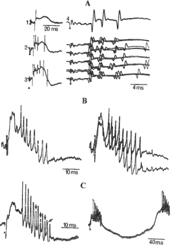
The synaptically elicited response of an interneuron usually
consists of a high-frequency repetitive (200-10001 sec, usually
400-600'/sec) spike barrage (Figures 3, 4, and 7). Such a burst
response is by no means necessary, as there are some types of
spinal interneurons that do not exhibit high-frequency barrages
to an afferent (Eccles, Eccles, & Lundberg, 1960) or a ventral
root volley (Willis & Willis, 1966). But these seem to be
excep-10 ms
Figure 3. Synaptic activation of putative interneurons in VL thalamus
and motor cortex of cat. Cells A and B: high-frequency barrage discharges
elicited in VL thalamus by precruciate cortical shocks (modified from
Steriade, Apostol, & Oakson, 1971; Steriade, Wyzinski, & Apostol, 1972).
Cell C was recorded in the motor precruciate area and driven by VL
shocks at increasing intensities (from 8 to 15 V); the 30-sweep sequence
(with reduced gain) shows the responses to 10 V stimulation; note the
progressive increase in number of spikes, shortening of latency and
increased intra-burst frequency with increase in stimulation strength
(modified from Steriade et al., 1974).
tions when considering the stereotyped burst patterns described,
following the discovery of cord Renshaw cells (Renshaw, 1946)
in the cerebellum (Eccles, Llinas, & Sasaki, 1966), inferior olive
(Llinas, Baker, & Sotelo, 1974), brain stem RF (Hikosaka &
Ka-wakami, 1976), VB (Andersen, Eccles, & Sears, 1964), and VL
(Marco, Brown, & Rouse, 1967; Steriade, Apostol, & Oakson,
1971; Steriade, Wyzinski, & Apostol, 1972) thalamus, lateral
ge-niculate (LG) (Burke & Sefton, 1966; Fukuda & Iwama, 1970;
Sakakura, 1968), olfactory bulb (Shepherd, 1963b, 1970),
hip-pocampus (Andersen, Eccles, & Loyning, 1964; Andersen,
Gross, L^m<^, & Sveen, 1969), and motor (Renaud & Kelly, 1974;
Stefanis, 1969; Steriade, Deschenes, & Oakson, 1974; Steriade et
al., 1974; Sukhov, 1968), somatosensory (Innocenti & Manzoni,
1972; Steriade & Yossif, 1977), and parietal association (Steriade,
Kitsikis, & Oakson, 1978) cortices. The prolonged spike barrage
THE BEHAVIORAL AND BRAIN SCIENCES (1978), 3
469
https://doi.org/10.1017/S0140525X00076147
A
40ms
Figure 4. Response patterns of putative interneurons in primary
so-matosensory area (SI) and parietal association cortex of cat. Cell A was
recorded in SI, stimulation applied to underlying white matter in animal
with lesion in VB thalamic complex; stimulation in 1, 2, and 3 was 0.09,
0.1, and 0.12 mA, respectively; note marked spike inactivation in the
mid-dle of the evoked burst in 3; in 4, same intensity as in 2: single sweep and
superimposed sweep sequences with reduced gain (modified from
Steriade and Yossif, 1977). Parietal association neurons B and C were
driven by stimulation of LP (B) and LI (C) thalamic nuclei; note spike
inactivation and fragmentation (arrows in C) within the spike barrage; the
lower speed sweep, with half gain, depicts both early and late events
evoked by the LI shock; note also that spike barrages occurred
im-mediately after the early depth-negative (depolarizing) wave that was
associated with synaptically evoked discharges in identified output cells
(modified from Steriade, Kitsikis &Oakson, 1978).
is often provoked by increasing stimulus intensity slightly above
that eliciting a single spike response (Figure 4A). But other
interneurons do not significantly increase the number of
dis-charges as a function of stimulus intensity, and a prolonged
bar-rage may occur at surprisingly constant latencies for each spike,
even at low intensity levels (Figures 3C and 4). With respect to
this, it was suggested by Shepherd (1970, p. 544; see his Figure
6B) that "impulse discharge is a very stereotyped affair,
inde-pendent to a certain extent of the amount of depolarization
im-pressed on the cell by the afferent volley." The spike amplitude
may diminish in the middle of the high-frequency burst of an
interneuron, and full discharge may even by reduced to abortive
spikes (Figure 4C). This "inactivation" process, described in
cerebellar cells as a consequence of strong depolarization
(Granit & Phillips, 1956) suggests that protracted EPSPs
un-derlie the repetitive spike activity of interneurons (Eccles,
Ec-cles, Iggo, & Lundberg, 1961; EcEc-cles, Fatt, & Koketsu, 1954;
Willis, 1971).
Should the high-frequency repetitive spike activity be
considered a distinct feature of interneurons, thus assuring a
clear-cut differentiation from synaptically evoked single spike
responses in output cells? An unequivocally positive answer to
this question is not available and some reservations are needed.
First, this distinction is valid in the analysis of responses to well
synchronized (electrical) stimuli applied to central pathways, as
peripheral (much less synchronized) stimulation may evoke
repetitive responses in output neurons too. With this
methodo-logical requirement, there is common agreement that even if a
small percentage of output cells (for example, dorsal
spino-cere-bellar tract (Kuno, 1969; Kuno & Miyahara, 1971) and cuneate
neurons (Calvin & Loeser, 1975, Galindo, Krnjevic, &
Schwartz, 1968)) may discharge spike doublets or triplets in
response to single shocks, prolonged burst discharges almost
never occur. "No one has reported recording a burst discharge
from a motoneuron" (Willis, 1971, p. 43). This applies not only to
spinal cord motoneurons. Striking differences have also been
ob-served between burst responses of interneurons (Shepherd,
1963b, 1970) and single orthodromic spikes (even after
increas-ing the intensity strength) in mitral cells (Shepherd, 1963a) of the
olfactory bulb. The same contrast has been repeatedly
em-phasized when investigating synaptically elicited responses in
hippocampal (Eccles, 1969), LG (Burke & Sefton, 1966;
Sakakura, 1968), VL (Steriade, Apostol, & Oakson, 1971;
Steriade, Wyzinski, & Apostol, 1972), and neocortical type I
versus type II neurons (Steriade, Deschenes, & Oakson, 1974;
Steriade et al, 1974; Steriade, Kitsikis, & Oakson, 1978;
Steri-ade & Yossif, 1977).
There are, of course, instances in which an output cell may
40 ms 40ms
Figure 5. Incremental responses of augmenting type in cortical neurons
of cat. A and B, two SI cells driven by VB thalamic stimulation at 1/sec (1)
and 10/sec (2). Note: orthodromic repetitive discharges evoked by 10/sec
shocks (with spike inactivation in A) occurring at longer latencies than
single spike discharges to 1/sec stimuli; the response to the first shock in
the 10/sec train is marked by arrow (modified from Steriade & Yossif,
1974). Neuron C was recorded from the parietal association cortex; two
100 msec-delayed shocks (10/sec stimulation) were applied to the LI
thalamic nucleus, and focal slow waves (fifty averaged sweeps in 1) and
unit discharges (fifty-sweep dotgram in 2) were recorded by the
mi-croelectrode; note that the response to the second shock had decreased
amplitude of the early slow depolarizing wave (a,l) and, relatedly,
decreased probability of firing in the first (a) part of the unitary response
(in 2), simultaneously with increased amplitude of b slow wave and
increased number of spikes in the secondary (b) repetitive firing
(modified from Steriade, Kitsikis & Oakson, 1978).
exhibit repetitive discharges in response to central stimuli.
1. In some cases, the group of repetitive discharges occurs
separately, a few milliseconds after the early, orthodromically, or
antidromically evoked single spike (Steriade, 1976; Steriade,
Deschenes, & Oakson, 1974; Steriade & Yossif, 1977). Two
pathways may be envisaged in such types of responses, the burst
probably reflecting activities in cortical excitatory interneurons
engaged in parallel by the testing volley. This is supported by
in-dependent alterations of the early and late parts of the response
in some experimental conditions (Steriade, 1976).
2. The augmenting responses of cortical output cells to 10/sec
thalamic stimulation may also consist of several discharges at
volleys following the first in a train (Figure 5), without, however,
reaching the long bursts of interneurons (Steriade & Yossif,
1974). In this case, too, the peculiar susceptibility of type II cells
to this kind of stimulation is probably at the basis of the spike
doublets or triplets in type I neurons. The greater involvement of
nonpyramidal, compared to pyramidal cells, in the mechanism
underlying the transition from primary to augmenting responses
was revealed in both the neocortex (Purpura, Shofer, &
Musgrave, 1964) and hippocampus (Andersen, Gross, L0m0, &
Sveen, 1969). One is tempted to speculate that the paradoxical
finding of increased latency associated with increased number
of spikes in response to the second and subsequent stimuli in a
train of 10/sec shocks (Figure 5) would result from simultaneous
blockade of direct thalamo-cortical excitation of output cells
combined with increased activities of excitatory interneurons
responsible for the secondary depolarization. This may well be
supported by firing changes from the first to the second shock at
10/sec in those cortical cells that exhibit two distinct components
of unitary responses; the second stimulus induces a decreased
probability of firing in the first (monosynaptically evoked)
dis-charge and, simultaneously, a spectacular increase of secondary,
repetitive spikes (Figure 5C).
What structural features and mechanisms are responsible for
the repetitive firing and underlying prolonged depolarization?
Several nonexclusive possibilities may be entertained. One of
them invokes regenerative excitation by positive feedback,
involving transmission of both depolarizing slow potentials and
spikes in a group of interconnected elements, leading to
syn-chrony of the network. This mechanism, described for initiation
and termination of burst activity in trigger-group neurons of
mollusks, is intrinsic to nonrectifying electrotonic coupling, and
may apply to some other coupled systems (Getting & Willows,
1974). Networks of interconnected interneurons are present in
the LG of monkeys (Pasik, Pasik, & Hamori, 1976). Since
evi-dence for such systems and mechanisms is still scarce in the
mammalian brain, the discussion will call upon the following
explanations, (a) Residual presynaptic action has been thought to
determine the amount of repetitive discharges in Renshaw cells:
when enzymatic destruction of synaptic transmitter is prevented,
a single synaptic volley may induce, instead of the usual 50
msec-burst, a rhythmic discharge lasting as long as 2 sec (Eccles,
Eccles, & Fatt, 1956). (b) The other explanation considers the
in-trinsic properties of the postsynaptic cell and arises from two
types of observations: the membrane properties of Renshaw
interneurons are altered for periods longer than that of the
presence of the chemical transmitter (Longo, Martin, & Unna,
1960); and identical time courses of transmitter action may affect
various postsynaptic neurons dissimilarly. This would imply that
multiple spike discharges are characteristic for some types of
cells and are possibly due to a smaller posthyperpolarization,
absence of susceptibility to inhibitory action, or other similar
fac-tors. The reader is referred to Calvin's (1974, 1975) papers for
elaborated models of multiple spike production. Differential
properties have been found in two populations of cortical output
cells, fast-conducting and slow-conducting PT neurons (Calvin
& Sypert, 1976; Koike et al, 1970; Takahashi, 1965), but such a
study has not yet been performed to distinguish long-axoned
from short-axoned neurons.
2.3.2 Spontaneous firing. Reports on background discharge of
interneurons are exceptionally few. A suitable analysis of
fluc-tuations in rate and pattern of spontaneous firing requires a
nonanesthetized animal, at best a chronically implanted
prepara-tion. Two papers describe spontaneous discharge of LG
tha-lamic (Sakakura, 1968) and precentral motor cortex (Steriade,
Deschenes, & Oakson, 1974) interneurons, as identified by
repetitive responses to volleys setting in motion afferent and/or
recurrent collateral pathways during sleep and wakefulness in
behaving animals. While statistical analyses of discharge rates
and patterns, indispensable in a study of spontaneous firing, are
only anecdotal in both Sakakura's work (1968) and our own
(Steriade, Deschenes, & Oakson, 1974), three findings deserve
attention in these studies, namely the lower mean-rate of firing of
interneurons compared to those of output cells (Sakakura, 1968),
arrest of firing on arousal (Sakakura, 1968; Steriade, Deschenes,
& Oakson, 1974), and a great proportion of very short (<10 msec
or even <5 msec) and very long intervals regardless of changes
in vigilance level (Steriade, Deschenes, & Oakson, 1974). All
these characteristics rendering interneurons basically different
from output cells have been confirmed in the statistical analyses
of association cortex type I and type II neurons reported in the
present article (see part 3).
2.3.3 "Injured" or "epileptic" cells? Since interneurons have
been described as discharging in bursts with "inactivation"
processes, much care should be taken to reject units
mechanically damaged by the microelectrode, as cell injury
creates conditions that may simulate the discharge pattern of an
"interneuron." Fortunately, criteria for the recognition of a
genuinely injured neuron are not lacking. Injury is certainly
more frequently expected when impaling nerve cells than with
extracellular recording. It is therefore not surprising that
experi-menters who use mostly intracellular electrodes (a) have more
chances to miss small-sized interneurons, and (b) are susceptible
to more anxiety when considering whether cells are "healthy" or
"unhealthy." But it is obviously necessary to document clearly
the "health" of units when describing interneuronal repetitive
responses. This would imply rejection of elements exhibiting
unusually high rates of discharge and illustrations with very fast
sweeps, allowing one to detect other signs of injury such as
notches on the first discharge in a high-frequency burst (the
suc-cessive ones may sometimes be "normally" fragmented due to
inactivation processes; see arrows in Figure 4C) and abnormal
duration and configuration of spikes. An injured cell cannot
sus-tain repetitive firing for periods longer than 10-15 min (Wyler &
Fetz, 1974), while a study of changes in activity of a bursting
interneuron during a full sleep-waking cycle requires long
dura-tion recordings (more than 1 hr). If these cells were "injured," it
remains mysterious why they stopped bursting on awakening
from sleep, continued to be silent for rather long periods of
arousal (10 sec [Sakakura, 1968], 7-15 sec [Steriade, Deschenes,
& Oakson, 1974]), and then progressively recovered the burst
pattern with repeated transitions from W to S (see Figure 12).
Besides these long-term recordings, an argument against the
burst being a mere injury artifact is its modification by afferent
driving (Calvin & Loeser, 1975). Furthermore, identified output
cells do not exhibit burst responses, nor do they discharge
spon-taneously with spike bursts during W (Evarts, 1964; Steriade,
Deschenes, & Oakson, 1974; Steriade et al., 1974) as
interneurons do (Steriade, Deschenes, & Oakson, 1974; Steriade
et al., 1974) (see also part 3). "Injury" is unlikely to be limited to
interneurons.
Studies of the Seattle group (Calvin, Sypert, & Ward, 1968;
Ward, 1969; Wyler, 1974; Wyler & Fetz, 1974; Wyler, Fetz, &
Ward, 1975) devoted to epilepsy have reported bursting patterns
in chronic alumina foci. As we did not attempt to render animals
epileptic, our subjects did not exhibit spontaneously recurring
focal and/or generalized motor seizures, nor did surface EEG or
focal slow waves simultaneously recorded with unit discharges
THE BEHAVIORAL AND BRAIN SCIENCES (1978), 3
471
https://doi.org/10.1017/S0140525X00076147from the cortical depth show EEG correlates of epilepsy.
Nonetheless, it is possible that chronic experiments with
multiple microelectrode penetrations might create conditions
leading to increased glial forms and "partial denervation" of
neuronal elements, which are thought to result in hyperactivity
of epileptic cells (Ward, 1969). The histology of the explored
cortical areas did not show evidence for this picture in our
ex-periments. In fact, what seems sufficient to preclude structural
epileptic changes in the recorded area is that nonoutput bursting
cells were recorded along the same track as antidromically
identified, output, nonbursting cells. In any event, there are
several striking differences in rate and pattern between epileptic
neurons and "healthy" interneurons. (a) The firing rate of
epileptic cells (see Figure 10-2 of Ward, 1969) is incomparably
higher than that of interneurons, and these recordings in
epileptic monkeys "were carried out only when they were
be-haviorally awake" (Ward, 1969, p. 268), a state when
interneurons are either silent or discharge at very low rates (see
3.1.2). (b) When comparing normal to epileptic cells, Calvin et al.
(1968) found in the latter a "long first-interval" and a late mode
due to the stereotyped silent periods between bursts, which are
not defining features of interneurons.
There are certainly a few resemblances between interneuronal
and epileptic bursts, and wyler et al. (1974; Wyler & Fetz, 1974;
Wyler, Fetz, & Ward, 1975) have discussed our findings on
cortical interneurons in relation to their own data on "epileptic"
cells. Moreover, some authors have inferred a role for
interneurons in the genesis of epileptic activity (Pollen, 1964),
but, again, type II cells, believed to be perhaps the most
im-portant elements of the epileptic neuronal aggregate, remained
inaccessible because of technical limitations in obtaining stable
recordings from interneurons (Prince, 1972). I have reported (see
1973, 1974) that during drowsiness, when spontaneous firing and
synaptic responsiveness of type II cells are enhanced, the spike
barrages of about 1/5 of precentral interneurons in the normal,
nonepileptic monkey become explosive as a consequence of VL
thalamic stimulation, leading to development in the cortical
depth of focal paroxysmal activity exhibiting a spikewave
pat-tern, with poor or no reflection at the cortical surface. PT neurons
recorded in the same region and even along the same track did
not exhibit such paroxysms. Thus, "normal cells were brought
into epileptic activities presumably as a consequence of both
their membrane properties and their extensive connections
fa-voring reverberation in closed neuronal chains" (Steriade, 1974,
p. 259). The same conclusion was drawn in studies of cortical
so-matosensory interneurons (Steriade & Yossif, 1974) with respect
to the progressive, unbroken transition between normal
responses and pathologic, paroxysmal events, with a striking
re-semblance between configuration of responses in the final stage
of stimulation and that of subsequently developing
self-sus-tained, epileptic-like activity.
2.4 Output-interneuronal circuits
As discussed above, output cells, and, with much less certainty,
interneurons can be identified by means of their response
properties. These two cellular types reciprocally articulate to
form complex circuits. The most extensively investigated
interneuronal organization is that of the spinal cord, due to the
work of the Swedish school in the last two decades. Yet, when
asked what he means by "identified," Lundberg (1969, p. 245)
emphasized that "great caution should be exercised in ascribing
interneurons with a certain type of response as belonging to a
given pathway." More recently, Jankowska (1975), who has
contributed greatly to the morphological identification of
in-tracellularly recorded spinal cord interneurons, has stressed the
difficulties of axonal staining with Procion Yellow and
con-cluded: "To define the role of these interneurons in a more
com-plete way, however, requires fuller knowledge of their target
cells and techniques allowing a detailed study of axonal
projec-tions" (p. 244).
If such is the case in the spinal cord, what will be found in the
thalamus and cortex? The most popular circuit accounting for
postsynaptic inhibition and rhythmically occurring spindle
waves in specific thalamic nuclei (experiments done on VB) was
based on excitation of local interneurons through the recurrent
collaterals of thalamo-cortical axons, and synchronous IPSPs
were believed to be elicited by ramification of interneurons
act-ing back on thalamic output neurons through inhibitory synapses
Figure 6. Diagrams showing postulated synaptic connections of axon
recurrent collaterals of PT cells (top) and of specific afferent fibers
(bot-tom). Inhibitory interneurons together with their inhibitory synapses on
the somata of the PT cells are shown in black. All other stellate cells and
the PT cells are assumed to be excitatory and are shown open. The arrows
indicate directions of impulse propagation. Note that, as suggested by the
experimental evidence, both the excitatory and inhibitory pathways can
include interpolated excitatory interneurons (from Eccles, 1969, courtesy
ofCh. C.Thomas).
on their soma (Andersen, Eccles, & Sears, 1964; Eccles, 1969).
As far as the recurrent collaterals of thalamo-cortical axons are
concerned, Scheibel et al. (1973) found them "in only 15 to 20
percent of all the cells . . . in the adult cat thalamus"; this would
be "an inadequate substrate for the powerful recursive effects
as-cribed to them" (p. 302). In concurrent studies using Golgi
tech-nique in the lateralis posterior (LP) thalamic nucleus, "the initial
collaterals of projection cells could be traced occasionally to
Golgi type II interneurons; however, regarding the sparseness of
this collateral arborization, it has little significance in the
neuronal connectivity" (Hajdu, Somogyi, & Tombol, 1974, p. 93).
The same difficulties for the recurrent collateral inhibitory
pathway arose from morphologic studies on the LG, which rather
supported the feed-forward type of inhibition, with retinal
af-ferents presynaptic to dendritic appendages of Golgi II cells
(Pasik,etal., 1973).
The diagrams used to depict synaptic connections within
output-interneuronal neo-(Eccles, 1969) and allocortical circuits
(Spencer, 1969) almost invariably postulate type II cells excited
by recurrent collaterals of pyramidal (type I) neurons. This can
be seen in the operational diagrams of Figure 6, which illustrate
the neocortical basic circuitry according to Eccles (1969). While
the importance of recurrent collaterals of thalamo-cortical axons
has been questioned, especially following the discovery of
dendritic synapses established in thalamic nuclei by type II cells
in almost all combinations (Famiglietti, 1970; Morest, 1971;
Pasik, Pasik, & Hamori, 1976; Ralston & Herman, 1969;
Scheibel, Davies, & Scheibel, 1973), the richness of synaptic
contacts made by recurrent collaterals of PT neurons has been
repeatedly mentioned, since Ramon y Cajal (1955), continuing
with Lo rente de No (1938) and other contemporary
neuroanatomists (Scheibel & Scheibel, 1970; Szentagothai,
1975; Tombol, 1975). The emphasis is on engagement of
interneurons by recurrent collaterals of output cells, and on
peri-cellular basketlike nests made by type II (inhibitory) synapses
around the somata of type I neurons (Colonnier, 1966;
Szentagothai, 1975). The shortest latency IPSPs are produced
via a bisynaptic pathway, with an intercalated inhibitory
interneuron, but, generally, inhibition is achieved by longer
pathways, with excitatory interneurons preceding inhibitory
ones (Figure 6).
Activity of excitatory interneurons activated by recurrent
collaterals of PT axons and acting back on PT cells was thought
to result in reexcitation of the latter (Chang, 1955). Unit
record-ings actually revealed "extra impulses" elicited at a few (6-10)
msec following the PT-evoked antidromic spike (Li, 1958).
However, without precautions to prevent spread of current from
the PT to ascending pathways, the "extra impulses" might be
as-cribed to contamination of lemniscal fibers. The activity of
ex-citatory interneurons is probably responsible for a period of
increased responsiveness, lasting about 20-30 msec following an
antidromic testing stimulation of the pes pedunculi, provided
that this was achieved in a preparation with a transected medial
lemniscus (Steriade & Yossif, 1977). It should be stressed that all
these facts are suggestive only of the existence of excitatory type
II cells interposed in the basic cortical circuit. The crucial
evi-dence will come from experiments utilizing cellular stimulation
and recording of cortical excitatory interneurons and output cells
belonging to the same neuronal pool.
The same is valid for inhibitory interneurons. The close
time-relation between high-frequency repetitive discharges of
interneurons and a focal positive wave, reflecting summated
hy-perpolarizing potentials in the neuronal pool, is commonly
regarded in the literature as indicative of the inhibitory nature of
the recorded bursting interneurons. Since, however, there is as
yet no criterion to differentiate electrophysiologically
excita-tory from inhibiexcita-tory type II cells, and as the former represent
the most powerful source of activation of the latter, some of the
bursting cells might be considered as excitatory local neurons,
driving the immediate progenitors of postsynaptic inhibition.
Renaud and Kelly (1974) recorded from pairs of neighboring PT
and non-PT cells and suggested, on the basis of reciprocal
changes in these two neuronal types, the inhibitory nature of the
latter. The latencies in their negative cross-correlograms ranged
from 1.9 msec to 7.3 msec. If short latencies (<2 msec) are taken
to imply monosynaptic inhibition of PT cells by non-PT cells, in
the case of longer latencies non-PT elements may be regarded as
type II excitatory cells driving inhibitory interneurons. The
con-nections of excitatory with inhibitory type II cells certainly do
not exhaust the yet undeciphered complexity of cortical neuronal
circuits.
When looking at Eccles (1969) diagrams in Figure 6, one may
be convinced that different interneurons are required for the
feedback and feed-forward collateral mechanisms. The goal of
those diagrams however is pedagogic. In fact, experiments
conducted on precentral motor cortex of primates have revealed
that the same type II cell may subserve both recurrent and
af-ferent collateral pathways (Steriade, Deschenes, & Oakson,
1974). The two interneurons illustrated in Figure 7 could be
driven from the pes pedunculi at latencies (<1.3 msec in A, <1
msec in B) indicating monosynaptic activation through recurrent
collaterals of fast PT fibers, and both were also activated at
slightly longer latencies (1.5-2.3 msec) from the specific
thalamic nucleus. Greater latency of thalamically elicited
repeti-tive discharges (compared to those evoked by peduncular
shocks) precludes spread of current to the internal capsule.
Summing up: It is possible to identify cortical type I cells by
5ms
Figure 7. Monosynaptic activation of putative interneurons in precentral
motor cortex of monkey by antidromic stimulation in the pes pedunculi
(PP) and by thalamic (Th) stimulation at the border between VB and VL
nuclei. A and B: two different interneurons. Left column in both cases is
PP stimulation; right column, Th stimulation. A: the PP testing shock was
delivered in 2 and 3 at a double and triple intensity relative to the liminal
one used in 1; intensity for superimposition of several traces in 4 (spikes
depicted with gain double of that in 1-3) was the same as in 2. Same unit
(A) was driven from thalamus (Th); 10 successive sweeps (with reduced
gain) and, below, superimposition of several traces. B: PP-evoked (left)
and thalamically elicited (right) responses depicted in the top
superim-positions (1) and, below, in 10-sweep sequences (2). Vertical bar (at right):
0.8 mV for A, 2 mV for B (modified from Steriade, Deschenes, & Oakson,
1974).
THE BEHAVIORAL AND BRAIN SCIENCES (1978), 3
473
https://doi.org/10.1017/S0140525X00076147
antidromic invasion, to know some of their target structure(s),
and to estimate their size on the basis of the conduction
ve-locities of their axons. Without morphological confirmation, the
features of evoked and spontaneous firing are not sufficient to
recognize unequivocally cortical type II cells; however, when
all electrophysiological criteria are met, it can be strongly
in-ferred that a locally ramifying neuron has been encountered.
There is as yet no definitive sign to differentiate
electrophysio-logically an excitatory from an inhibitory type II cell in the
cere-bral cortex.
3 Cortical output cells and interneurons during the
sleep-waking cycle
Our initial observation of the strikingly dissimilar alterations in
the firing rate of cortical output cells and interneurons as a
conse-quence of changes in vigilance level was made during
experi-ments on encephale isole cats. An "arousing" stimulation of the
2