Carcinogenesis vol.16 no. 10 pp.2553-2560, 1995
Influence of amino acids on the formation of mutagenic/
carcinogenic heterocyclic amines in a model system
Maria A.EJohansson3, Laurent B.Fay1, Gian A.Gross1,
Kjell Olsson2 and Margaretha Jagerstad
Department of Applied Nutrition and Food Chemistry, Chemical Centre, Lund University, PO Box 124, S-221 00 Lund, Sweden, 'Nestec Ltd, Nestlf Research Centre, PO Box 44, Vers-chez-les-Blanc, CH-1000 Lausanne 26, Switzerland and 2Department of Chemistry, Swedish University of
Agricultural Sciences, PO Box 7015, S-750 07 Uppsala, Sweden 'To whom correspondence should be addressed
Mixtures of creatinlne, glucose and various single amino acids
were heated at 180°C for 10 min in an aqueous model system.
The heated mixtures all showed mutagenic activity, ranging
from 80 to 2400 TA98 revertant colonies/ujnol creatinine with
metabolic activation. Testing of HPLC fractions for mutagenic
activity showed each mixture to contain several mutagenic
components, some of which corresponded to known
hetero-cyclic amines and others to unknown compounds. The
presence of amlno-3-methyl-irnidazo[4,5-/|quinoxaline,
3$-dimethylimidazo[4,5-/|quinoxaline and
2-amino-3,7,8-trimethylimid^zo[4,5-/|quinoxaline in most of the
samples was established using HPLC with photodiode array
detection and liquid chromatography/mass spectrometry with
electrospray interface and single ion monitoring. In addition,
3,4,8-trimethyllniidazo[4,5-/]quinoxaline,
2-amino-l-methyl-6-phenylimidazo[4,5-6]pyridine,
3-amlno-l,4-di-methyl-5//-pyrido[4,3-fc]indole and
3-amino-l-methyI-5ff-pyrido[4,3-*]indole and the co-mutagenic compounds
9H-pyrido[3,4-*]indole and l-methyl-9H-9H-pyrido[3,4-*]indole
were detected in some samples.
Introduction
The major food mutagens isolated to date from cooked meat
and fish products are heterocyclic amines (HAs*). Since first
reported by Sugimura et al. (1), much effort has been devoted
to identifying and quantifying these compounds and to
under-standing the mechanisms of their formation (for reviews see
2,3). Most of the mutagenic heterocyclic amines formed
at normal cooking temperatures are imidazoquinolines or
imidazoquinoxalines (IQ compounds), imidazopyridines (e.g.
2-amino-l-methyl-6-phenylimidazo[4,5-b]pyridine; PhIP) or
imidazofuropyridines (4-6).
Many HAs are multipotent carcinogens in long-term rodent
bioassays (for a review see 7), with
2-amino-3-methylimid-•Abbreviations: HA(s), heterocyclic amine(s); PhIP, 2-amino-1
-methyl-6-phenylimidazo[4,5-6)pyridine; IQ, 2-amino-3-methylimidazo|4,5-/]quinoline; IQx, 2-amino-3-methylimidazo[4,5-/lquinoxaline; MelQ, 2-anuno-3,4-dimethylimidazo[4,5-./]quinoline; MelQx, 2-amino-3,8-dimethylimidazo[4,5-/Iquinoxaline; 4,8-DiMeIQx, amino-3,4,8-trimethylimidazo[4,5-/]quinox-aline; LC/MS, liquid chromatography/mass spectrometry; 7,8-DiMeIQx, 2-amino-3,7,8-trimethylimidazo[4,5-/|quinoxaline; Glu-P-1, 2-amino-6-methyl-dipyrido[l,2-a:3',2'-rf|imidazole; Glu-P-2, 2-aminodipyrido[l,2-<i:3',2'-<] imidazole; Trp-P-1, 3-amino-l,4-dimethyl-5//-pyrido[4,3-b]indole; Trp-P-2, 3-amino-l-methyl-5//-pyrido[4,3-fc]indole; harman, l-methyl-9//-pyrido[3,4-6]indole; norharman, 9H-pyrido[3,4-i]indole.
azo[4,5-/]quinoline (IQ) also positive in primates (8).
Epidemi-ological studies have shown a relationship between the
consumption of fried meat products and an elevated risk of
colon and other cancers (9-13). The HAs are animal
carcino-gens and the IARC classified several of them as possible or
probable (IQ) human carcinogens (14-16). Efforts to minimize
their formation in cooked foods are therefore of great
impor-tance and a better understanding of the formation mechanism
is needed. Simple modelling experiments have been useful in
providing basic information on precursors, inhibitors and the
effect of reaction conditions and a good correlation between
the formation of HAs in cooked foods and model systems has
been observed (for reviews see 3,17).
All details of the formation mechanism of HAs have not
yet been clarified. Jagerstad et al. (18) suggested that IQ
compounds may be produced via the Maillard reaction from
creatin(in)e, amino acids and hexoses present in foods of
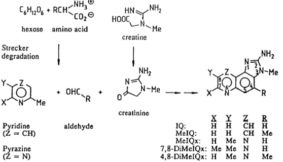
animal origin (see Scheme 1). This hypothesis has been
confirmed by heating creatin(in)e, amino acids and sugars in
various model systems, resulting in the formation of IQ,
2-amino-3-methylimidazo[4,5-/lquinoxa]ine (IQx),
2-amino-3,4-dimethylimidazo[4,5-/lquinoline (MelQ),
2-amino-3,8-dimethyl-imidazo[4,5-/]quinoxaline (MelQx),
2-amino-3,4,8-trimethyli-midazo[4,5-/|quinoxaline (4,8-DiMeIQx) and PhIP (for
reviews see 3,19). Also,
l4C-labelled glucose was shown to
be incorporated into IQx, MelQx and 4,8-DiMeIQx using an
aqueous model system (20). Alternative routes for the formation
of HAs may exist, since their formation from creatin(in)e and
amino acids has also been observed in the absence of sugar
in dry heating model experiments. However, the yield of HAs
in the absence of sugar is usually lower (for a review see 3).
According to the hypothesis illustrated in Scheme 1, amino
acids act as precursors of HAs partly by serving as a nitrogen
source in pyridine or pyrazine formation (18). However,
mutagenic activity and HA yield previously reported in model
systems vary depending on the particular amino acid used (for
reviews see 3,17). These variations were not completely
explained and, so far, the ability to produce mutagenic activity
in a model system has been investigated for less than half the
naturally occurring amino acids, and even fewer amino acids
have been tested as precursors of HAs.
This study was performed to further investigate the effect
of various amino acids on the yield and nature of HAs formed
in a model system and to increase the understanding of the
reactions behind the formation of HAs. Model mixtures, each
containing creatinine, glucose and one of the most common
amino acids, were heated at 180°C for 10 min. The heated
mixtures were tested for mutagenic activity using Salmonella
typhimurium TA98 with metabolic activation. Furthermore, the
model mixtures were purified using the propylsulphonic
acid-silica gel tandem extraction method (21) and analysed for
known HAs using HPLC wkh a photodiode array and
fluores-cence detection and liquid chromatography/mass spectrometry
(LC/MS) with electrospray ionization and single ion
mon-itoring. The HPLC fractions were tested for mutagenic activity.
®
V
HOOC
hexose amino acid ^Me
creatine
Strecker
degradation
Y-^Z
Pyridine
(Z =• CH)
Pyrazine
(Z = N)
NH
2• OHL +
aldehyde
creatinine
IQ:
MelQ:
MelQx:
7,8-DiMeIQx:
4,8-DiMeIQx:
X
H
H
H
Me
H
H
H
Me
Me
Me
Z
CH
CH
N
N
N
£
H
Me
H
H
Me
Scheme 1. Suggested pathway for the formation of imidazoqumolines and imidazoquinoxalines (18).Materials and methods
Chemicals
AJI chemicals and solvents were of HPLC or analytical grade. The solvents, e.g. acetonitrile, methanol and dichloromethane, were purchased from Merck AG (Darmstadt, Germany). Water was obtained from a Milli-Q water purifica-tion system (Millipore, Bedford, MA). Creatinine and amino acids were obtained from Sigma Chemical Co. (St Louis, MO) and glucose from BDH Chemicals Ltd (Dorset, UK). Synthetic IQ, IQx, MelQ, MelQx, 4,8-DiMeIQx, amino-3,7,8-trimethylimidazo[4,5-/]quinoxa]ine (7,8-DiMeIQx), PhIP, 2-amino-6-methyl-dipyrido[l,2-a:3',2'-rf]imidazole (Glu-P-1), 2-aminodipyr-ido[l,2-a:3',2'-<flimidazole (Glu-P-2), 3-amino-l,4-dimethyl-5//-pyrido[4,3-b]indole (Trp-P-1), 3-amuio-l-rnethyl-5//-pyrido[4,3-6]indole (Trp-P-2) and 1-methyl-9//-pyrido[3,4-i>]indole (harman) were obtained from Toronto Research Chemicals (Downsview, Ontario, Canada) and 9//-pyrido[3,4-6]indole (norhar-man) from Aldrich (Steinheim, Germany). The materials used for propylsulph-onic acid-silica gel tandem extraction (Extrelut and BondElut, e.g. PRS and C]g) were obtained from Merck AG (Darmstadt, Germany) and Analytichem International (Sorbent, Vastra FrOlunda, Sweden).
Sample preparation
Samples were prepared by heating creatinine, glucose and various amino acids in an aqueous model system as previously described (22). In brief, creatinine (0.9 mmol), glucose (0.45 mmol) and either (0.9 mmol) glycine, alanine, valine, leucine, isoleucine, serine, threonine, aspartic acid, asparagine, glutamic acid, glutamine, lysine, arginine, histidine, phenylalanine, tyrosine, tryptophan, cysteine, cystine, methionine, proline or hydroxyproline were dissolved in 2.5 ml water and heated for 10 min at 180°C in sealed test tubes. A blank sample (without any amino acid) was prepared concurrently.
After heating the samples were purified using the solid phase extraction method of Gross (21) with some minor modifications (22). Using this method a polar extract (containing the IQ-type HAs and glutamic acid pyrolysates) and a non-polar extract (containing pyridoindoles) were obtained. Only the polar extract was analysed for HAs in this study.
HPLC fractionation for mutagenic activity profiles
Extract residues obtained after purification were dissolved in 250 (il HPLC buffer A (see below) and aliquots (30 u.1) w e r e injected (Varian 9100
Autosampler) into a Varian 9010 Liquid Chromatograph with a photodiode array UV detector (Varian 9065, Polychrom) equipped with a ToyoSoda TSK Gel ODS 80TM column (250X4.6 mm i.d., 5 nm particle size; Varian, Stockholm, Sweden) and a pre-column (Supelguard LC-18-DB, 20X4.6 mm i.d.) and eluted with a mobile phase of 10 mM aqueous triethylamine adjusted with acetic acid to pH 3.2 (A) or pH 3.6 (B) and acetonitrile (C). A gradient of 5-15% C in A for 10 min, then 15-25% C in B for 10 min and finally 25-55% C in B for 5 min was used. The flow rate was 1 ml/min and the effluent was monitored at 263 nm. Fractions were collected every 30 s between 8 and 30 min and lyophilized before assaying for mutagenic activity.
Table I. Mutagenic activity of model mixtures containing creatinine,
glucose and various amino acids heated at 180°C for 10 min Amino acid Mutagenic activity1
Cysteine Cystine Threonine Lysine Serine Alanine Histidine Asparagine Tyrosine Arginine Glutamine Glycine Aspartic acid Methionine Isoleucine Proline Valine Leucine Hydroxyproline Tryptophan Glutamic acid Phenylalanine 2420 2251 796 556 483 478 439 432 322 314 306 290 271 262 261 240 240 198 177 102 92 83
*TA98 revertants/(imol creatinine, with metabolic activation from the linear portion of dose-response curves from replicate platings.
Mutation assay
The mutagenic activity of the lyophilized HPLC fractions was tested as described by Ames et al. (23) using Salmonella strain TA 98 with the addition of 0.5 ml S9 mix containing 5% chlorophene-induced rat liver/plate (24). The fractions were tested at single doses. Crude heated model mixtures were tested in duplicate at three different doses to establish dose-response curves. The number of revertants/nmol original creatinine was calculated from the linear part of the curve (25). Synthetic MelQx was used as a positive control (50 000 revertants/jlg). The colonies were counted in an automated colony counter using the software Cream™ (Kem-En-Tec, Copenhagen, Denmark). The spontaneous reversion rate was 30-35 revertants/plate. A fraction or sample was considered mutagenic if it induced revertants to twice the background level.
Identification and quantification of HAs using HPLC
The HAs extracted from the heated model systems were identified and quantified by HPLC employing a Hewlett Packard 1090M system containing
Influence of amino adds on HA formation I
If „
KMr
7KtKk UMIift•
n
CMI
l h
« m1
KM1
=
Pli
IJ
—CTTtmm KMa
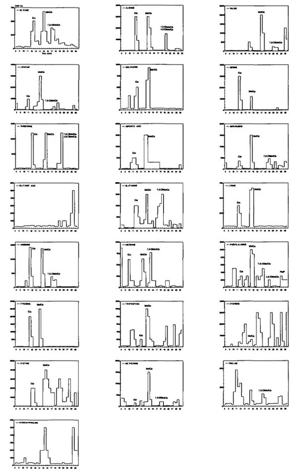
Figure 1. Mutagenic activity in TA98 with metabolic activation in fractions from HPLC separation of heated model mixtures containing various amino acids.
Mutagens corresponding to the retention times of HAs are indicated. Retention times of synthetic HAs are: Glu-P-2, 11.3 min; IQ, 11.6 min; IQx, 11.8 min; MelQ, 14.3 min; MelQx, 15.1 min; Glu-P-1, 15.3 min; 7,8-DiMeIQx, 17.2 min; 4,8-DiMeIQx, 18.3 min; norharman, 21.1 min; harman, 23.1 min; Trp-P-2,
MelQx mAU 30 20 10 0 IQx 7,8-OIMelQx 10.0 '12.5 '15.0 17.5 2 0 0 Time (mln] mAU 20-1 15 10 MclQx mAU 1.5 1.0 0.5 7,8-DIMelQx 7 0 0 250 300 2 0 0 7 5 0 300 Wavelength (nm) 200 7 5 0 '300
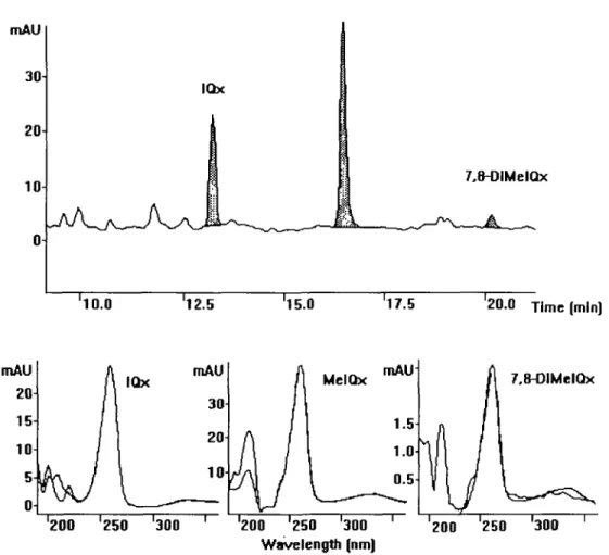
Figure 2. Expanded region of a UV chromatogram (wavelength 263 nm) from HPLC analysis of a heated model mixture containing tyrosine. Peaks
corresponding to known HAs are indicated. On-line recorded UV spectra compared with those of synthetic IQx, MelQx and 7,8-DiMeIQx. a photodiode array and a time programmable fluorescence detector (HP
1046A) connected in series. Chromatographic conditions were as above. UV detection was performed at 263 nm while excitation/emission wavelengths were 360/450 nm for Glu-P-1 and Glu-P-2, 300/440 nm for harman and norharman, 265/410 nm for Trp-P-1 and Trp-P-2 and 315/390 nm for PhlP. Aliquots of 10 nl from 200 nl of the purified samples were injected.
HAs were identified by comparing the retention times of the peaks with those of synthetic compounds, namely IQ, IQx, MelQ, MelQx, 4,8-DiMeIQx, 7,8-DiMeIQx, PhJP, Glu-P-1, Glu-P-2, Trp-P-1, Trp-P-2, norharman and harman, obtained under the same conditions. In addition, some extracts were also spiked with synthetic compounds before injection. UV spectra of synthetic compounds obtained under the same conditions, together with literature data, were used to confirm the identities of the HAs.
The amounts of HAs were estimated by comparing the HPLC peak area of the chromatographed sample with that of a known amount of standard. The amounts were not corrected for incomplete extraction recovery.
LC/MS analysis of HAs
HPLC was performed with a Waters 600-MS pump, a Waters 490-MS UV detector and a Waters 717 autosampler injecting 20 ul of the sample. The column was a ToyoSoda TSK Gel ODS 80TM (250X4.6 mm i.d., 5 nm particle size), protected with a pre-column containing the same stationary phase. The mobile phases were as follows: solvent A, 10 mM ammonium acetate adjusted with HC1 to pH 3.2; solvent B, 10 mM ammonium acetate adjusted with HC1 to pH 4.0; solvent C, acetonitrile. The linear gradient program was as follows: 0-10 min, 5-17% C in A; 10-10.1 min exchange of buffer B for A; 10.1-22 min, 17-55% C in B; 22-25 min, 55-90% C in B. The flow rate was 1 ml/min and 50% of the flow was split off before entering the mass spectrometer. MS was performed with a Finnigan TSQ-700 mass spectrometer (Bremen, Germany) equipped with a Finnigan electrospray interface working at the high voltage of 4.5 kV. The manifold temperature was 70°C and the heated capillary was set at 250°C. Nitrogen was used as sheath gas at a pressure of 4.8 bar. The compounds were detected after monitoring the protonated molecular ions [M + H]+.
Results
Formation of mutagenic activity in model mixtures
The heated model mixtures all showed mutagenic activity in
Salmonella typhimurium TA98 with metabolic activation, as
shown in Table I. The mutagenic activity ranged from 80 to
2400 revertants/(i.mol creatinine. Mixtures containing cysteine,
cystine or threonine showed the highest response, while the
lowest was found in mixtures containing tryptophan, glutamic
acid or phenylalanine. No detectable mutagenic activity was
formed in the blank sample.
HPLC fractions were tested for mutagenic activity to
deter-mine the number of mutagenic compounds and to compare
the elution times of the mutagenic components with those of
known HAs. Figure 1 shows the mutagenic activity profiles
for heated samples containing different amino acids. Each
profile contained two to seven mutagenic peaks, some of which
elute at similar retention times to those of known HAs found
in the pyrolysis products of foods. The mutagenic compounds
co-eluting with known HAs are indicated in Figure 1.
Identification of mutagenic compounds
By comparing the retention times of the mutagenic peaks with
those of known HAs and by using HPLC UV spectrometry
and LC/MS analysis, the presence of several known HAs in
the various model systems was established. Figures 2 and 3
show chromatograms obtained from a heated model sample
containing tyrosine from HPLC and LC/MS analyses
respect-Influence of amino adds on HA formation
A
100, 50 1«fr 100 so 100 60 100 60-100 60 100-60 100 60 100 60 100! 60 Olu-P-2 tQ du-P-1 HoJQ E+06 3.723 E+O« 1.093 E+06 HeKtx 7,6-dllMQx 4,8-dUUKht AoC Trp-P-2 nVz:1M BoAaCA
E+08 1.704 E+oe 2.613 E+O8 1.328 E+O8 1224 E+Of 2.626 E+O8 Trp-P-1 E+06 3.764 E+06 3.426 1 0 * 0 13:20 16:40 20:00 23:20 26:40 30:00 Ratantton dm* (mdn)B
100 10O 6a 100-60 100 60 m/z:200 mfc214 « * . nWz:22a! f
MatQxI f
PhIP 7,8-dHMQjl E+06 1.0*8 E+06 1.936 E+03 3.184 E+04 1.803 1ChOO 13:20 16:40 2040 23:20 Ratantion d i m (mln)Figure 3. LC7MS analysis of a model system. (A) Selected ion monitoring
of reference HAs at the respective mJz [M+H]+ ions. (B) Analysis of a
sample containing tyrosine. The chromatogram displays the expanded region containing IQx, MelQx, PhIP and 7,8-DiMeIQx (shadowed).
ively. A comparision of UV absorption spectra of the tyrosine
sample and those of synthetic references is also shown in
Figure 2. As shown in Table II, IQx, MelQx and 7,8-DiMeIQx
were present in most of the heated model mixtures. In addition,
4,8-DiMeIQx was detected in samples containing alanine,
threonine, lysine, phenylalanine or methionine. We failed to
confirm the presence of IQx with LC/MS in some samples,
due to unstable signals (Table II). Norharman and harman
were formed in samples containing isoleucine, arginine,
phenyl-Table II. Estimated amounts (jimol/mol creatinine) of IQx, MelQx and
7,8-DiMeIQx formed in heated mixtures of glucose, creatinine and amino acids analysed using HPLC with photodiode array detection
Amino acid IQx MelQx 7,8-DiMeIQx
Glycine Alanine Valine Leucine Isoleucine Serine Threonine Aspartic acid Asparagine Glutamine Lysine Arginine Histidine Phenylalanine Tyrosine Tryptophan Cysteine Cystine Methionine Proline 3.1 ± 0.8 5.0" c c" c 6.5* 4.5' c" 2.9s 1.4 c" 3.7 ± 0.3 1.8 1.5 ± 0.3 2.6 ± 2.3 0.7 0.1 0.9* 3.6 ± 1.2 5.4 ± 5.1 4.7 ± 3.5 2.4 2.7 ± 0.7 3.0 ± 1.7 9.2 ± 2.1 0.8 ± 0.6 1.5 0.6 7.7 ± 3.7 4.1 ± 0.7 3.4 ± 3.2 1.9 ± 0.7 3.6 ± 2.1 5.0 ± 3.5 c c 2.7 ± 0.5 0.3 0.7 cb c c 0.1 c 0.2 c c ca c* 0.1 c c c c ' "Not detected using LC/MS.
bc, co-eluting compounds interfered with peak area determination. Amounts are means from the analysis of duplicate samples. LC/MS with electrospray interface and single ion monitoring was used for the detection of the compounds.
alanine or tryptophan. Norharman was also formed in a model
mixture with tyrosine. PhIP was found in model mixtures
containing phenylalanine, isoleucine or tyrosine. Trp-P-1 and
Trp-P-2 were found in a model mixture containing tryptophan.
Trp-P-1 was also detected in the isoleucine sample. IQ, MelQ,
Glu-P-1 and Glu-P-2 were not found in any of the samples.
No known HAs were found in the glutamic acid and
hydroxy-proline samples or in the blank sample. However, several
unknown mutagenic components were present in the heated
samples.
Quantification of HAs
The amounts of HAs in the samples were estimated from the
UV and fluorescence chromatograms and are shown in Table
II for IQx, MelQx and 7,8-DiMeIQx; their maximum yields
were 6.5, 9.2 ± 2.1 and 0.7 umol/mol creatinine respectively.
Samples containing arginine, glycine or tyrosine produced
large amounts of IQx. Most MelQx was formed in the sample
containing threonine, followed by samples containing lysine,
tryptophan and alanine. The alanine-containing sample
pro-duced 1.1 (jjnol 4,8-DiMeIQx/mol creatinine, while 7.3 ± 4.2
(xmol PhlP/mol creatinine were produced in the
phenylalanine-containing sample. Due to co-eluting compounds, it was
impossible to estimate the amounts of HAs, especially
7,8-DiMeIQx and 4,8-7,8-DiMeIQx, in some of the samples. The
amounts of norharman, harman, Trp-P-1 and Trp-P-2 were not
determined. The amounts of HAs given in Table II are not
corrected for incomplete extraction recovery. Extraction of
similar model mixtures gave a recovery of 52-97% for
MelQx (19,22).
Discussion
The hypothesis for the formation of IQ compounds in Scheme
1 postulated that creatine forms the 2-aminoimidazo part by
cyclization and dehydration. The quinoline/quinoxaline part of
the molecule arises from pyridines or pyrazines and Strecker
aldehydes, formed in the Maillard reaction (18). Surprisingly,
neither IQ nor MelQ was found in any of the samples. This
implies mat no pyridines are formed in die model system. IQ
and MelQ have previously been detected in model systems
with fructose (26,27) and in dry heating experiments (for a
review see 3).
Amino acids act as precursors of HAs by serving as a
nitrogen source in pyrazine formation (28). They also seem to
provide the carbon and, in some cases, the methyl group at
position 4 (18). According to the hypothesis, glycine is a
precursor of IQ, MelQx and 7,8-DiMeIQx, while alanine might
give MelQ and 4,8-DiMeIQx. However, in this study IQx,
MelQx and 7,8-DiMeIQx were formed from most amino acids,
including alanine, when heated with glucose and creatinine in
the model system. Moreover, 4,8-DiMeIQx was formed not
only from alanine, but also from threonine, lysine,
phenylalan-ine and methionphenylalan-ine.
Some of these results are hard to reconcile with the
hypo-thesis illustrated in Scheme 1. However, several amino acids
are derivatives of alanine carrying an electropositive group
(E), e.g. a heteroatom with a free electron pair or an aromatic
ring with TI electrons. These amino acids, e.g. serine, threonine,
cysteine, cystine, phenylalanine, tyrosine, tryptophan and
histi-dine, might undergo retroaldolization to glycine,
NH
3H - E - C H
2CH
-NH
3= CH
2" + C H
2- C O
2'
thereby explaining the formation of IQx, MelQx and
7,8-DiMeIQx. The other retroaldolization product (E = CH
2) of
threonine is acetaldehyde, which could explain the formation
of 4,8-DiMeIQx from threonine. Less reactive retroaldolization
products are expected from aromatic amino acids. This might
explain why IQx compounds with an aryl group at position 4
have never been observed. It is more difficult to guess which
reactions valine, leucine, isoleucine, methionine, asparagine,
glutamine, aspartic acid, glutamic acid, proline and
hydroxy-proline undergo in the model system. One possibility might
be that these molecules fragment through free radical reactions.
Clearly, the observed IQx compounds do not always fit the
hypothesis in Scheme 1 and other reaction pathways cannot
be excluded. The divergence might be explained by the
preferred formation of other HAs, such as PhIP from
phenylal-anine and Trp-P-1 and Trp-P-2 from tryptophan.
Pyrazines arise through cyclodimerization of aminodeoxy
sugars, formed from a-dicarbonyl compounds and amino acids
in the Strecker degradation (29). The yield and species of
pyrazines vary with the amino acid (28, 30-33). Sugars are
known to fragment through retroaldol or related reactions. If
fragmentation precedes pyrazine formation, glyoxal,
methyl-glyoxal and/or biacetyl serve as a-dicarbonyl sources.
Methyl-glyoxal is the most common source, giving rise to
2,5-dimethylpyrazine, which is die precursor of MelQx and
4,8-DiMeIQx. If glyoxal and methylglyoxal co-dimerize,
pyrazine, which is the precursor of IQx, is formed. If
methyl-glyoxal and biacetyl co-dimerize, trimethylpyrazine, which is
the precursor of 7,8-DiMeIQx, is obtained. Consequently, a
mixture of glyoxal, methylglyoxal and biacetyl may have been
present in our model mixtures, thus giving rise to IQx, MelQx,
4,8-DiMeIQx and 7,8-DiMeIQx. However, the
methylpyraz-ines may also have formed through initial cyclodimerization,
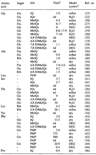
Table n i .Literature data on heterocyclic amines in i amino acids and creatin(in)e with and without sugar Amino acid Gly Ala Leu Ser Thr Lys Tyr Phe Pro Sugar Fru Glc Glc Fru Glc Glc Glc Glc Glc Glc Glc Fru Glc Rib -Fru Glc Rib -Glc Glc Glc Glc Glc Rib Rib -Glc Glc Glc Glc -Glc -Glc -HA IQ IQx MelQx MelQx MelQx MelQx MelQx 4,8-DiMeIQx 4,8-DiMeIQx 7,8-DiMeIQx 7,8-DiMeIQx MelQ MelQ MelQ MelQ 4,8-DiMeIQx 4,8-DiMeIQx 4,8-DiMeIQx PhIP IQ IQx MelQx IQx MelQx MelQx 4,8-DiMeIQx 4,8-DiMeIQx MelQx 4,8-DiMeIQx MelQx IQ IQ MelQx 4,8-DiMeIQx PhIP PhIP PhIP PhIP PhIP PhIP IQ Yield1 1.0 nd 4.4 6-7 4 8.8-17.9 7-10 nd nd 1.1 nd nd 0.9 1.8 nd 1.9-2.6 4.2 1.5 nd 3.7 2.7 nd nd nd 9 nd 30 4.2 26.1 nd 3.0 13.5 nd nd 3.6 735 560 nd 20.9 6.4 0.4 nodel systems Model system2 reflux H2O reflux reflux DEG H2O H2O DEG H2O reflux DEG reflux reflux reflux dry reflux reflux reflux dry dry dry dry H2O reflux H2O reflux H2O reflux reflux dry dry dry DEG DEG reflux dry dry dry DEG DEG dry from Ref. no. (27) (22) (38) (27) (35) (19) (20) (35) (19) (39) (35) (26) (40) (40) (34) (26) (40) (40) (34) (41) (41) (34) (20) (42) (20) (42) (20) (40) (40) (34) (43) (43) (44) (44) (45) (42) (42) (34) (44) (44) (46) 'Yield in nmol/mmol creatin(in)e.
nd = Not determined.
2Reflux = reflux-boiling in diethylene glycol/water (5:1) at 125-128°C for
2 h; H2O = heated in H2O in sealed test-tubes at 180°C for 10 min;
DEG = heated in deithylene glycol/water (5:1) at 180°C for 10 min; dry = dry heating at 180°C for 1 h.
followed by fragmentation of the pyrazine substituents.
All amino acids tested produced mutagenic activity to a
varying extent when heated with creatinine and glucose in a
model system. The mutagenic activity was generally higher
than reported earlier. When various amino acids, creatine and
glucose were boiled under reflux in aqueous diethylene glycol
for 2 h at 128
CC only samples containing threonine, glycine
or lysine produced mutagenic activity exceeding 200 TA98
revertants/nmol creatine (18). Mixtures containing cysteine
and cystine, which produced the highest mutagenic activity in
the present study, only showed weak mutagenic activity in our
previous study. One explanation might be that a closed model
system was used in the present study, preventing volatile
intermediates from vaporizing. In another study by Overvik
et al. (34) various amino acids produced much lower mutagenic
activity after dry heating (1 h at 200°C) with creatine in the
absence of sugar. In that study samples with serine or threonine
Influence of amino acids on HA formation
showed the highest mutagenic activity. However, serine and
threonine are known to decarboxylate and produce pyrazines
through dimerization when heated (29).
Comparing our results with previous studies on the formation
of HAs in model systems containing various amino acids (see
Table HI) shows both similarities and differences. However,
different kinds of model systems have been used, e.g. dry
heating at 180°C for 2 h, reflux boiling in diethylene glycol/
water at 125-128°C for 2 h, heating in diethylene glycol/water
at 180°C for 10 min in open test tubes and heating in water
at 180°C for 10 min in sealed test tubes.
To our knowledge, this is the first study reporting the
formation of IQx and 7,8-DiMeIQx from most amino acids
tested (for a review see 3). In addition, several unknown
mutagenic components were present in the heated samples.
New mutagenic compounds have been first identifed in model
systems and later identified in cooked foods (35-37). Much
still remains to be done before the complex reaction
mechan-isms behind the formation of HAs is totally elucidated and the
unknown mutagenic compounds are identified.
Acknowledgements
We thank Mr Santo AH for his excellent technical assistance. This study was supported by the Swedish Cancer Foundation (1824-B93-10XBC) and the Swedish Council for Forestry and Agricultural Research (50.00440/91).
References
1. Sugimura,T. et al (1977) Mutagens-carcinogens in food, with special reference to highly mutagenic pyrolytic products in broiled foods. In Hiatt,H.H., WatsonJ.D. and WinstenJ.A. (eds), Origins of Human Cancer. Cold Spring Harbor Laboratory Press, Cold Spring Harbor, NY, pp. 1561-1577.
2.FeltonrI.S. and Knize,M.G. (1991) Occurrence, identification and bacterial
mutagenicity of heterocyclic amines in cooked foods. Mutat. Res., 259, 205-217.
3. Skog.K. (1993) Cooking procedures and food mutagens: a literature review.
Fd Chem. Toxicol., 31, 655-675.
4. Sugimura,T. and Sato,S. (1983) Mutagens—carcinogens in food. Cancer
Res., 43 (suppl.), 2415s-2421s.
5.FeltonJ.S. and Hatch.F.T. (1986) Mutagens in cooked foods. Energy
TechnoL Rev., 1-15.
6. Knize,M.G., Roper,M., Shen,N.H. and FeltonJ.S. (1990) Proposed structures for an amino-dimethylimidazofuropyridine mutagen in cooked meat. Carcinogenesis, 11, 2259—2262.
7.Ohgaki,H., Takayama,S. and Sugimura,T. (1991) Carcinogenicities of heterocyclic amines in cooked food. Mutat. Res., 259, 399^12. 8. Adamson.R.H., Thorgeirsson,U.P., Snyderwine.E.G., Thorgeirsson.S.S.,
ReevesJ., DalgaitLD.W, Takayama,S. and Sugimura,T. (1990) Carcino-genicity of 2-amino-3-methylimidazo[4,5-/|quinoline in nonhuman primates: induction of tumors in three macaques. Jpn. J. Cancer Res., 81,
10-14.
9.Norell,S.E., AhlbomA, Erwald,R., Jacobson.G., Lindberg-NavierJ., Olin.R., Tbrnberg.B. and Wiechel.ICL. (1986) Diet and pancreatic cancer: a case-control study. Am. J. Epidemiol, 124, 894-902.
lO.Schiffman.M.H. and FeltonJ.S. (1990) Fried foods and risk of colon cancer. Letter to the Editor. Am. J. Epidemiol., 131, 376-378.
11. Steineck,G., Hagman.U., Gerhardsson de Verdier.M. and Norell.S.E. (1990) Vitamin A supplements, fried foods, fat and urothelial cancer. A case-referent study in Stockholm in 1985-87. Int. J. Cancer, 45, 1006-1011. 12.Steineck,G., Gerhardsson de Verdier,M. and Overvik,E. (1993) The
epidemiological evidence concerning intake of mutagenic activity from fried surface and the risk of cancer cannot justify preventive measures.
Eur. J. Cancer Prev., 2, 293-300.
13.Willett,W.C, Stampfer,M.J., Colditz.G.A., Rosner.B.A. and Speizer.F.E. (1990) Relation of meat, fat and fiber intake to the risk of colon cancer in a prospective study among women. New EngL J. Med., 1664-1672. 14.IARC (1988) Some Naturally Occurring and Synthetic Food Components,
Furocoumarines and Ultraviolet Radiation. IARC monographs on the
evaluation of the carcinogenic risk of chemicals to humans Vol. 40, International Agency for Research on Cancer, Lyon, France.
15. IARC (1993) Some Naturally Occurring Aromatic Amines and Mycotoxins.
IARC monographs on the evaluation of the carcinogenic risk of chemicals to humans Vol. 56, International Agency for Research on Cancer, Lyon, France.
16.Layton,D.W., Bogen,KX, Knize,M.G., Hatch.F.X, Johnson.V.M. and FeltonJ.S. (1995). Cancer risk of heterocyclic amines in cooked foods: an analysis and implications for research. Carcinogenesis, 16, 39-52. 17.JagerstadJvI., Skog.K., Grivas.S. and Olsson.K. (1991) Formation of
heterocyclic amines using model systems. Mutat. Res., 259, 205—218. 18.JSgerstad,M., Laser ReuterswardA, Oste.R., Dahlqvist,A., Grivas.S.,
01sson,K. and Nyhammar.T. (1983) Creatinine and Maillard reaction products as precursors of mutagenic compounds formed in fried beef. In Waller.G.R. and Feather.M.S. (eds), The Maillard Reaction m Foods and
Nutrition. ACS Symposium Series 215, American Chemical Society,
Washington, DC, pp. 507-520.
19.Johansson,M. and Jagerstad,M. (1993) Influence of oxidized deep-frying fat and iron on the formation of food mutagens in a model system. Fd
Chem. Toxicol., 31, 971-979.
20. Skog.K. and Jagerstadjvl. (1993) Incorporation of carbon atoms from glucose into the food mutagens MelQx and 4,8DiMeIQx using R e -labelled glucose in a model system. Carcinogenesis, 14, 2027-2031. 21.Gross,G.A. (1990) Simple methods for quantifying mutagenic heterocyclic
aromatic amines in food products. Carcinogenesis, 11, 1597-1603. 22. Johansson,M., Skog.K. and JagerstacLM. (1993) Effects of edible oils and
fatty acids on the formation of mutagenic heterocyclic amines in a model system. Carcinogenesis, 14, 89-94.
23.Ames,B.N., McCannJ. and Yamasaki.E. (1975) Methods for detecting carcinogens and mutagens with the Salmonella mammalian-microsoma] mutagenicity test Mutat. Res., 31, 347-364.
24. Maron.D.M. and Ames.B.N. (1983) Revised methods for the Salmonella mutagenicity test Mutat. Res., 113, 173-215.
25. Bjeldanes.L.F, Grose,K.G., DavisJ>.H., Stuermer.D.H., Healy.S.K. and FeltonJ.S. (1982) An XAD-2 resin method for efficient extraction of mutagens from fried ground beef. Mutat. Res., 105, 43—49.
26.Grivas.S., Nyhammar.T., Olsson.K. and Jagerstadjvl. (1985) Formation of a new mutagenic DiMelQx compound in a model system by heating creatinine, alanine and fructose. Mutat. Res., 151, 171-183.
27. Grivas.S., Nyhammar.T., Olsson.K. and Jagerstadjvl. (1986) Isolation and identification of the food mutagens IQ and MelQx from a heated model system of creatinine, glycine and glucose. Fd Chem., 20, 127-136. 28. Koehler.P.E., Mason.M.E. and NewellJ.A. (1969) Formation of pyrazine
compounds in sugar-amino acid model systems. J. Agric. Fd Chem., 17, 393-396.
29. Baltes.W. and Bochmann.G. (1987) Model reactions on roast aroma formation. 1. Reaction of serine and threonine with sucrose under the conditions of coffee roasting and identification of new coffee aroma compounds. /. Agric. Fd Chem., 35, 340-346.
30. Koehler,P.E. and Odell.G.V. (1970) Factors affecting the formation of pyrazine compounds in sugar-amine reactions. J. Agric. Fd Chem., 18, 895-898.
31.AdrianJ. (1976) La reaction de Maillard vue sous Tangle nutrionnel.
Revue Francaise Diititique, 76, 7-30.
32. Piloty.M. and Baltes.W. (1979) Untersuchungen zur Reaktion von Aminosfluren mit cc-Dicarbonylverbindungen. Z Lebensm. Unters. Forsch.,
168, 368-373.
33. Arnokh'A, Arnoldi.C, Baldi.O. and GriffiniA (1988) Flavor components in the Maillard reaction of different amino acids with fructose in cocoa butter—water. Qualitative and quantitative analysis of pyrazines. J. Agric.
Fd Chem., 36, 989-992.
34. Overvik.E., Kleman,M., Berg,I. and GustafssonJ.-A. (1989) Influence of creatine, amino acids and water on the formation of the mutagenic heterocyclic amines found in cooked meat Carcinogenesis, 10,2293—2301. 35. Skog.K. and Jagerstad,M. (1990) Effect of monosaccharides and disaccharides on the formation of food mutagens in model systems. Mutat.
Res., 25, 263-272.
36. KnizeJvI.G., Hopmans.E. and HappeJ.A. (1991) The identification of a new heterocyclic amine mutagen from a heated mixture of creatine, glutamic acid and glucose. Mutat. Res., 260, 313-319.
37. Skog.K., KnizeJvi.G., FeltonJ.S. and Jagerstadjvl. (1992) Formation of new heterocyclic amine mutagens by heating creatinine, alanine, threonine and glucose. Mutat. Res., 268, 191-197.
38. Jagerstad,M., Olsson.IC, Grivas.S., Negishi.C, Wakabayashi.K., Tsuda^l, Sato.,S. and SugimuraJ". (1984) Formation of 2-amino-3,8-dimethylimidazo[4,5-/]quinoxaline in a model system by heating creatinine, glycine and glucose. Mutat. Res., Y2&, 239-244.
39.Negishi,C, Wakabayashi.K., Tsuda>i., Sato.S., SugimuraJ., SaitoJL, Maeda,M. and Jagerstadjvl. (1984) Formation of 2-amino-3,7,8-trimethylimidazo[4,5-/|quinoxaline, a new mutagen, by heating a mixture
of creatine, glucose and glycine. Mutat. Res., 140, 55-59.
40. Muramatsu,M. and Matsushima,T. (1985) Formation of MelQx and 4,8-DiMelQx by heating mixtures of creatinine, amino acids and monosaccharides (abstract). Mutat. Res., 147, 266-267.
41.Knize,M.G., ShenJM.H. and FeltonJ.S. (1988) The production of mutagens in cooked foods. Proc. Air Pollution Control Ass., April, 88-130. 42.Negishi,C, Wakabayashi.K., YamaizumiJ., Saito.H., Sato.S. and
JSgerstad,M. (1985) Identification of 4,8-DiMeIQx, a new mutagen (abstract). Mutat. Res., 147, 267-268.
43. FeltonJ.S. and Kmze,M.G. (1990) Heterocyclic-amine mutagens/ carcinogens in foods. In Cooper.C.S. and Grover.P.L. (eds), Handbook of
Experimental Pharmacology, Vol. 94/1. Springer-Verlag, Berlin, Germany,
pp. 471-502.
44.Skog,K. and JSgerstad.M. (1991) Effect of glucose on the formation of PhIP in a model system. Carcinogenesis, 12, 2297-2300.
45. Shioyajvl, Wakabayashi.K., Sato.S., Nagaojd. and SugimuraJ". (1987) Formation of a mutagen, 2-amino-l-methyl-6-phenylimidazo[4,5-ft]pyridine (PhIP) in cooked beef, by heating a mixture containing creatinine, phenylalanine and glucose. Mutat. Res., 191, 133—138. 46. Yoshida,D., Sato.Y. and Mizusaki.S. (1984) Isolation of
2-amino-3-methyl-imidazo[4,5-/]quinoline as a mutagen from the heated product of a mixture of creatine and proline. Agnc. Biol. Chem., 48, 241-243.
Received on March 7, 1995; revised on June 16, 1995; accepted on June 16, 1995
![Figure 3. LC7MS analysis of a model system. (A) Selected ion monitoring of reference HAs at the respective mJz [M+H] + ions](https://thumb-eu.123doks.com/thumbv2/123doknet/14890987.649223/5.904.90.460.81.876/figure-analysis-model-selected-monitoring-reference-respective-ions.webp)