Cite this article as: Czerny M, Reser D, Eggebrecht H, Janata K, Sodeck G, Etz Cet al. Aorto-bronchial and aorto-pulmonary fistulation after thoracic endovascular aortic repair: an analysis from the European Registry of Endovascular Aortic Repair Complications. Eur J Cardiothorac Surg 2015;48:252–7.
Aorto-bronchial and aorto-pulmonary
fistulation after thoracic
endovascular aortic repair: an analysis from the European Registry
of Endovascular Aortic Repair Complications
Martin Czerny
a,†,*, Diana Reser
a,†, Holger Eggebrecht
b, Karin Janata
c, Gottfried Sodeck
c, Christian Etz
d,
Maximilian Luehr
d, Fabio Verzini
e, Diletta Loschi
e, Roberto Chiesa
f, Germano Melissano
f, Andrea Kahlberg
f,
Philippe Amabile
g, Wolfgang Harringer
h, Rolf Alexander Janosi
i, Raimund Erbel
i, Jürg Schmidli
j,
Piergiorgio Tozzi
k, Yutaka Okita
l, Ludovic Canaud
m, Ali Khoynezhad
n, Gabriele Maritati
o,
Piergiorgio Cao
p, Tilo Kölbel
qand Santi Trimarchi
ra Department of Cardiovascular Surgery, University Hospital Zurich, Zurich, Switzerland bCardioangiological Center Bethanien, Frankfurt, Germany
c Department of Emergency Medicine, Medical University of Vienna, Vienna, Austria dHeart Center Leipzig, Leipzig, Germany
e Vascular and Endovascular Surgery Unit, Hospital S. Maria Misericordia, Perugia, Italy f Department of Vascular Surgery, Scientific Institute H San Raffaele, Milan, Italy g Department of Interventional Radiology, AP-HM–Hôpital Nord, Marseille, France hDepartment of Cardiovascular and Thoracic Surgery, Braunschweig, Germany i Department of Cardiology, West-German Heart Center Essen, Essen, Germany
j Department of Cardiovascular Surgery, Inselspital, University Hospital Berne, Berne, Switzerland k Department of Cardiac Surgery, CHUV, Lausanne, Switzerland
l University, Division of Cardiovascular Surgery, Kobe University, Kobe, Japan
mDepartment of Vascular and Thoracic Surgery, Arnaud de Villeneuve Hospital, Montpellier, France nDivision of Cardiothoracic Surgery Cedars Sinai Medical Center, Los Angeles, CA, USA
oThe Leeds Teaching Hospitals NHS Trust, Leeds, UK pSan Camillo Hospital, Rome, Italy
qUniversitäres Herzzentrum Hamburg, Hamburg, Germany
r IRCCS Policlinico San Donato, Thoracic Aortic Research Center, Milan, Italy
* Corresponding author. Department of Cardiovascular Surgery, University Hospital Zurich, Rämistrasse 100, 8091 Zurich, Switzerland. Tel: +41-442-551804; fax: +41-442-554467; e-mail: martin.czerny@usz.ch (M. Czerny).
Received 19 August 2014; received in revised form 30 September 2014; accepted 16 October 2014
Abstract
OBJECTIVES: To learn upon incidence, underlying mechanisms and effectiveness of treatment strategies in patients with central airway
and pulmonary parenchymal aorto-bronchialfistulation after thoracic endovascular aortic repair (TEVAR).
METHODS: Analysis of an international multicentre registry (European Registry of Endovascular Aortic Repair Complications) between 2001 and 2012 with a total caseload of 4680 TEVAR procedures (14 centres).
RESULTS: Twenty-six patients with a median age of 70 years (interquartile range: 60–77) (35% female) were identified. The incidence of
either central airway (aorto-bronchial) or pulmonary parenchymal (aorto-pulmonary)fistulation (ABPF) in the entire cohort after TEVAR in
the study period was 0.56% (central airway 58%, peripheral parenchymal 42%). Atherosclerotic aneurysm formation was the leading
indica-tion for TEVAR in 15 patients (58%). The incidence of primary endoleaks after initial TEVAR wasn = 10 (38%), of these 80% were either type
I or type III endoleaks. Fourteen patients (54%) developed central left bronchial tree lesions, 11 patients (42%) pulmonary parenchymal lesions and 1 patient (4%) developed a tracheal lesion. The recognized mechanism of ABPF was external compression of the bronchial tree in 13 patients (50%), the majority being due to endoleak formation, further ischaemia due to extensive coverage of bronchial feeding
ar-teries in 3 patients (12%). Inflammation and graft erosion accounted for 4 patients (30%) each. Cumulative survival during the entire study
period was 39%. Among deaths, 71% were attributed to ABPF. There was no difference in survival in patients having either central airway
or pulmonary parenchymal ABPF (33 vs 45%, log-rankP = 0.55). Survival with a radical surgical approach was significantly better when
compared with any other treatment strategy in terms of overall survival (63 vs 32% and 63 vs 21% at 1 and 2 years, respectively), as well as
in terms offistula-related survival (63 vs 43% and 63 vs 43% at 1 and 2 years, respectively).
†Thefirst two authors contributed equally to this work.
© The Author 2014. Published by Oxford University Press on behalf of the European Association for Cardio-Thoracic Surgery. All rights reserved. –257
CONCLUSIONS: ABPF is a rare but highly lethal complication after TEVAR. The leading mechanism behind ABPF seems to be a continuing external compression of either the bronchial tree or left upper lobe parenchyma. In this setting, persisting or newly developing endoleak
formation seems to play a crucial role. Prognosis does not differ in patients with central airway or pulmonary parenchymalfistulation.
Radical bronchial or pulmonary parenchymal repair in combination with stent graft removal and aortic reconstruction seems to be the most durable treatment strategy.
Keywords:Thoracic endovascular aortic repair• Aorto-bronchial fistulation • Complications • Treatment
INTRODUCTION
Since its broad introduction in the late 1990s, thoracic endovascular aortic repair (TEVAR) has gained widespread acceptance for the treatment of various types of acute and chronic thoracic aortic
pathology [1–3]. However, as indications were further broadened,
several limitations of and complications by the method became
ap-parent [4–6]. Some of these are very rare, reports are merely
anec-dotical and it is difficult to develop an algorithm to understand,
anticipate and thereby prevent them [7–9]. One of these
complica-tions is aorto-bronchial or aorto-pulmonaryfistulation (ABPF) after
TEVAR presenting either as central airway (aorto-bronchial) or as
pulmonary parenchymal (aorto-pulmonary)fistulation [10–13].
The aim of this study was to learn upon incidence, underlying mechanisms and effectiveness of treatment strategies in patients with ABPF after TEVAR.
METHODS
Patients
The records of 26 patients having developed ABPF after TEVAR between 2001 and 2012 were analysed. The median age was 70
years [interquartile range (IQR): 60–77]. The cumulative caseload of
all 14 participating centres in this time period was 4680. Thirty-five percent of patients were female and 42% had already undergone any kind of previous open aortic surgery in various segments. The primary underlying aortic pathology for TEVAR was thoracic aortic aneurysm formation in 58% of patients.
Definition of aorto-bronchial or aorto-pulmonary
fistulation
ABPF was defined as any communication between the thoracic aorta and the central airways or the pulmonary parenchyma post-TEVAR. Patients where ABPF could have been already present at the time of TEVAR or patients having native ABPF, either due to the underlying aortic pathology or due to any other reasons, were excluded.
Parameters
From the patients’ charts, individual clinical data, including any kind
of previous aortic surgery, underlying aortic pathology and extension of aortic disease were collected. Procedural data included the index TEVAR procedure, elective or emergent intervention, number of prostheses, covered length and landing zones according to current
definitions. Additionally, detailed variables included the presence or
absence of mediastinal haematoma at the time of TEVAR, the time
interval between TEVAR and the diagnosis of ABPF, clinical symp-toms, diagnostic modalities, treatment strategies, survival and causes of death.
Statistical methods
Continuous data are presented as the median and IQR (range from the 25th to the 75th percentile). Discrete data are given as counts and percentages. In regard to the fortunate rare incidence of this particular complication, we intentionally did not perform extensive statistical comparisons between treatment groups or forced data into regression analysis, as these procedures would
have resulted in extensive confidence intervals of no obvious
clin-ical benefit. However, despite the known clinical heterogeneity
between centres, we tried to answer two clinical questions by stat-istical means by forming two groups of daily clinical interest: one,
the impact of the localization of afistula, and second, if patients
at risk might benefit from a radical ‘one-stop-show’ approach.
Overall andfistula-related survival were assessed via the method
by Kaplan and Meier. Calculations were performed with SPSS 20.0 for Mac OsX (IBM SPSS, Inc., NY, USA).
RESULTS
Incidence and prevalence
Out of 4680 patients, 26 patients with newly developed ABPF after TEVAR were identified. The prevalence was 0.56% (range: 0.12– 3.31). The incidence was 0.40/1000 interventions/year (range: 0.08–2.36).
Patient demographics
Descriptive characteristics of the patient cohort are given in
Table1. Eleven patients (42%) had already previous aortic repair
in various segments (Table1). Atherosclerotic aneurysm formation
was the most frequent indication for TEVARn = 15 (58%) (Table1).
Index thoracic endovascular aortic repair
procedure and lesion diameter
Operative data of the ABPF cohort at the time of primary TEVAR
are given in Table2. Half of the patients (n = 13) underwent
emer-gency TEVAR. Among the ABPF group, several different landing zones were present, necessitating arch vessel overstenting in 35%
of cases (Table2). The median covered length was 152 mm (IQR:
150–200). Eight patients (31%) had a mediastinal haematoma at
the time of TEVAR (Table2). Of 15 patients having undergone
A
O
RTIC
SURGER
TEVAR for atherosclerotic aneurysm formation, 7 underwent elect-ive TEVAR. Their median aneurysm sac diameter was 9.2 cm. The remaining patients underwent emergency TEVAR with a median sac diameter of 5.6 cm.
Perioperative data
Acute kidney injury was seen in 2 patients (8%) and 8 patients (31%) required prolonged intubation. Ten patients (38%) had
persisting endoleaks after TEVAR. The majority (80%) were type I and type III endoleaks.
Presentation of aorto-bronchial or
aorto-pulmonary
fistulation
The median time interval between the initial TEVAR procedure and the development of ABPF was 310 days (IQR: 28–1065). Three
patients developed simultaneous aorto-oesophageal fistulation
(Table3). Twenty-four patients (92%) had haemoptysis as leading
clinical sign. The diagnosis was confirmed by computed
tomog-raphy (CT) in 23 patients (89%) with additional endoscopy in 13
patients (50%) (Table3).
Diagnostic information and recognized
mechanisms of aorto-bronchial or
aorto-pulmonary
fistulation
Periaortic haematoma was the leading sign on CT scans (65%) fol-lowed by periaortic air (39%) and lung haemorrhage (31%)
(Table4). Fourteen patients (54%) developed central left bronchial
treefistulation, 11 patients (42%) developed pulmonary
parenchy-malfistulation and 1 patient (4%) developed tracheal fistulation
(Table 4). External compression of the bronchial tree was the
recognized mechanism of ABPF in 13 patients (50%) whereas per-sisting or newly developing endoleaks were common in
contribut-ing (Table4). An ischaemic aetiology mainly due to overstenting
of feeding bronchial arteries was the recognized mechanism in 3
patients (12%) followed by inflammation and graft erosion in 4
patients (30%) each.
Management
Ten patients (39%) were deemedfit for open repair. A
conserva-tive strategy was chosen in 5 patients (19%). Redo-TEVAR as the
Table 2: Initial interventional characteristics of the cohort
N overall = 26 Initial procedure
Emergency, (%) 13 (50)
Overstenting of arch vessels,n (%) 9 (35) Previous vessel transposition,n (%) 4 (15) Proximal bare metal springs,n (%) 21 (81)
Criado zone 0,n (%) 4 (15) Criado zone 1,n (%) 2 (8) Criado zone 2,n (%) 6 (23) Criado zone 3,n (%) 12 (46) Criado zone 4,n (%) 2 (8) Stent coverage
Number of prostheses, median (range) 1 (1–2) Coverage in mm, median (IQR) 152 (150–200) Oversizing factor in percentages, median (range) 15 (7–20) TEVAR extending below TA transition,n (%) 4 (15%) Mediastinal haematoma at diagnosis,n (%) 8 (31%) Intraoperative hypotension,n (%) 1 (4%) Unless otherwise indicated, data are number (percentage). IQR: interquartile range.
Table 1: Descriptive characteristics of the cohort
N overall = 26 Demographics
Age, median (IQR) 70 (60–77)
Female,n (%) 9 (35)
Chronic health conditions and risk factors
Hypertension,n (%) 20 (77)
Coronary artery disease,n (%) 6 (23)
Pulmonary disease,n (%) 8 (31)
Renal disease,n (%) 3 (12)
Previous CABG,n (%) 1 (4)
Previous aortic surgery/intervention,n (%) 11 (42) Ascending aortic replacement,n (%) 2 (18) Aortic arch replacement,n (%) 3 (27) Descending aortic replacement,n (%) 5 (46) Abdominal aortic replacement,n (%) 4 (36) Underlying pathology
Aneurysm,n (%) 15 (58)
Chronic type B aortic dissection,n (%) 4 (15) Penetrating atherosclerotic ulcer,n (%) 1 (4) Anastomotic aneurysm,n (%) 1 (4) Traumatic aortic injury,n (%) 4 (15) Intramural haematoma,n (%) 1 (4) Unless otherwise indicated, data are number (percentage). IQR: interquartile range.
Table 3: Presentation of ABPF
N overall = 26 Timing and coincidence with ABPF
Days since initial TEVAR procedure, median (IQR) 310 (28–1065) Simultaneous aorto-esophageal fistulation,n (%) 3 (12%) Clinical presentation
Fever of unknown origin,n (%) 7 (27%)
Haematemesis,n (%) 15 (58%) Haemoptysis,n (%) 24 (92%) Shock,n (%) 6 (23%) Pain,n (%) 4 (15%) Dyspnoea,n (%) 7 (27%) Other,n (%) 4 (15%) Diagnostics Computed tomography,n (%) 23 (89%) MRI,n (%) 1 (4%)
Confirmation via endoscopy,n (%) 13 (50%) Serum CRP (C-reactive protein) levels (mg/dl) 4.5 (0.8–7.8) Unless otherwise indicated, data are number (percentage).
only treatment strategy was followed in 7 patients (27%). Bronchial and/or pulmonary parenchymal repair without any aortic repair was performed in 2 patients (8%) whereas bronchial and/or pul-monary parenchymal repair in combination with redo-TEVAR was done in another 2 patients (8%). Bronchial and/or pulmonary par-enchymal repair, stent graft removal and aortic reconstruction were performed in 8 patients (30%). Finally bronchial repair and oesophagectomy and carotid-carotid bypass with tracheal
stent-ing were performed in 1 patient each (8%) (Table5).
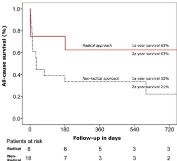
Outcome and follow-up
Overall survival was 39%. Twelve deaths (71%) were ABPF-related. There was no difference in survival with regard to central bronchial
or pulmonary parenchymal fistulations (33 vs 45%, log-rank
P = 0.55). There was a substantial difference in survival regarding
the conceptual treatment approach being significantly better for a
radical approach than for a non-radical approach with regard to overall survival (63 vs 32% and 63 vs 21% at 1 and 2 years,
respect-ively) as well as with regard tofistula-related survival (63 vs 43% and
63 vs 43% at 1 and 2 years, respectively) (Fig.1).
COMMENT
ABPF is a rare but highly lethal complication after TEVAR. The leading mechanism behind ABPF seems to be a continuing external compression of either the bronchial tree or left upper lobe paren-chyma. In this setting, persisting or newly developing endoleak formation seems to play a crucial role. Prognosis does not differ in
patients with central airway or pulmonary parenchymalfistulation.
Radical bronchial or pulmonary parenchymal repair in combination with stent graft removal and aortic reconstruction seems to be the most durable treatment strategy.
The incidence of ABPF in this series was very low. This agrees
with previously published reports being mainly casuistics [10–13].
The percentage of patients who had previous aortic repair in this series is high and underlines the multisegmental nature of the disease. Atherosclerotic aneurysm formation was the leading indi-cation for primary TEVAR in the majority of patients, followed by chronic type B aortic dissection and traumatic aortic injury. This
reflects the distribution of acute and chronic thoracic aortic
path-ologies as is known in tertiary care centres.
Fifty percent of patients underwent initial emergency TEVAR. As is known from previous European Registry of Endovascular Aortic Repair Complications reports, the incidence of adjacent organ injury is high in patients undergoing emergency TEVAR
[7–9]. Reasons are extensive mediastinal haematoma formation,
which may compress the oesophagus or the bronchial tree as well as phases of continuing hypotension, thereby causing end-organ ischaemia. As a consequence, staged mediastinal haematoma
evacuation to prevent secondary organ fistulation has been
recommended previously [9].
The median time interval between initial TEVAR and the devel-opment of ABPF was 310 days, suggesting a substantially slower
disease process than in aorto-oesophagealfistulation (AOF) where
Table 4: Diagnostic information
N overall = 26 Findings by imaging Periaortic haematoma,n (%) 17 (65%) Periaortic air,n (%) 10 (39%) Lung haemorrhage,n (%) 8 (31%) Lung consolidation,n (%) 7 (27%) Haematothorax,n (%) 5 (19%)
Bronchial wall erosion,n (%) 2 (8%) Mediastinal haematoma,n (%) 4 (15%) Localization of ABPF
Central left bronchial tree,n (%) 14 (54%) Pulmonary parenchyma,n (%) 11 (42%)
Trachea,n (%) 1 (4%)
Recognized mechanism of ABPF
External compression of bronchial tree,n (%) 13 (50%)
Ischaemic,n (%) 3 (12%)
Inflammation,n (%) 4 (15%)
Graft erosion,n (%) 4 (15%)
Endoleak,n (%) 9 (35%)
Table 5: Management and outcome of ABPF
N overall = 26 Clinical assessment
Fit for open repair,n (%) 10 (39) Management
Conservative,n (%) 5 (19)
TEVAR,n (%) 7 (27)
Bronchial or pulmonary parenchymal repair, no
aortic treatment,n (%) 2 (8)
Bronchial or pulmonary parenchymal repair, TEVAR,n (%)
2 (8) Bronchial or pulmonary parenchymal repair,
stent graft removal, aortic reconstruction,n (%) 8 (30) Bronchial repair and oesophagectomy,n (%) 1 (4) Carotideo-carotid bypass and tracheal stenting 1 (4) Outcome
Survival,n (%) 10 (39)
Death related to ABPF,n (%) 12 (71) Unless otherwise indicated, data are number (percentage).
Figure 1:Overall survival according to the approach.
A
O
RTIC
SURGER
the time interval between initial TEVAR and the development
of AOF was 90 days [9]. The chronic pressure exertion on the
bronchus or the pulmonary parenchyma initially does not cause the same injury as it might chronic pressure is exerted on the oesophagus, which is an organ more susceptible to ischaemia. Clinical signs of ABPF corresponded to what would be expected of a leading sign of any kind of haemoptysis. Diagnosis was primarily carried out by CT scanning with an additional 50% of patients
undergoing endoscopy for confirmation.
Notably, the number of endoleaks after TEVAR was very high with the majority being type I and type III. There seems to be a direct correlation between aneurysmal sac extension and growth, mediastinal haematoma formation and secondary central airway
or pulmonary parenchymalfistulation. As a consequence, correct
indications, long landing zones and respecting anatomy in
patients scheduled for TEVAR cannot be overemphasized [4]. In
addition, we feel that it is important not to scotomize the third option in treating patients with acute and chronic thoracic aortic pathology if open surgery is not an option and TEVAR might represent a trade-off, namely conservative therapy.
Although TEVAR has progressively evolved to become the first choice of thoracic aortic management for several clinical scenarios, many of the current indications are not adequately substantiated and proved. It is vital to verify if these extended indications are reasonable and rational or if they are offered as an option due to the inability to perform regular open surgery. In addition, type I and type III endoleaks have to be regarded as treatment failures, necessitating an urgent management that
should not be delayed [4].
However, external compression of the bronchial tree might also occur without endoleak formation. In particular in patients with a
very large aneurysm diameter, per se, which already alters the
geometry of the bronchial tree, TEVAR might further increase pressure exertion due to aneurysmal sac thrombosis with lack of aortic wall elasticity. It was interesting to observe that aneurysm sac diameter in elective patients undergoing TEVAR in this series was impressively large, thereby substantiating this hypothesis. It
might be wise to reflect if in these clinical scenarios, an open
sur-gical approach might be the better strategy or to add a saccotomy for decompression during the initial days after TEVAR.
Another factor exacerbating the process might be the additional stenting of major airways already externally compressed. This approach might increase the ischaemic burden of the bronchial
wall by exerting additional pressure from inside (Fig. 2) [13].
Oversizing might also play a role especially in patients where graft
erosion was the leading mechanism behind ABPF formation [12].
Oversizing is a matter of the underlying pathology, where acute type B aortic dissection will require minimal oversizing and atherosclerotic aneurysms will require more extensive oversizing. Recommended ranges do merely represent approximations learned from adverse events. In general, it might be stated that any kind of oversizing >30% is inappropriate.
A variety of approaches to treat ABPF were chosen in this series
ranging from a conservative approach to an orthotopic fullfix of
the disease process with bronchial and/or pulmonary parenchy-mal repair, stent graft removal and orthotopic or heterotopic
aortic reconstruction. Redo-TEVAR to seal thefistulation from the
aortic side was a common treatment strategy as was the combin-ation of redo-TEVAR and any kind of bronchial and/or pulmonary parenchymal repair.
Interestingly, survival in patients with central airway and pul-monary parenchymal ABPF did not differ. One would expect
pulmonary parenchymalfistulas to be the more benign ones but
we did not observe that. There was a substantial difference with regard to outcome according to the treatment strategy chosen. Any approach either conservative or interventional with or without bronchial and/or pulmonary parenchymal repair was associated with poor outcome. Merely a radical surgical approach with complete removal of the infected material, bronchial and/or pulmonary parenchymal repair and any kind of orthotopic or het-erotopic aortic reconstruction was associated with durable success
(Fig.3) [14]. Thesefindings substantiate the concept that a
conser-vative strategy in patients with graft infection is palliative, although
this is currently suggested otherwise [15].
Limitations and strengths
Without doubt, secondary organ injury is not limited to TEVAR
alone and secondary organ fistulation also occurs after open
surgery. Patient number is limited and there is a selection bias in this work as this approach to learn about the disease naturally picks only a mere percentage of ABPFs occurring worldwide within the last decade. Furthermore, the treatment strategy is Figure 2:Left main bronchus before and after stenting due to compression from the aneurysmal sac as a mechanism of aorto-bronchialfistula formation— axial view.
strongly influenced by the suitability or non-suitability for a radical approach and thereby precludes a full-fix of the problem in many of these patients. Nevertheless, this series was able to present an initial systematic approach to learn about the inci-dence, underlying mechanisms and effectiveness of treatment
strategies. Finally, to the best of our knowledge, this is the first
report stratifying ABPF into central airway and pulmonary paren-chymal lesions.
In sum, ABPF is a rare but highly lethal complication after TEVAR. The leading mechanism behind ABPF seems to be a continuing external compression of either the bronchial tree or left upper lobe parenchyma. In this setting, persisting or newly developing endo-leak formation seems to play a crucial role. Prognosis does not differ in patients with central airway or pulmonary parenchymal fistulation. Radical bronchial or pulmonary parenchymal repair in
combination with stent graft removal and aortic reconstruction seems to be the most durable treatment strategy.
Conflict of interest: none declared.
REFERENCES
[1] Grimm M, Loewe C, Gottardi R, Funovics M, Zimpfer D, Rodler Set al. Novel insights into the mechanisms and treatment of intramural hematoma affecting the entire thoracic aorta. Ann Thorac Surg 2008;86: 453–6.
[2] Czerny M, Funovics M, Sodeck G, Dumfarth J, Schoder M, Juraszek Aet al. Long-term results of thoracic endovascular aortic repair in atherosclerotic aneurysms involving the descending aorta. J Thorac Cardiovasc Surg 2010; 140(6 suppl):S179–84.
[3] Nienaber CA, Kische S, Rousseau H, Eggebrecht H, Rehders TC, Kundt G et al. INSTEAD-XL trial. Endovascular repair of type B aortic dissection: long-term results of the randomized investigation of stent grafts in aortic dissection trial. Circ Cardiovasc Interv 2013;6:407–16.
[4] Dumfarth J, Michel M, Schmidli J, Sodeck G, Ehrlich M, Grimm Met al. Mechanisms of failure and outcome of secondary surgical interventions after thoracic endovascular aortic repair (TEVAR). Ann Thorac Surg 2011; 91:1141–6.
[5] Canaud L, Ozdemir BA, Patterson BO, Holt PJ, Loftus IM, Thompson MM. Retrograde aortic dissection after thoracic endovascular aortic repair. Ann Surg. 2014;260:389–95.
[6] Törnqvist P, Resch T. Endoleaks after EVAR and TEVAR: indications for treatment and techniques. J Cardiovasc Surg 2014;55(2 Suppl 1):105–14. [7] Eggebrecht H, Thompson M, Rousseau H, Czerny M, Lönn L, Mehta RH
et al. European Registry on Endovascular Aortic Repair Complications. Retrograde ascending aortic dissection during or after thoracic aortic stent graft placement: insight from the European registry on endovascular aortic repair complications. Circulation 2009;120(11 Suppl):S276–81. [8] Czerny M, Eggebrecht H, Sodeck G, Verzini F, Cao P, Maritati Get al.
Mechanisms of symptomatic spinal cord ischemia after TEVAR: insights from the European Registry of Endovascular Aortic Repair Complications (EuREC). J Endovasc Ther 2012;19:37–43.
[9] Czerny M, Eggebrecht H, Sodeck G, Weigang E, Livi U, Verzini Fet al. New insights regarding the incidence, presentation and treatment options of aorto-oesophagealfistulation after thoracic endovascular aortic repair: the European Registry of Endovascular Aortic Repair Complications. Eur J Cardiothorac Surg 2014;45:452–7.
[10] Luehr M, Etz CD, Nozdrzykowski M, Garbade J, Lehmkuhl L, Schmidt A et al. Emergency open surgery for aorto-oesophageal and aorto-bronchial fistulae after thoracic endovascular aortic repair: a single-centre experi-ence. Eur J Cardiothorac Surg 2015;47:374–83.
[11] Chiesa R, Melissano G, Marone EM, Marrocco-Trischitta MM, Kahlberg A. Aorto-oesophageal and aortobronchialfistulae following thoracic endo-vascular aortic repair: a national survey. Eur J Vasc Endovasc Surg 2010;39: 273–9.
[12] Hyhlik-Dürr A, Geisbüsch P, Hakimi M, Weber TF, Schaible A, Böckler D. Endovascular aortic surgery: management of secondary aortobronchial and aorto-enteralfistulas. Chirurg 2009;80:947–55.
[13] Abdul-Ghani A, Pisipati S, McWilliams R, Page RD. Aorto-bronchialfistula following aortic and bronchial stenting of a thoracic aneurysm. Eur J Cardiothorac Surg 2006;29:419–21.
[14] Czerny M, von Allmen R, Opfermann P, Sodeck G, Dick F, Stellmes Aet al. Self-made pericardial tube graft: a new surgical concept for treatment of graft infections after thoracic and abdominal aortic procedures. Ann Thorac Surg 2011;92:1657–62.
[15] de Donato G, Setacci F, Galzerano G, Ruzzi U, Borrelli MP, Mazzitelli G et al. Prosthesis infection: prevention and treatment. J Cardiovasc Surg 2014;55:779–92.
Figure 3:Intraoperative view of same patient depicting central airwayfistulation as well as explanted stent grafts and explanted bronchial stents.
A
O
RTIC
SURGER