Nephrol Dial Transplant (1994) 9: 1063-1065
Editorial Comment
Nephrology
Dialysis
Transplantation
Cell adhesion molecules and inflammatory renal diseases
R. P. Wuthrich
Abteilung far Nephrologie, Universitatsspital, Zflrich, Switzerland
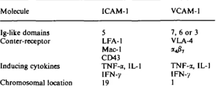
Cell adhesion molecules (CAMs) are a group of cell surface molecules capable of binding leukocytes through specific counter-receptors. CAMs are members of the large immunoglobulin superfamily by virtue of their extracellular immunoglobulin-like domains, and are expressed in the kidney in endothelial cells, mesang-ial cells, and renal tubular epithelmesang-ial cells (TEC) [1]. Table 1 summarizes the characteristics of two major renal CAMs, ICAM-1 and VCAM-1. Intercellular adhesion molecule-1 (ICAM-1) is a monomeric unpaired cell-surface glycoprotein of 76—114 kDa, and is found in many different cell types in various tissues. Vascular cell adhesion molecule-1 (VCAM-1) is an inducible monomeric cell-surface glycoprotein of 110 kDa which is not only expressed on vascular endothelium as its name implies, but also on extravas-cular sites such as mesangial cells and TEC.
ICAM-1 and VCAM-1 are induced in the kidney in immunologically mediated renal diseases. This induc-tion is caused by interferons and certain cytokines, namely TNF-a and IL-1. Cytomegalovirus is also able to enhance directly ICAM-1 expression on cultured proximal TEC [2], and perhaps also in vivo.
The expression of ICAM-1 is enhanced in various inflammatory renal disease states, and it has been speculated that upregulation of CAMs confers increased 'stickiness' to the kidney, thereby promoting leukocyte-mediated renal injury [3]. In human renal allograft rejection, ICAM-1 expression is prominent on renal TEC [4,5], paralleling MHC class II expres-sion [6]. Increased glomerular ICAM-1 expresexpres-sion can be demonstrated in rapidly progressive glomerulo-nephritis, lupus nephritis and some other
glomeru-Table 1. Characteristics of the cell adhesion molecules ICAM-1 and VCAM-1
Molecule ICAM-1 VCAM-1
Ig-like domains Conter-receptor Inducing cytokines Chromosomal location 5 LFA-1 Mac-1 CD43 TNF-a, IL-1 IFN-y 19 7, 6 or 3 VLA^ TNF-a, IL-1 IFN-y 1
lonephritides [7]. The enhanced glomerular ICAM-1 expression in these diseases is sometimes accompanied by increased tubular ICAM-1 staining at the luminal side. The increased tubular ICAM-1 expression correl-ates with the presence of LFA-1 positive intratubular leukocytes [7]. VCAM-1 expression is also enhanced in inflammatory renal diseases, including crescentic nephritis, vasculitis, interstitial nephritis, and allograft rejection [8,9]. Up-regulation of VCAM-1 is particu-larly striking in proximal TEC and endothelial cells.
In our own studies we investigated ICAM-1 and VCAM-1 expression in murine models of autoimmune renal injury and found that CAMs are up-regulated in the kidney of MRL/lpr and (NZBxNZW^ mice with lupus nephritis [3,10]. Low constitutive expression is found in normal mice in glomeruli and proximal tubules. Up-regulation in autoimmune strains is par-ticularly prominent in proximal tubules, endothelium and mesangial cells. TNF-a and IL-1 are present at high levels in the kidneys of autoimmune MRL/lpr and (NZBxNZW)F! mice [11,12], and could be dir-ectly responsible for the increased expression of ICAM-1 and VCAM-1 in the kidney of these auto-immune strains.
In vitro we have demonstrated that ICAM-1 and
VCAM-1 are rapidly induced in cultured TEC in response to TNF-a, IL-1 and IFN-y [10,13]. Peak expression occurs within 6 h, and involves translation of mRNA and de-novo transcription. It also appears that a down-regulation-resistant type of protein kinase C is involved in the induction in response to the cytokines TNF-a and IL-1 [14,15]. Commonly used immunosuppressants such as cyclosporin A, steroids and azathioprine are unable to block the cytokine-induced expression of ICAM-1 and VCAM-1 in vitro; this does not exclude an inhibitory effect in vivo through reduced expression of cytokines [16].
Cytokine-stimulated monolayers of TEC become more adhesive for leukocytes (T cells, monocytic cells). This adherence can be blocked with monoclonal anti-bodies (MAb) targeting ICAM-1 and VCAM-1, and also by blocking the counter-receptors LFA-1 and VLA-4 respectively. Antigen presentation by TEC can also be inhibited with anti-ICAM-1 and anti-LFA-1 monoclonal antibody treatment in vitro [13]. Others have shown that the cytotoxic action of T cells for O 1994 European Dialysis and Transplant Association-European Renal Association
1064
TEC can be inhibited with anti-ICAM-1/anti-LFA-l MAb [17]. Thus, TEC expression of adhesion molec-ules such as ICAM-1 confers enhanced stickiness and immune reactivity for T cells, and may therefore pro-mote immune renal injury.
What is the in-vivo significance of these inducible CAMs in renal injury? Certain CAMs have a well-known structural (architectural) function in the kidney in that they play an essential role in cell-cell contact. Thus, NCAM (neural cell adhesion molecule) is a Ca2+-independent cell-cell adhesion molecule member of the immunoglobulin superfamily that causes renal tubular cell binding through homophilic interaction among NCAM molecules [18]. Cadherins such as uvomorulin (E-cadherin) and N-cadherin are Ca2+ -dependent CAMs with structural function and cell-polarity-inducing characteristics in the kidney [19]. Both NCAM and the cadherins also play an important role in morphogenesis and nephron development. CD31 (also termed PECAM-1), a 6 Ig extracellular domain member of the immunoglobulin superfamily, is another example of a CAM that is capable of homophilic (and possibly heterophilic) cell-cell inter-action in the kidney [20], thus playing a role in kidney structure.
ICAM-1 and VCAM-1, however, do not appear to have primarily a structural function. We have shown that kidney cryostat section from MRL/lpr mice dis-play a significant increase in the adherence for T cells and monocytes when compared with sections obtained from normal mice [3]. This adherence can be blocked to various degrees with antibodies targeting ICAM-1 or VCAM-1. These experiments directly demonstrate that the kidney parenchyma becomes more sticky for inflammatory cells in murine autoimmune renal dis-ease, and that this enhanced stickiness is caused pre-dominantly by ICAM-1 and VCAM-1.
Several MAb against different adhesion molecules have been tested for therapeutic intervention in experi-mental models of renal inflammation. Anti-ICAM-1 and anti-LFA-1 MAb have already been tried in human kidney allograft rejection.
A murine anti-human ICAM-1 MAb (R6.5) has been used both prophylactically and therapeutically in Cynomolgus monkeys receiving kidney allografts [21]. While this monoclonal antibody clearly prolongs car-diac and renal allograft survival in Cynomolgus mon-keys, its clinical usefulness needs to be confirmed. A small human trial using the same ICAM-1 antibody (BIRR1, previously termed R6.5) showed limited use-fulness in controlling allograft rejection and possibly reperfusion injury [22]. An anti-LFA-1 antibody used in a small clinical trial to treat acute renal transplant rejection showed no effectiveness [23].
A recent study using mouse heart allografts nicely demonstrated that tolerance could be induced against major histocompatibility barriers when anti-ICAM-1 and anti-LFA-1 monoclonal antibodies were used together [24]. Although the mechanisms of tolerance induction in this animal model remain to be deter-mined, this promising study may have important
con-R. P. WOthrich
sequences for prevention and treatment of human renal allograft rejection.
In a recent study of hereditary autoimmune tubulo-interstitial nephritis in the mouse (kdkd mouse of the CBA/Ca strain) it was shown that ICAM-1 is overex-pressed in the kidney of these mice with interstitial nephritis [25]. Treatment with an anti-ICAM-1 MAb reduced the leukocyte infiltration, and also reduced proteinuria. In rat models of nephrotoxic serum neph-ritis and crescentic glomerulonephneph-ritis it could be shown that treatment with anti-ICAM-1 or anti-LFA-1 MAb reduced proteinuria and glomerular inflamma-tion [26-28].
In summary, the cell adhesion molecules ICAM-1 and VCAM-1 play an important role in immune-mediated renal diseases. By conferring increased adhes-iveness to the renal parenchyma they promote the adhesion of inflammatory leukocytes. Targeting these adhesion molecules with MAb may show promising effects in various renal diseases and in allograft rejection.
References
1. Wuthrich RP. Intercellular adhesion molecules and vascular cell adhesion molecule-1 and the kidney. J Am Soc Nephrol 1992; 3: 1201-1211
2. van Dorp WT, van Wieringen PA, Marselis-Jonges E et aJ. Cytomegalovirus directly enhances MHC class I and ICAM-1 expression on cultured proximal tubular epithelial cells.
Transplantation 1993; 55: 1367-1371
3. Wuthrich RP. Vascular cell adhesion molecule-1 (VCAM-1) expression in murine lupus nephritis. Kidney Int 1992; 42: 903-914
4. AndeTsen CB, Blaehr H, Ladefoged S, Larsen S. Expression of ICAM-1 in human renal allografts and cultured human tubular cells. Nephrol Dial Transplant 1992; 7: 147-154
5. Faull RJ, Russ GR. Tubular expression of intercellular adhesion molecule-1 during renal allograft rejection. Transplantation 1989; 48: 226-230
6. Bishop GA, Hall BM. Expression of leukocyte and lymphocyte adhesion molecules in the human kidney. Kidney Int 1989; 36:
1078-1085
7. Lhotta IC, Neumayer HP, Joannidis M, Geissler D, K.6nig P. Renal expression of intercellular adhesion molecule-1 in different forms of glomerulonephritis. Clin Sci 1991; 81: 477-481 8. Mampaso F, Sanchez-Madrid F, Molina A, Bricio T, Liano F,
Alvarez V. Expression of adhesion receptor and counter-receptors from the leukocyte-endothelial adhesion pathways LFA-l/ICAM-1 and VLA-4/VCAM-1 on drug-induced tubulo-interstitial nephritis. Am J Nephrol 1992; 12: 391-392
9. Seron D, Cameron JS, Haskard DO. Expression of VCAM-1 in the normal and diseased kidney. Nephrol Dial Transplant 1991; 6: 917-922
10. Wuthrich RP, Jevnikar AM, Takei F, Glimcher LH, Kelley VE. Intercellular adhesion molecule-1 (ICAM-1) expression is upreg-ulated in autoimmune murine lupus nephritis. Am J Palhol 1990; 136: 441^t50
11. Boswell JM, Yui MA, Burt DW, Kelley VE. Increased tumor necrosis factor and IL-1/? gene expression in the kidneys of mice with lupus nephritis. J Immunol 1988; 141: 3050-3054 12. Brennan DC, Yui MA, Wuthrich RP, Kelley VE. Tumor necrosis
factor and IL-1 in New Zealand Black/White mice. J Immunol 1989; 143: 3470-3475
13. Jevnikar AM, Wuthrich RP, Takei F et al. Differing regulation and function of ICAM-1 and class II antigens on renal tubular cells. Kidney Int 1990; 38: 417-425
Cell adhesion molecules and inflammatory renal diseases
by murine renal tubular epithelial cells is transcriptionally regu-lated and involves protein kinase C. Renal Physiol Biochem 1992; 15: 302-306
15. Wuthrich RP, Jenkins TA, Snyder TL. Regulation of cytokine-stimulated vascular cell adhesion molecule-1 (VCAM-1) expres-sion in renal tubular epithelial cells. Transplantation 1993; 55:
172-177
16. Wuthrich RP, Sekar P. Effect of dexamethasone, 6-mercaptopurine and cyclosporine A on ICAM-1 and VCAM-1 expression. Biochem Pharmacol 1993; 46: 1349-1353
17. Suranyi MG, Bishop GA, Clayberger C el al. Lymphocyte adhesion molecules in T cell-mediated lysis of human kidney cells. Kidney Int 1991; 39: 312-319
18. Nouwen EJ, Dauwe S, Van der Biest I, de Broe ME. Stage- and segment-specific expression of cell-adhesion molecules NCAM, A-CAM, and L-CAM in the kidney. Kidney Int 1993; 44: 147-158
19. Geiger B, Ayalon O. Cadherins. Annu Rev Cell Biol 1992; 8: 307-332
20. Fuggle SV, Sanderson JB, Gray DW, Richardson A, Morris PJ. Variation in expression of endothelial adhesion molecules in pretransplant and transplanted kidneys—correlation with intra-graft events. Transplantation 1993; 55: 117-123
21. Cosimi AB, Conti D, Delmonico FL et al. In vivo effects of monoclonal antibody to ICAM-1 (CD54) in nonhuman primates with renal allografts. / Immunol 1990; 144: 4604-4612
1065 22. Haug CE, Colvin RB, Delmonico FL et al. A phase I trial of immunosuppression with anti-ICAM-1 (CD54) mAb in renal allograft recipients. Transplantation 1993; 55: 766-773 23. Le Mauff B, Hourmant M, Rougier JP et al. Effect of
anti-LFA-1 (CD lla) monoclonal antibodies in acute rejection in human kidney transplantation. Transplantation 1991; 52: 291-296
24. Isobe M, Yagita H, Okumura K, Ihara A. Specific acceptance of cardiac allograft after treatment with antibodies to ICAM-1 and LFA-1. Science 1992; 255: 1125-1127
25. Harning R, Pelletier J, Van G, Takei F, Merluzzi VJ. Monoclonal antibody to MALA-2 (ICAM-1) reduces acute autoimmune nephritis in kdkd mice. Clin Immunol Immunopathol 1992; 64: 129-134
26. Kawasaki K, Yaoita E, Yamamoto T, Tamatani T, Miyasaka M, Kihara I. Antibodies against intercellular adhesion molecule-1 and lymphocyte function-associated antigen-1 pre-vent glomerular injury in rat experimental crescentic glomerulo-nephritis. J Immunol 1993; 150: 1074-1083
27. Mulligan MS, Johnson KJ, Todd R F et at. Requirements for leukocyte adhesion molecules in nephrotoxic serum nephritis.
J Ctin Invest r993; 577-587
28. Nishikawa K_, Guo Y, Miyasaka M et al. Antibodies to ICAM-l/LFA-1 prevent crescent formation in rat autoimmune glomerulonephritis. / Exp Mtd 1993; 177: 667-677