-specific expression of a
FM/??/(3-galactosidase fusion gene in
transgenic mice
Martin Hergersberg*, Koichi Matsuo1, Max Gassmann2, Walter Schaffner1, Bernhard
Luscher3, Thomas Rulicke4 and Adriano Aguzzi5
Institut fur Medizinische Genetik, Universitat Zurich, Ramistr. 74, CH-8001 Zurich, 1lnstitut fur Molekularbiologie II, 2Physiologisches Institut, and 3Pharmakologisches Institut, Universitat Zurich, Winterthurerstr. 190, CH-8057 Zurich, "Biologisches Zentrallabor, Universitatsspital Zurich, Sternwartstr. 6, CH-8091 Zurich and 5lnstitut fur Neuropathologie, Universitatsspital Zurich, Schmelzbergstr. 12, CH-8091 Zurich, Switzerland
Received October 12, 1994; Revised and Accepted December 19, 1994
Fragile X syndrome is one of the most common genetic causes of mental retardation, yet the mechanisms controlling expression of the fragile X mental retardation gene FMR1 are poorly understood. To identify sequences regulating FMR1 transcription, transgenic mouse lines were established using a fusion gene consisting of an E.coli (3-galactosidase reporter gene (/acZ) linked to a 2.8 kb fragment spanning the 5-region of FMR1. Five transgenic mouse lines showed lacZ expression in brain, in particular in neurons of the hippocampus and the granular layer of the cerebellum. Expression of the reporter gene was also detected in Leydig cells and spermatogonia in the testis, in many epithelia of adult mice, and in the two other steroidogenic cell types, adrenal cortex cells and ovarian follicle cells. Embryonic tissues which showed strong activity of the reporter gene included the telencephalon, the genital ridge, and the notochord. This expression pattern closely resembles the endogenous one, indicating that the 5' FMR1 gene promoter region used in this study contains most c/s-acting elements regulating FMR1 transcription.
INTRODUCTION
Fragile X syndrome, an X-linked inherited disease and the most common form of monogenic inherited mental retardation, is mainly due to a block in the transcription of the fragile X mental retardation 1 gene (FMR1). Other clinical features of the fragile X syndrome are postpubertal macroorchidism in male patients, a longish face, large ears and several symptoms reminiscent of a connective tissue dysplasia (1). The only neuroanatomic alteration reported in the brains of affected patients is a reduction in size of the posterior vermis of the cerebellum (2). A characteristic feature of the pathogenesis is the expansion of a CGG-trinucleotide repeat, located upstream of the translation initiation site in the first exon of FMR1, and a concomitant cytosine methylation of the CpG island encompassing the 5'-end of this gene (3,4). It has been postulated that this erroneous methylation of the putative promoter region results in the inhibition of transcription of the
FMR1 gene, and that the absence of the FMR1 gene product
leads to the various disease symptoms (3,4). The causative role of the FMR1 gene for fragile X syndrome has been confirmed by the description of patients suffering from mental retardation similar to the fragile X syndrome who carried deletions in this gene (5-7). Moreover, FMR1 mRNA and
protein were found to be strongly reduced or absent in cells and tissues derived from fragile X syndrome patients (8-11).
FMR1 consists of 17 exons spanning 38 kb of chromosome
Xq27.3, and the corresponding 4.8 kb transcript is subjected to alternative splicing resulting in several isoforms of the protein, including the most abundant 69 kDa isoform (12-14). The FMR1 gene product is an RNA-binding protein that binds to various sequences with different affinities. It has been estimated that about 4% of all mRNAs expressed in the fetal human brain are bound by the FMR1 protein (15,16). Indeed, in a mentally retarded male patient with the symptoms of fragile X syndrome, no CGG-repeat amplification or methylation in
FMR1 was found, however, a point mutation was identified in FMR1 which inhibited binding of the FMR1 protein to RNA
(17,18). Recently, an animal model for the fragile X syndrome has been generated by targeted inactivation of the murine
Fmrl gene in embryonic stem cells. The resultant knockout
mice did not express the FMR1 protein and showed increased testicular weight, learning deficits, and hyperactivity (19).
FMR1 mRNA has been detected by reverse
transcriptase-mediated PCR and Northern blot analyses in a variety of tissues including fetal brain, liver and pancreas, and adult
brain, testis and lymphocytes (9,14,20). In situ hybridization studies localized endogenous human and murine mRNA in multiple tissues of pre- and postnatal origin. Postnatally FMRl transcripts were observed in the central nervous system, in particular in the hippocampus and the cerebellum, and also in the testis (21,22). Highest FMRl protein expression was observed in neurons as confirmed by immunostaining (8,11).
More information about the regulation of developmental and tissue-specific transcription of FMRl will contribute to our understanding of the pleiotropic phenotype of the fragile X syndrome and of the molecular mechanisms leading to transcriptional silencing of FMRl in affected patients. An established procedure to study the regulation of gene expression in an organism is the generation of transgenic mouse lines using a DNA construct consisting of a reporter gene linked to the relevant regulatory fragment (23). In an attempt to define cw-acting sequences regulating expression of FMRl, we gener-ated five transgenic mouse lines by introducing a fusion gene consisting of the putative 5'-regulatory region of FMRl fused to lacZ into the mouse genome and analyzed expression of the reporter gene in developing and adult mice.
RESULTS
Generation of transgenic mice expressing a FMRl/lacZ fusion gene
The fusion gene FMRl/lacZ contains a fragment from the upstream region of FMRl linked to the lacZ reporter gene (Fig. 1). The 2830 bp FMRl sequence includes the 5'-region of the CpG island and ends 18 bp upstream of the translation initiation codon. As a major transcription start site of FMRl has been reported to lie about 320 bp upstream of the translation initiation codon, approximately 300 bp of 5'-untranslated sequences are therefore included (12). To direct the reporter gene product to the nucleus and thereby increase the resolution of the histological analysis, we used a reporter gene containing the nuclear localization signal from the large T antigen of simian virus 40 (SV40) at the N-terminus of p-galactosidase (24). Injection of the fusion gene into zygote pronuclei resulted in 43 newborn mice from which six were identified to be transgenic by Southern blot analysis of tail DNA using a lacZ probe. The six independent mouse lines and the approximate copy number of the transgene (in parentheses) within these lines were: 22 [1-5], 35 [1-5], 38 [5-20], 41 [50-100], 42 [50-100], and 43 [1-5]. Mouse line 38 did not give rise to transgenic offspring and was excluded from further analysis.
Transgene expression in the central nervous system
The regions of the brain expressing the reporter gene were identified by overnight whole-mount staining of perfusion-fixed brains with Bluo-Gal, a substrate for P-galactosidase, followed by paraffin-embedding and sectioning, or by staining of frozen sections (23). The expression level was estimated from staining intensity. Varying concentrations of the reporter gene product were observed in different parts of the brain (Fig. 2 a - c ; summarized in Table 1). Although the expression level was variable between the different transgenic mouse lines, the expression pattern between the five lines was very similar (Tables 1 and 2). This implies that the observed pattern was due to intrinsic properties of the FMRl promoter rather
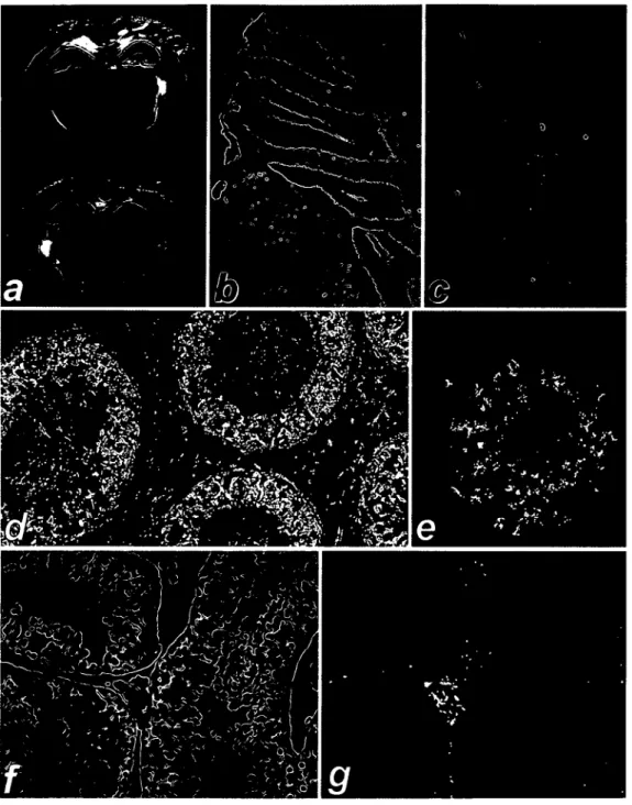
than to positional effects after integration of the transgene. Both the expression pattern and relative expression level in tissues were similar or identical between mice of the same line. The hippocampus and the granular layer of the cerebellum showed the strongest signals (Fig. 2a, b). In brain slices of perfused mice, the hippocampus showed intense staining as early as two hours after incubation, while in other parts of the brain little or no staining was visible (Fig. 2a). Nevertheless, overnight staining revealed a lower level of lacZ expression in other neurons, e.g. in the bulbus olfactorius (Fig. 2c). Staining was also observed in the retinal bipolar neuronal layer and in the nervus vagus in the oesophagus (not shown).
Transgene expression in other adult tissues
The lacZ expression was analyzed in testicular and ovarian tissues (Fig. 3; summarized in Table 2). When visualized by polarized light, the sensitivity of the lacZ assay is increased and allows a detailed histological analysis of the expression pattern (25). Figures 2f,g and 3d,e show the same tissue specimens in bright field and in dark field with polarized light, respectively, thus illustrating the increase in sensitivity. In testis, expression was detected in the interstitial Leydig cells (Fig. 2f,g). Staining of spermatogonia was strong near the surface of whole-mount stained testis and in frozen sections (Table 2), and the seemingly exclusive staining of Leydig cells in Figure 2g results from better diffusion of (3-galactosidase substrate in the interstitium compared to diffusion into the seminiferous tubuli. In female mice, staining was observed in granulosa cells of the ovarian follicles (Fig. 2e). LacZ activity was found only in a subgroup of cells at later stages of follicle development, suggesting that FMRl might be expressed in a regulated manner during follicle maturation. The oocytes did not show significant staining. Heart, liver, skeletal and smooth muscle did not show (J-galactosidase activity (summarized in Table 2). Staining was observed in a number of epithelia, most prominently in epithelia lining the organs of the urogenital system, e.g. urethra, vas deferens and epididymis (Fig. 2d), and to a lesser degree in the endothelia of the blood vessels and the epithelia of Bowman's capsule in the kidney. Other epithelia expressing lacZ were the dermis, the epithelia of hair follicles, and epithelia in the salivary glands and the trachea.
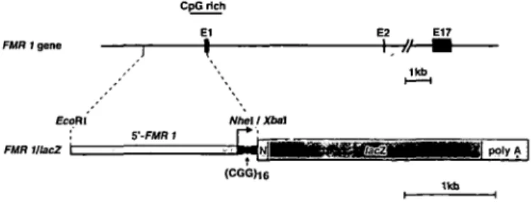
FMR 1 gene
CpG rich
E1 E2 E17
(CGGhe
Figure 1. Structure of the 5'-end of the FMRl gene and of the FMRl/lacZ
fusion gene injected into mouse oocytes. The restriction sites used for the construction of the fusion gene are indicated. The narrow part of the construct is the 5'-fragment of the human FMRl gene. The CpG island, the approximate transcription start site (arrow), and the CGG-repeat (sphere) are marked, as well as the nuclear localization signal (N) and the polyadenylation signal (polyA) from SV40 adjacent to the E.coli lacZ gene.
Transgene activity during embryonic development
Expression of the FMRlllacL fusion gene was assayed in transgenic embryos from line 22 at midgestation (Figs 3 and 4; summarized in Table 3). In an 11.5 day old embryo, expression of the transgene was more widespread than in adult
tissues although not ubiquitous. A strong specific signal was observed in the notochord, preceding the lacZ expression in developing intervertebral discs, which contain cells invaginated from the notochord (Fig. 3c). Expression in cells of the developing nervous system was also specifically regulated:
Figure 2. Expression of the lacZ reporter gene in tissues of adult transgenic mice. Using Bluo-Gal as chromogenic p-galactosidase substrate, the reaction product can either be visualized as blue stain in normal light (a, b, c, and 0, or as a bright stain in polarized light (d, e, and g). (a) The frontal brain section in a 10 week old mouse (line 22) showed intense lacZ activity of the hippocampus after staining for two hours, (b) The cerebellum revealed lacZ expression in the granular layer, as well as in singular cells in the molecular layer (frozen section, line 41). (c) A frontal section of the bulbus olfactorius showed tissue-specific lacZ expression in a cone-shaped layer of neurons (frozen section, line 43). (d) The P-galactosidase staining of the epididymis revealed strong lacZ expression in the epithelium. The sperm cells in the lumen of the epididymal tubules show also some expression of the reporter gene (paraffin embedded section after whole-mount staining, line 35). (e) In an ovary, lacZ expression was visible in a subgroup of follicle cells, but not in the oocyte. Adjacent follicles in earlier stages of the ovarian cycle did not show lacZ activity (paraffin embedded section after whole-mount staining, line 41). (f) The same testis section in bright field (paraffin embedded section after whole-mount staining, line 43). (g) Testis section in polarized light, showing lacZ expression in a group of Leydig cells.
Table 1. Expression of the lacZ reporter gene in brain tissues of the five transgenic mouse lines
Tissue Line 22 Line 35 Line 41 Line 42 Line 43 Bulbus olfactorius Caudate putamen Cerebellum (granular layer) Corpus callosum Choroid plexus Hippocampus Neocortex (neurons of the pyramidal layer) Retina (neuronal layer)
The estimation of staining intensities was done qualitatively, using four categories: ' + ': single cells visualized with polarized light; ' + + ': majority of all cells or strong staining of single cells with polarized light, weak staining in bright field; ' + + + ': staining in a tissue in bright field; ' + + + + ': strong staining in bright field
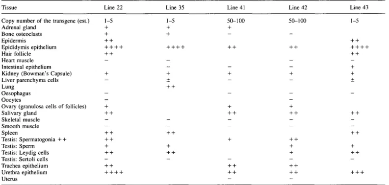
Table 2. Expression of lacZ reporter gene in different tissues of the five transgenic mouse lines
Tissue Line 22 Line 35 Line 41 Line 42 Line 43 Copy number of the transgene (est.) 1-5
Adrenal gland + Bone osteoclasts + Epidermis + + Epididymis epithelium + + -Hair follicle + + Heart muscle — Intestinal epithelium
Kidney (Bowman's Capsule) + Liver parenchyma cells — Lung
Oesophagus Oocytes -Ovary (granulosa cells of follicles) + Salivary gland + + Skeletal muscle -Smooth muscle — Spleen + + Testis: Spermatogonia + + + + Testis: Sperm + Testis: Leydig cells + + Testis: Sertoli cells -Trachea epithelium + + Urethra epithelium + + -Uterus 1-5 + + 50-100 + 50-100 1-5
The estimation of staining intensities was as described in the legend for Table 1
particularly strong expression was detected in the telence-phalon, whereas other structures, e.g. the neural tube and the dorsal root ganglia, showed weak or no lacL activity (Fig. 3a—c). A strong lacL signal was also detected in different developing organs, e.g., the pericardium and the tongue (Fig. 3d, e), and in particular in the genital ridge (Fig. 4).
DISCUSSION
After introduction of the FMRl/lacL fusion gene into the mouse germline, similar expression patterns of the reporter gene P-galactosidase were found in a number of tissues of transgenic offspring from five independent founders. We observed that the tissue-specificity and the level of transgene expression were regulated similarly to the endogenous gene: in the brain, the neurons of the hippocampus and the cerebellum showed the highest p-galactosidase activity. This pattern resembles transcription of the endogenous gene found in the
brain of human fetuses (21) and adult mice (22). Since the hippocampus is involved in establishing memory, it is tempting to speculate that the absence of FMR1 protein in hippocampal neurons might cause some of the symptoms of mental retard-ation observed in fragile X syndrome patients. In addition, strong expression of the transgene occurs in the cerebellum. Significantly, the only neuroanatomical alteration found in affected individuals so far is a reduction in size of the posterior vermis of the cerebellum (2). Different methods were used for tissue preparation and staining, resulting in a broad range of detectable staining intensities, and most tissues were analyzed with several techniques. In particular, polarized light darkfield imaging increased sensitivity in paraffin sections approximately 10-fold compared to bright field, and paraffin embedding and frozen sections required sections of different thickness also affecting staining intensities.
During fetal development, transcription of the human and mouse gene has been reported to be more widespread than in
Figure 3. Expression of FMRIAacZ in embryonic tissues at embryonic day 11.5 (mouse line 22). (a) Longitudinal section through the head of a whole mount stained, paraffin-embedded embryo, photographed in polarized light. Intense staining of the telencephalon is visible, (b) The same section as in (a) shows lacZ expression in the hindbrain (Hb) adjacent to the fourth ventricle (4th ve). The arrows point to P-galactosidase activity in the roof of the 4th ventricle, (c) Neural tube, notochord, and spinal ganglia in polarized light (transverse section). No staining is visible in the cells of the neural tube, the neural crest and the spinal ganglia. LacZ expression is specifically restricted to the notochord (arrow). Some staining is also found in the mesoderm of the somites (lower left), (d) A longitudinal section of the thorax, showing the developing tongue, the pericardium (Pc), the atrioventricular junction of the heart (Ht), the liver (Li), and the stomach (St). (e) The same section as in (d) in polarized light shows particularly strong staining in the tongue, the pericardium, and mesenchymal tissues below the heart and liver.
adult tissues, but the transcription pattern has been controversial (22,26). In an 11.5 day old embryo, we observed many of the features described for endogenous Fmrl transcription, most notably the enhanced expression at the genital ridge and in the telencephalon revealing tissue specific expression of the transgene. In addition, lacZ gene product was found in develop-ing organs such as the pericardium, which do not express the gene in adults.
In testis, expression of the transgene was found in different stages of spermatogenesis and in the interstitial Leydig cells. Expression in spermatogonia was enhanced, but in whole-mount stained testes the lacL activity seemed to be weak in those tubuli not directly exposed to the staining solution. This observation might be explained by a reduced diffusion rate of the substrate into the tubuli. No transgene expression was found in Sertoli cells, and no differences in lacL activity have yet been observed betwen different seminiferous tubuli. We detected a weak lacL signal in mature sperm of transgenic mice. However, this signal may have resulted from remnants of P-galactosidase expressed during earlier stages of spermato-genesis rather than from transgene expression in mature sperm.
The testicular expression pattern of endogenous Fmrl showed enhanced gene transcription in the periphery of seminiferous tubules, which was either located to Sertoli cells (22) or to spermatogonia (26). Different tubuli showed different levels
of Fmrl transcription (22,26). The FMR1 protein was identified
in human spermatogonia, but not in sperm, by immunostaining (8). Transcription or expression of the endogenous gene in interstitial cells was not observed (8,22,26). Fmrl transcription was found in ovarian follicle cells in one study (22), but this result was not confirmed in another publication (26). Our transgenic animals showed lacZ activity in ovarian follicle cells. In conclusion, the expression of the FMRl/lacZ transgene is observed in several cell types in genital organs, where the transcription pattern of the endogenous gene is either a matter of debate, or has not been observed yet. Enhanced lacZ expression was found in the three cell types synthesizing androgens, namely Leydig cells, ovarian follicle cells and adrenal cortex cells (27). It is tempting to speculate that the
FMR1 protein has a function in steroid synthesis, and that its
absence might affect the function of the peritubular Leydig cells. The macroorchidism of fragile X syndrome patients
Figure 4. Expression of the FMRl/lacZ fusion gene in the genital ridge of
the murine embryo at embryonic day 11.5 (line 22). (a) Longitudinal section in normal light, (b) The same section in polarized light showing strong (3-galactosidase activity in the cells of the genital ridge.
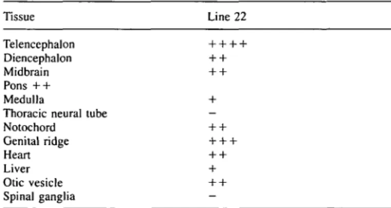
Table 3. Expression pattern of the reporter gene in embryonic tissue of mouse
line 22 Tissue Line 22 Telencephalon Diencephalon Midbrain Pons + + Medulla
Thoracic neural tube Notochord Genital ridge Heart Liver Otic vesicle Spinal ganglia
seems to result from an increase in peritubular tissue (28,29), although in the enlarged testis of the Fmrl null mutant mice no apparent changes in cell number or size have been detected (19). It remains to be elucidated, whether the FMR1 protein plays a physiological role in the Leydig cells as suggested by the transgene expression. Another discrepancy between the
expression patterns of the transgene and the endogenous Fmrl was a lack of lacZ signal in the oesophagus epithelium of adult transgenic mice (11). In summary, these differences suggest that some regulatory sequences might be missing from the 2.8 kb 5'-sequence of FMR1 used in our construct. We also observed that the expression level of the transgene is not directly related to its copy number, suggesting that our trans-gene does not contain a locus control region (30). Indeed, the mouse lines carrying the highest copy number of the transgene showed relative low expression level. This phenomenon might result from inactivation of long strands of tandem repeats of transgenes (31). Additional regulatory elements might be located in the 9.9 kb first intron of FMR1, which contains the 3' part of the CpG island excluded from the transgene. For example, enhancer sequences have been identified in the first and second introns of the gene for the intermediate filament nestin, directing expression to differentiating muscle cells and neural precursors, respectively (32). Independent regulatory elements acting on a CpG-rich promoter have also been identified in the Thy I gene. Expression of fusion genes in transgenic mice was more tissue-specific, when sequences located downstream from the transcriptional initiation site were included (33). In contrast, the CpG-rich promoter of the rat calmodulin gene II was active in neurons and testis of transgenic mice generated with a construct harboring 294 bp of 5'-upstream sequence plus 68 bp of 5'-untranslated leader sequence of calmodulin (34,35). Taken together, the similarity observed between the expression pattern of the transgene and the endogenous gene in mouse and in human strongly suggests that not all, but most of the cw-regulatory sequences of
FMR1 are present within our construct. This notion is further
supported by the similar expression patterns between the independent transgenic mouse lines.
The mechanism(s) which regulate FMR1 transcription are not yet understood. A patient with the clinical phenotype of the fragile X syndrome carrying a 1.6 kb deletion upstream and adjacent to the CGG repeat of FMR1 confirms that these sequences are essential for the expression of the gene (6). It is also known that CpG-rich promoters are present in active chromatin (36), and can be activated by constitutively bound transcription factors in response to multiple stimuli in a cell-specific manner in various cell types including neurons (37). In the case of FMR1, no factors binding to the promoter region have been described so far. However, several factors were found, which bind to the CGG-repeat of FMR1, but the significance of these protein-DNA interactions for FMR1 gene expression remains to be elucidated (38). In preliminary experiments, we found binding sites for putative transcription factors in the FMR1 promoter region by gel mobility shift assays (M.H., unpublished observations). Mutagenesis of these binding sites will help to identify functional regulatory sequences in FMRL Further characterization of elements controlling FMR1 gene expression using transgenic model systems like the one described here may contribute to the understanding of the molecular mechanisms of the fragile X syndrome of mental retardation.
MATERIALS AND METHODS
Cloning of the fusion gene
pNL(Not2X) is a derivative of the pNL plasmid that expresses the E.coli lacZ gene with a nuclear localization signal of the SV40 large T antigen at the
N-terminus of the P-galactosidase and an SV40 polyadenylation signal downstream of the open reading frame (24). The 5.1 kb £coRI-fragment from the genomic FMRI clone pE5.1 containing 16 CGG-triplets (39; a gift of Dr D. Nelson, Houston, USA) was blunt ended with Klenow DNA polymerase and recut with Nhe\, 18 bp upstream of the ATG codon of the FMRI open reading frame (12). Before ligation with the £coRI-Mid-fragment of FMR1, pNL(Not2X) was cut with Sph\, blunt ended with Klenow DNA polymerase, and digested with Xba\ in the pNL(Not2X) polylinker.
Generation and identification of transgenic mice
The fusion gene was excised by Not\ restriction digestion, separated from vector-derived sequences by agarose gel electrophoresis and purified by DNA extraction with Prep-A-Gene (Biorad) and filtration through a Millipore filter (pore size 0.45 nm). The isolated fragment was injected into the pronuclei of fertilized oocytes of B6C3F1 heterozygote donor mice, which were transferred into foster mothers. Transgenic animals were identified by Southern blotting of //mdlll-digested tail DNA using the E.coli lacZ gene as a probe. Transgenic founder mice were allowed to mate overnight with nontransgenic animals. Gestational days were counted from the presence of a vaginal plug in the morning (embryonic day 0.5).
Analysis of lacZ expression
Animals were anesthetized by inhalation of Methoxyfiurane (Metofane, Arovet AG), and perfused through the left ventricle with PBS containing 5 IU/ml heparin, followed by 4% PBS-buffered paraformaldehyde (23). Perfusion-fixed tissues were directly stained for 12-48 h at 37°C in PBS with 5 mM K3Fe(III)(CN)6, 5 mM I^FefHXCN^, 2 mM MgCl2, 0.02% NP-40, 0.01%
sodium-deoxycholate, and 1 mg/ml Bluo-Gal (Life Technologies; 100 mg/ml dissolved in dimethylformamide). The stained tissues were postfixed in 4% PBS-buffered paraformaldehyde, embedded in paraffin, sectioned in 5 urn sections and counterstained with hematoxylin. Frozen sections were also made from tissues of perfused animals, which were frozen in isopentan (Fluka). Sections of 15-20 u.m were mounted on glass slides, fixed for 5 min in 4% PBS-buffered paraformaldehyde, washed for 5 min in PBS, and stained under identical conditions as whole tissues (23). After staining the sections were washed 10 min in PBS, 10 min in water, covered with Crystal Mount (Biomeda) and analyzed under bright field and polarized light. The embryos were fixed in PBS containing 0.2% glutaraldehyde, 2 mM MgCl2, 5 mM
EGTA, and 0.02% NP-40, stained, sectioned and analyzed as described above.
ACKNOWLEDGMENTS
We thank Dr D.Nelson (Houston) for providing plasmid pE5.1, and Dr W.Klein (Houston) for plasmid pNL. We also thank Drs D.Konecki and A.Poustka for discussion, D.Mahrer and H.Bratsowol for animal care, I.Einsch-enk and M.Konig for excellent technical help, Dr O.Clay and J.Silke for critical reading of the manuscript, and C.Gasser for the art work. We acknowledge Dr S.Brandner, U.Hoffmann, M.Koedood and Dr P.Mitchell for important suggestions during the course of this work, and A.Schinzel and one reviewer for helpful comments on the manuscript. M.H. thanks S.H. for support. A.A. was supported by a grant of the European Union (No. BMHI-CT93-II42) and M.G. by the Swiss National Science Foundation (grant 31-36369.92).
REFERENCES
1. Fryns,J. (1989) In Davies,K.E. (ed.), The Fragile X Syndrome. Oxford University Press, pp. 1-39.
2. Reiss,A., Aylward.E., Freund.L., Bryan.N. and Joshi.P.K. (1991) Neuroanatomy of fragile X syndrome: The posterior fossa. Ann. Neuroi, 29, 26-32.
3. MandeU. and Heitz.D. (1992) Molecular genetics of the fragile-X syndrome: a novel type of unstable mutation. Curr. Opin. Genet. Dev., 2, 422-430.
4. Warren S.T. and Nelson.D.L. (1994) Advances in molecular analysis of fragile X syndrome. JAMA, 271, 536-542.
5. Gedeon,A.K., Baker.E., Robinson,H., Partington.M.W., Gross,B., Manca.A., Korn,B., Poustka,A., Yu,S., Sutherland.G.R. and Mulley,J.C. (1992) Fragile X syndrome without CGG amplification has an FMR-I deletion. Nature Genet., 1, 341-344.
6. Meijer,H., de Graaff.E., Merckx,D.M.L., Jongbloed,R.J.E., de Die-Smu!ders,C.E.M., EngelenJ.J.M., FrynsJ., Curfs.P.M.G. and Oostra,B. (1994) A deletion of 1.6 kb proximal to the CGG repeat of the FMR1
gene causes the clinical phenotype of the fragile X syndrome. Hum. Mol.
Genet., 3, 615-620.
7. Wohrle.D., Kotzot,D., Hirst.M.C, Manca,A., Korn.B., Schmidt,A., Barbi,G., Rott,H.-D., Poustka.A., Davies.K.E. and Steinbach.P. (1992) A microdeletion of less than 250 kb, including the proximal part of the FMR-1 gene and the fragile-X site, in a male with the clinical phenotype of fragile-X syndrome. Am. J. Hum. Genet., 51, 299-306.
8. Devys.D., Lutz.,Y., Rouyer.N., Bellocq,J.-P. and Mandel,J.L. (1993) The FMR-1 protein is cytoplasmic, most abundant in neurons and appears normal in carriers of a fragile X premutation. Nature Genet., 4, 335-340. 9. Pieretti,M., Zhang,F., Fu,Y., Warren,S.T., Oostra,B.A., Caskey.C.T.and Nelson.D.L. (1991) Absence of expression of the FMR-1 gene in fragile X syndrome. Cell, 66, 817-822.
10. SutcliffeJ.S., Nelson,D.L., Zhang.F., Pieretti.M., Caskey.C.T., Saxe,D. and Warren.S.T. (199.2) DNA methylation represses FMR-1 transcription in fragile X syndrome. Hum. Mol. Genet., 1, 397^100.
11. Verheij.C, Bakker,C.E., de Graaff.E., KeulemansJ., Willemsen.R., Verkerk.A.J.M.H., Galjaard.H., Reuser,A.J.J., Hoogeveen,A.T. and Oostra.B.A. (1993) Characterization and localization of the FMR-1 gene product associated with fragile X syndrome. Nature, 363, 722-724. 12. Ashley.C.T., SutcliffeJ.S., Kunst.C.B., Leiner,H.A., Eichler,E.E.,
Nelson.D.L. and Warren.S.T. (1993) Human and murine FMR-1: alternative splicing and translational initiation downstream of the CGG-repeat. Nature Genet., 4, 244-251.
13. Eichler.E.E., Richards.S., Gibbs.R.A. and Nelson.D.L. (1993) Fine structure of the human FMRI gene. Hum. Mol. Genet., 2, 1147-1153. 14. Verkerk.AJ.M.H., de Graaff.E., de Boulle.K., Eichler.E.E., Konecki.D.S.,
Reyniers.E., Manca.A., Poustka.A., Willems.P.J., Nelson.D.L. and Oostra, B.A. (1993) Alternative splicing in the fragile X gene FMRI. Hum. Mol.
Genet., 2, 399-404.
15. Ashley.C.T., Wilkinson,K.D., Reines.D. and Warren.S.T. (1993) FMRI protein: conserved RNP family domains and selective RNA binding.
Science, 262, 563-566.
16. Siomi.H., Siomi.M.C, Nussbaum.R.L. and Dreyfuss.G. (1993) The protein product of the fragile X gene, FMRI, has characteristics of an RNA-binding protein. Cell, 74, 291-298.
17. De Boulle.K., Verkerk.A.J.M.H., Reyniers,E., Vits.L., HendrickxJ., van Roy.B., van den Bos.E, de Graff.E., Oostra,B.A. and Willems, P.J. (1993) A point mutation in the FMR-1 gene associated with fragile X mental retardation. Nature Genet., 3, 31-36.
18. Siomi.H., Choi.M., Siomi.M.C., Nussbaum.R.L. and Dreyfuss.G. (1994) Essential role for KH domains in RNA binding: impaired RNA binding by a mutation in the KH domain of FMRI that causes fragile X syndrome.
Cell, 11, 33-39.
19. The Dutch-Belgian Fragile X Consortium (1994) FMRI knockout mice: a model to study fragile X mental retardation. Cell, 78, 23-33. 20. Verkerk.AJ.M.H., Pieretti.M., SutcliffeJ.S., Fu,Y.-H., Kuhl.D.P.A.,
Pizzuti.A., Reiner,O., Richards.S., Victoria.M.F., Zhang.F., Eussen.B.E., van Ommen,G.-J.B., Blonden.L.A.J., Riggins.G.J., ChastainJ.L., Kunst.C.B., Galjaard,H., Caskey,C.T., Nelson.D.L., Oostra.B.A. and Warren, S.T. (1991) Identification of a gene (FMR-1) containing a CGG repeat coincident with a breakpoint cluster region exhibiting length variation in fragile X syndrome. Cell, 65, 905-914.
21. Abitbol.M., Menini,C, Delezoide,A.-L., Rhyner.T., Vekemans,M. and Mallet, J. (1993) Nucleus basalis magnocellularis and hippocampus are the major sites of FMR-1 expression in the human fetal brain. Nature
Genet., 4, 147-153.
22. Hinds.H.L., Ashley.C.T., SutcliffeJ.S., Nelson.D.L., Warren.S.T., Housman,D.E. and Schalling,M. (1993) Tissue specific expression of FMR-1 provides evidence for a functional role in fragile X syndrome.
Nature Genet., 3, 36-43.
23. Bonnerot.C. and NicolasJ.-F. (1993) In DePamphilis.M.L., & Wassarman, P.M. (eds.), Methods in Emymology 225. Academic Press, San Diego, pp. 451-469.
24. Gan,L., Wessel,G.M. and Klein.W.H. (1990) Regulatory elements from the related spec genes of Strongylocentrotus purpuratus yield different spatial patterns with a lacZ reporter gene. Development. Bioi, 142, 346-359.
25. Aguzzi.A. and Theuring.F. (1994) Improved in situ b-galactosidase staining for histological analysis of transgenic mice. Histochemistry, 102, 477-481.
26. Bachner.D., Manca.A., Steinbach.P., Wohrle,D., Just.W., Vogel.W., Hameister.H. and Poustka.A. (1993) Enhanced expression of the murine FMR-1 gene during germ cell proliferation suggests a special function in both the male and the female gonad. Hum. Mol. Genet., 2, 2043-2050.
27. Gill,G.N. (1987) In Felig.R, BaxterJ.D., Broadus,A.E. and Frohmann, L.A. (eds.), Endocrinology and Metabolism. McGraw-Hill, New York, pp. 59-81.
28. Cantu.J., Scaglia, H.E., Medina, M., Gonzalez-Diddi, M., Morato, T., Moreno,M.E. and Perez-Palacios,G. (1976) Inherited congenital normofunctional testicular hyperplasia and mental deficiency. Hum.
Genet., 33, 23-33.
29. Turner.G., Eastman.C, Casey,J., McLeay.A., Procopis.P. and Turner,B. (1975) X-linked mental retardation associated with macroorchidism. /.
Med. Genet., 12, 367-371.
30. Grosveld,R, Blom van Assendelft.G., Greaves.D.R., and Kollias.G. (1987) Position-independent, high-level expression of the human {J-globin gene in transgenic mice. Cell, 51, 975-985.
31. Palmiter.R.D. and Brinster.R.L. (1986) Germ-line transformation of mice.
Ann. Rev. Genet., 20, 4 6 5 ^ 9 9 .
32. Zimmermann,L., Lendahl,U., Cunningham,M., McKay.R., Parr.B., Gavin,B., Mann.J., Vassileva,G. and McMahon,A. (1994) Independent regulatory elements in the nestin gene direct transgene expression to neural stem cells or muscle precursors. Neuron, 12, 11-24.
33. Vidal,M., Morris.R., Grosveld.F. and Spanopoulou.E. (1990) Tissue-specific control elements of the Thy-1 gene. EMBO J., 9, 833-840. 34. Matsuo.K., Ikeshima,H., Shimoda,K., Umezawa,A., Hata.J., Maejima,K.,
Nojima.H. and Takano.T. (1993) Expression of the rat calmodulin gene II in the central nervous system: a 294-base promoter and 68-base leader segment mediates neuron-specific gene expression in transgenic mice.
Mol Brain Res., 20, 9-20.
35. Ikeshima,H., Shimoda,K., Matsuo.K., Hata,J., Maejima,K. and Takano,T. (1994) Spermatocyte-specific transcription by calmodulin gene II promoter in transgenic mice. Mol. Cell. Endocrinoh, 99, 49-53.
36. Tazi,J. and Bird.A. (1990) Alternative chromatin structure at CpG islands.
Cell, 60, 909-920.
37. Karin,M., and Smeal,T. (1992) Control of transcription factors by signal transduction pathways: the beginning of the end. Trends Biochem. Sci., 17,418-422.
38. Richards,R.I., Holman.K., Yu,S. and Sutherland,G.R. (1993) Fragile X syndrome unstable element, p(CCG)n, and other simple tandem repeat sequences are binding sites for specific nuclear proteins. Hum. Mol.
Genet., 2, 1429-1435.
39. Fu,Y., Kuhl.D.P.A., Pizzuti.A., Pieretti,M., Sutcliffe,J.S., Richards,S., Verkerk,A.J.M.H., Holden,J.J.A., Fenwick.R.G., Warren,S.T., Oostra,B.A., Nelson,D.L. and Caskey,C.T. (1991) Variation of the CGG repeat at the fragile X site results in genetic instability: resolution of the Sherman paradox. Cell, 67, 1047-1058.