Arch Orthop Trauma Surg (2007) 127:967–970 DOI 10.1007/s00402-007-0392-x
123
O R T H O P A E D I C O U T C O M E A S S E S S M E N TConservative management of bilateral femoral neck fractures
in a child with autosomal dominant osteopetrosis
Andreas H. Krieg · B. M. Speth · H. Y. Won · P. D. Brook
Received: 15 March 2007 / Published online: 17 July 2007 © Springer-Verlag 2007
Abstract Management of minimally displaced femoral neck fractures in paediatric patients with autosomal domi-nant osteopetrosis (ADO) remains unclear as only small numbers have been reported. There are no detailed reports on successful conservative treatment. Common causes of failure in this particular area include non-union and devel-opment of coxa vara. Although there are no quantitative studies, case reports have inXuenced most authors to rec-ommend operative treatment. It is well recognised that operative treatment of osteopetrotic bone is challenging. Problems arise intraoperatively due to the bone hardness, and postoperatively due to altered biomechanics and defec-tive remodelling. This case of a child with ADO who suVered two asynchronous compression-side stress frac-tures in the femoral neck demonstrates that non-operative management can be satisfactory. After 8 weeks with partial weight-bearing the fractures were stable. At the latest fol-low-up 2.5 and 4 years after the fractures the patient pre-sented with an excellent clinical and radiological outcome. There was no development of coxa vara.
Keywords Osteopetrosis · Fracture · Femoral neck · Children
Abbreviations
ADO Autosomal dominant osteopetrosis NSA Neck-shaft angle
Introduction
Osteopetrosis is a rare inherited skeletal disease with defec-tive osteoclast function. Osteoclast diVerentiation and activa-tion is multifactorial and depends on progenitor cell competence as well as signals from osteoblasts and the extra-cellular microenvironment. Because of these complex inter-actions, bone resorption and remodelling can be inhibited in varying degrees of severity [15, 16, 20]. At least nine forms of osteopetrosis exist. Four clinical types of osteopetrosis have been reported in the literature: the malignant osteopet-rosis (autosomal recessive) which appears during infancy and is often fatal from haematological manifestations, the inter-mediate form (autosomal recessive), the mild form (autoso-mal dominant) which is the biggest group, and a very rare fourth type associated with renal tubular acidosis [16].
Patients with the common mild variant of autosomal dominant osteopetrosis (ADO) have full life expectancy but suVer many orthopaedic problems. The main acute problem is the high incidence of fractures because of the highly brit-tle nature of such dense sclerotic bones. The fractures are commonly located at the neck of femur, the upper third of the femur shaft and tibia [11, 18, 20].
Further problems are the development of coxa vara after femoral neck fractures and the high risk of osteomyelitis particularly at the mandible and after operative treatment [11, 13]. The risk of osteomyelitis is higher due to immuno-deWciency and less vascularity in the long bones [20].
While upper extremity fractures heal without complica-tions with conservative treatment, surgical management is A. H. Krieg (&) · B. M. Speth
Pediatric Orthopaedic Department,
University Children’s Hospital Basel (UKBB), Römergasse 8, 4005 Basel, Switzerland e-mail: andreas.krieg@ukbb.ch
H. Y. Won · P. D. Brook
Department of Orthopaedic Surgery, Women’s and Children’s Hospital, 72 King William Road, North Adelaide, SA 5006, Australia
968 Arch Orthop Trauma Surg (2007) 127:967–970
123
especially indicated for acute femoral neck fractures according to the Pediatric Orthopedic Society of North America (POSNA) survey [2]. The management of stress fractures of the femoral neck remains unclear.
Stress fractures are partial or complete disruptions of the bone secondary to an inability to withstand repetitive, non-violent loads. There are two types of stress fractures: the “fatigue” fracture occurs when bone is in normal condition and “insuYciency” fracture be a result in bones with deW-cient mineral or elastic resistance [22]. Busch [5] recom-mended that femoral neck stress fractures in young active patients should be treated aggressively and may require internal stabilization.
To our knowledge, there are no detailed reports about successful conservative treatment of stress fractures in patients with osteopetrosis. Reported causes of failure include non-union and development of coxa vara in the femoral neck. Although there have been no quantitative studies, case reports have inXuenced most authors to rec-ommend operative treatment in femoral neck fractures [2, 10, 21]. We present a patient managed non-operatively with bilateral asynchronous femoral neck fractures.
Case report
The patient presented at age Wve with a 3 days history of left leg pain and limping. There was no history of infection or trauma. Radiological examination of the left hip showed an osteosclerotic bone with cortical thickening and medul-lary calciWcations. There was no evidence of a fracture. The family history revealed marble bones in both the maternal grandmother and a maternal uncle. Skeletal and genetic examination conWrmed the diagnosis of ADO tarda.
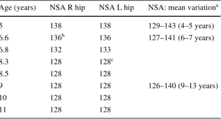
For the assessment of a coxa vara we measured the neck-shaft angle (NSA). At the time of Wrst presentation at the age
of Wve the patient had a bilateral NSA of 138° (Table1). This served as our baseline for subsequent follow-ups.
At the age of 6 years 7 months the patient presented with a history of few months pain in the right groin. The radiographs showed a femoral neck stress fracture at the compression-side (inferior aspect of the neck) with a medially displaced epiphy-sis (Fig.1a). There was no associated causative trauma. At this age the NSA measured 136° on both sides.
Treatment options were discussed with the parents and according to the literature research at this time [2, 5, 10], we recommended internal Wxation with a valgus osteotomy. The parents were concerned about the surgical complica-tions and therefore asked for the patient to be managed con-servatively as an option prior to surgery.
Consequently we treated the patient with partial weight bearing on crutches. After 8 weeks the patient had no limp or pain, and was able to resume all normal activities with a full range of motion in the right hip. Interestingly, X-rays and computer tomographs at that time did not show any callus formation (Fig.1b).
Table 1 Femoral neck-shaft angles (NSA) for the patient and
refer-ence values
a According to Paley [19] b Time of Wrst
c
Second fracture
Age (years) NSA R hip NSA L hip NSA: mean variationa
5 138 138 129–143 (4–5 years) 6.6 136b 136 127–141 (6–7 years) 6.8 132 133 8.3 128 128c 8.5 128 128 9 128 128 126–140 (9–13 years) 10 128 128 11 128 128
Fig. 1 a Femoral neck fracture with medial slip of the epiphysis at the
time of Wrst fracture. b Eight weeks later the X-Ray still shows the frac-ture line and no callus formation. c Four months after fracfrac-ture with
consolidation. d Complete remodelling of the right femoral neck 7 months after fracture
Arch Orthop Trauma Surg (2007) 127:967–970 969
123
Radiographs after 4 months showed healing of theverti-cal fracture line (Fig.1c), and at 7 months the fracture was united and the femoral neck remodelled (Fig.1d).
Eighteen months after the right femoral neck stress frac-ture the patient started limping on the left side after playing at school without previous trauma. The radiographs of the 8 years 4 months old boy revealed a similar compression-side stress fracture (Salter–Harris type 2) at the left femoral neck (Fig.2a). This fracture was also treated conservatively with partial weight bearing on crutches for a period of 8 weeks. At this time the radiographs did not show any cal-lus formation at the left femoral neck (Fig.2b) but he was free of pain with a full range of motion and weight bearing. First radiological signs of union at the left femoral neck were evident at the 5 months radiographs (Fig.2c).
In another follow-up examination 9 months after the sec-ond femoral neck fracture the 9-year-old patient was asymptomatic with a full range of motion in both hips. Union was radiologically conWrmed. After 2.5 years at the left side and after 4 years at the right side, the radiographs revealed completely remodelled femoral necks with a NSA of 128° on both sides (Fig.2f).
Our analysis of the patient’s NSAs and comparison with established age-matched ranges over a period of 4 years, did not show any evidence for the development of coxa vara (Table1).
Discussion
Osteopetrosis, in its autosomal dominant form (ADO) is a benign variant and therefore often is incidentally discov-ered as in our case. Patients may be asymptomatic (45%),
present with osteomyelitis of the mandible (10%), cranial nerve palsy (10–20%), bone pain (20–25%) or early hip osteoarthritis [2, 4, 6, 10, 11, 17]. Our patient presented with hip pain in combination with limping. We deWned the lesions as stress fractures as there was no associated causi-tive trauma.
Typical fracture patterns in osteopetrosis are transverse fractures perpendicular to the direction of stress because the dense but disorganised osteopetrotic bones cope with com-pression adequately, but are weak to tension.
Fracture healing has been studied histologically with specimens from up to 1 year post fracture [7]. In this report the biopsy showed that disorganised woven bone with no lamellar or Haversian systems persists with very low quan-tities of osteoclasts and osteocytes.
Surgical Wxation is the recommended treatment espe-cially in the femur neck. Internal Wxation was thought to address the risks of non-union, subsequent coxa vara and avascular necrosis [2, 10, 14, 21]. It is with these consider-ations that internal Wxation was recommended as the treat-ment even for undisplaced fractures [2, 5]. Series in the past using K-wires, Steinmann pins, blade plates, dynamic com-pression plates and screws have given reasonable results. Primary valgus osteotomy could also be performed if varus preceded the fracture [2, 11, 19]. Technical issues with dril-ling of the hard bone were commonly encountered [6, 10–
12, 18]. Longer operating time was needed and changing over of multiple drills helped to reduce the heat. Drill and chisel breakage were also reported [1].
Altered local biomechanics caused by the hardware cre-ated problems like peri-prosthetic fracture and re-fracture through screw holes after removal of the implant [12]. In Greene’s report, the pin tracks were still evident on
Fig. 2 a Stress fracture at the
left femoral neck. b Left hip 8 weeks after fracture and par-tial-weight bearing. c Patient at 5 months after fracture at the left-side with callus formation.
d, e 1.5 years post L fracture and
3 years post R fracture. f Com-plete remodelling of both femo-ral necks at the latest follow-up 2.5 years (left) and 4 years (right) after the fractures
970 Arch Orthop Trauma Surg (2007) 127:967–970
123
radiographs 2.5 years after their removal [10]. For osteopet-rotic patients, the lack of remodelling in surrounding bone to cope with new stress distribution makes them more sus-ceptible to this complication [7, 12].
Reported problems with conservative management includes delayed or non-union, with subsequent coxa vara deformity [11, 13]. Bilateral proximal femoral non-union was reported by Alexander [1] in 1923.
Armstrong et al. [2] (POSNA survey) reported about closed reduction and internal Wxation with pins or compres-sion screws in four acute femoral neck fractures. Also they found no evidence of healing at 6 months in a series of three conservative treated femoral neck fractures in two patients. They were eventually internally Wxed with pri-mary valgus osteotomy. In their study two stress lesions were successfully treated with protection only.
Several authors have devised classiWcations for stress fractures of the femoral neck [3, 8, 9]. Stress fractures at the superior cortex of the femoral neck are tension fractures and are associated with a risk of displacement [8]. Fractures at the compression side at the inferior aspect of the femoral neck rarely show displacement. In bones with previously normal bone biology compression fractures are therefore usually treated conservatively by bed rest and a non-weight-bearing regimen.
In our case the patient was fully weight bearing after 8 weeks on both occasions because he was clinically asymptomatic by that time but still showed the fracture line without callus formation (Figs.1b, 2b). The right side was consolidated after 4 months and the left side after 5 months (Figs.1c, 2c). The early mobilization did not inXuence the
healing nor cause development of coxa vara. Radiological analysis of radiographs consistently shows the NSA mea-surements to be within the normal range for age matched population (Table1).
This case demonstrates that successful conservative treatment of undisplaced femoral neck stress fractures in ADO is possible. Management of femoral neck stress frac-tures in paediatric patients with ADO remains contentious. Both surgical and non-operative treatments present their own challenges. While coxa vara can develop, early pro-phylactic Wxation or primary valgus osteotomy are proba-bly unnecessary in children.
The outcome of our patient is clinically and radiologi-cally successful. The lesson we involuntarily learned from this case led us to the conclusion that in the future we would treat a patient with the same problem in a stepwise regimen beginning with conservative treatment with regu-lar radiographic follow-ups at 2 weeks, 1, 2, 4 and 6 months. In case of ongoing pain, absent callus formation
after 6 months or progressive deformity we would consider an operative intervention. More reporting of experiences are needed in the future to help bring consensus.
References
1. Alexander WG (1923) Report of a case of so-called ‘‘marble bones’’with a review of the literature and a translation of an article. Am J of Roentgenol 10:280–301
2. Armstrong DG, NewWeld JT, Gillespie R (1999) Orthopedic man-agement of osteopetrosis: results of a survey and review of the lit-erature. J Pediatr Orthop 19(1):122–132
3. BlickenstaV LD, Morris JM (1966) Fatigue fracture of the femoral neck. J Bone Joint Surg Am 48(6):1031–1047
4. Bollerslev J, Mosekilde L (1993) Autosomal dominant osteopetro-sis. Clin Orthop Relat Res 294:45–51
5. Busch MT (2001) Sports medicine in children and adolescents. In: Lovell WW, Winter RB, Morrissy RT, Weinstein SL (eds) Lippin-cott. Williams & Wilkins, Philadelphia pp 1277–1279
6. Cameron HU, Dewar FP (1977) Degenerative osteoarthritis asso-ciated with osteopetrosis. Clin Orthop Relat Res 127:148–149 7. de Palma L, Tulli A, Maccauro G, Sabetta SP, del Torto M (1994)
Fracture callus in osteopetrosis. Clin Orthop Relat Res 308:85–89 8. Devas MB (1965) Stress fractures of the femoral neck. J Bone
Joint Surg Br 47(4):728–738
9. Fullerton LR Jr, Snowdy HA (1988) Femoral neck stress fractures. Am J Sports Med 16(4):365–377
10. Greene WB, Torre BA (1985) Femoral neck fracture in a child with autosomal dominant osteopetrosis. J Pediatr Orthop 5(4):483–485
11. Griss P, Schafer T (1988) Therapy of bone and joint changes in Albers–Schonberg osteopetrosis. Orthopade 17(5):411–419 12. Gupta R, Gupta N (2001) Femoral fractures in osteopetrosis: case
reports. J Trauma 51(5):997–999
13. King RE, Lovejoy JF Jr (1973) Familial osteopetrosis with coxa vara. A case report J Bone Joint Surg Am 55(2):381–385 14. Lam SF (1971) Fractures of the neck of the femur in children.
J Bone Joint Surg Am 53(6):1165–1179
15. Marks SC Jr., Schmidt CJ (1978) Bone remodeling as an expres-sion of altered phenotype: studies of fracture healing in untreated and cured osteopetrotic rats. Clin Orthop Relat Res 137:259–264 16. McCarthy EF, Frassica FJ (2000) Genetic diseases of bones and
joints In: Pathology of bone and joint disorders. WB Saunders, Philadelphia
17. McKusick VA (1972) Heritable disorders of connective tissue. Mosby, St. Louis, Mo
18. Milgram JW, Jasty M (1982) Osteopetrosis. A morphological study of twenty-one cases. J Bone Joint Surg Am 64(6):912–929 19. Paley D (2002) Principles of deformity correction. Springer,
Berlin
20. Shapiro F (1993) Osteopetrosis. Current clinical considerations. Clin Orthop Relat Res 294:34–44
21. Song KS, Kim HK (2005) Femoral neck fracture in a child with autosomal-dominant osteopetrosis: failure of spica cast treatment and successful outcome by internal Wxation. J Orthop Trauma 19(7):494–497
22. St Pierre P, Staheli LT, Smith JB, Green NE (1995) Femoral neck stress fractures in children and adolescents. J Pediatr Orthop 15(4):470–473