ORIGINAL ARTICLE
Strontium ranelate improves bone strength
in ovariectomized rat by positively influencing bone
resistance determinants
S. D. Bain&C. Jerome&V. Shen&I. Dupin-Roger&
P. Ammann
Received: 1 July 2008 / Accepted: 1 December 2008 / Published online: 19 December 2008
# International Osteoporosis Foundation and National Osteoporosis Foundation 2008
Abstract
Summary Treatment of adult ovariectomized (OVX) rats with strontium ranelate prevented vertebral biomechanics degradation as a result of the prevention of bone loss and micro-architecture deterioration associated to an effect on intrinsic bone material quality. Strontium ranelate influ-enced the determinants of bone strength by prevention of ovariectomy-induced changes which contribute to explain strontium ranelate antifracture efficacy.
Introduction Strontium ranelate effects on the determinants of bone strength in OVX rats were evaluated.
Methods Adult female Sprague–Dawley rats were OVX, then treated daily for 52 weeks with 125, 250, or 625 mg strontium ranelate/kg. Bone strength, mass, micro-architecture, turnover, and intrinsic quality were assessed.
Results Strontium ranelate prevented ovariectomy-induced deterioration in mechanical properties with energy necessary for fracture completely maintained vs. SHAM at 625 mg/ kg/day, which corresponds to the clinical dose. This was related to a dose-dependent effect on bone volume, higher trabeculae number, and lower trabecular separation in strontium ranelate vs. OVX. Load and energy required to induce lamella deformation were higher with strontium ranelate than in OVX and in SHAM, indicating that the bone formed with strontium ranelate is able to withstand greater damage before fracture. Bone formation was maintained high or even increased in strontium ranelate as shown by mineralizing surfaces and alkaline phosphatase while strontium ranelate led to reductions in deoxypyridinoline.
Conclusion Strontium ranelate administered at 625 mg/kg/day for 52 weeks prevented OVX-induced biomechanical properties deterioration by influencing the determinants of bone strength: it prevented bone loss and micro-architecture degradation in association with an effect on intrinsic bone quality. These beneficial effects on bone contribute to explain strontium ranelate antifracture efficacy.
Keywords Bone biomechanics . Bone intrinsic quality . Bone micro-architecture . Ovariectomized rat .
Strontium ranelate
Introduction
Postmenopausal osteoporosis is still a major public health problem. Until now, most efforts to reduce the risk and DOI 10.1007/s00198-008-0815-8
DO00815; No of Pages
S. D. Bain
Department Orthopaedics/Sports Medicine, University of Washington,
Washington, USA
e-mail: [email protected] C. Jerome
Think Bone Consulting, Inc.,
P.O. Box 1611, Langley, WA 98260, USA e-mail: [email protected] V. Shen MDS Pharma Services, 22011 30th Dr. SE, Bothell, WA 98021-4444, USA e-mail: [email protected] I. Dupin-Roger
Institut de Recherches Internationales Servier, Courbevoie, France
e-mail: [email protected] P. Ammann (*)
Service of Bone Diseases (WHO Collaborating Center for Osteoporosis Prevention), Hopital Cantonal, rue Micheli du Crest,
1211 Geneva 14, Switzerland
incidence of fractures have focused on therapies that either preserve skeletal mass by inhibiting osteoclastic bone resorption or reverse bone loss by stimulating osteoblastic
bone formation [1]. An ideal therapy for the reversal of
bone fragility would be a single therapeutic engineered to possess both antiresorbing and bone-forming properties
[2].
Strontium ranelate, composed of two stable strontium atoms and ranelic acid, represents a new paradigm in the
treatment of osteoporosis [3]. In phase III studies in
postmenopausal osteoporotic women, strontium ranelate (2 g/day) reduced the risk of vertebral fracture, the risk of nonvertebral fracture, and the risk of hip fracture over
3 years [4,5]. It has been shown to be effective in reducing
bone loss and/or increasing bone mass and/or bone
resistance in intact mice [6], rats [7], and monkeys [8] as
well as in short-term rat models of osteopenia, including
ovariectomy-induced bone loss [9,10] and immobilization
[11]. Furthermore, in vitro studies have shown that
strontium ranelate has an original mechanism of action
acting by reducing osteoclastic bone resorption [12, 13]
while at the same time stimulating osteoblastic bone
formation [14].
The clinical aim of an anti-osteoporotic treatment is to decrease the risk of fracture, an event related to bone resistance which is driven by parameters including bone mass, bone size and shape, bone turnover, bone micro-architecture, and intrinsic bone tissue quality including
damage accumulation [15]. Valid techniques for
nonin-vasive assessment of bone strength in human patients are currently not available and fracture risk reduction is only an indirect marker of bone strength. Therefore, evalua-tion of the drug-induced effects on these variables in animals can yield valuable insights into the underlying mechanisms that influence the antifracture efficacy of a therapy. The ovariectomized (OVX) rat is a well-recognized and validated model of bone loss that closely resembles the osteoporosis observed in postmenopausal
women [16–18]. Therefore, the current study was carried
out to evaluate the long-term bone efficacy and safety of strontium ranelate (125, 250, and 625 mg/kg/day for 12 months) on all the determinants of bone strength in OVX adult Sprague–Dawley rats, a relevant model of osteoporosis, in order to better understand clinical antifracture efficacy of this anti-osteoporotic agent. The response to strontium ranelate was evaluated using determinations of bone strength (using biomechanical testing) and of the determinants influencing bone strength: bone mass and architecture (using
micro-computed tomography [μCT] and static histomorphometry),
bone turnover (using dynamic histomorphometry and bone turnover markers), and intrinsic bone tissue quality (using nano-indentation).
Materials and methods Animals and treatment
The protocol for this experiment was approved by the Institutional Animal Care and Use Committee at SkeleTech where the study was performed. Six-month-old virgin female Sprague–Dawley rats (Harlan Sprague Dawley, Indianapolis, IN, USA) were randomized to treatment groups based on body weights and ovariectomy or sham surgery was performed. Treatment was initiated on the day following ovariectomy or sham surgery. Animals were individually housed in rooms with controlled temperature and relative humidity and an alternating 12-h dark/light photoperiod. Animals were fed PMI Certified Rodent Diet 5002 containing 0.80% calcium, 0.60% phosphorus, and
2.2 IU of vitamin D3per gram of feed. Water was available
ad libitum.
Three OVX treatment groups (SR125, SR250, and SR625; n=30 animals each) were administered strontium ranelate (S12911-2, Technologie Servier, Orléans, France) at dose levels of 125, 250, and 625 mg/kg/day. These doses were based on previous studies demonstrating that strontium ranelate was effective in preventing OVX-induced bone loss
and at improving bone resistance [7,9]. Strontium ranelate
was prepared weekly as a suspension in the vehicle (0.5% carboxymethylcellulose sodium salt, medium viscosity; Spectrum Chemical, Gardena, CA, USA). The control sham-operated (SHAM) group (n=24 animals) and OVX group (n=25 animals) were administered the vehicle. All treatments were administered daily by gavage (10 mL/kg) for 52 weeks.
During treatment, body weights were obtained weekly and the animals were individually pair-fed according to the average daily food consumption of the SHAM control animals. Prior to necropsies performed on week 52, all animals received a fluorochrome-labeling regimen on days 16/15 and 5/4 prior to killing to deposit double fluorochrome labels on mineralizing surfaces (calcein green, 12 mg/kg IP). At the time of necropsy, the final body weights were recorded and the animals were sedated with ketamine/xylazine and then humanely killed while still under sedation. Following killing, the uterus was removed and weighed to verify the success of ovariectomy. The lumbar vertebrae (L2 to L5) were then excised.
Bone mechanical properties
Biomechanical tests were performed in a blinded manner on LV5 vertebral body specimens using an Instron mechanical testing machine (Instron 4465 retrofitted to 5500) interfaced to a personal computer with Merlin II machine software. For each sample, the vertebral arch,
pedicle, and cranial and caudal ends of each vertebral body were removed using a low-speed diamond saw to obtain a vertebral body specimen with two parallel surfaces and a height of approximately 4 mm (width and height of the vertebral body were measured using digital calipers). The specimens were then placed between two platens and a load was applied at a displacement rate of 6 mm/min until failure. The load–displacement curve for each test was recorded: the maximum load at failure (expressed in newtons) as well as the yield load at the transition between the elastic and plastic phases of the deformation were manually selected (changes detected on the tangent). Machine software was then used to calculate the stiffness (slope of the elastic part of the curve, expressed in newtons per millimeter) and total energy absorbed (total energy absorbed, expressed in millijoules). All calculations were according to established
formulae [15].
Bone histomorphometry
Bone specimens for histomorphometry (L3) were fixed in cold, 70% ethanol immediately after collection. After trimming, the bone samples were dehydrated, infiltrated, and embedded in methyl methacrylate plastic composite
[19]. L3 in the sagittal plane were sectioned at 5 and 10μm
using a Reichert–Jung motorized rotary microtome equipped with a tungsten carbide microtome knife. The
5-μm sections were stained with Goldner’s trichrome for
bright field microscopy and the 10-μm sections were left unstained for epifluorescence microscopy.
Bone histomorphometry was performed using an OsteoMeasure software program version 4.00c (OsteoMetrics, Atlanta, GA, USA) interfaced with a Nikon Eclipse E400 light/ epifluorescent microscope and video subsystem. The sampling site of the secondary spongiosa of the vertebral body was performed on an area approximately 1×2 mm within the central portion of the vertebral body, extending dorsoventrally within the marrow space, but excluding the endocortical surfaces. Vigorous validation process was performed prior to the histomorphometry measurement based on Good Laboratory Practices requirements. In particular, only one user performed the measurements with an intra-user precision of 0.72% for BV/TV measurement as an example. The measurements were performed at the middle one third of the lumbar vertebral body and, on average, the marrow cavity of the lumbar vertebral body is about 1.2–1.6×5–6 mm and it seems then hard to fit a region of interest much bigger than 1×2 mm in the cancellous bone of a rat vertebral body. The region of interest may be smaller than usual but the results are quite representative as they are always measured at the same place in each lumbar vertebral body and the sample size (20 to 27) in each treatment group added further assurance. Furthermore, this 2-D histomorpho-metric measurement method allowed demonstrating a 49.1%
OVX-induced bone loss, in accordance with the results found
in 3-D histomorphometry, assessed by μCT. We, therefore,
consider that this method allowed validating the model of ovariectomy and the following profound deterioration of bone architecture and that the results obtained in strontium ranelate-treated groups can be interpreted. Static parameters (trabecular volume [BV/TV; percent]; trabecular number [Tb.N; number per millimeter]; trabecular thickness [Tb.Th; micrometers]; trabecular spacing [Tb.Sp; micrometers]; osteoid volume [OV/TV; percent]) and dynamic parameters (mineralizing surface [MS/BS; percent]; mineral apposition rate [MAR; micrometers per day]; bone formation rate [BFR/BS; cubic micrometers per square micrometer per year]) were measured in a blinded manner in cancellous bone and were calculated according to the recommendations of the American Society
for Bone and Mineral Research committee [20].
Bone nano-indentation
Proximal and distal plateaus of L2 vertebral bodies were cut transversally. The samples were exposed to ultrasound in Deconnex® bath in order to remove the marrow content and rinsed with water. The samples were embedded in poly-methyl methacrylate and the faces of the proximal cut were
polished and finished with a 0.25-μm diamond spray. The
specimens were then hydrated in a 9 g/L NaCl solution for 16 h and nano-indentation tests were performed in wet conditions. A Nano Hardness Tester (CSM Instruments, Peseux, Switzerland) equipped with a three-sided pyramidal Berkovich indenter was used. Five indentations were performed along the center of the posterior cortex of the vertebrae and five indentations in trabecular nodes close to the posterior cortex. The indenter was pressed into the specimens to a 900-nm maximum depth at a rate of 76 mN/ min. At maximum load, a 5-s holding period was imposed. The applied load and the penetration depth were continu-ously recorded during the loading and unloading cycle. Hardness (H) and modulus (Eit) were directly calculated from this load–displacement curve using the method
previously described [21, 22]. In addition, the dissipated
energy that occurred during the deformation of bone was also estimated from this curve.
Bone microtomography
At the end of these evaluations, the embedded L2 vertebral
samples were measured by μCT. Parameters of mass and
architecture of the secondary spongiosa of the L2 vertebral body were investigated with a high-resolution
microcom-puter tomography system (μCT 40, Scanco Medical,
Bassersdorf, Switzerland). Three-dimensional images of L2 vertebral body were acquired with a voxel size of
images were segmented using a low-pass filter to remove noise and a fixed threshold to extract the mineralized bone phase. The trabecular and cortical parts of the vertebrae were separated with semi-automatically drawn contours. From the binarized images, structural indices were assessed. Relative bone volume (BV/TV), trabecular number (Tb.N), thickness (Tb.Th), and separation (Tb.Sp) were calculated by measuring directly the 3-D distances in the trabecular network. The mean cortical thickness (Cort.Th.) was also evaluated.
Serum and urine biochemistry
Animals were sedated with a ketamine/xylazine anesthetic for blood sampling from the retro-orbital sinus. Serum samples were obtained on the day of surgery and again during weeks 8, 26, and 52. The effectiveness of ovariec-tomy was confirmed by radioimmunoassay determination of serum levels of 17beta-estradiol (Diagnostic Products, Los Angeles, CA, USA). To assess bone formation, serum alkaline phosphatase (ALP) was determined colorimetrically on a Roche Cobas Mira auto-analyzer (Roche Diagnostic Systems, Somerville, NJ, USA). In order to assess exposure to circulating strontium ranelate, serum concentrations of strontium were measured at the end of the 52-week treatment period by inductively coupled plasma atomic emission spectrometry (Vista apparatus, Varian).
Urinary samples were collected at week 0 and again during weeks 8, 26, and 52. Prior to urine collections, the animals were placed in metabolic cages and deprived of food for an overnight fast period of 18 h. Centrifuged urine samples were measured for deoxypyridinoline (DPD) by enzyme-linked immunosorbent assay (Quidel, San Diego, CA, USA) to assess bone resorption. The DPD values were normalized to the urinary creatinine, which was determined colorimetrically on a Roche Cobas Mira auto-analyzer (Roche Diagnostic Systems, Somerville, NJ, USA).
Statistical analyses
Results are presented as the means±standard deviations (SD) for all parameters measured and comparisons between groups were done using analysis of variance (ANOVA)
[23]. When an overall treatment effect was shown by
ANOVA, significant differences vs. the OVX group or the SHAM group were evaluated using Dunnett’s multiple
comparison procedure or a Fisher test (for μCT and
nano-indentation results). For the statistical evaluation of the nano-indentation results, a mean value of five indents was obtained in each bone envelope for each rat and these values were used to calculate the level of significance of the difference between the groups.
Results Animals
Treatment of OVX rats with 125, 250, or 625 mg/kg/day of strontium ranelate was well-tolerated and safe as it did not have any significant or adverse effects on animal survival, food consumption, body weight changes, clinical signs during the study, terminal body weights, or gross pathology at necropsy (data not shown). Despite pair-feeding, the body weights in all OVX animals (OVX, SR125, SR250, and SR625) slightly increased (from 7.3% to 9.2%) compared to the SHAM rats. However, strontium ranelate had no effect on the body weight gain, confirming that the treatment was well-tolerated. Success of ovariectomy was confirmed at necropsy by the significantly decreased uterine weights and by the lack of detectable levels of serum estradiol in all OVX rats whatever the time point and regardless of treatment, in contrast with SHAM rats exhibiting mean estradiol levels of 8.9±0.5 pg/mL at killing.
Serum strontium concentrations
SHAM and OVX endogenous serum strontium concentra-tions were similar. After strontium ranelate oral administration for 52 weeks, serum strontium concentration changes were
dose-dependent (Table 1). However, the serum strontium
concentrations increased 3.4 times between the dose levels of 125 and 625 mg/kg/day, while the given dose increased five times. Therefore, serum strontium concentrations did not increase proportionally to the dose administered, indicating saturation of the strontium absorption processes.
Bone mechanical properties
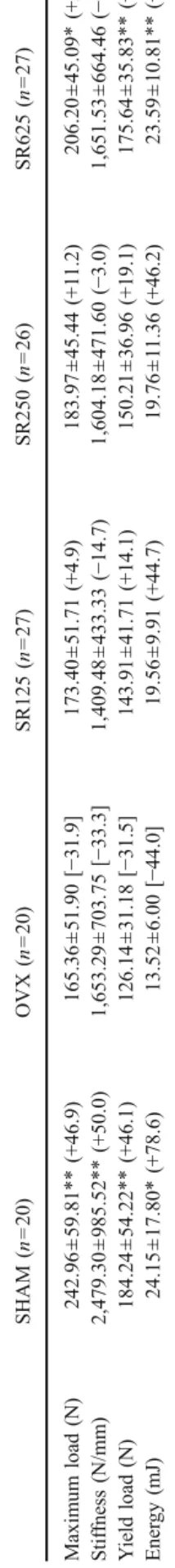
Compared to SHAM animals, compressive testing of L5 vertebral bodies of OVX animals showed significantly lower
Table 1 Serum strontium concentration (in nanograms per milliliter) obtained at the end of a 52-week treatment with strontium ranelate at 125, 250, and 625 mg/kg/day in OVX rats
SHAM (n=20) OVX (n=20) SR125 (n=29) SR250 (n=26) SR625 (n=28)
37.8±16.4 55.6±58.4 2,609.9±601.6 4,594.1±1,137.4 8,999.2±2,269.9
maximal load (−31.9%, p<0.01, Table2), stiffness (−33.3%,
p<0.01), and energy absorbed (−44.0%, p<0.05). Similarly,
the yield load was −31.5% lower (p<0.01). Strontium
ranelate-treated OVX rats showed a dose-dependent higher maximal load (up to +24.7%, p<0.05) of the vertebral bodies
when compared to OVX controls (Fig. 1a). This was
accompanied by a complete prevention of the ovariectomy-induced energy lost in animals treated with 625 mg/kg/day, while stiffness in strontium ranelate-treated animals was unchanged from the values in the OVX animals treated with
vehicle (Fig.1b; Table2).
Bone mass and architecture
2-D histomorphometry of the L3 lumbar vertebra
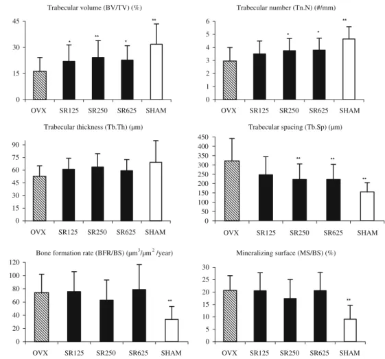
Ovariectomy exerted negative effects on cancellous bone structural properties, evidenced by lower BV/TV, Tb.N, and Tb.Th and higher Tb.Sp in OVX vs. SHAM groups for the
lumbar vertebra (Figs. 2 and 3). Treatment of OVX rats
with 125, 250, or 625 mg/kg/day strontium ranelate prevented the effects of ovariectomy with significant dose-dependent higher BV/TV and Tb.N and lower Tb.Sp
T able 2 Mechanical strength testing of L5 lumbar vertebra in OVX rats treated with strontium ranelate SHAM (n = 20) OVX (n = 20) SR125 (n = 27) SR250 (n = 26) SR625 (n = 27) Maximum load (N) 242.96 ± 59.81** (+46.9) 165.36 ± 51.90 [− 31.9] 173.40 ± 51.71 (+4.9) 183.97 ± 45.44 (+1 1.2) 206.20 ± 45.09* (+24.7) Stif fness (N/mm) 2,479.30 ± 985.52** (+50.0) 1,653.29 ± 703.75 [− 33.3] 1,409.48 ± 433.33 (− 14.7) 1,604.18 ± 471.60 (− 3.0) 1,651.53 ± 664.46 (− 0.1) Y ield load (N) 184.24 ± 54.22** (+46.1) 126.14 ± 31.18 [− 31.5] 143.91 ± 41.71 (+14.1) 150.21 ± 36.96 (+19.1) 175.64 ± 35.83** (+39.2) Ener gy (mJ) 24.15 ± 17.80* (+78.6) 13.52 ± 6.00 [− 44.0] 19.56 ± 9.91 (+44.7) 19.76 ± 1 1.36 (+46.2) 23.59 ± 10.81** (+74.5) V alues represent the mean±SD. The positive or negative values in parentheses beside the mean values for SHAM and SR groups indicate the percent change vs. OVX. The values in square brackets beside the mean values for the OVX indicate the percent change vs. SHAM *p < 0.05 compared to OVX; ** p < 0.01 compared to OVX 0 50 100 150 200 250 300 OVX SR125 SR250 SR625 SHAM Maximum Load (N) 0 5 10 15 20 25 30 OVX SR125 SR250 SR625 SHAM Energy (mJ) * * ** **
b
a
Fig. 1 a Maximum load (in newtons) and b energy (in millijoules) of L5 lumbar vertebra obtained by a compression test in OVX rats treated with strontium ranelate at 125, 250, and 625 mg/kg/day for 52 weeks. Values represent the mean±SD; n=20–27 animals per group. *p<0.05 compared to OVX control group; **p<0.01 compared to OVX control group
compared to OVX control (Figs.2and3). Furthermore, the mean values for Tb.Th in rats treated with 250 mg/kg/day strontium ranelate were as much as +20.2% greater in the lumbar vertebra vs. values in the OVX group, but statistical significance was not achieved.
3-D histomorphometry of the L2 lumbar vertebra
In the OVX control group, BV/TV, Tb.N, and Tb.Th were markedly and significantly decreased (−39.8%, −22.4%, and
−7.1%, respectively; Fig.4). As a consequence, Tb.Sp was
significantly increased to +29%. The treatment by strontium ranelate dose-dependently prevented these micro-architecture deteriorations with a significantly higher BV/TV, Tb.N, and Tb.Th than in OVX control animals (up to +36.3%, +12.8%, and +7.6%, respectively, at the highest dose tested, p<0.05
to p<0.001) and a lower Tb.Sp (up to −12.5% for SR625, p<0.001). No statistical changes of the cortical thickness were induced by ovariectomy and by strontium ranelate as well. Intrinsic bone tissue quality
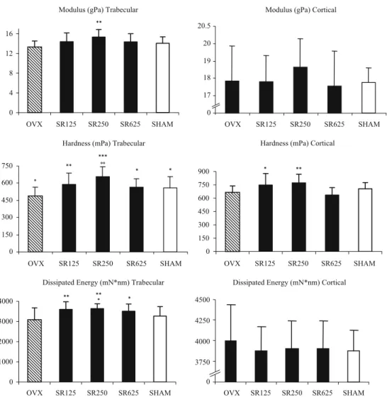
At the level of the trabecular bone, OVX decreased all the parameters of intrinsic bone tissue quality assessed by the
nano-indentation technique (Fig. 5). The differences
be-tween OVX and SHAM control groups were statistically significant for hardness (−13.4%, p<0.05). In contrast, compared to the OVX control group, all the intrinsic bone tissue quality parameters were significantly higher in rats treated with strontium ranelate whatever the dose level. The highest values were systematically observed in the group receiving SR250, +15.9% for modulus (p<0.01), +35.7% for
a
b
c
e
d
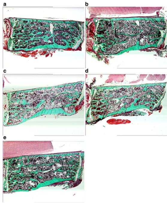
Fig. 2 L3 lumbar vertebrarep-resentative pictures of each group (Goldner’s trichrome staining, ×10 magnification). a SHAM animal; b OVX animal; c, d, and e strontium ranelate-treated animals with 125, 250, and 625 mg/kg/day, respectively
hardness (p<0.001), and +18.4% for dissipated energy (p<0.01). Furthermore, in the same strontium ranelate-treated group, SR250, the values were also significantly higher than in SHAM controls for hardness (+17.5%) and dissipated
energy (+11.2%). At the level of cortical bone (Fig.5), no
significant differences between the OVX control group and the SHAM control group were observed. However, the hardness was significantly higher in OVX rats treated with strontium ranelate at doses of 125 and 250 mg/kg/day compared to OVX control group (+13.4%, p < 0.05, and +17%, p<0.01, respectively).
Bone turnover
Dynamic histomorphometry of the L3 lumbar vertebra As expected, ovariectomy produced significantly higher dynamic indices of bone formation in the lumbar vertebra
(Fig. 3), including bone mineralizing surfaces (MS/BS;
+128.8%) and bone formation rate normalized to bone surface (BFR/BS; +121.2%). These changes were consis-tent with the expected increase in bone formation that accompanies increased bone turnover in estrogen-deficient animals. In rats treated with strontium ranelate, MS/BS and
BFR/BS of the lumbar vertebra remained elevated at the high turnover level observed in OVX control rats.
Serum alkaline phosphatase
Treatment of OVX animals with strontium ranelate increased serum levels of ALP, a marker of bone formation, dose-dependently with statistically significant increases of 30.0%, 25.9%, and 23.9% in the SR625 group at the 8-, 26-, and 52-week time points, respectively (p<0.01 compared to
OVX; Table3).
Urinary deoxypyridinoline
Ovariectomy resulted in increased mean levels of urinary DPD, a biomarker of bone resorption at the 8-, 26-, and
52-week time points (Table 3) with a maximal effect 8 weeks
after ovariectomy. In addition, there was a notable age-related decline in this parameter that appeared to reach a steady state by the 26-week time point (i.e., around the age of 1-year-old). Strontium ranelate treatment reduced the DPD levels by 20.4%, 13.9%, and 21.1% (p<0.05 compared to
OVX for SR625, Fig.1b) in the SR125, SR250, and SR625
groups, respectively, at the 8-week time point.
Trabecular volume (BV/TV) (%) 0 15 30 45 OVX SR125 SR250 SR625 SHAM Trabecular number (Tn.N) (#/mm) 0 1 2 3 4 5 6 OVX SR125 SR250 SR625 SHAM Trabecular thickness (Tb.Th) (µm) 0 15 30 45 60 75 90 OVX SR125 SR250 SR625 SHAM Trabecular spacing (Tb.Sp) (µm) 0 50 100 150 200 250 300 350 400 450 OVX SR125 SR250 SR625 SHAM ** * ** * ** ** ** Mineralizing surface (MS/BS) (%) 0 5 10 15 20 25 30 OVX SR125 SR250 SR625 SHAM Bone formation rate (BFR/BS) (µm3/µm2 /year)
0 20 40 60 80 100 120 OVX SR125 SR250 SR625 SHAM ** * * ** ** Fig. 3 L3 lumbar vertebra 2-D
histomorphometry indices in OVX rats treated with strontium ranelate. Values are expressed as the mean±SD; n=20–27 animals per group. *p<0.05 compared to OVX; ** p<0.01 compared to OVX
Discussion
The results indicate that ovariectomy decreased lumbar vertebra bone strength by altering bone mass, bone micro-architecture, and intrinsic bone tissue quality, which is consistent with previously published findings observed in
this model [16–18,24–30]. In contrast, in the conditions of
this study, there were no reductions in the mechanical strength of the midshaft femur in OVX control rats 52 weeks after ovariectomy when compared to SHAM control rats (mean maximum load±SD =182.64±17.85 N for OVX vs. 171.41±17.47 N for SHAM). This may be explained by the lack of OVX-induced cortical bone loss (no change of cortical width, data not shown) and by an increase in cross-sectional geometries between SHAM and OVX animals (data not shown), as previously described in
this model [31,32]. The combination of these two effects is
consistent with the lack of differences in long bone strength between the SHAM and OVX control groups as the resistance to bending loads is primarily governed by the
midshaft’s geometric configuration. Therefore, the absence
of effects of ovariectomy on long bones midshaft cortical strength preclude any interpretation in our experimental conditions.
Compared to vehicle-treated OVX controls, the treat-ment of OVX rats with strontium ranelate for 1 year at doses of 125, 250, and 625 mg/kg/day was well-tolerated and safe. These dose levels lead to mean serum strontium concentrations which correspond, respectively, to 0.25-, 0.44-, and 0.87-fold the median serum strontium concen-trations observed in patients administered the therapeutic
dose of 2 g/day (i.e., 10,560 ng/mL, [4]) and receiving
1,500 mg calcium/day. As illustrated in the present study, serum strontium concentrations did not increase propor-tionally to the dose administered (serum strontium concen-trations increased of only 3.4 times when dose levels increased of five times), indicating a saturation of the strontium ranelate absorption processes when doses in-crease. As the same transport properties that have been developped and extensively used for studies of calcium
absorption also apply to strontium [33], it is assumed that
some portions of strontium are absorbed into blood from the intestinal lumen via passive nonsaturable diffusion while another portion proceeds by an active saturable
transport [34] which can explain why calcium and
strontium can compete during their absorption process.
Interestingly, this is illustrated in a recent publication [35]
where OVX rats receiving 150 mg/kg/day of strontium Trabecular number (Tb.N) (#/mm) 0 1 2 3 OVX SR125 SR250 SR625 SHAM Trabecular volume (BV/TV) (%) 0 5 10 15 20 25 30 35 OVX SR125 SR250 SR625 SHAM Trabecular spacing (Tb.Sp) (µm) 0 150 300 450 OVX SR125 SR250 SR625 SHAM °°° °°° °°° * °° *** *** °°° °°° °°° °° * *** ° ° * * °° °°° °°° * °° *** *** Trabecular thickness (Tb.Th) 0 70 75 80 85 90 95 OVX SR125 SR250 SR625 SHAM Cortical Thickness (Ct.Th) (mm) 0 0.2 0.22 0.24 OVX SR125 SR250 SR625 SHAM Fig. 4 L2 lumbar vertebra 3-D
histomorphometry indices assessed byμCT in OVX rats treated with strontium ranelate. Values represent the mean±SD; n=12 animals per group. *p<0.05 compared to OVX; **p<0.01 compared to OVX; ***p<0.001 compared to OVX. °p<0.05 compared to SHAM; °°p<0.01 compared to SHAM; °°°p<0.001 compared to SHAM
ranelate with a normal calcium diet (1.19% Ca) exhibited, as expected, a sevenfold less serum strontium concentration than the same dose given with a calcium-deficient diet (0.1% Ca) due to the competition in the absorption process
of both cations. In the Fuchs et al. paper [35], when normal
calcium diet was given to OVX rats, the serum strontium concentration obtained at 150 mg/kg/day was comparable to the one observed in the present study at the dose of 125 mg/kg/day. Nevertheless, these concentrations repre-sent a fourfold lower strontium concentration than the one observed in treated patients at the therapeutic dose which explains their lack of efficacy. It is also interesting to note
that, in this recent publication [35], strontium ranelate at
150 mg/kg/day (but administered with a calcium-deficient diet) exhibited no efficacy in OVX rats while leading to a higher serum strontium concentration than the efficient dose of 625 mg/kg/day in the present paper (administered with normal calcium diet). This confirms that no beneficial effect on bone is possible if the amount of calcium available from the diet is not sufficient. It is indeed described in the literature that ovariectomy worsens the calcium deficiency-induced hyperparathyroidism leading to decreases in bone
calcium content, bone mineral density, and bone strength
[36,37]. Therefore, as recommended for all drugs used for
osteoporosis treatment, a normal calcium intake is neces-sary and only serum strontium concentrations obtained in OVX rats fed a normal calcium diet are relevant for human therapeutic interpretation.
In the present study, at the dose of 625 mg/kg/day, leading in OVX rats fed a normal calcium diet to serum strontium concentration close to those observed in treated patients, the maximum load and total energy absorbed by the vertebra before fracture were equivalent to SHAM animals, indicating a complete prevention of the ovariectomy-induced loss of bone strength.
In the strontium ranelate-treated OVX rats, BV/TV values were intermediate between those of OVX and SHAM control groups, as indicated by the significant difference between the BV/TV measured by 2-D and 3-D histomorphometry and the OVX and SHAM controls. The prevention in BV/TV values degradation was accompanied by corresponding dose-dependent positive effects of strontium ranelate on bone micro-architecture as evidenced by higher Tb.N and Tb.Th. Importantly, the two different methods used
Modulus (gPa) Trabecular
0 4 8 12 16 OVX SR125 SR250 SR625 SHAM
Hardness (mPa) Trabecular
0 150 300 450 600 750 OVX SR125 SR250 SR625 SHAM
Hardness (mPa) Cortical
0 150 300 450 600 750 900 OVX SR125 SR250 SR625 SHAM
Dissipated Energy (mN*nm) Trabecular
0 1000 2000 3000 4000 OVX SR125 SR250 SR625 SHAM ** *** °° ° ** * * * ** ** ° *
Dissipated Energy (mN*nm) Cortical
0 3750 4000 4250 4500 OVX SR125 SR250 SR625 SHAM Modulus (gPa) Cortical
0 17 18 19 20 20.5 OVX SR125 SR250 SR625 SHAM **
Fig. 5 Intrinsic trabecular and cortical bone quality testing of L2 lumbar vertebra in OVX rats treated with strontium ranelate. Values represent the mean±SD; n=12 animals per group. *p<0.05 compared to OVX; **p<0.01 compared to OVX; ***p<0.001 compared to OVX. °p<0.05 compared to SHAM; °°p<0.01 compared to SHAM
to assess these parameters, namely, 2-D and 3-D histomorph-ometry, led to the same results, suggesting a negligible
influence of strontium on bone μCT assessment in these
conditions. It is important to note that bone strength was assessed at the level of the whole vertebral body; the measured bone strength reflects both the contribution of the trabecular and cortical compartments on bone resistance. In contrast, micro-architecture was assessed, whatever the method used, only at the trabecular level which can explain why the partial effect on micro-architecture cannot totally explain the full rescue of the biomechanical effect on bone strength.
Evaluation of the bone biomarkers and dynamic indices of bone formation provide further insight regarding stron-tium ranelate’s beneficial action on bone. Interestingly, the effects on bone resorption markers of strontium ranelate after ovariectomy combined a modest decrease in urinary DPD (observed mainly at 625 mg/kg/day and at the 8-week time point when ovariectomy induced the maximal increased level of bone resorption), in line with a decreased
osteoclastic bone resorption [12,13,38] observed together
with a dose-dependent increase in the serum levels of ALP
in OVX animals, consistent with previous data [9]. Taken
together, these observations indicate that the ability of strontium ranelate to prevent the ovariectomy-induced bone loss is due to a combination of its inhibitory effects on bone resorption and its ability to stimulate and/or maintain high levels of bone formation. Maintaining elevated indices of bone formation in OVX animals provides evidence for an
effect on bone formation [9, 14, 38] and the dynamic
indices of bone formation such as mineralizing surface and bone formation rate are coherent with the bone markers changes observed. However, cellular parameters (such as osteoclast and osteoblast number and surface) were not measured in this study because their changes, 52 weeks after ovariectomy when the adult rats were more than 18 months old, were anticipated to be undetectable
compared to SHAM controls. It is indeed described in
Wronski et al. [26] that, from 150 days after ovariectomy,
osteoblast surfaces and osteoclast surfaces in OVX rats declined to SHAM control levels. Furthermore, osteoblasts are difficult to identify in old animals and this can result in a high variability of the cellular parameters. Static cellular parameters are not indicative of cellular activity and corresponding bone formation and resorption rates. They can only give information at a given time, without allowing an extrapolation for the whole treatment period. In contrast, dynamic fluorochrome-based parameters are meaningful as they provide strong evidence for a high bone formation rate. In OVX strontium ranelate-treated rats, fluorochrome-based indices of bone formation were maintained high while bone mass decrease was prevented, indicating that the level of bone formation exceeds the level of bone resorption.
In younger OVX rats [9], strontium ranelate decreased
histomorphometric indices of bone resorption to the levels in SHAM animals; in contrast to this inhibitory effect on bone resorption, osteoblast surfaces were as high in OVX rats treated with strontium ranelate as those in control OVX rats treated with vehicle. The positive strontium ranelate effect on bone mass and bone micro-architecture can then be related to the cellular effect observed in previous in vitro and in vivo studies which indicate that strontium ranelate
acts through a dual mechanism of action [38], namely,
reduction of osteoclastic bone resorption [12, 13] and
stimulation of osteoblastic bone formation [14].
The contribution of intrinsic bone tissue quality, using nano-indentation tests in wet conditions, was assessed at the level of the dorsal cortex of the vertebral body and of the trabecular nodes close to this region. Previous studies indicated that this latter area of the vertebral body is markedly affected by low protein regimen or treatments of
osteoporosis [21]. The results of the present study confirm
Table 3 Bone formation (ALP) and bone resorption markers (urinary DPD) in OVX rats treated with strontium ranelate
SHAM (n=20–22) OVX (n=22–25) SR125 (n=28–30) SR250 (n=27–30) SR625 (n=27–30) ALP (IU/L) Baseline 162±22 (+1.3) 160±33 [−1.2] 159±37 (−0.6) 161±23 (+0.6) 161±42 (+0.6)
Week 8 159±26 (−0.6) 160±36 [+0.6] 156±34 (−2.5) 178±34 (+11.3) 208±35** (+30.0) Week 26 153±24 (−3.2) 158±28 [+3.3] 156±28 (−1.3) 161±30 (+1.9) 199±47** (+25.9) Week 52 170±33 (−5.6) 180±53 [+5.9] 182±53 (+1.1) 193±39 (+7.2) 223±48** (+23.9) DPD (nM/mM creatinine) Baseline 27.7±9.9 (−17.6) 33.6±17.3 [+21.3] 36.9±33.2 (+9.8) 31.9±12.0 (−5.1) 37.0±17.6 (+10.1) Week 8 27.5±14.9** (−74.0) 105.7±37.8 [+284.4] 84.1±27.5 (−20.4) 91.0±34.2 (−13.9) 83.4±30.4* (−21.1) Week 26 15.4±5.4** (−49.3) 30.4±11.8 [+97.4] 30.3±8.8 (−0.3) 33.5±14.4 (+10.2) 31.5±7.1 (+3.6) Week 52 13.6±5.0** (−66.3) 40.4±18.1 [+197.1] 38.1±15.0 (−5.7) 37.3±18.0 (−7.7) 43.1±19.3 (+6.7) Values represent the mean±SD. The positive or negative values in parentheses beside the mean values for SHAM and SR groups indicate the percent change vs. OVX. The values in square brackets beside the mean values for the OVX indicate the percent change vs. SHAM
that trabecular bone intrinsic quality is affected by OVX and that the treatment with strontium ranelate at each investigated dose resulted in a significant higher hardness and dissipated energy in this type of bone. Whatever the strontium ranelate dose, these values were higher than in SHAM controls. These results indicate that the load and energy required to induce a given deformation of a bone lamella are markedly increased by strontium ranelate treatment in OVX rats. The bone tissue formed under strontium ranelate treatment shows improved intrinsic quality properties, suggesting that it is able to withstand greater damage before fracture. The fact that cortical bone is poorly affected by OVX is not surprising, since in rodents there is little remodeling of the cortical bone and since its vascularization is poorly developed. Furthermore, the treatment started in mature rats in which no periosteal apposition could be observed. However, even in these conditions, strontium ranelate was able to increase hardness at two dose levels. This is in line with a previous study performed in intact rats treated during all their life with strontium ranelate where a mild but significant effect of the treatment was observed at the level of the cortical bone
[39].
How strontium ranelate totally prevented and even improved the OVX-induced deterioration of intrinsic bone tissue quality is still under investigation. Indeed, strontium ranelate does not affect crystal properties and characteristics
as shown in vivo [40, 41]: the normal mineralization
process and the mean degree of mineralization of bone are preserved, and when strontium takes the place of calcium, only one calcium ion out of ten is substituted by one strontium ion in the hydroxyapatite crystal lattice. The majority of strontium present in bone is adsorbed onto the
surface of the crystal, in the nonapatitic hydrated layer [42].
This leads to the hypothesis that this strontium localization in the bone tissue could potentially lead to a better cohesion between the mineral and the protein matrix and/or to a direct or indirect effect (through the cellular activity) on the spatial orientation of the crystals in the lamellae or between the lamellae themselves.
In conclusion, strontium ranelate effects on bone mass, on trabecular micro-architecture, and on the intrinsic properties of the material allow explaining, at the dose level of 625 mg/kg/day, the prevention of OVX-induced biomechanical deterioration. Long-term strontium ranelate treatment prevents the OVX-induced deterioration of bone mechanical properties acting on all their main determinants in adult rats at a therapeutic equivalent human dose level. This therapeutic effect was associated with a bone formation rate maintained at a high level together with a slight decrease of bone resorption. Taken together, obser-vations reported in this study support the efficacy and the safety of strontium ranelate and contribute to explain
strontium ranelate antifracture efficacy for the treatment of postmenopausal osteoporosis.
Acknowledgements The authors would like to acknowledge Craig Bailey, Matt Heggem, Ryan Leininger, and Debbie Puerner for their contributions to the study management and mechanical testing procedures, Tina Bailey for her performance and analysis of the bone biomarker assays, Hellen Zheng and Chung Liu for the performance of the bone histomorphometry, and Isabelle Badoud for performing the nano-indentation tests andμCT analysis.
Conflicts of interest None.
References
1. Delmas PD (2002) Treatment of postmenopausal osteoporosis. Lancet 359:2018–2026
2. Riggs BL, Parfitt AM (2005) Drugs used to treat osteoporosis: the critical need for a uniform nomenclature based on their action on bone remodeling. J Bone Miner Res 20:177–184
3. Reginster J-Y, Deroisy R, Jupsin I (2003) Strontium ranelate: a new paradigm in the treatment of osteoporosis. Drugs Today 39:89–101
4. Meunier PJ, Roux C, Seeman E, Ortolani S, Badurski JE, Spector TD, Cannata J, Balogh A, Lemmel EM, Pors-Nielsen S, Rizzoli R, Genant HK, Reginster JY (2004) The effects of strontium ranelate on the risk of vertebral fracture in women with postmenopausal osteoporosis. N Engl J Med 350:459–468 5. Reginster JY, Seeman E, de Vernejoul MC, Adami S, Compston J,
Phenekos C, Devogelaer JP, Diaz-Curiel M, Sawicki A, Goemaere S, Sorensen OH, Felsenberg D, Meunier PJ (2005) Strontium ranelate reduces the risk of non vertebral fractures in postmeno-pausal women with osteoporosis: TROPOS study. J Clin Endocrinol Metab 90:2816–2822
6. Delannoy P, Bazot D, Marie P (2002) Long-term treatment with strontium ranelate increases vertebral bone mass without delete-rious effect in mice. Metabolism 51:906–911
7. Ammann P, Shen V, Robin B, Mauras Y, Bonjour JP, Rizzoli R (2004) Strontium ranelate improves bone resistance by increasing bone mass and improving architecture in intact female rats. J Bone Miner Res 19:2012–2020
8. Buehler J, Chappuis P, Saffar JL, Tsouderos Y, Vignery A (2001) Strontium ranelate inhibits bone resorption while maintaining bone formation in alveolar bone in monkeys (Macaca fascicu-laris). Bone 29:176–179
9. Marie PJ, Hott M, Modrowski D, De Pollak C, Guillemain J, Deloffre P, Tsouderos Y (1993) An uncoupling agent containing strontium prevents bone loss by depressing bone resorption and maintaining bone formation in estrogen-deficient rats. J Bone Miner Res 8:607–615
10. Grynpas MD, Hamilton E, Cheung R, Tsouderos Y, Deloffre P, Hott M, Marie PJ (1996) Strontium increases vertebral bone volume in rats at a low dose that does not induce detectable mineralization defect. Bone 18:253–259
11. Hott M, Deloffre P, Tsouderos Y, Marie PJ (2003) S12911-2 reduces bone loss induced by short-term immobilization in rats. Bone 33:115–123
12. Baron R, Tsouderos Y (2002) In vitro effects of S12911 on osteoclastic function and bone marrow macrophages differentia-tion. Eur J Pharmacol 450:11–17
13. Takahashi N, Sasaki T, Tsouderos Y, Suda T (2003) Strontium ranelate inhibits osteoclastic bone resorption in vitro. J Bone Miner Res 18:1082–1087
14. Canalis E, Hott M, Deloffre P, Tsouderos Y, Marie PJ (1996) The divalent strontium salt S12911 enhances bone cell replication and bone formation in vitro. Bone 18:517–523
15. Turner CH, Burr DB (1993) Basic biochemical measurements of bone: a tutorial. Bone 14:595–608
16. Wronski TJ, Lowry PL, Walsh CC, Ignaszewski LA (1985) Skeletal alterations in ovariectomized rats. Calcif Tissue Int 37:324–328
17. Wronski TJ, Dann LM, Scott KS, Crooke LR (1989) Endocrine and pharmacological suppressors of bone turnover protect against osteopenia in ovariectomized rats. Endocrinology 125:810–816 18. Kalu DK (1991) The ovariectomized rat model of postmenopausal
bone loss. Bone Miner 15:175–192
19. Bain SD, Impeduglia T, Rubin CT (1990) Cement line staining in undecalcified thin sections of cortical bone. Stain Technol 65:1–5 20. Parfitt AM, Drezner MK, Glorieux FH, Kanis JA, Malluche H, Meunier PJ, Ott SM, Recker RR (1987) Bone histomorphometry: standardization of nomenclature, symbols, and units. J Bone Miner Res 2:595–610
21. Hengsberger S, Ammann P, Legros B, Rizzoli R, Zysset P (2005) Intrinsic bone tissue properties in adult rat vertebrae: modulation by dietary protein. Bone 36:134–141
22. Oliver WC, Pharr GM (1992) An improved technique for determining hardness and elastic modulus using load and displacement sensing indentation experiments. J Mater Res 7:1564–1583
23. Steel RGD, Torrie JH (1980) Principles and procedures of statistics: a biometrical approach, 2nd edn. McGraw-Hill, New York, NY 24. Parfitt AM, Oliver I, Villanueva AR (1979) Bone histology in
metabolic bone disease: the diagnostic value of bone biopsy. Orthop Clin North Am 10:329–345
25. Wronski TJ, Walsh CC, Ignaszewski LA (1986) Histologic evidence for osteopenia and increased bone turnover in ovariec-tomized rats. Bone 7:119–123
26. Wronski TJ, Cintron M, Dann LM (1988) Temporal relationship between bone loss and increased bone turnover in ovariectomized rats. Calcif Tissue Int 43:179–183
27. Wronski TJ, Dann LM, Scott KS, Cintron M (1989) Long-term effects of ovariectomy and aging on the rat skeleton. Calcif Tissue Int 45:360–366
28. Yamazaki I, Yamaguchi H (1989) Characteristics of an ovariec-tomized osteopenic rat model. J Bone Miner Res 4:13–22 29. Wronski TJ, Dann LM, Horner SL (1989) Time course of
vertebral osteopenia in ovariectomized rats. Bone 10:295–301
30. Boyce RW, Wronski TJ, Ebert DC, Stevens ML, Paddock CL, Youngs TA, Gundersen HJG (1995) Direct stereological estima-tion of the three-dimensional connectivity in rat vertebrae: effect of estrogen, etidronate and risedronate following ovariectomy. Bone 16:209–213
31. Turner RT, Vandersteenhoven JJ, Bell NH (1987) The effects of ovariectomy and 17 beta-estradiol on cortical bone histomorph-ometry in growing rats. J Bone Miner Res 2:115–122
32. Bagi CM, Mecham M, Weiss J, Miller SC (1993) Comparative morphometric changes in rat cortical bone following ovariectomy and/or immobilization. Bone 14:877–883
33. Comar CL, Wasserman RH (1966) Strontium. Mineral Metabolism 2:523–571
34. Perault-Staub AM (1990) Extracellular calcium homeostasis. Elsevier, Amsterdam
35. Fuchs RK, Allen MR, Condon KW, Reinwald S, Miller LM, McClenathan D, Keck B, Phipps RJ, Burr DB (2008) Strontium ranelate does not stimulate bone formation in ovariectomized rats. Osteoporosis Int 19(9):1331–141
36. Zhang Y, Lai W-P, Wu C-F, Favus MJ, Leung PC, Wong MS (2007) Ovariectomy worsens secondary hyperparathyroidism in mature rats during low-ca diet. Am J Physiol Endocrinol Metab 292:E723–E731
37. Creedon A, Cashman KD (2001) The effect of calcium intake on bone composition and bone resorption in young growing rat. Br J Nutr 86:453–459
38. Marie PJ, Ammann P, Boivin G, Rey C (2001) Mechanisms of action and therapeutic potential of strontium in bone. Calcif Tissue Int 69:121–129
39. Ammann P, Badoud I, Barrauld S, Dayer R, Rizzoli R (2007) Strontium ranelate treatment improves trabecular and cortical intrinsic bone tissue quality, a determinant of bone strength. J Bone Miner Res 22:1419–1425
40. Boivin G, Deloffre P, Berat B, Panczer G, Boudeulle M, Mauras Y, Allain P, Tsouderos Y, Meunier PJ (1996) Strontium distribution and interactions with bone mineral in monkey iliac bone after strontium salt (S12911) administration. J Bone Miner Res 11:1302–1311 41. Farlay D, Boivin G, Panczer G, Lalande A, Meunier PJ (2005)
Long-term strontium ranelate administration in monkeys preserves characteristics of bone mineral crystals and degree of mineralisation od bone. J Bone Miner Res 20:1569–1578
42. Cazalbou S, Combes C, Rey C (2002) S12911 treatment maintains bone mineral crystal characteristics. J Bone Miner Res 17(Suppl 1):S376–S377