Megalin in normal tissues Ziak, Meier, and Roth
SHORT COMMUNICATION
Megalin in normal tissues and carcinoma cells
carries oligo/poly
a
2,8 deaminoneuraminic acid as a
unique posttranslational modification
Martin Ziak, Mirjam Meier and Jürgen Roth*
Division of Cell and Molecular Pathology, Department of Pathology, University of Zürich, CH-8091 Zürich, Switzerland
In rat kidney, megalin, a member of the low density lipoprotein receptor gene family, is the sole glycoprotein which carries oligo/polya2,8 deaminoneuraminic acid (KDN) as a posttranslational modification. We have investigated immunoprecipi-tated megalin from rat brain, lung and placenta, mouse yolk sac carcinoma and megalin synthesizing carcinoma cell lines, for presence of this unique glycan structure. Our immunoblot analysis revealed the presence of oligo/polya2,8 KDN on megalin in all the studied normal tissues and carcinoma cells. Furthermore, it is demonstrated to be part of oligosaccharides O-glycosidically linked to megalin.
Keywords: oligo/polya2,8 deaminoneuraminic acid, megalin, lung, brain, placenta, F9 cells, L2 cells
Introduction
In previous studies, we demonstrated the presence of oligo/poly a2,8 linked deaminoneuraminic acid (oligo/poly a2,8 KDN) in various embryonic, postnatal developing and adult mammalian tissues by immunohistochemistry and immunoblot analysis [1,2] with the use of the monoclonal antibody mAb.kdn8kdn [3]. The existence of KDN in mam-malian tissues was furthermore confirmed by gas liquid chromatography analysis [1], and a sensitive fluorescent probe for HPLC [4] detection of KDN in tissues [5]. West-ern blot analysis of extracts of various rat tissues revealed the presence of a single reactive band in each tissue studied. The oligo/poly a2,8 KDN was found on a single 150 kDa glycoprotein except for a single⬎350 kDa glycoprotein in kidney [1,2].
Recently, we isolated and purified the oligo/poly a2,8 KDN bearing glycoprotein from rat kidney [6] and identi-fied it as being megalin, a member of the low-density lipo-protein receptor gene family [7]. The presence of oligo/poly a2,8 KDN on kidney megalin was confirmed by combined immunoprecipitation / immunoblot analysis and on
RAP-affinity purified megalin. Further supporting evidence was obtained by immunoelectron microscopy revealing an identical subcellular distribution of oligo/poly a2,8 KDN and megalin in rat kidney proximal tubules [6].
Megalin represents a major membrane protein of kidney proximal tubules but is additionally found in rat lung, cho-roid plexus and microvasculatur of brain, placenta, yolk sac epithelia, ciliary epithelium of the eye, parathyroid as well as inner ear [8]. Furthermore, by immunoblotting megalin was demonstrated in mouse F9 teratocarcinoma cells [9] and L2 rat yolk sac carcinoma cells [10].
Here, we report that megalin from various normal rat tissues, carcinoma tissue and carcinoma cell lines carries oligo/poly a2,8 KDN which is part of O-glycosidically linked oligosaccharides.
Materials and methods
Materials
For the detection of oligo/poly a2,8 KDN, purified IgM of the mouse monoclonal antibody mAb.kdn8kdn was used [3]. The hybridoma cell line 2G-5 [3] was kindly supplied by Dr. Ken Kitajima (University of Nagoya, Nagoya Japan). Polyclonal antibodies against purified rat kidney megalin were raised in rabbits as described previously [11] and IgG
Glycoconjugate Journal 16, 185–188 (1999) © 1999 Kluwer Academic Publishers. Manufactured in The Netherlands
*To whom correspondence should be addressed: Tel:⫹41 1 255 50 90. Fax:⫹41 1 255 44 07. E-mail: juergen.roth@pty.usz.ch
fractions prepared using Protein A Sepharose CL-4B (Pharmacia, Uppsala, Sweden). Alkaline phosphatase-con-jugated donkey anti-mouse IgM (affinity-purified) was purchased from Jackson ImmunoResearch Laboratories, Inc. (West Grove, PA, USA) and frozen rat tissues from Pel Freez Biologicals (Rogers, AR, USA). Mouse yolk sac tu-mor tissue and the L2 cell line were kindly supplied by Dr. Ulla Wewer (University Institute of Pathological Anatomy, Copenhagen, Denmark). Mouse F9 teratocarcinoma cells were obtained from the American Type Culture Collection (Rockville, MD). Protease inhibitor cocktail tablets, recom-binant N-Glycosidase F and digoxigenin-conjugated Con-canavalin A were from Boehringer (Mannheim, Germany). Sephacryl S-400, XK 16/100 column were purchased from Pharmacia (Uppsala, Sweden). All other reagents were of analytical grade and purchased from Fluka (Buchs, Swit-zerland).
Cell cultures
L2 cells were grown in Dulbecco’s modified Eagle’s me-dium (DMEM) supplemented with high glucose and 10% fetal calf serum at 37⬚C in a humid atmosphere containing 5% CO2. Mouse F9 teratocarcinoma cells were cultured in gelatin coated (0.1%) flasks in DMEM with high glucose and 15% fetal calf serum. To induce differentiation, F9 cells were incubated for 72 hours with 1 lM retinoic acid and 1 mM dibutyryl cyclic AMP.
Megalin immunoprecipitation
Protein A Sepharose CL-4B was incubated with PBS con-taining 1% BSA and 0.05% of Triton X-100 and Tween 20 for 1 h at 4⬚C, followed by incubation with anti-megalin antibodies for 18 h at 4⬚C. Rat kidney (0.7 g), lung (4.2 g), brain (25 g), placenta (4.2g), mouse yolk sac carcinoma tissue (4.2 g) and pellets of L2 and F9 cells (200 lg each) were homogenized in PBS containing protease inhibitors and Triton X-100 (final concentration 1%). After 60 min on ice, the homogenate was centrifuged at 100,000⫻ g for 60 min. The soluble extract was incubated with the anti-megalin / Protein A Sepharose CL-4B overnight at 4⬚C. Following two washes with PBS containing 0.1% Triton X-100, the Protein A Sepharose CL-4B was pelleted, placed in SDS-PAGE sample buffer and heated. The super-natant was analyzed by SDS-PAGE and Western blotting using anti-oligo/poly a2,8 KDN or anti-megalin antibodies as described previously [6].
Partial purification of megalin from mouse yolk sac
carcinoma tissue
A mouse yolk sac carcinoma tissue homogenate was ap-plied onto a XK 16/100 gel filtration column packed with Sephacryl S-400. Fractions, immunoreactive for oligo/poly
a2,8 KDN were pooled and used for Western blot and lectin blot analysis.
b-elimination, N-Glycosidase F treatment and
lectin blotting
For b-elimination, PVDF strips containing partially puri-fied megalin from yolk sac carcinoma tissue were incu-bated with 0.1 N sodium hydroxide for 24 h at 37⬚C. As control, strips were incubated with PBS under the same conditions as described above. For N-Glycosidase F treat-ment, nitrocellulose strips with megalin were blocked with 1% BSA in 50 mM sodium acetate buffer (pH 5.5) fol-lowed by incubation with 5 U of N-Glycosidase F for 16 h at 37⬚C. For lectin blotting, strips were incubated with digoxigenin-conjugated Concanavalin A for 1 h followed by alkaline phosphatase-conjugated polyclonal sheep anti-digoxigenin Fab⬘ fragments (150 U/ml). Color reaction was performed using nitroblue tetrazolium/BCIP-phosphate as substrates.
Results and discussion
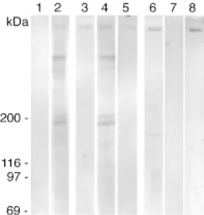
In our previous studies [6] the⬎350 kDa oligo/poly a2,8 KDN bearing glycoprotein from rat kidney was identified as being megalin. Megalin is the most abundant mem-brane protein in proximal tubules of rat kidney but it is also expressed in other specialized epithelia [8] albeit at much lower levels. Immunoprecipitated megalin from rat lung, brain and placenta, from mouse yolk sac carcinoma tissue as well as L2 cells and from mouse F9 teratocarci-noma cells, induced to differentiate (data not shown) ex-hibited immunoreactivity for oligo/poly a2,8 KDN (Fig. 1). Thus, megalin present in these normal rat tissues and in carcinoma cells carries oligo/poly a2,8 KDN as a unique posttranslational modification. These findings on megalin are similar to those for a 150 kDa glycoprotein in lung which also carries oligo/poly a2,8 KDN both in normal human and rat embryonic and postnatal lung and in hu-man lung carcinomas [2]. Furthermore, KDN has been de-tected in both normal human ovary and ovarian carcinoma cells but no data have been reported regarding its protein carrier [12].
b-elimination on stripes containing partially purified megalin from mouse yolk sac tissue resulted in the absence in immunoreactivity for oligo/poly a2,8 KDN. In contrast N-glycosidically linked oligosaccharides on megalin are not affected by this treatment as demonstrated by the reactiv-ity with Conconavalin A (Fig. 2). Furthermore, N-Glycosi-dase F treatment increased the immunoreactivity for oligo/poly a2,8 KDN (Figure 2) indicating that oligo/poly a2,8 KDN is part of O-glycosidically linked oligosaccha-rides on megalin from mouse yolk sac carcinoma tissue. These data are in agreement with previously described results obtained from purified rat kidney megalin [6].
Megalin has been found to bind a number of different ligands (for a review see [13]) in vitro. What may be a possible function of oligo/poly a2,8 KDN on megalin? Due to its localization in clathtrin-coated pits megalin was sug-gested to act as an endocytic receptor in kidney. Dependent on the organ localization of megalin and the composition of the surrounding fluids it can be anticipated that the nature of the ligands will vary from one organ to another. For example, in proximal tubules it was suggested that megalin is involved in the reabsorption of filtered proteins [14] and Ca2⫹-ions [15]. In type II pneumocytes, megalin
seems to be responsible for the clearance of protease / protease inhibitor complexes [16,17] from the alveolar space [18]. Charge interactions between negatively charged complement-type repeats and basic regions of the ligands seems to be important for receptor / ligand interaction. Furthermore, megalin is not only a Ca2⫹binding protein but Ca2⫹-ions are essential for ligand binding. In vitro
oligo/poly a2,8 KDN binds Ca2⫹-ions highly preferentially
[19]. Therefore, we speculate that the polyanionic nature of oligo/poly a2,8 KDN together with the negatively charged complement-type repeats may be important for ligand binding to megalin and for the receptor activity in the various tissues. Studies are in progress to clarify this pro-posal.
References
1 Ziak M, Qu B, Zuo X, Zuber C, Kanamori A, Kitajima K, Inoue S, Inoue Y, Roth J (1996) Proc Natl Acad Sci USA 93: 2759–63. 2 Qu B, Ziak M, Zuber C, Roth J (1996) Proc Natl Acad Sci USA
93: 8995–8.
3 Kanamori A, Inoue S, Xulei Z, Zuber C, Roth J, Kitajima K, Ye J, Troy FA, Inoue Y (1994) Histochemistry 101: 333–40.
4 Hara S, Takemori Y, Yamaguchi M, Nakamura M, Ohkura Y (1987) Anal Biochem 164: 138–45.
5 Inoue S, Kitajima K, Inoue Y (1996) J Biol Chem 271: 24341–4. 6 Ziak M, Kerjaschki D, Farquhar M, G., Roth J (1999) J Am Soc
Nephrol. 10: 203–09.
7 Saito A, Pietromonaco S, Loo AK, Farquhar MG (1994) Proc Natl
Acad Sci USA 91: 9725–9.
8 Zheng G, Bachinsky DR, Stamenkovic I, Strickland DK, Brown D, Andres G, McCluskey RT (1994) J Histochem Cytochem 42: 531–42.
9 Czekay RP, Orlando RA, Woodward L, Adamson ED, Farquhar MG (1995) J Cell Sci 108: 1433–41.
10 Lundstrom M, Orlando RA, Saedi MS, Woodward L, Kurihara H, Farquhar MG (1993) Am J Pathol 143: 1423–35.
11 Guhl B, Ziak M, Roth J (1998) Histochem Cell Biol 110: 603–11. 12 Inoue S, Lin SL, Chang T, Wu SH, Yao CW, Chu TY, Troy FA,
Inoue Y (1998) J Biol Chem 273: 27199–204.
13 Farquhar MG, Saito A, Kerjaschki D, Orlando RA (1995) J Amer
Soc Nephrol 6: 35–47.
Figure 2. b-elimination and N-Glycosidase F treatment on partially pu-rified megalin from yolk sac carcinoma tissue.b-elimination resulted in the absence of immunoreactivity for oligo/polya2,8 KDN (lane 1). Lectin blot using Concanavalin A afterb-elimination (lane 2). Immunoblot for oligo/poly a2,8 KDN (lane 3), Concanavalin A blot (lane 4) and im-munoblot for megalin (lane 5) after incubation of the stripes with PBS at 37⬚C for 24 h. Increased immunoreactivity for oligo/polya2,8 KDN after N-Glycosidase F treatment (lane 6) and absence of labeling for Conca-navalin A (lane 7). Immunoblot for megalin after N-Glycosidase F treat-ment (lane 8).
Figure 1. Immunoprecipitated megalin from rat kidney (lane 1), lung (lane 2) brain (lane 3) and placenta (lane 4), mouse yolk sac carcinoma (lane 5) and L2 cells (lane 6) exhibited immunoreactivity for oligo/poly a2,8 KDN. Molecular mass standards were as follows: myosin (200 kDa), b-galactosidase (116 kDa), phosphorylase b (97 kDa), bovine serum albumin (69 kDa).
14 Moestrup SK, Christensen EI, Nielsen S, Jorgensen KE, Bjorn SE, Roigaard H, Gliemann J (1994) Ann NY Acad Sci 737: 124–37. 15 Christensen EI, Gliemann J, Moestrup SK (1992) J Histochem
Cytochem 40: 1481–90.
16 Stefansson S, Kounnas MZ, Henkin J, Mallampalli RK, Chappell DA, Strickland DK, Argraves WS (1995) J Cell Sci 108: 2361–8. 17 Poller W, Willnow TE, Hilpert J, Herz J (1995) J Biol Chem 270:
2841–5.
18 Willnow TE, Hilpert J, Armstrong SA, Rohlmann, A, Hammer, RE, Burns DK, Herz J, (1996) Proc Natl Acad Sci USA 93: 8460–4. 19 Shimoda Y, Kitajima K, Inoue S, Inoue Y (1994) Biochemistry 33:
1202–8.
Received 4 November 1998, revised 19 February 1999, accepted 3 March 1999.
