DQCUMENT
FFICE 2gNT
ROOM 36-412
RESEARCH LAK)j'R.'kl'",g,''
' , L't:t il'I<Us
NCSMASSACHUS 'i, .':
.i
f
E HNLOGY
.iL.,"
CAMBRIDGE. ME
v i .39,
U.S.A.
BIOELECTRIC CONTROL OF PROSTHESES
RALPH ALTER
7
a
TECHNICAL REPORT 446
DECEMBER I, 1966
I
MASSACHUSETTS INSTITUTE OF TECHNOLOGY
RESEARCH LABORATORY OF ELECTRONICS
CAMBRIDGE, MASSACHUSETTS
The Research Laboratory of Electronics is an interdepartmental laboratory in which faculty members and graduate students from numerous academic departments conduct research.
The research reported in this document was made possible in part by support extended the Massachusetts Institute of Tech-nology, Research Laboratory of Electronics, by the JOINT SER-VICES ELECTRONICS PROGRAMS (U.S. Army, U.S. Navy, and U.S. Air Force) under Contract No. DA 36-039-AMC-03200(E); additional support was received from the Liberty Mutual Insurance Company.
Reproduction in whole or in part is permitted for any purpose of the United States Government.
Qualified requesters may obtain copies of this report from DDC.
MASSACHUSETTS
INSTITUTE OF TECHNOLOGY
RESEARCH LABORATORY OF ELECTRONICS
Technical Report 446
December 1, 1966
BIOELECTRIC CONTROL OF PROSTHESES
Ralph Alter
Submitted to the Department of Electrical Engineering, M. I. T.,
August 23, 1965, in partial fulfillment of the requirements for
the degree of Doctor of Science.
(Manuscript received September 28, 1965)
Abstract
Externally powered prostheses have been studied for many years, in efforts to provide more effective rehabilitation of amputees. In order to take advantage of the benefits offered by external power in prostheses, however, the mode of control of the prosthesis by the amputee must also be improved. Conventional control methods require a high degree of mental concentration by the amputee on his prosthesis, because of at least two important factors: (i) the mode of control of a prosthesis motion conventionally is different from control of the corresponding normal action; and (ii) there is no feedback of sensation from the prosthesis to the patient except through the visual sense.
Bioelectric control offers the possibility that control of the prosthesis action can be similar to control of the corresponding body action, which was lost through the amputation. The muscles in the body produce an electrical signal, called the electromyogram (emg), which can be sensed directly from the surface of the skin. Several experimental prostheses have been developed pre-viously, which use emg signals for control purposes. The major shortcoming of most of these devices has been the lack of graded control of their behavior.
A system has been developed which utilizes surface emg signals from the biceps and triceps of an amputee's arm to provide graded control of an elbow prosthesis. The development included as an intermediate step the control of a simulated forearm in a digital computer, in real time. In its present form the signal processing consists of full-wave rectification and lowpass filtering of the emg signals from biceps and triceps muscles; an actual mechanical elbow prosthesis can now be voluntarily controlled through the subject's emg signals.
The performance of the system in its present form is described, and its indications for future work are outlined. Foremost among these is the need for feedback to the patient of (at least) position information from the prosthesis, outside of the visual sense. One method by which it may be possible to accomplish this in an inherently normal manner is suggested.
The use of nerve signals as the control signal for a prosthesis offers many potential advan-tages over the use of the emg signal; the practical problems involved in observing nerve signals, however, combined with the lack of information as to how to properly interpret them, makes this approach infeasible now. Preliminary experiments aimed at deriving the necessary information in order that nerve signals can ultimately be used are described here. The results are still inconclusive.
TABLE OF CONTENTS
I.
INTRODUCTION
1
II.
AMPUTEES AND PROSTHESES: THE PROBLEM
3
2.1 General Background
3
2.2 Standard Means of Prosthesis Control
4
2.3 Cineplasty
7
2.4 External Power
7
2. 5 Bioelectric Control
9
III.
PHYSIOLOGY
13
3.1 Nerve
13
3.2 Muscle
16
3.3 Muscle Spindles
18
3.4 Organization of the Neuromuscular System
18
3.5 Conscious Control and Sensation
21
IV.
INVESTIGATION OF NERVE SIGNALS
25
4.1 Reasons for our Interest
25
4. 2 Other Recordings of Gross Nerve Potentials
28
4. 3 Our Attempts to Measure Nerve Signals
29
4.4 Conclusions on the Nerve Experiments
35
V.
USE OF MUSCLE SIGNALS FOR PROSTHESIS CONTROL
37
5.1 Factors Affecting Use of Muscle Signals
37
5.2 Data on EMG Signals
39
5.3 Proposed EMG-Controlled Prosthetic System
45
VI.
DEVELOPMENT OF THE PROSTHETIC SYSTEM
47
6.1 Simulation of the Proposed System
47
6.2 The Prosthetic Arm
57
6.3 EMG-Controlled Prosthetic System
59
VII. EVALUATION OF THE SYSTEM - SUGGESTIONS FOR
THE FUTURE
65
7.1 Over-all System Performance
65
7.2 Mechanical Portion of the System
68
7.3 EMG Signal Processing
69
7.4 Feedback to the Amputee
71
CONTENTS
APPENDIX I
Equipment Used in Attempts to Record Gross
Nerve Potentials
APPENDIX II Method Used for Measuring Power Spectra of
Bioelectric Signals
Acknowledgment
Bibliography
iv
75
77
78
79
I. INTRODUCTION
For many years, the standard means of control of the action of a prosthesis has been through gross body motions of the amputee. In the early 1950's the late Norbert Wiener observed that the biological signals that controlled actions which had been lost in amputation, were still present in the body of the amputee. Professor Wiener sug-gested that certain of these signals could be used to control a prosthesis, instead of using gross body motions. The main advantage of such an approach is that its mode of control would be similar to the control of the corresponding body section that had been amputated. Thus one could logically expect that the amputee's ability to control the device would exceed his ability to control a conventional prosthesis.
Interest in Professor Wiener's idea grew slowly in this country. In Britain and the Soviet Union, however, at least two groups of workers began work in the field. By 1955, the British had reported some progress, and the Russians reported their first ac-complishments in 1959.24,90 The Russian work received a great deal of publicity and, in 1961, on a visit to Russia, Dr. M. Glimcher witnessed a demonstration of the pros-thesis that had been developed.
Upon Dr. Glimcher's return, he approached Professor Wiener, who suggested that Professor Amar G. Bose, of the Department of Electrical Engineering at M.I.T., might be willing to undertake a research project in this direction. This author expressed interest in the idea, and it was agreed that the work would be undertaken under Pro-fessor Bose's guidance, as this author's Doctor of Science thesis research. Financial
support was obtained from the Liberty Mutual Insurance Company, whose interest stemmed from their long-time involvement at the forefront of rehabilitation of the
disabled.
In this research effort we continually emphasized a fundamental approach to the problem. The work began in February 1962, and included from the outset one year of
study in physiology, prosthetics, and related fields. As a result of this work, we under-took to carry out two investigations simultaneously. Through the study of the relevant physiology it became clear that the use of nerve signals for control purposes could potentially offer considerably improved rehabilitation, as compared with that possible through the use of muscle signals. Thus, despite the practical difficulties involved in the study and use of nerve signals, we elected to investigate the various factors affect-ing their application to prosthesis control. Some preliminary data on nerve signals have been obtained.
It is clear, however, that for use in the immediate future, the electromyographic signal (emg) is the most practical bioelectric signal for control purposes. Thus we set out to study, in parallel with the investigation of nerve signals, the properties of the muscle signal. This began with an investigation of the spectral characteristics of the
emg signal, as a function of muscle tension. The study of physiology, combined with the experiences of others in prosthetics, have led us to emphasize that the control
behavior in the prosthesis should be similar to that in a normal arm. Our interest in this ideal of normal behavior caused us to choose a very flexible form of signal pro-cessor, the digital computer, for our experimental emg-controlled prosthesis. Although it was clear that the ultimate control system must be small and compact, and therefore simple, we chose to place as few restrictions as possible on the initial form of the system. In this way the fundamental issues relating to the control problem could most easily be identified and studied.
II. AMPUTEES AND PROSTHESES: THE PROBLEM
2.1 GENERAL BACKGROUND
Mechanical devices have been devised for many years to assist persons who have, through various medical disorders or accidents, or congenitally, been deprived of some measure of normal physical ability. ' There are examples of such prostheses
dating from medieval times. For surgical amputation of an arm or leg, prostheses have taken many forms, and all have had certain deficiencies, for which scientists have not yet found completely satisfactory solutions. Generally speaking, the shortcomings of the arm prostheses that are now clinically available are the following.
1. The prostheses have far fewer degrees of freedom than the normal arm for which they are intended to substitute. Thus they perform certain tasks in an awkward manner, and in some cases only with great difficulty for the amputee.
2. The controls (i.e., the actions of the amputee which control motions of the pros-thesis) for a given prosthesis motion are not related to the actions of a normal person which cause the corresponding motion of a normal arm. For example, flexion of the "elbow" of a prosthesis may result only from movement of the shoulder of the amputee; whereas in a normal person elbow and shoulder motions can be independent. The re-sult of this is that the amputee must learn an entirely new pattern of activity in order to make the prosthesis useful to him, and his ultimate performance is often limited because the degree of relearning which is required is so great, and the constraints of
the control system are so severe.
3. The only sensation that the amputee receives from the prosthesis is that which he can obtain from visual observation of its performance, and from any extraneous noises that it makes or forces that it transmits back through the socket and harness.
We will see that this is significantly less sensory information than is present in a real arm. The degree of visual concentration which is required to properly operate present protheses precludes development of more complex, more flexible devices because the patient could not adequately control a device more complex than one of the current types. Furthermore, it is now very difficult for an amputee to carry on a conversation or engage in any other activity that reduces his conscious concentration on the prosthesis, while simultaneously operating his prosthesis; while for a normal person a large num-ber of common activities involving his arm can be carried on while he is talking and performing other activities perfectly naturally, and with very little concentration on or attention to his arm motions.
Although recent concerted efforts in prosthesis design have been historically re-lated to the World Wars, the number of civilian amputees now far exceeds the number of amputees resulting from war injuries. A recent estimate places the number of civilian amputees at approximately one-half million, while the veteran amputees of all wars number approximately 40,000.50 Nevertheless, it was immediately following
3
World War II, through the efforts of the National Research Council in this country, and simultaneously in other countries, that scientific research was channelled into this field. By comparison of the engineering sophistication of many of the machines that have been developed by modern science with that of current prostheses, one cannot
avoid feeling that the area of prosthesis development has been largely ignored. It seems clear that a concerted effort cannot fail to provide significant improvement in the
capabilities of the prostheses available for amputees. It is probably for these reasons that the present broad effort on the over-all problems of the amputee has developed.
Arm amputees vary over a wide range in many ways. For example, amputations may be due to injuries leaving stumps in various stages of injury themselves. Amputa-tions may be due to diseases leaving the muscles and nerves in the stump and the surrounding area of the person in various stages of damage. An amputation may be congenital, and the existing stump in such a case may not closely resemble that of a normal person. The strength and psychological state of amputees vary widely, depend-ing on age, cause of amputation, and so forth. A patient may have other injuries or disabilities that constrain the way in which he may be fitted. Finally, an upper-extremity amputation may be at any of at least six functionally different levels, as shown in Fig. 1, from a wrist disarticulation, in which case normal wrist rotation is usually retained, to a removal of the whole arm and shoulder (interscapulothoracic), in which case no arm or shoulder function exists at all. The breadth of these variations, of course, only increases the difficulty of building one or a few types of devices to serve all cases; the individual cases will often impose constraints on the type of prosthesis which can be fitted, which may preclude the application of any "standard" device. 2.2 STANDARD MEANS OF PROSTHESIS CONTROL
An important evaluation with respect to any amputee is the number of possible sites which he can provide for control of the prosthesis. By this is meant the number of motions or actions which he can control, and which either are of no present use to him because of his amputation or are less important than some of the actions that have been lost in the amputation and can be restored by a prosthesis. The intention is to retrain the patient to use these actions to control other actions in the prosthesis. The three most common control motions for an above-elbow prosthesis, for example, are
135
flexion and extension of the shoulder, and the shoulder shrug. The concept of using motions to control a given activity of the prosthesis which are not associated with such activities in a normal person has been accepted for many years, largely because very little choice was possible. In most cases the amputation leaves so little of the normal activity which it is desired to restore that there is no possibility of prosthesis control by the remainder.
Generally speaking, the longer the stump, the greater the number of possible con-trol sites, and the smaller the number needed, because of the relatively lesser degree
of disability in these cases. Thus the extremely short stumps are the ones that are
From Human Limbs and Their
Substi-tutes by P. E. Klopsteg and P. D.
Wilson.
Copyright
1954
McGraw-Hill Book Company, Inc. Reproduced
by permission of McGraw-Hill Book
Company.
Fig. 1. Major sites of amputation in the upper extremity.
most discouraging, since the patient needs a great amount of help and has the least residual function with which he can be helped.
The types of controls which are most common consist of a cable anchored to some point on the harness which is supporting the prosthesis, connected at its other end to
5
--the segment of --the pros--thesis which it is desired to move. The point on the harness which is used is carefully chosen so that by some motion that the amputee is able to perform he can exert tension on the cable. At most, three such motions have been successfully used in one above-elbow prosthesis, as shown in Fig. 2. Considering the number of degrees of freedom in the human arm this is very little indeed. We must
remember, however, that the restoration of even one degree of freedom to an amputee is considered by him to be a considerable improvement, especially if the choice of action to be restored and action to be used in controlling it are carefully tailored to the patient's needs. On the other hand, it seems apparent that any effort to find improved ways of providing this assistance, which may make possible far greater help for the handicapped, is justified.
Above-elbow triple control with shoulder saddle. Chest strap a buckles near front apron of shoulder saddle c and passes around opposite axilla to give rise to shrug-control cable d. Strap b
gives vertical suspension to a. Straps e and f are suspensors passing through D rings on the lateral and medial aspects of the socket. Arm-extension control g runs from shoulder saddle to elbow lock. Arm-flexion control h passes around socket and attaches to forearm lever i.
From Human Limbs and Their Substitutes by P. E. Klopsteg and P. D. Wilson. Copy-right ) 1954 McGraw-Hill Book Company, Inc. Reproduced by permission of McGraw-Hill Book Company.
Fig. 2. Triple-control harness for an above-elbow amputee.
Besides the problem of using a very few control sites to control a device that should have a rather large number of degrees of freedom, one other factor plays an important part in prosthesis design. That is, that the control site must have available enough force to deliver to the prosthesis, and it must be able to impart to the control cable sufficient excursion to operate the prosthesis motion for which it is intended. Of course, by appropriate lever arrangements force can be traded for excursion, but the product of the two, force times excursion, must be sufficient for the required prosthesis action. This requirement has led to the abandonment of a great number of otherwise satisfactory control sites.
When an actual gross body motion is used to control a prosthesis, this motion is coupled to the prosthesis so that in addition to its normal action as before, that motion also controls the desired action of the prosthesis. Thus the amputee is in the position of having to perform some unrelated body motion in order to control the prosthesis. The external effect of this is undesirable. It has long been considered an important goal to control a prosthesis directly from the force of some muscle, rather than through the motion of some part of the body. If this were possible, any muscle which could be flexed would be a potential control site. The same technique could possibly be applied to
the muscles in the stump which formerly moved a portion of the body which was ampu-tated, and if these muscles were then connected to control the corresponding prosthesis
action, then the resulting over-all effect could logically be expected to be more natural for the amputee than any other possible means of control of that action.
2.3 CINEPLASTY
A technique was developed which could make these goals attainable. It is called cineplasty, and dates in its earliest forms from before World War I. It consists of surgically modifying a muscle by creating a skin-lined "tunnel" through it, through which a pin can be inserted, which in turn can operate a control cable to a prosthesis.8 8 Cineplastic tunnels have been made in biceps and/or triceps, in the pectoral, and with less success in the forearm flexor and extensor groups. They have been most success-ful in the biceps in the case of below-elbow amputations, in which case the normal
in-sertion of the biceps into the stump of the forearm is released, leaving to other
muscles the job of flexing the elbow, and the biceps "muscle motor" is used to control closing of the terminal device. Because of harnessing problems, however, cineplasty has not been as successful as one might hope in using muscles to control the prosthesis motion corresponding to the real motion those muscles formerly controlled.
Despite its potential, the cineplastic technique has fallen into disuse in recent years, at least in the United States. The cineplastic tunnels have been found to be very difficult to keep clean, and are a frequent source of irritations and infections. Further-more, they involve a surgical modification to the body which amputees and their
families have found difficult to accept. Hence cineplastic operations, which were at one time quite prevalent in this country, are now virtually unknown.
2.4 EXTERNAL POWER
Another important concept has been the application of external power, controlled by a very small motion at some control site, for operation of the prosthesis.1 For example, the bulge of a muscle as it tenses would be a possible control site if it were able to impart to the prosthesis sufficient force and excursion for prosthesis operation. By connecting such a control site to operate an electrical switch, which in turn operates an electrically powered prosthesis, or to operate a mechanical valve which in turn applies pressurized carbon dioxide to a gas-powered prosthesis, as examples, it would be possible to relax considerably the restrictions on the choice of acceptable control sites.5 3,8 5,1 0 5,14 9
7
The application of external power provides one other major advantage: In cases of severe disability, such as, for example, a bilateral shoulder disarticulation (both arms completely removed at the shoulder), there are clearly very few, if any, available control sites capable of the power necessary for conventional prostheses. By careful training, however, the patient can learn to operate one or a few motions as controls for an externally powered prosthesis. A "sequential" control (which uses one control site to control several actions, in a fixed sequence, so chosen that the common activities requiring multiple actions need these actions in the order in which they occur on the control) can often be fitted to one control site. The combination of this with perhaps one other control site for some one function, then can provide adequate rehabilitation for a case which would otherwise be very discouraging. For the severe disabilities, the application of corresponding techniques to a conventional (body-powered) prosthesis is a serious burden on the amputee, and is rarely acceptable.
The application of external power to prosthetics has been the subject of research for many years. Gas- and electric-powered devices of many types have been developed both on this continent and in Europe. As early as 1919, there was a report of an effort to use an electromagnetic solenoid as the means of converting electrical power to the mechanical power required for a prosthesis. The poor control characteristics of
such a device, along with the considerable weight which it required in order to supply reasonable power, made it impractical. The development of modern light-weight motors was required before electrically powered prostheses were to be practical. A project was undertaken at the International Business Machines Corporation in 1946, to develop an electric arm, and that effort eventually gave rise to five different models, all of which had in common the fact that a very few motions were used to control both the
switching of the mechanical power from one "joint" to the next, and the turning on and off of this power.7 ' 8 1 Thus these motions had to be sequential, and were therefore quite complex from the point of view of the amputee. The result was that the operation was not natural for the patient and undoubtedly would have required a great deal of time to adapt to. Amputees have consistently rejected or been very slow to adapt to
prostheses which required sequential actuation of a very few control sites for control of more degrees of freedom of the prosthesis, preferring instead devices with fixed joints in some places, and passive motion only at others, combined with perhaps con-ventional control of only the terminal device, when necessary.
For completeness, one other development deserves mention here. That is the 85,105
work on gas-powered devices in most recent years. This work has been stimulated also by modern developments in equipment, in this case by new valves and gas cylinders, which combine low weight with high strength and therefore high capacity. Thus although
power-to-weight ratios for batteries have increased markedly in recent years, the comparison between the use of electric and gas power for externally powered prostheses has not caused gas power to be rejected.8
The results of the various research efforts in the application of external power to prosthetics has shown that such techniques are feasible in many cases, from the point of view of the equipment weight and volume considerations. It is possible to provide the power mechanisms, and the necessary power supply sources, n packages with the capacity for a normal day's activity, at least for upper-extremity prostheses.
It has become clear, however, that new developments are needed in the way in which amputees must control their prostheses. The interface between the man and his prosthesis must be made less abrupt. The equipment must in its mode of control take account of the limitations and needs of the amputee. The concept of sequential control, by using a very few control sites for control of a large number of motions, does not meet these requirements. Thus the application of external power alone does not provide the most satisfactory prostheses. Indeed, by making possible the movement of several more joints than would be possible with conventional prostheses, it challenges the imagination of the researcher with the problem that the amputee is actually less able to control the "improved" device than he would be to control a conventional prosthesis.
One reaction to this result has been an aversion to "complexity" in prostheses. This was well-founded in the day of the "IBM arm", which was admittedly very complex, both in construction and in control. But it is a contention of this author that complexity in and of itself is completely irrelevant to the amputee; rather it is the complexity of the control operations which the amputee must perform, for motions which may in fact be very simple, which matters. One of the assumptions that is basic to the work reported here is that, although complexity in the prosthesis itself ought to be avoided when
possible for many good reasons (such as cost and trouble-free performance), one of those reasons is not the fear that the amputee will reject the prosthesis. Complexity from the point of view of the control required to operate the prosthesis, however, should definitely be avoided.
2.5 BIOELECTRIC CONTROL
It has been shown that many amputees are potentially capable of learning wholly new patterns of activity in order to control a prosthesis whose mode of control is very different from that of a real arm. For example, the present standard prostheses all require such a readjustment, and there are many patients who operate them remarkably well indeed. It is admittedly difficult to find a fair basis for comparing the performance of a normal arm with that of a prosthesis, and it is probable that at least in the details of the motions considered, the real arm will always be superior. It is another conten-tion of this author that a prosthesis design that requires a minimal adjustment by the patient to the control mode of the prosthesis is preferable to one requiring considerable adjustment, for the reasons that (1) a large proportion of patients will not be completely successful in learning a wholly new mode of control, and (2) even the patient who is best adjusted to his prosthesis will still be limited in his performance by the limitations of the control system, and by the limits of his ability to produce all of the various control
9
-signals necessary. Thus we propose that, if such an ideal were attainable, the proper method of controlling an artificial arm would be to control it in an identical manner to that in which a real arm is controlled, specifically the real arm that was just amputated.
This would, if it were possible, provide a measure of precision and coordination which may never be attainable by other means.
The ideal manner in which such a scheme might be implemented would be to elec-trically sense human nerve or brain signals, which were intended by the brain to operate a muscle or a joint which was no longer operative because of the amputation. This signal could then be used to control the corresponding action in the prosthesis. By this means any motion, however complex, which had been learned by the patient before his amputation, would again be possible for him after his amputation. A related possi-bility is that electrical stimuli might be applied to the person, which could give him the same sensations (touch, feel, proprioception) that he formerly had in his real arm.
As we shall see, there are many different ways in which this broad suggestion can be interpreted. Early in the 1950's, Norbert Wiener suggested the use of an electrical signal generated by muscles when they contract, called the "electromyogram" (abbre-viated emg). For practical reasons, most of the effort toward applying bioelectric control to externally powered prostheses has utilized the emg for the control signal. This signal can be sensed by very simple means; for example, almost any type of electrode taped to the surface of the skin will suffice for observation of the emg. In 1955, Battye, Nightingale, and Whillis described an experimental prosthetic device, using emg as the control signal. This was a prosthetic hook, potentially suitable for a forearm amputee, and used surface electrodes to sense the emg signal. The forearm flexor and extensor groups of muscles, which are normally flexors and extensors of the fingers and wrist, generated the signals which controlled the prosthesis. This device had the capability only of being open or closed under control of the emg signal. No control or force, velocity or any intermediate positions was possible. Those authors did discuss several different possible procedures that might yield a graded control, which would be more satisfactory for the amputee. They suggested either a position or a velocity control servo, and discussed some of the advantages and disadvantages of both types. They do not appear to have considered the normal relations between muscular activity and the emg signals; nor did they emphasize the restoration of an action to the amputee in such a way that his control of it was similar to the control of the normal arm he had lost.
Around 1956, a similar prosthesis was developed in Russia, by Kobrinsky
et al.2 4 ' 9 0 9 3 Details have been lacking for many years, but the best available informa-tion indicates that the earliest version was quite similar in its capabilities to the
British prosthesis.54 The Russians have continued to develop their device, however, and the latest version of which this author is aware operates as follows: The emg signals (from a pair of opposing muscles or muscle groups) are processed in such a way that the prosthetic hand either closes or opens at one constant velocity, or stays in
any intermediate position. Thus the amputee can control the position of his terminal device, but he still cannot control its velocity; and only through spurious mechanical slack in the mechanism is he able to control the force that the device is exerting.1 4 0 Nevertheless, this device has apparently found wide acceptance in the Soviet Union; it
is understood that at least 200 are in use. This version is known to have been licensed to both British and Canadian groups.
A recent development has been announced by Bottomley and Cowell. In their case, a prosthetic hand or hook is controlled by emg; amplitude information in the emg signal is used, and the amputee is able to control both velocity and force. This device is a significant step in the direction of providing normal behavior for the emg-controlled prosthetic terminal device. It uses both a force and a velocity feedback loop, and the
amplitude of the processed emg signal is made proportional to velocity while the hand is open, that is, while the force that it is exerting is negligible. When the hand
en-counters resistance, however, while it is closing on some object, the force feedback loop dominates the behavior, and the force with which the object is clasped is then pro-portional to the amplitude of the processed emg signal. The latest information available to this author is that this device has not yet been widely used; reactions of amputees to its behavior would be interesting and valuable.
It has been the purpose of the research described in this report to study the problems associated with providing for amputees graded control of prostheses, by using voluntarily generated bioelectric signals as the control source and an external supply of power to operate the prosthesis. In order to create an experimental situation that could be studied without the severe mechanical constraints inherent in the lower extremity, we decided to focus our attention initially on arm rather than leg prostheses. Considering the foregoing summary of the work in related areas which has preceded our effort, it is considered of prime importance that the behavior of a normal arm be our model, and that every effort be made to duplicate the control aspects of a normal arm in the prosthesis. It will be seen that feedback from sensory organs in a normal arm contributes significantly to control of the arm, and certain aspects of the control of the prosthesis are limited by the lack of feedback (other than visual) to the amputee. The topic of feedback will be discussed further, although time has not allowed us to devote significant attention to this area.
11
----J1
III. PHYSIOLOGY
Our basic problem, of utilizing bioelectric signals for prosthesis control, led directly to a need for information about the various signals involved in the body in the control of muscle action. Our emphasis on the concept of restoring normal control to an amputee, to whatever extent possible, raised questions about the normal neuro-muscular control in the body. By a study of physiology, the various signals present in the system, and their interactions, could be identified, and those most interesting for bioelectric control in an amputee could be studied further. We shall now summarize the
information available on the organization of the neuromuscular system, and the details of the important components in that system. This information bears both directly and indirectly on the remainder of the work discussed in this report.
3.1 NERVE
All of the activities of life, including both voluntary and involuntary activities, in-volve in their performance a multiplicity of devices in the body, called nerves.5 2 1 5 0 This includes thinking, receiving any sensation, such as touch, smell, vision, and performing any motion. The brain itself is, for the most part, a collection of nerves. In the sense in which the term is being used here, a nerve consists of a cell body, an axon, and, depending on the type and function of the nerve, it may also include dendrites
or some nerve ending appropriate to the function of the nerve. In this sense, the term "nerve" is synonymous with neuron.
The cell body of a nerve is the source of nourishment for the nerve; a section of a nerve which is separated from the cell body degenerates rapidly. The axon, often re-ferred to as the nerve fiber, conducts a nerve impulse in either direction, although for any given nerve axon only one direction of transmission is used by the body. For a neuron in the brain, spinal cord, or a motor nerve in some peripheral area of the body, there are dendrites serving to receive nerve impulses from other neurons. Depending on the state of a given neuron, which of the other neurons that connect to its dendrites are actually transmitting nerve impulses, and in what sequence, a nerve impulse may be initiated in the given neuron to travel down its axon. For a sensory nerve, there are specialized nerve endings such as Pacinian corpuscles, Golgi tendon organs, and many others, which serve to measure the variable appropriate to the type of nerve, and cause the axon to transmit a signal to the central nervous system which will be interpreted as the proper sensation.
In the peripheral nervous system, which is the portion of the nervous system out-side of the brain and spinal cord, the term nerve has another meaning: In this area of the body, many nerve axons communicate between muscles and sensory endings and the spinal cord, and the paths along which they do so each contain thousands of such axons. The group of nerve fibers which travel a common path, collectively are also referred to as a nerve. Thus the radial nerve in man, for example, is one of the main nerves in the
13
---arm, and contains both sensory and motor nerve fibers. A sensory nerve fiber is one that conducts impulses from some sensory ending toward the central nervous system, and is also referred to as an afferent fiber. Motor fibers, which conduct signals from the central nervous system to some muscle, are often referred to as efferent fibers.
Little is known about the details of the action of any of the parts of the nerve, with the single exception of the axon. The axon of the squid has been extensively studied because it is large and has therefore been susceptible to physiological techniques that cannot be applied to the neural elements found in the human body, since they are much
69-73
smaller. There appears to be a great deal of similarity between the types of components found in human and other mammalian nervous systems.
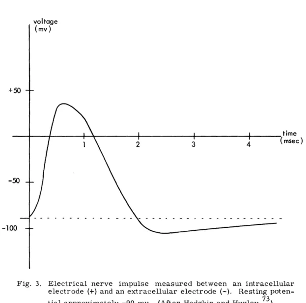
The nerve axon, in the "resting" state (in the absence of a nerve impulse), has a potential of approximately -90 mv as measured from the inside of the axon membrane (+) to the exterior liquid (-). This potential is maintained by an ion transport mechanism for which models exist but which is not thoroughly understood. During the transmission of a nerve impulse, the nerve membrane is depolarized in one region at a time, this depolarization being propagated from one point on the axon to the adjacent one by the ion currents resulting from the depolarization at the first point. At the peak of the depolarization curve, the potential of the axon reaches approximately +30 mv, measured as before. Just after a nerve impulse has passed a given point on a nerve axon, there is a period during which that region of the nerve membrane is less sensitive to stimula-tion than before. This interval is referred to as the refractory period. A sketch of the potential of a nerve impulse being propagated along a nerve axon is shown in Fig. 3.
The point at which a nerve ends on the dendrites or the cell body of another nerve is called a synapse. There appear to be both excitatory and inhibitory types of such connections, the former being those at which the impinging of a nerve impulse is likely to cause the generation of another such impulse in the axon of the succeeding nerve, and the latter being synapses at which the appearance of a nerve impulse will tend to inhibit the generation of such a second impulse. The stimulus is thought to be transmitted across the synapse by an electrical depolarization of the membrane of the succeeding nerve. Details of the action of many aspects of nerve activity are not well understood because experimental techniques are still not capable of the required precision, small size, and so forth.
Nerve fibers in mammals range in diameter from as small as 0.3 micron (1 micron = 10 meters) to as large as 22 microns. Roughly speaking, the larger the fiber di-ameter, the faster the conduction rate. Nerve conduction rates range from less than 1 meter to more than 100 meters per second. Conducting rates of nerve fibers fall in certain groups; depending on the nerve trunk considered as many as 4 or 5 of these groups can be identified, and they are labelled, in order of decreasing conduction rate, alpha, beta, gamma, . . . .
voltage
time msec)
Fig. 3. Electrical nerve impulse measured between an intracellular electrode (+) and an extracellular electrode (-). Resting poten-tial approximately -90 myv. (After Hodgkin and Huxley. 73)
15
4
-1(
3.2 MUSCLE
There are two major classes of muscle in mammals: skeletal and cardiac (smooth) 151
muscle. Skeletal muscle is responsible for all voluntary movements, and concerns us most in this work. Cardiac muscle is to a greater extent involuntarily controlled; examples are the heart and other visceral bodies.
A skeletal muscle consists of thousands of thin filaments, called muscle fibers. A muscle is usually innervated by a branch of one nerve, consisting of several hundred motor nerve axons, and some sensory nerve fibers. Each muscle fiber is innervated once by one of the motor nerve axons, which also innervates other muscle fibers. The set of muscle fibers innervated by any one nerve fiber is called a motor unit. Although perhaps not all muscle fibers in a motor unit contract each time a nerve impulse is received along the nerve fiber that innervates them, it is understood that only when an impulse is received along that nerve fiber will the muscle fiber contract.
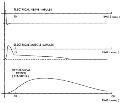
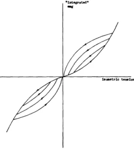
The connection between a motor nerve fiber and a muscle fiber is called a motor end plate. The means of transmission of the nerve stimulus to the muscle fiber at the end plate is fairly well established to be by chemical means. The muscle-fiber mem-brane is depolarized by the appearance of the nerve impulse and, by means which are probably similar to propagation of a nerve impulse, this depolarization is propagated throughout the muscle fiber. The result is an electrical impulse somewhat similar in shape to that of a nerve axon, but longer in terms of spike duration than in the case of nerve. The conduction rate is of the same order of magnitude as that of the motor nerves that innervate the muscle, being about 5 meters per second in mammalian muscle. Beginning during the electrical impulse, however, and continuing for a con-siderable time afterwards, the muscle fiber produces also a mechanical twitch of tension. When the fiber is held at a fixed length during the twitch (an isometric con-traction), the relations among the arriving nerve impulse, the muscle electrical response, and the mechanical twitch are shown qualitatively in Fig. 4.
Electrically, a muscle exhibits a refractory period similar to that of nerve. This refractory period probably plays little part in the usual function of muscle, since the "logic" functions of nerve do not appear to exist in muscle. Mechanically, however, the muscle twitches can superimpose and add to a maximum tension that considerably exceeds maximal twitch tension. This is referred to as tetanus; there is evidence that a stimulus rate of 50 per second or more is often required before the twitches fuse completely and the maximal tetanus tension is attained.1 51
In any voluntary contraction, the familiar uniformity of tension is attained by ex-citing many motor units in a muscle in a "random" pattern, in such a way that the total tension over the whole muscle is approximately the same at any instant. The macro-scopic effect is then the smooth muscular activity with which we are familiar. For any motor unit whose average excitation frequency is less than that which results in
complete tetanus, the average tension which it is contributing to the total muscle tension
ELECTRICAL NERVE IMPULSE
i
10 TIME (msec )
\ ELECTRICAL MUSCLE IMPULSE
10 TIME ( msec ) MECHANICAL TWITCH ( TENSION ) 10 100 TIME ( msec )
Fig. 4. Comparison of the nerve impulse, the electrical muscle impulse,
and the mechanical muscle twitch. Qualitative relations only are
shown for isometric conditions. (After Woodbury and Ruch. 51)
can be varied by varying its excitation rate. Another way in which the total muscle
tension can be varied is by varying the number of motor units which are active in the
contraction.
There is evidence that both of these methods of gradation of muscle activity are
used in the body. At low tensions, there are apparently only a few motor units active,
and the over-all muscle force is varied by varying the discharge frequencies of these
few motor units. At high degrees of tension, however, other motor units are recruited,
and apparently their discharge frequency is not varied as widely as was that of the
motor units active at low tensions. Rather, gradation of tension at high levels is
obtained largely by varying the number of active motor units.l8
It is interesting to note here that, in voluntary contractions, excitation rates to
given motor units sufficient to cause complete tetanus are not always observed;
maxi-mum rates that have been observed are in the range 35-40 pulses per second, whereas
complete tetanus often requires a rate as high as 50-60 pulses per second. Hence it is
probably only in maximal efforts that any significant number of motor units are
stimulated tetanically.
1 4 1When any muscle fiber is stimulated, the depolarization of its membrane produces
an electric field whose potential appears across any pair of electrodes in the vicinity.
The combined effects of the fields of all muscle fibers in a muscle, as observed with a
pair of electrodes neither of which is inside a muscle fiber, is the electromyogram
(emg). Whereas the electrical membrane potential is fairly well defined, the emg
signal seen in any voluntary contraction is the interaction of the potentials created by
a great many muscle fibers, all being excited in some random manner. Thus the emg
17
- ·T
does not have well-defined characteristics; the geometry of the relations among the
various muscle fibers and the electrodes, and the electrical properties of the tissue
surrounding the muscle, are so complex that it is impractical to attempt to formulate
the field equations in order to predict the emg signal. Even if this were possible, the
statistics of the distribution of stimuli to the various motor units of a muscle are not
known.
3.3 MUSCLE SPINDLES
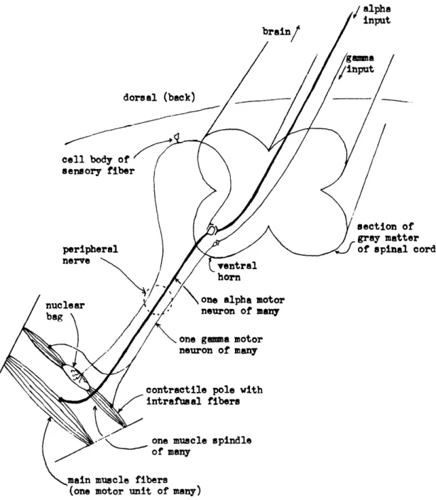
There is a small number of devices distributed within the muscle which are not
active in producing tension, but are sensory in nature.
5 2'
5 7'
1 20These are called
muscle spindles. The central portion of a muscle spindle (called the nuclear bag) is
elastic and can be stretched but cannot actively contract. Connecting to either end of
the nuclear bag are two contractile poles, consisting of a few small muscle fibers
(called intrafusal fibers). The nuclear bag is innervated by a large fast-conducting
sensory nerve fiber. The intrafusal fibers are innervated by small, slow-conducting
motor nerve fibers (gamma efferents).
Because of the relative ease with which they can be found and worked with (relative
to other sensory devices), the muscle spindles are probably the best understood of
sensory structures. The sensory nerve apparently transmits nerve impulses at a rate
that is logarithmically related to the length of the nuclear bag. Thus the muscle spindle
contributes to a position sense. The spindles seem to exhibit only slight adaptation,
120
and this only after a period of several minutes.
Spindles stretch parallel to the primary muscle fibers in the muscle, connecting to
the tendons or bones in much the same way as the remainder of the muscle does. Thus
there are two ways in which the nuclear bag can be stretched: (1) by lengthening the
muscle which then would lengthen the nuclear bag accordingly, and (2) by excitation of
the intrafusal fibers by the gamma efferent nerve fibers, in which case even if the
over-all muscle length remains constant, the nuclear bag will be forced to lengthen.
3.4 ORGANIZATION OF THE NEUROMUSCULAR SYSTEM
The well-known knee-jerk response to a tap on the tendon just below the kneecap
is an example of the action of a reflex arc which is a very common type of connection
in the neuromuscular system. Studies of the reflex arc have contributed vastly to our
understanding of the mechanism that provides proprioceptive control of joint
position.
4 3'
6 2'
6 3'
7 7'
1 0 0 1 02The ability to hold a joint in some constant position
without conscious attention to its position (and with only very small movements about
that position) is likely to be the main function of the reflex arc, and certainly is a main
function of the muscle spindle. The muscle spindle is the prime component in the
postural position control system.
The gray matter of the spinal cord is sketched in section in Fig. 5. Motor neurons
are located in the ventral horn, and travel down the peripheral nerve from there to the
brain
dorsal (back)
cell body of '
sensory fiber
peripheral
nerve
\
r
cord
ventral
horn
Fig. 5. Section of the spinal cord in man, and the important peripheral pathways.
muscle. Sensory fibers enter the spinal cord by the dorsal root, their cell bodies being
located in the dorsal-root ganglion. The sensory fibers that concern us most at this
time are those innervating the nuclear bags of the muscle spindles of a given muscle.
There is considerable weight of evidence to indicate that these spindle signals, upon
entering the spinal cord, directly form excitatory synapses with the alpha motor
neurons for the muscles from which they came.
The effect of such a connection can be understood as follows: Assume that the
muscles and nervous system are in some stationary stable state, and that some external
19
__^1__111_·11__^__1__ --- I· - ··I I
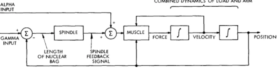
influence forces some small change upon this situation, such as the tap on the tendon, which momentarily stretches some muscle. Then the spindles in that muscle will sense an increase in muscle length, and return an increased nerve impulse rate along their respective sensory nerve fibers. This signal will then in turn cause the average impulse rate along the alpha motor nerve fibers of the muscle which was just stretched to in-crease. The result will be an increase in the tension in that muscle, causing the joint in question to move in a direction so as to shorten the muscle, tending to reduce the spindle discharge rate, and thereby returning the system to its former state. At the same time, there is evidence that the alpha motor neurons for the antagonist muscles are forced to decrease their average discharge rate, although this may not be due to a direct connection from the spindles. This will decrease the tension in those muscles, tending once again to assist in returning the joint to its former position. The position feedback control loop so formed is diagrammed schematically in Fig. 6.
COMBINED DYNAMICS OF LOAD AND ARM
ALPHA
;ITION
Fig. 6. Reflex control loop
Of course the description above is grossly oversimplified in several ways. In the first place, there are rarely only two muscles, an agonist and an antagonist, for a given joint. Second, there are several muscle spindles in each muscle, and many alpha motor-nerve fibers. Nevertheless, the action of the reflex arc seems to be consistent with the oversimplified description given.
The "control loop" shown in Fig. 6 contains two inputs. The one that is understood to be relevant in the postural type of performance just outlined is the gamma input. This corresponds to a signal applied to the gamma motor fibers from the central nervous system, and it can be seen that this corresponds to a different "set point" of desired position for the joint. Thus the postural control system can be used with a different input signal to regulate position at any point.
The second input to the neuromuscular control loop shown in Fig. 6 is labelled the "alpha input," and is intended to represent a direct input from higher centers in the brain to the muscle, by which a rapid action can be caused, without the relatively long time delays that are involved in the gamma system. It is thought by most researchers that this alpha input is the signal used for rapid voluntary motions.
From the way in which the gamma input and muscle spindle are interconnected, however, it is clear that the gamma-nerve signals could be used as voluntary command signals for slow movements from some initial position to some final one. Whether or
20
not the gamma signals are so used is not universally agreed upon. Many apparently
believe that the gamma input is activated for postural control only. If this were so, then
at least near the termination of some voluntary motion the spindles would be activated
(through the gamma system) to provide a reference point for the steady position that
should follow the motion, but during the motion the spindles should have no effect. Some
132
unpublished experiments by Stark and Rushworth seem to support this theory.
Never-theless, Granit, Merton, and others appear to believe that the gamma system is used for
all slow carefully controlled motions.
4 2'
4 4 5 8,
5 9'
1 0 9It has been pointed out by Hunt
that the gamma and alpha systems appear to receive simultaneous stimuli.
7 9His
inter-pretation is that this serves to keep the spindle operating in a sensitive portion of its
characteristic. Merton suggests that this is exactly the type of behavior to be expected
if the gamma system is actually being used for servo operation during the voluntary
109
motion.
There is one other type of sensory organ associated with muscular activity which
has been identified specifically. That is the Golgi tendon organ, which is located in the
muscle tendons where it measures muscle tension. It has been found that the Golgi
tendon organ has a high threshold, thereby producing only small signals until the muscle
tension reaches high levels. There is a clinical observation tending to correlate with
these data on high threshold of the Golgi tendon organ, called the "clasp-knife" reflex.
1 2 0In extraordinary feats of exertion of muscles, it is not an unusual experience that the
muscles should suddenly "give," temporarily losing their capability to provide the
forces expected of them. This action is similar to that of a clasp knife, in which a
great deal of force is required to begin the motion of closing the knife, after which the
knife suddenly "snaps" closed. This and other data indicate that the Golgi tendon organ
acts as an inhibitor of the alpha motor neurons, at high muscle tensions; its function
is thought to be mainly that of a "safety valve," serving to release the tension in the
muscles when this tension reaches such a high level that damage to the muscle could
result.
3.5 CONSCIOUS CONTROL AND SENSATION
A voluntary motion exists in the conscious thought in terms of a functional rather
than a specific description. That is, the act of grasping some object is thought of in
just those terms, and a general idea of the shape and perhaps weight of the object, along
with its position in space relative to the person is the type of information which exists
on a conscious level. "Lower" in the brain, however, the concept of grasping is
trans-lated to a preprogrammed sequence of individual muscle motions, based on the person's
experience since birth in grasping objects, which will enable the grasping action to
come close enough to the object that local feedback can complete it. Clearly, this
translation occurs between the conscious level and the level of the lower motor neurons,
since they are specialized to the specific muscles they control. It is understood that
some major portion of this "coding" is performed in the cerebellum, an area of the brain
which apparently is an important link in the coordination of muscle activities. 42,44,58
21
In any case, at the level in the spinal cord where the specific motor signals leave the cord and travel down the proper peripheral nerve to the muscle, the motor neurons for the correct muscles are activated in the proper sequence. The signals are carried along the nerves to the muscles, and they begin to contract in the required manner. This operation may or may not involve regulation by the spindles. As the hand contacts the object, the sense of touch relays this information back to the spinal cord, and to higher levels, and the closing of the hand is coordinated.
Many other senses are involved in activities related to muscle actions. Certainly, the visual sense is one of the most active, being not only the source of the idea that initiates many actions (i.e., the sight of an object might suggest reaching for it), but also furnishing continuous feedback of position information with which the remainder of the motion can be optimized. There are joint position receptors in joints, whose function it is to report joint position to the central nervous system, so that we may
"feel" position without visual observation. Certainly, the sense of touch is involved in grasping and other actions. The ability to feel shape, temperature, and pressure, is due to the existence of specialized receptors in skin for sensing these variables, and for various types of muscle activities all of these sensory signals play a part.
It is of interest to consider what sense we have on a conscious level, that is, what sensory information existing in the body is in such a form that the person can be consciously aware of it. Certainly, it is true that we can feel the positions of many joints in the body, at least for short times. For example, with one's eyes closed it is possible to bring the forearm to some specified angle with the upper arm, to a
sur-prising degree of accuracy. It is apparently the opinion of many researchers that the spindle signals, which could provide this information, do not reach a conscious level. We conclude that the conscious feel of joint position is probably due to the joint receptors alone.
This author and several others had the occasion to perform a very crude experiment which, if repeated in a well-controlled manner, would cast doubt on the conclusion that
conscious sensation of joint position is due only to the joint receptors. We were fortunate to have the opportunity to examine an amputee whose amputation was very long, but still slightly above the elbow. That is, he had no part of the lower arm or the elbow joint remaining, but otherwise his upper arm was complete. We asked him to attempt to show us, by positioning his other (normal) arm, in what position he felt as if his amputated arm was at each instant. We then commenced to move his biceps and triceps muscles by external pressures through the skin, including pushing in a direction on the skin opposite to that in which the underlying muscle was being pushed. It is un-fortunate that we were ill-prepared for this experiment, for we were not able to
anesthetize the skin in order to be absolutely certain that no skin sensation was con-fusing our conclusions. In every case when the biceps was displaced upwards, and the triceps was moved in the opposite direction, he claimed to "feel" that the missing arm was being raised. This applied for small and large displacements and in all cases of
various attempts to mask any skin sensation by forcing the skin in different directions. This also applied when the triceps was left slack, and only the biceps was moved. Furthermore, he had remarkable sensation not only of direction, but of magnitude, for he could follow displacements varying from very small to very large, and he demon-strated that his sensation was as if the missing forearm had moved through the complete range of positions. Finally, we were able to have him feel that his missing forearm was being supported in one position for a period in excess of 60 seconds, with no observable adaptation, which tends to eliminate the skin as a source of the position sense which he demonstrated.
As a result of the experiment described above, we suggest that (1) either there are other position sensors in the body, not in the skin, whose signals are transmitted to a conscious level, which have not been found, or (2) the muscle-spindle signals do reach a conscious level. In order to be absolutely certain of these conclusions, the experiment should be repeated on several other subjects, and with better controls than we had. It is certain in this case, however, that the conscious sensation of position of his missing arm was not derived from information transmitted by the joint receptors, since these were not present in the subject.
It is questionable whether any direct feedback of force (or weight) information from sensory organs to a conscious level exists. Consider, for example, the usual method used to compare the weights of two objects: One "hefts" them, then "catches" them. Thus the response in any position sensor could be the source of the signal which is measured. Consider also the fact that when one attempts to apply a constant tension to
a spring balance, without looking at the balance, a rapid, wide drift in the force applied will be observed.
On the other hand, one still has some conscious "feel" of the force that he is applying against an infinite load, for example. And it is very easy to distinguish two very different weights. It is possible that this sensation of force is only an indirect one, derived from a knowledge of the conscious effort being applied to support the weight. Another possibility is that the pressure sensors in the skin, which are assumed to adapt very rapidly, are maintained in an active state by small motions of the hand which is applying the force, thereby yielding a continuing measurement of the force that is applied. This would then apply most clearly when a significant surface area of the person's skin was in continuing contact with the object against which the force was being applied.
There are a great many details about the coordination of motions with the responses of the many different sensory receptors in man, which are unknown. Thus we find ourselves potentially limited by our lack of knowledge, in our desire to restore lost function in a normal way to an amputee. On the other hand, it should be reasonably clear from our discussions that many aspects of the control and coordination of a normal arm are not provided by present prostheses. It is reasonable to say that present prostheses can provide the mechanical capabilities for the more importance actions, but the lack
23