Disrupting Pre-SMA Activity Impairs Facial Happiness Recognition: An Event-Related
TMS Study
Vincent Rochas
1,2,, Lauriane Gelmini
1, Pierre Krolak-Salmon
3,4, Emmanuel Poulet
1, Mohamed Saoud
1, Jerome Brunelin
1,5and
Benoit Bediou
6,71
EA4615-SIPAD, Université Lyon 1, CH Le Vinatier, Bron F-69677, France
2Functional Brain Mapping Laboratory, Department of
Fundamental Neuroscience, University of Geneva, CH-1206 Geneva, Switzerland
3Memory Center of Lyon, Hôpital des
Charpennes, Hospices Civils de Lyon, Lyon, France
4Lyon Neuroscience Research Center, INSERM U1028, CNRS UMR5292, Brain
Dynamics and Cognition Team, Lyon F-69000, France
5Institut Universitaire en Santé Mentale de Québec, Université Laval,
Québec, Canada
6Swiss Center for Affective Sciences (CISA), University of Geneva, CH-1205 Geneva, Switzerland and
7Faculté
de Psychologie et des Sciences de l
’Education FPSE, University of Geneva, CH-1205 Geneva, Switzerland
Address correspondence to Vincent Rochas, Functional Brain Mapping Laboratory, Department of Fundamental Neuroscience, University of Geneva, CMU, Rue Michel-Servet 1, 1206 Geneva, Switzerland. Email: vincent.rochas@unige.ch
V.R. and L.G. contributed equally to this work.
It has been suggested that the left pre-supplementary motor area
( pre-SMA) could be implicated in facial emotion expression and
recognition, especially for laughter/happiness. To test this
hypoth-esis, in a single-blind, randomized crossover study, we investigated
the impact of transcranial magnetic stimulation (TMS) on
perform-ances of 18 healthy participants during a facial emotion recognition
task. Using a neuronavigation system based on T1-weighted
mag-netic resonance imaging of each participant, TMS (5 pulses, 10 Hz)
was delivered over the pre-SMA or the vertex (control condition) in
an event-related fashion after the presentation of happy, fear, and
angry faces. Compared with performances during vertex
stimu-lation, we observed that TMS applied over the left pre-SMA speci
fi-cally disrupted facial happiness recognition (FHR). No difference
was observed between the 2 conditions neither for fear and anger
recognition nor for reaction times (RT). Thus, interfering with
pre-SMA activity with event-related TMS after stimulus presentation
produced a selective impairment in the recognition of happy faces.
These
findings provide new insights into the functional implication
of the pre-SMA in FHR, which may rely on the mirror properties of
pre-SMA neurons.
Keywords: facial emotion recognition, happiness, mirror neurons,
pre-SMA, transcranial magnetic stimulation
Introduction
Facial expressions are a key feature for communication within
and across species (
Darwin 1872
). This type of non-verbal
communication is based on the expression, perception, and
recognition of facial emotions. Facial emotion recognition
(FER) appears to be closely related to social functioning
(
Hooker and Park 2002
;
Addington et al. 2006
), and is
im-paired in a variety of psychiatric and neurological conditions,
such as schizophrenia (
Bediou, Franck et al. 2005
,
Bediou
et al. 2007
), major depressive disorder (
Bediou,
Krolak-Salmon et al. 2005
), Parkinson
’s disease (
Lachenal-Chevallet
et al. 2006
), as well as fronto-temporal dementia and
Alzhei-mer
’s disease (
Bediou, Ryff et al. 2009
). FER is also impaired
in healthy individuals with heightened risk for developing
schizophrenia (
Bediou, Ryff et al. 2009
). Hence, better
under-standing the neural mechanisms implicated in FER is of
primary importance.
Numerous imaging studies have investigated the cerebral
networks implicated in FER. It has been suggested that
complex expressions that contain blends of emotions may
fully be recognized by simulating the perceived expression,
either overtly or covertly, and sensing the emotion produced
by that simulation (
Adolphs 2001
). Thus, FER mechanisms
may involve both facial mimicry (i.e. the motor simulation of
another
’s expression) and empathy (i.e. the sensory simulation
of the feelings associated with another
’s emotional expression;
Iacoboni 2009
). Motor and somatosensory cortical areas may
be involved in the motor components of simulation (e.g. facial
mimicry), whereas the amygdala and insula may be involved
in the sensory components of simulation (e.g. empathy), and
both may contribute to FER (
van der Gaag et al. 2007
).
Consistent with this embodied view of emotion recognition
(
Niedenthal 2007
), lesion of the somatosensory cortex
(
Adolphs et al. 2000
), amygdala (
Adolphs et al. 1994
;
Calder
et al. 1996
) and insula (
Calder et al. 2000
) produces
impair-ments in FER, possibly re
flecting the role of the sensory
simu-lation mechanisms or empathy in FER. A peak of activation in
the pre-supplementary motor area ( pre-SMA) has been
ob-served during a task of facial emotion observation in healthy
subjects (
Carr et al. 2003
), and both the recognition and the
generation of happy and sad expressions activate the pre-SMA
(
Seitz et al. 2008
), consistent with a role in motor (mimicry)
and sensory (empathy) simulations. The fact that the neural
responses to the observation and execution of (dynamic)
smiles overlap in the premotor cortex and somatosensory
cortex (
Hennenlotter et al. 2005
) is further consistent with a
role for these regions in both the recognition and the motor
(mimicry) simulation of this emotion. Furthermore, electrical
stimulation of the left pre-SMA with intracranial subdural
elec-trodes in 2 epileptic patients has consistently produced
laugh-ter (
Fried et al. 1998
;
Krolak-Salmon et al. 2006
). In both
studies, laughter was accompanied by a sensation of
merri-ment or mirth, and patients gave a different explanation for it
each time. In addition,
Krolak-Salmon et al. (2006)
recorded
intracranial evoked potentials in the same epileptic patient
during the presentation of emotional faces. In 2 different
blocks, the patient had to pay attention to gender or emotion.
Between 150 and 450 ms after the presentation of an
emotion-al face (during both tasks), a selective response to happy faciemotion-al
expression was recorded by the electrode implanted in the left
pre-SMA. These studies suggest that the pre-SMA may
partici-pate in FER via a mirror communicative activity involved in
both the detection and the production of facial emotional
© The Author 2012. Published by Oxford University Press. All rights reserved.Cerebral Cortex July 2013;23:1517–1525 doi:10.1093/cercor/bhs133
expression, especially happiness/laughter. However, these
results were obtained in the epileptic brains. Although the
pre-SMA was not a part of the patients
’ seizures, functional
reorganization cannot be excluded. Further studies in healthy
subjects are essential to conclude a real and systematic
impli-cation of the pre-SMA in FER, and especially in facial
happi-ness recognition (FHR). Because of its cortical location,
non-invasive and reversible inhibition of the pre-SMA with
transcranial magnetic stimulation (TMS) may be used during
an FER task to assess its causal implication in FHR.
At the interface between neuropsychology and functional
neuroimaging, TMS appears to be a suitable means to
non-invasively investigate the cerebral function. According to
Fara-day
’s principle, a brief current flows through the stimulation
coil producing a transient magnetic
field that penetrates the
cranium. As a result of this induced magnetic
field, an eddy
current occurs in the brain, transiently and reversibly
perturb-ing activity in the affected cortical region. Thus, usperturb-ing a
perturb-and-measure approach, TMS gives the opportunity to
infer about the necessity (but not the suf
ficiency) of the
integ-rity of a particular brain region for a given behavior (
Paus
2005
;
Brunelin et al. 2006
). For example, TMS over the medial
prefrontal cortex has been shown to reversibly modify the
analysis of facially expressed anger (
Harmer et al. 2001
),
suggesting a crucial role for this region in the recognition of
facial anger. Similarly, TMS over the right occipital face area
and TMS over the face region of the right somatosensory
cortex (relative to the
finger region) have been shown to
inter-fere with the processing of emotional facial expressions but
not facial identity (
Pitcher et al. 2008
). Conversely, TMS of the
right superior temporal sulcus impaired the processing of the
gaze direction without affecting expression processing (
Pour-tois et al. 2004
). Based on the past results and its excellent
temporal resolution, an event-related TMS protocol appears
appropriate to investigate the role of the pre-SMA in FER.
Our study aims to clarify the role of the pre-SMA in FHR.
Given 1) the robust activation of the pre-SMA in response to
happy faces (
Krolak-Salmon et al. 2006
;
Seitz et al. 2008
) and
2) the laughter and the merriment sensation elicited by
elec-trical stimulation of the pre-SMA in epileptic patients (
Fried
et al. 1998
;
Krolak-Salmon et al. 2006
), we hypothesized that
the pre-SMA is implicated in FER, especially in the recognition
of happiness.
Materials and Methods
Subjects
A total of 20 right-handed (average right-handedness score: 97.10; standard deviation (SD) = 4.81%; Edinburgh Handedness Inventory;
Oldfield 1971) healthy volunteers (12 males and 8 females) aged between 20 and 34 years (mean age = 24.61; SD = 3.74; and years of education = 17; SD = 2) were enrolled in this single-blind, randomized crossover study in return for payment (100€). Postgraduate and graduate students were recruited through“word-of-mouth,” according to the following general non-inclusion criteria, which were evaluated during a medical interview: 1) a story of neurological issues (e.g. epi-lepsy), 2) a personal or familial psychiatric disorder history (axis I of the diagnostic and statistical manual of mental disorders DSM IV), 3) uncorrected vision, 4) pregnancy, 5) TMS contraindication (e.g. met-allic prosthesis, pacemaker), and 6) medication intake. All these ex-clusion criteria were evaluated during a personal medical interview with a psychiatrist (Personal medical interviews were undertaken by psychiatrists, Emmanuel Poulet and Mohamed Saoud.). Past and
current histories of psychiatric disorders were assessed throughout the clinical interview using the Mini-International Neuropsychiatric Interview semi-standardized evaluation (MINI version 4.4). A familial history was evaluated at the knowledge of the participant and consul-tation of hospital records. All participants were naive to the FER task, the TMS tool, and the presented stimuli. They all gave their written consent after a complete description of the study procedure. A local ethical committee (CPP Sud-Est IV) had approved the study design and consent procedure. The subjects were told that they could with-draw from the study at any time, and 1 male subject did so before the TMS protocol (n = 19).
Facial Stimuli
The images were taken from a standard set of facial emotion pictures (Ekman and Friesen 1976). Each stimulus was obtained by morphing 2 black and white facial pictures (1 neutral and 1 emotional, in differ-ent proportions) from a same iddiffer-entity. Morphing construction permits the creation of an ecological variation in facial emotional intensity and to test the subjects’ performances on various levels of difficulties, thus providing a more sensitive FER measure than classical tests (Bediou, Ryff et al. 2009). Moreover, the task should be neither too hard nor too easy to perform to increase the probability of 1) disrupt-ing the psychological process studied and 2) inferrdisrupt-ing a functional implication of the cerebral area stimulated in the evaluated function. In our study, morphed faces were generated between a neutral face and a happy, angry, or fearful face, for 8 identities (4 men and 4 women). The choice of the relevant morphing proportions was based on the results of a pilot study in an independent sample of 8 healthy volunteers without TMS. Seven levels of morphing between each emotional and neutral faces were used. Our pilot data showed that anger was recognized with greatest difficulty. As a consequence, we used a greater proportion of the expressive face in each anger morph-ing (20%, 30%, 40%, 50%, 60%, 70%, and 80% for fear and happiness; 30%, 40%, 50%, 60%, 70%, 80%, and 90% for anger), in order to keep task difficulty equal between emotions across intensity levels. The pilot data showed that with these morphing levels, recognition accu-racy did not differ significantly between happiness (71.33; SD = 6.53), fear (65.07; SD = 12.72), and anger (66.33; SD = 12.76), F = 1.19, P = 0.34. The total set of stimuli comprised 168 faces (8 identities × 3 emotions × 7 morphs), the order of which was randomized between series and across subjects. The digitized size, brightness, and contrast of images were standardized.
FER Task
FER task was run by the software“Presentation v0.55” (Neurobeha-vioral Systems Inc., Canada), which presented the different images, recorded subjects’ responses, and controlled the TMS device con-nected to the computer. Figure 1A depicts the timeline of an exper-imental trial. Each trial started with a central whitefixation cross on a dark background (duration: 1000, 1250, 1500, or 1750 ms, randomly selected) attracting subjects’ attention while minimizing the response anticipation and motor preparation. Then, a facial stimulus appeared during 50 ms on a black background. Each stimulus was followed by a black screen (500 ms) during which event-related TMS was applied (1 pulse every 100 ms). This was in turn followed by the response screen (2000 ms). Subjects were instructed to maintainfixation, visu-alize each picture, and judge as quickly and as accurately as possible whether the target face expressed happiness, fear, anger, or neutrality (no emotion), by pressing 1 of 4 possible response buttons (up, down, left, and right arrows) with their right-handfingers.
Experimental Procedure
In order to standardize experimental conditions, all participants were seated in a padded armchair at a 60 cm distance from the 17-inch computer monitor, with their head held in place comfortably by a headrest. The subject’s resting motor threshold (RMT) was deter-mined and study started by a practice block with the same task but with the different stimuli (other identities) and without TMS, in order to familiarize with the task and procedure. Thus, FER under TMS
treatment was measured on 4 occasions: 2 sessions ( pre-SMA and vertex), each comprising 2 series of 168 faces. The 168 faces were randomized. Faces were the same for each series and each session. For the 2 sessions, once the participant had completed the practice block, the FER task started with afirst series of 168 stimuli, lasting ∼10 min. Then, a second series of the same 168 faces was shown, while TMS was applied on the same site as for thefirst series. Short breaks (5–10 min depending on the subject’s comfort) were inserted between each series to avoid fatigue and to prevent overheating of the stimulator. Thus, for each TMS session, FER was divided into 2 series. Because low frequency (LF) repeated TMS (rTMS) research de-monstrated delayed and extended effects in time on several indices of emotional processing (vanHonk et al. 2002), the 2 sessions were sep-arated by 15 days. During thefirst session, the subject received the TMS pulses over the vertex or the left pre-SMA, and inversely during the second session (Fig. 1B). The order of TMS stimulation sites (vertex− pre-SMA or pre-SMA − vertex) was randomized between par-ticipants. This had 2 main advantages. First, it allowed us to compare the performance in the 2 TMS conditions within the same subjects. Secondly, it allowed us to control for any potential order/training effect.
TMS Procedure Neuronavigation
We localized the 2 TMS sites using a frameless stereotaxic system (Softaxic Optic;http://www.softaxic.com/) to guide the TMS coil po-sitioning over the brain, by means of individual high-resolution T1-weighted magnetic resonance imaging (MRI) transformed in the Talairach space. We targeted the sites based on the Talairach coordi-nates (x, y, z) for either the left pre-SMA (−6, 15, 58) or the vertex
(0,−30, 70). Once MRI co-registration and cortical target localization were successful, infrared tracking was used to monitor the position of the coil with respect to the participant’s brain.
Resting Motor Threshold
The RMT was determined as the minimal intensity of electromagnetic stimulation that produced a visible inch adductor contraction in at least 5 times out of 10 TMS pulses. Afigure-8 coil was placed over the participant’s left motor cortex hand area with the coil held tangen-tially to the skull and the handle-pointing posterior and down. Single pulses were delivered to the motor cortex, with the intensity of the stimulation adjusted until a muscle movement in the right hand was visually observed. The location of the stimulation was adjusted to locate the inch adductor. Furthermore, it has been demonstrated that rTMS delivered to the primary motor cortex (M1) produces intensity-dependent increases in brain activity locally and has associated effects in distant sites with a known connection (Speer et al. 2003). In order to be the more accurate over the pre-SMA and limit the impact of TMS over the network interconnected to the pre-SMA, the intensity of TMS wasfixed to 80% RMT, which is known to produce a more focal effect (Wagner et al. 2009). In addition, a moderate intensity of stimulation tends to limit the peripheral discomforts and muscles contraction. TMS Pulses
All TMS pulses were delivered by a MagPro X100 magnetic stimulator (MagVenture, Denmark) with a 70 mmfigure-8 coil at 80% intensity of each subject’s RMT. Stimulations were controlled through the Pres-entation v0.55 software installed on a computer connected to the stimulator. Based on previous work, (Krolak-Salmon et al. 2006) event-related stimulations were delivered in trains of 5 pulses (1 pulse every 100 ms) during the 500 ms after the picture presentation (i.e. thefirst pulse was synchronized with the vertical offset). Each partici-pant received 3360 pulses during the whole protocol, which lasted ∼2 h (168 stimuli × 5 pulses × 2 series × 2 sessions; 1680 pulses during each session, separated by 2 weeks).
Subjective Ratings
TMS can perturb subjects’ mood if used daily and repeatedly ( Brune-lin et al. 2007). Thus, subjects were asked to report their mood on the Norris’ 16-item visual analog scale (VAS; Norris 1971) before, between, and after each series of each session (3 measures for each session).
Data Analysis
Post Experimental Coil Positioning
Post experimental visual inspection of the coil localization was con-ducted to ensure that the stimulation site corresponded to the desired one. The data from 1 female subject had to be excluded from statisti-cal analysis because the coil lostatisti-calization was substantially different at the end compared with the beginning of the session (n = 18; 1 subject having withdrawn before the TMS protocol). As a result, statistical analyses were conducted on data from 18 participants: 10 received TMS over the vertex and then the pre-SMA, and 8 received the inverse sequence ( pre-SMA− vertex).
Statistical Analysis
To directly test our a priori hypothesis that TMS over the pre-SMA impairs the recognition of happiness selectively, we subtracted data ( performances P and reaction times RT) in the vertex condition from data in the pre-SMA condition (see also Romei et al. 2011 for a similar approach). This procedure cancels any individual side-effects due to TMS treatment (e.g. sounds, feelings, stress). The obtained ( pre-SMA − vertex) value provides a quantitative measure of the modification in FER induced by pre-SMA stimulation compared with vertex stimu-lation for each participant. A positive value indicates an increase for the pre-SMA compared with the vertex, whereas a negative value indi-cates a reduction for the pre-SMA compared with the vertex. Statistical analyses were performed on these pre-SMA− vertex difference scores Figure 1. (A) Timeline of a TMS experimental trial. Each stimulus appeared during
50 ms and ±1400 ms after crosshairfixation. After stimulus offset, 5 single pulses of TMS were applied during a black screen (500 ms). The subjects were instructed to answer as quickly and as accurately as possible once the response screen appeared (2000 ms or until response). Responses were followed by a black screen (500 ms), preceding the next trial. (B) Crossover TMS stimulation protocol. During thefirst session, a subject received the TMS pulses over the vertex or the left pre-SMA, and inversely during the second session. The two sessions were separated by a washout period of 15 days. Short breaks (5–10 min depending on the subject’s comfort) were inserted between each series to avoid fatigue and to prevent overheating of the stimulator. The order of the TMS conditions was randomized between subjects.
for both the performance (i.e., difference in percent correct responses, delta-P) and reaction time (i.e., difference in ms between the response screen onset and the onset of the subject’s response, delta-RT). Only correct trials were considered in the analysis of RT. Considering that FER accuracy was equal across emotions in our pilot study without TMS stimulation, we then tested whether the delta-P and delta-RT differed significantly from zero, using 2-tailed 1-sample Student t-test with a significance threshold at P = 0.05 with Bonferroni correction. We predicted a significant change in FHR in the pre-SMA compared with the vertex condition resulting in a delta-P significantly different from zero for happiness but not for fear and anger.
To investigate the impact of TMS on mood, VAS ratings before and after the session were compared using a repeated-measures multi-variate analysis of variance (MANOVA) with the within-subject factors session ( pre vs. post stimulation) and TMS ( pre-SMA vs. vertex), and items (the various aspects rated) as multiple dependent variable. Con-sidering the risk of low statistical power because of the limited sample size, we therefore averaged the 16 items into an overall mood score and submitted this value to the repeated-measure analysis of variance (ANOVA) examining the impact of TMS and session only as a double-check.
To assess the adequacy of our crossover design (and rule-out any possible order/training effect), task performance in the first and second sessions (all emotions, morphings and TMS conditions col-lapsed) were compared using 2-tailed paired Student t-test. To quan-tify a possible training effect within each session, task performance for the first and the second series of each session (all emotions, morphings, and TMS conditions collapsed) were compared using 2-tailed paired Student t-test.
RESULTS
Effects of TMS on FER
Preliminary Considerations
No statistically signi
ficant difference was highlighted between
the performance in the
first session compared with the
second, t(17) =
−0.04, P = 0.97, and between the performance
in the
first compared with the second series of each session,
t(17) =
−1.21, P = 0.24. In the absence of order effect (i.e., no
difference in FER performance between sessions 1 and 2),
training effect (i.e., no difference in FER performance
between series 1 and 2 of each session), and TMS effect on
mood (i.e., no difference in VAS ratings before and after
TMS), subsequent analyses examined the impact of TMS on
FER data ( pre-SMA
− vertex) collapsed across series and TMS
conditions, irrespective of stimulation sequence order (vertex
− pre-SMA or pre-SMA − vertex). Although our study was not
designed to test gender differences in FER, or in the effect
of TMS on FER, exploratory (i.e., uncorrected) analyses
re-vealed signi
ficant differences in FER between men (n = 10,
mean = 54.73, SD = 5.75) and women (N = 8, mean = 63.04,
SD = 8.04), t(16) = 2.56, P = 0.021, in line with previous
studies (e.g.
Montagne et al. 2005
). Signi
ficant gender
differ-ences in FER were found for happiness and fear in the
pre-SMA condition, and for fear in the vertex condition, but
not for anger. Overall accuracy also differed between men
and women, in both the vertex and the pre-SMA conditions
(see Supplementary Table 1). Critically, however, there was
no gender difference in the effect of TMS on FER when the
pre-SMA data were subtracted from the vertex data (see
Sup-plementary Table 2).
Effects of TMS on FER Performance
On average (i.e., all emotions and morphings collapsed),
par-ticipants
’ performance was 57.62% (SD = 10.54%) in the
pre-SMA condition and 59.24% (SD = 11.30%) in the vertex
condition, a difference that was statistically signi
ficant, t(17) =
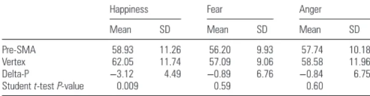
−2.20, P = 0.04. As reported in Table
1
and illustrated in
Figure
2
, the delta-P for happiness differed signi
ficantly from
zero, t(17) = 2.95, P = 0.009, whereas the delta-P for fear and
anger did not, t(17) = 0.56, P = 0.59; t(17) = 0.53, P = 0.60,
respectively.
As expected, subjects had more dif
ficulty in identifying
happiness in the pre-SMA TMS condition than in the vertex
TMS condition, whereas fear and anger recognition were not
signi
ficantly affected by TMS over the left pre-SMA compared
with the TMS over the vertex.
Effects of TMS on RTs
RT data for each emotion and TMS condition (i.e., collapsed
across all morphs), as well as the pre-SMA
− vertex difference
in RT (delta-RT) are summarized in Table
2
. None of the
delta-RT value differed signi
ficantly from zero, suggesting that
TMS did not affect RTs.
Effects of TMS on Mood
We found no effect of TMS on mood. A repeated-measures
MANOVA with the within-subject factors session ( pre vs. post
stimulation) and TMS ( pre-SMA vs. vertex), and items as
mul-tiple dependent variable yielded a signi
ficant main effect of
item (F = 280, P < 0.001). Importantly, however, there was no
signi
ficant effect or interaction with the factors session and
Table 1
The mean score (expressed in percentage of correct responses) for the different emotions (happiness, fear, and anger, all morphings collapsed) in the vertex and pre-SMA conditions, delta-P (%; pre-SMA− vertex) and 2-tailed 1-sample Student t-test (P values; n = 18) all morphings collapsed. Only the delta-P for happiness differed significantly from zero (P = 0.009)
Happiness Fear Anger
Mean SD Mean SD Mean SD
Pre-SMA 58.93 11.26 56.20 9.93 57.74 10.18
Vertex 62.05 11.74 57.09 9.06 58.58 11.96
Delta-P −3.12 4.49 −0.89 6.76 −0.84 6.75
Studentt-test P-value 0.009 0.59 0.60
Figure 2. Delta-P values ( percentage of correct responses in the pre-SMA condition minus percentage of correct responses in the vertex condition) for each subject and each emotion (averaged across all morphings). The delta-P for happiness differed significantly from zero, whereas the delta-P for fear and anger did not (see text for statistics).
TMS (all F
’s <1), suggesting that our TMS protocol did not
sig-ni
ficantly affect participants’ mood. We note, however, that
running this analysis with our limited sample size bears the
risk of low statistical power. We therefore averaged the 16
items into an overall mood score and submitted this value to
repeated-measure ANOVA examining the impact of TMS and
session. This analysis showed very similar results; there were
no signi
ficant main effect of TMS, and no TMS × session
inter-action (all F
’s <1). A marginal effect of session, F = 3.47,
P = 0.08, suggested that mood varied between the beginning
and the end of each session, probably due to fatigue.
Criti-cally though, this effect was not affected by the TMS
con-dition. VAS ratings before and after each TMS session did not
differ signi
ficantly (before pre-SMA: mean = 51.57, SD = 34.17;
after pre-SMA: mean = 52.17, SD = 33.92; before vs. after
pre-SMA: t(15) =
−0.44, P = 0.67; before vertex: mean = 50.54,
SD = 32.21; after vertex: mean = 51.68, SD = 33.99, before vs.
after vertex: t(15) =
−1.14, P = 0.27), suggesting that subjects’
mood was not affected by TMS. There was no signi
ficant
cor-relation between FER and mood (all R
’s <0.34, P’s >0.17).
In summary, compared with TMS over the vertex, TMS over
the pre-SMA impaired selectively the recognition of happy
facial expressions, without affecting the recognition of anger
and fear, and without affecting RTs and mood.
Discussion
The primary goal of the present study was to assess, using an
interference technique (TMS), whether the pre-SMA is
in-volved in FHR. We hypothesized that compared with TMS
over the vertex, TMS over the left pre-SMA would speci
fically
interfere with the recognition of happiness. As predicted, we
showed that TMS over the left pre-SMA impaired the
recog-nition of happy faces, without affecting the recogrecog-nition of
fearful and angry faces, and without affecting RT. There was
no evidence that TMS pulses delivered during this study led
to undesirable short- and long-term effects, and none of the
subjects included in our study reported the adverse event.
Moreover, we found no effect of TMS on mood, and no
relationship between mood and FER. Hence, reduced
happi-ness recognition following TMS stimulation of the left
pre-SMA compared with the vertex, may be attributed to the
perturbation of neural activity in the pre-SMA or in a broader
neural network including this structure. Although the precise
mechanism(s) by which the pre-SMA may be involved in the
recognition of facial happiness remains unclear, our study
provides the
first evidence for a direct relationship between
pre-SMA activity and recognition accuracy for happy faces in
healthy subjects.
Previous studies examining the impact of rTMS of lateral
prefrontal cortical areas (PFC) on mood suggest that the
effects are opposed depending on the hemisphere stimulated
(left vs. right) and on the frequency of stimulation [LF vs. high
frequency (HF)]. In healthy volunteers, left PFC HF
stimu-lation increases self-rated sadness (
George et al. 1996
;
Pascual-Leone et al. 1996
;
Dearing et al. 1997
), whereas HF
stimulating of the right PFC increases self-rated happiness
(e.g.
George et al. 1996
;
Pascual-Leone et al. 1996
), though
negative results have also been reported (
Mosimann et al.
2000
). However, rTMS has been successfully used to treat
depressive symptoms in patients with a major depressive
dis-order with 2 main approaches: HF rTMS of the left
dorsolat-eral prefrontal cortex or LF rTMS of the right dorsolatdorsolat-eral
prefrontal cortex (
George et al. 1999
;
Klein et al. 1999
;
Post
et al. 1999
;
Eche et al. 2012
). These therapeutic effects are
con
firmed by several large-scale clinical trials and a number
of meta-analyses (see
Padberg and George 2009
;
Fitzgerald
2011
for recent reviews). Interestingly, rTMS also has
latera-lized effects on facial expressions in depressed patients. In
particular, the frequency of laughter was increased after
stimulation of the left PFC and decreased following
stimu-lation of the right PFC (
Padberg et al. 2001
). In sum, similar
effects are found by either stimulating the left prefrontal
cortex with HF or inhibiting the right prefrontal cortex with
LFs, but opposite effects are found with the same stimulation
protocol in depressed patients and healthy controls.
Here, we stimulated a different but connected region (the
left pre-SMA) using 5 pulses of event-related TMS at 10 Hz
(transient lesion) and found no effect on mood, making it
un-likely that the TMS-induced perturbation of happiness
recog-nition is an indirect consequence of the impact of TMS on
mood. Although the disruption of activity in the left pre-SMA
impaired the recognition of happiness without any short-term
effect on mood, it is plausible that the modi
fication in FER—
in this case in happiness recognition
—would affect mood in
the long-term (e.g. with a prolonged rTMS treatment), similar
to what is observed following antidepressants administration
in both healthy volunteers (
Harmer et al. 2004
) and depressed
patients (
Harmer, O
’Sullivan et al. 2009
), in which changes in
facial expression processing (especially fear and happiness
recognition) are observed several days or weeks before
changes in mood or depressive symptoms, and actually
pre-dicting these changes (
Harmer et al. 2003
,
Hammer, Goodwin
et al. 2009
).
In addition to its effects on mood, rTMS of the dorsolateral
PFC has been shown to affect attention and physiological
responses in healthy volunteers (
van Honk et al. 2003
). LF
rTMS of right prefrontal areas reduces attention to
(un-masked) fearful faces (
van Honk et al. 2002
) and increases
at-tention towards angry faces (
d
’Alfonso et al. 2000
), whereas
left rTMS diverts attention away from angry faces. However,
the hemispheric lateralization of HF rTMS effects may depend
on additional factors, such as the sex and the valence and/or
motivational direction of the emotional expression (
Brüne
et al. 2006
), though in this study the authors stimulated the
left versus right temporal (not frontal) cortex and only
in-cluded healthy female subjects. However, the transient
modi-fication of FHR by left pre-SMA TMS cannot be accounted for
a general effect of TMS on attention for at least 2 reasons.
First, the disruption was speci
fic to happiness, and secondly,
there were no differences in RTs between emotions and no
Table 2
Mean and SD for RT (ms) for correct trials for the different emotions (happiness, fear, and anger, all morphings collapsed) in the vertex and pre-SMA conditions, delta-RT (ms; pre-SMA− vertex), and 2-tailed 1-sample Studentt-test (P values; n = 18). No statistical difference was highlighted
Happiness Fear Anger
Mean SD Mean SD Mean SD
Pre-SMA 413.69 161.43 613.87 204.19 548.67 144.18 Vertex 408.91 165.17 536.72 212.69 548.01 223.32 Delta-RT −4.79 128.40 −77.15 198.08 −0.66 187.34
effect of TMS on RTs. Thus, we surmise that the decrease in
FHR is caused by the impact of TMS on a selective
mimicry-like mechanism involving the mirror properties of the
preSMA, as discussed in more details here below. Our
find-ings extend the current literature on the neurobiology of FER
by showing that event-related TMS (as opposed to rTMS) of
the left pre-SMA can impair the recognition of happiness
se-lectively without any short-term modi
fications of mood and
attention. Previous studies already suggested an implication
of the somatosensory cortex in FER (
Pourtois et al. 2004
;
Pitcher et al. 2007
,
2008
,
2009
), and of the medial PFC in
anger (
Harmer et al. 2001
).
Recent work (
Mukamel et al. 2010
) suggests that some
neurons in the human pre-SMA show mirror properties
—that
is, discharging when executing a given motor act and when
observing the same action being performed by someone else.
An important element for understanding the selective impact
of pre-SMA stimulation on happiness recognition is the motor
aspect of facial emotional expressions. Facial expressions are
differentiated on the basis of the activity of speci
fic facial
muscles (
Ekman and Friesen 1978
). In particular, anger is
characterized by an increased activity of the Corrugator,
pro-ducing frowning (
Duchenne 1859
). Similarly, fear is
associ-ated by an increased activity of the Orbicularis oculi (and/or
frontalis) (
Duchenne 1859
) responsible for eyes-opening.
Unlike these 2 expressions involving mainly the eyes region,
happiness is easily recognizable via the contraction of the
Zy-gomaticus characterizing smiles (
Duchenne 1859
). Happiness
is also known to be particularly contagious (
Dimberg et al.
2000
). Passive viewing of happy faces induces contractions of
the Zygomaticus (
Hat
field et al. 1993
), suggesting that this
emotion is particularly keen to activate mirror neuron
mech-anisms. Importantly, the repertoire of the mirror neuron
system indeed extends from hand actions to a wide range of
body actions including facial actions (
Buccino et al. 2001
).
Furthermore, the left SMA (SMA-proper and pre-SMA), but
not the right, has a bilateral face representation essential in
producing facial expressions (
Fried et al. 1991
). Facial
happi-ness expression is intrinsically related to mouth movements,
suggesting that pre-SMA mirror neurons may potentially
dis-charge in relation to the mouth movement. Consistent with
this idea, increasing the intensity of an emotional expression
(i.e., morphing level) during passive viewing is associated
with increases in both the evoked neural and the facial
mus-cular activities involved in the expression of the perceived
emotion (
Achaibou et al. 2008
). Thus, the observed effect of
left pre-SMA stimulation on happiness recognition may be
due to an impact of TMS on the activity of pre-SMA mirror
neurons involved in the perception and production of mouth
movements, or in their simulation. Just like mirror neurons
located in the somatosensory cortex, mirror neurons in the
pre-SMA may be involved in embodied cognition, and more
speci
fically in the (motor) simulation mechanisms (e.g. facial
mimicry) that are known to facilitate FER (
Niedenthal 2007
),
and more particularly so for happiness (
Oberman et al. 2007
).
The fact that a signi
ficant proportion of mirror neurons in the
pre-SMA respond to communicative mouth movements
(
Mukamel et al. 2010
) brings further support for this
interpretation.
In our study, the 5 TMS pulses were applied over the left
pre-SMA (50, 150, 250, 350, and 450 ms) after the offset of
the facial stimulus (thus, between 100 and 500 ms after the
stimulus onset). Our results are thus consistent with past
elec-trophysiological studies showing a pre-SMA implication in
FHR between 150 and 450 ms after the stimulus onset
(
Krolak-Salmon et al. 2006
) or between 100 and 720 ms after
the stimulus onset (
Seitz et al. 2008
). Current models (e.g.
Adolphs 2002
) suggest that the information suf
ficient to
dis-tinguish faces from other objects is encoded within 120 ms,
whereas the construction of a detailed perceptual
represen-tation of a face requires
∼170 ms, and the conceptual
knowl-edge of the emotion signaled by the face, >300 ms.
Furthermore, information suf
ficient to distinguish among
different emotional expressions appears around 170 ms after
the onset of the stimulus, suggesting that responses to
emotional stimuli in visual cortices are modulated by a
feed-back from interconnected structures, such as the amygdala
and
orbitofrontal
cortex
(
Adolphs
2002
),
where
rapid
responses to facial expressions have been recorded (
Kawasaki
et al. 2001
;
Krolak-Salmon et al. 2004
). In line with this
model, activity differentiating between speci
fic emotional
expressions can be recorded between 250 and 550 ms after
the stimulus onset both intracranially (
Krolak-Salmon et al.
2003
,
2004
) and on the scalp (
Krolak-Salmon et al. 2001
;
Bediou et al. 2007
) and even before over frontocentral
electro-des (
Bediou, Eimer et al. 2009
).
Our results suggest a direct relationship between the
activity of the left pre-SMA and FHR. The pre-SMA may react
to happy faces very rapidly (within 100
–450 ms), most likely
via interactions with the orbitofrontal cortex, the amygdala,
and occipitotemporal areas. The amygdala and orbitofrontal
cortex may generate an emotional response in the subject, via
thalamic connections to motor structures (e.g. the pre-SMA;
Inase et al. 1996
), hypothalamus, and brainstem nuclei,
where components of an emotional response to the facial
expression can be activated (
Adolphs 2002
). This mechanism
may contribute to the generation of knowledge about another
person
’s emotional state, via the process of simulation by
motor mirror neurons, and would draw on somatosensory
related cortices in the right hemisphere for representing the
emotional changes in the perceiver (
Adolphs 2001
;
Pitcher
et al. 2008
). Further studies are needed to uncover the
dynamic functional connectivity of the pre-SMA with other
brain areas involved in FHR. Double-pulses of TMS with 50
ms between pulses could be delivered at different times from
the stimulus onset, in order to pinpoint the timing, and
caus-ality, of pre-SMA implication in FHR.
Because of our crossover TMS design, we were constrained
in the number of trials per subject and thereby in the number
of experimental conditions (i.e. emotions and morphing
levels). Various arguments guided our choice of emotional
expressions. First, fear, and anger differ from happiness on
valence and motivational direction, 2 of the main underlying
dimensions of emotion. Previous studies have found that the
effects of TMS on FER depend on the valence or motivational
categories of the emotions considered, and on the
lateraliza-tion of the stimulalateraliza-tion (d
’
Alfonso et al. 2000
;
Baeken et al.
2011
). Considering that we were targeting the left pre-SMA to
investigate its implication in happiness recognition ( positive
valence, approach motivation), our choice of fear (negative
valence, avoidance motivation), and anger (negative valence,
approach motivation) was motivated by the existence of
the competing theories about the lateralization of emotions
(
Davidson 2004
;
Harmon-Jones 2004
). Moreover, fear and
anger are known to attract strong attention, and together with
happiness, are generally recognized easier than other
nega-tive emotions, such as disgust or sadness. Although sadness
would have been the most intuitive emotion to oppose to
happiness, this emotion tends to be poorly recognized in FER
studies, especially when using morphed faces (Montagne
et al. 2007). In addition, the neural circuitry underlying the
perception and recognition of fear and anger is at least partly
established, whereas the neurobiology of sadness recognition
is much less clear. The neural basis of disgust recognition is
also partly known, but when used with anger, the 2 emotions
are less recognized. Thus, the fact that we observed a signi
fi-cant impairment in FHR following the disruption of the left
pre-SMA activity is further consistent with an involvement of
the left PFC in the recognition of a positive valence, and
approach-related, emotional expression.
Our control condition (TMS over the vertex) may be subject
of controversy. An appropriate sham should stimulate the
an-cillary aspects of TMS, such as scalp stimulation and acoustic
artifacts, as closely as possible to experimental TMS, but
should not result in cortical stimulation. Available sham coils
fail to truly mimic the peripheral sensations associated with
TMS easily, such that it becomes obvious to all subjects in a
crossover protocol whether they are receiving the real or
placebo stimulation. Furthermore, previous research has
shown that the performance and RTs in a FER task were not
affected by TMS over the vertex compared with a no-TMS
con-dition (
Pitcher et al. 2008
). For these reasons, we used the
same
figure-8 coil over the vertex for our TMS-control
con-dition. To our knowledge, the vertex is an appropriate control
site for TMS stimulation in a FER task (
Pitcher et al. 2008
) in
that it does not interfere with attentional processes, vision,
and emotion recognition. Moreover, our VAS analysis showed
that subjects
’ mood was not affected by TMS over the vertex.
In the current study, none of the subjects was able to say
whether he or she was stimulated on the vertex or pre-SMA.
Thus, modi
fications of FHR performance can reliably be
at-tributed to the functional TMS-induced perturbation of the
targeted cortical area, which is the only parameter changing
between the 2 sessions. As expected, TMS over the left
pre-SMA resulted in lower performance for happiness
recog-nition. Such an emotion-speci
fic impairment is compatible
with a selective involvement of the left pre-SMA in the
proces-sing of facial expressions of happiness (
Krolak-Salmon et al.
2006
).
In conclusion, we have demonstrated that the functional
integrity of the left pre-SMA is indispensable for the
recog-nition of happy but not angry and fearful faces. The present
research provides new insights into the functions of this
region and provides the
first direct link between the activity
of this region and the performance in a social cognitive
task. Combined to works disclosing the selective pre-SMA
reaction to happy faces, the present study supports the
exist-ence of mirror properties of pre-SMA neurons, which may
represent a neural basis of embodied FER mechanisms that
create a direct link between the sender and the receiver of a
social message.
Supplementary Material
Supplementary material can be found at: http://www.cercor. oxfourdjournals.org/.
Funding
This research was supported by 2 grants from Le Vinatier
Hospital CSR (Scienti
fic Council of Research; CSR 2006 and
2010).
References
Achaibou A, Pourtois G, Schwartz S, Vuilleumier P. 2008. Simul-taneous recording of EEG and facial muscle reactions during spon-taneous emotional mimicry. Neuropsychologia. 46(4):1104–1113. Addington J, Saeedi H, Addington D. 2006. Facial affect recognition: a
mediator between cognitive and social functioning in psychosi? Schizophr Res. 85:142–150.
Adolphs R. 2002. Neural systems for recognizing emotion. Curr Opin Neurobiol. 12:169–177.
Adolphs R. 2001. The neurobiology of social cognition. Curr Opin Neurobiol. 11:231–239.
Adolphs R, Damasio H, Tranel D, Cooper G, Damasio AR. 2000. A role for somatosensory cortices in the visual recognition of emotion as revealed by three-dimensional lesion mapping. J Neurosci. 20:2683–2690.
Adolphs R, Tranel D, Damasio H, Damasio A. 1994. Impaired recog-nition of emotion in facial expressions following bilateral damage to the human amygdala. Nature. 372:669–672.
Baeken C, Van Schuerbeek P, De Raedt R, De Mey J, Vanderhasselt MA, Bossuyt A, Luypaert R. 2011. The effect of one left-sided dor-solateral prefrontal sham-controlled HF-rTMS session on approach and withdrawal related emotional neuronal processes. Clin Neuro-physiol. 122:2217–2226.
Bediou B, Eimer M, d’Amato T, Hauk O, Calder AJ. 2009. In the eye of the beholder: individual differences in reward-drive modulate early frontocentral ERPs to angry faces. Neuropsychologia. 47:825–834.
Bediou B, Franck N, Saoud M, Baudouin JY, Tiberghien G, Dalery J, d’Amato T. 2005. Effects of emotion and identity on facial affect processing in schizophrenia. Psychiatry Res. 133(2–3):149–157. Bediou B, Henaff MA, Bertrand O, Brunelin J, d’Amato T, Saoud M,
Krolak-Salmon P. 2007. Impaired fronto-temporal processing of emotion in schizophrenia. Clin Neurophysiol. 37(2):77–87. Bediou B, Krolak-Salmon P, Saoud M, Henaff MA, Burt M, Dalery J,
d’Amato T. 2005. Facial expression and sex recognition in schizo-phrenia and depression. Can J Psychiatry. 50(9):525–33.
Bediou B, Ryff I, Milliery M, Hénaff MA, d’Amato T, Bonnefoy M, Vighetto A, Krolak-Salmon P. 2009. Impaired social cognition in mild Alzheimer disease. J Geriatr Psychiatry Neurol. 22(2):130–140. Brüne M, Bahramali H, Hennessy M, Snyder A. 2006. Are angry male
and female faces represented in opposite hemispheres of the female brain? A study using repetitive transcranial magnetic stimu-lation (rTMS). J Integr Neurosci. 5:187–197.
Brunelin J, Poulet E, Bediou B, Kallel L, Dalery J, D’amato T, Saoud M. 2006. Low frequency repetitive transcranial magnetic stimu-lation improves source monitoring deficit in hallucinating patients with schizophrenia. Schizophr Res. 81(1):41–45.
Brunelin J, Poulet E, Boeuve C, Zeoug-vial H, d’Amato T, Saoud M. 2007. Efficacy of repetitive transcranial magnetic stimulation (rTMS) in major depression: a review. Encephale. 33(2):126–134. Buccino G, Binkofski F, Fink GR, Fadiga L, Fogassi L, Gallese V, Seitz
RJ, Zilles K, Rizzolatti G, Freund HJ. 2001. Action observation acti-vates premotor and parietal areas in a somatotopic manner: an fMRI study. Eur J Neurosci. 13:400–404.
Calder AJ, Keane J, Manes F, Antoun N, Young AW. 2000. Impaired recognition and experience of disgust following brain injury. Nature Neurosci. 3:1077–1078.
Calder AJ, Young AW, Rowland D, Perrett DI, Hodges JR, Etcoff NL. 1996. Facial emotion recognition after bilateral amygdala damage: differentially severe impairment of fear. Cogn Neuropsychol. 13:699–745.
Carr L, Iacaboni M, Dubeau MC, Mazziotta JC, Lenzi GL. 2003. Neural mechanisms of empathy in humans: a relay from neural systems
for imitation to limbic areas. Proc Natl Acad Sci U S A. 100:5497–5502.
D’Alfonso AAL, van Honk J, Hermans E, Postma A, de Haan EHF. 2000. Laterality effects in selective attention to threat after repeti-tive transcranial magnetic stimulation at the prefrontal cortex in female subjects. Neurosci Lett. 280(3):195–198.
Darwin C. 1872. The expression of emotions in man and animals. New York: Philosophical Library.
Davidson RJ. 2004. What does the prefrontal cortex “do” in affect: perspectives on frontal EEG asymmetry research? Biol Psychol. 67(1–2):219–234.
Dearing M, George MS, Greenberg BD, Wassermann EM, Schlaepfer TE, Murphy DL, Hallett M, Post RM. 1997. Mood effects of prefron-tal repetitive high frequency TMS in healthy volunteers. CNS Spectr. 2:53–68.
Dimberg U, Thunberg M, Elmehed K. 2000. Unconscious facial reac-tions to emotional facial expressions. Psychol Sci. 11(1):86–89. Duchenne GBA. 1859. The mechanism of human facial expression.
New York: Cambridge University Press.
Eche J, Mondino M, Haesebaert F, Saoud M, Poulet E, Brunelin J. 2012. Low- vs high-frequency repetitive transcranial magnetic stimulation as an add-on treatment for refractory depression. Front Psychiatry. 3:13.
Ekman P, Friesen WV. 1978. Facial Action Coding System: a technique for the measurement of facial movement. Palo Alto, CA: Consulting Psychologists Press.
Ekman P, Friesen WV. 1976. Pictures of facial affect. Palo Alto, CA: Consulting Psychologists Press.
Fitzgerald PB, Daskalakis ZJ. 2011. A practical guide to the use of repetitive transcranial magnetic stimulation in the treatment of depression. Brain Stimulation. doi:10.1016/j.brs.2011.03.006. Fried I, Katz A, McCarthy G, Sass KJ, Williamson P, Spencer SS,
Spencre DD. 1991. Functional organization of human supplemen-tary motor cortex studied by electrical stimulation. J Neurosci. 11 (11):3656–3666.
Fried I, Wilson CL, MacDonald KA, Behnke EJ. 1998. Electric current stimulates laughter. Nature. 391:650.
George MS, Lisanby SH, Sackeim HA. 1999. Transcranial magnetic stimulation. Arch Gen Psychiatry. 56:300–311.
George MS, Wassermann EM, Williams WA, Steppel J, Pascual-Leone A, Basser P, Hallett M, Post RM. 1996. Changes in mood and hormone levels after rapid-rate transcranial magnetic stimulation (rTMS) of the prefrontal cortex. J Neuropsychiatry Clin Neurosci. 8:172–180. Harmer CJ, Goodwin GM, Cowen PJ. 2009. Why do antidepressants
take so long to work? A cognitive neuropsychological model of antidepressant drug action. Br J Psychiatry. 195:102–108.
Harmer CJ, Hill SA, Taylor MJ, Cowen PJ, Goodwin GM. 2003. Toward a neuropsychological theory of antidepressant drug action: increase in positive emotional bias after potentiation of norepinephrine activity. Am J Psychiatry. 160:990–992.
Harmer CJ, O’Sullivan U, Favaron E, Massey-Chase R, Ayres R, Rein-ecke A, Goodwin GM, Cowen PJ. 2009. Effect of acute antidepress-ant administration on negative affective bias in depressed patients. Am J Psychiatry. 166:1178–1184.
Harmer CJ, Shelley NC, Cowen PJ, Goodwin GM. 2004. Increased positive versus negative affective perception and memory in healthy volunteers following selective serotonin and norepi-nephrine reuptake inhibition. Am J Psychiatry. 161:1256–1263. Harmer CJ, Thilo KV, Rothwell JC, Goodwin GM. 2001. Transcranial
magnetic stimulation of medial-frontal cortex impairs the proces-sing of angry facial expressions. Nat Neurosci. 4:17–18.
Harmon-Jones E. 2004. Contributions from research on anger and cognitive dissonance to understanding the motivational functions of asymmetrical frontal brain activity. Biol Psychol. 67(1–2):51–76.
Hatfield E, Cacciopo J, Rapson RL. 1993. Emotional contagion. Curr Dir Psychol Sci. 2:96–99.
Hennenlotter A, Schroeder U, Erhard P, Castrop F, Haslinger B, Stoecker D, Lange KW, Caballos-Baumann AO. 2005. A common neural basis for receptive and expressive communication of plea-sant facial affect. NeuroImage. 26:581–591.
Hooker C, Park S. 2002. Emotion processing and its relationship to social functioning in schizophrenia patients. Psychiatry Res. 112:41–50.
Iacoboni M. 2009. Imitation, empathy, and mirror neurons. Ann Rev Psychol. 60:653–670.
Inase M, Tokuno H, Akazawa T, Takada M. 1996. Origin of thalamo-cortical projections to the presupplementary motor area ( pre-SMA) in the macaque monkey. Neurosci Res. 5:217–227. Kawasaki H, Adolphs R, Kaufman O, Damasio H, Damasio AR,
Granner M, Bakken H, Hori T, Howard MA. 2001. Single-unit responses to emotional visual stimuli recorded in human ventral prefrontal cortex. Nat Neurosci. 4:15–16.
Klein E, Kreinin I, Chistyakov A, Koren D, Mecz L, Marmur S, Ben-Shachar D, Feinsod M. 1999. Therapeutic efficacy of right pre-frontal slow repetitive transcranial magnetic stimulation in major depression. Arch Gen Psychiatry. 56:315–320.
Krolak-Salmon P, Fischer C, Vighetto A, Mauguiere F. 2001. Proces-sing of facial emotional expression: spatio-temporal data as as-sessed by scalp event-related potentials. Eur J Neurosci. 13(5): 987–994.
Krolak-Salmon P, Henaff MA, Isnard J, Tallon-Baudry C, Guenot M, Vighetto A, Bertrand O, Mauguiere F. 2003. An attention modu-lated response to disgust in human ventral anterior insula. Ann Neurol. 53:446–453.
Krolak-Salmon P, Hénaff MA, Vighetto A, Bauchet F, Bertrand O, Mauguière F, Isnard J. 2006. Experiencing and detecting happi-ness in humans: the role of the supplementary motor area. Ann Neurol. 59:196–199.
Krolak-Salmon P, Henaff MA, Vighetto A, Bertrand O, Mauguiere F. 2004. Early amygdala reaction to fear spreading in occipital, tem-poral, and frontal cortex: a depth electrode ERP study in human. Neuron. 42:665–676.
Lachenal-Chevallet K, Bediou B, Bouvard M, Thobois S, Broussole E, Vighetto A, Krolak-Salmon P. 2006. Emotional facial expression recognition impairment in Parkinson disease. Psychol Neuropsy-chiatr Vieil. 4(1):61–67.
Montagne B, Kessels RP, Frigerio E, de Haan EH, Perrett DI. 2005. Sex differences in the perception of affective facial expressions: do men really lack emotional sensitivity? Cogn Process. 6:136–141. Montagne B, Kessels RPC, De Haan EHF, Perrett DI. 2007. The
emotion recognition task: a paradigm to measure the perception of facial emotional expressions at different intensities. Percept Mot Skills. 104:589–598.
Mosimann UP, Rihs TA, Engeler J, Fisch H, Schlaepfer TE. 2000. Mood effects of repetitive transcranial magnetic stimulation of left pre-frontal cortex in healthy volunteers. Psychiatry Res. 94:251–256. Mukamel R, Ekstrom AD, Kaplan J, Iacaboni M, Fried I. 2010.
Single-neuron responses in humans during execution and observation of actions. Curr Biol. 20:750–756.
Niedenthal PM. 2007. Embodying emotion. Science. 316:1002–1005. Norris H. 1971. The action of sedatives on brain stem oculomotor
systems in man. Neuropharmacology. 10:181–191.
Oberman LM, Winkelman P, Ramachandran VS. 2007. Face to face: blocking facial mimicry can selectively impair recognition of emotional expressions. Soc Neurosci. 2(3–4):167–178.
Oldfield RC. 1971. The assessment and analysis of handedness: the Edinburgh inventory. Neuropsychologia. 9:97–113.
Padberg F, George MS. 2009. Repetitive transcranial magnetic stimu-lation of the prefrontal cortex in depression. Exp Neurol. 219:2–13.
Padberg F, Juckel G, Prassl A, Zwanzger P, Mavrogiorgou P, Hegerl U, Hampel H, Moller HJ. 2001. Prefrontal cortex modulation of mood and emotionally induced facial expressions: a transcranial mag-netic stimulation study. J Neuropsychiatry Clin Neurosci. 13:206–212.
Pascual-Leone A, Catala MD, Pascual-Leone Pascual A. 1996. Latera-lized effect of rapid-rate transcranial magnetic stimulation of the prefrontal cortex on mood. Neurology. 46:499–502.
Paus T. 2005. Inferring causality in brain images: a perturbation
approach. Philos Trans R Soc Lond B Biol Sci. 360:
Pitcher D, Charles L, Devlin JT, Walsh V, Duchaine B. 2009. Triple dissociation of faces, bodies, and objects in extrastriate cortex. Curr Biol. 19:319–324.
Pitcher D, Garrido L, Walsh V, Duchaine BC. 2008. Transcranial mag-netic stimulation disrupts the perception and embodiment of facial expressions. J Neurosci. 28:8929–8933.
Pitcher D, Walsh V, Yovel G, Duchaine B. 2007. TMS evidence for the involvement of the right occipital face area in early face proces-sing. Curr Biol. 17:1568–1573.
Post RM, Kimbrell TA, McCann UD, Dunn RT, Osuch EA, Speer AM, Weiss SR. 1999. Repetitive transcranial magnetic stimulation as a neuropsychiatric tool: present status and future potential. J Extra-corporeal Technol. 15:39–59.
Pourtois G, Sander D, Andres M, Grandjean D, Reveret L, Olivier E, Vuilleumier P. 2004. Dissociable roles of the human somatosen-sory and superior temporal cortices for processing social face signals. Eur J Neurosci. 20:3507–3515.
Seitz RJ, Schäfer R, Scherfeld D, Friederichs S, Popp K, Wittsack HJ, Azari NP, Franz M. 2008. Valuating other people’s emotional face expression: a combined functional magnetic resonance imaging and electroencephalography study. Neuroscience. 152:713–722.
Speer AM, Willis MW, Herscovitch P, Daube-Witherspoon M, Shelton JR, Benson BE, Post RM, Wassermann EM. 2003. Intensity-dependent regional cerebral blood flow during 1-Hz repetitive transcranial magnetic stimulation (rTMS) in healthy volunteers studied with H215O positron emission tomography: I. Effects of primary motor cortex rTMS. Biol Psychiatry. 54(8):818–825. Van der Gaag C, Minderaa RB, Keysers C. 2007. Facial expressions:
what the mirror neuron system can and cannot tell us. Soc Neuro-sci. 2:179–222.
Van Honk J, Schutter JLG, d’Alfonso AAL, Kessels RPC, de Haan EHF. 2002. 1 Hz rTMS over the right prefrontal cortex reduces vigilant attention to unmasked but not to masked fearful faces. Soc Biol Psychiatry. 52:312–317.
Van Honk J, Schutter DJ, Putman P, de Haan EH, d’Alfonso AA. 2003. Reductions in phenomenological, physiological and attentional indices of depressive mood after 2 Hz rTMS over the right parietal cortex in healthy human subjects. Psychiatry Res. 120:95–101. Wagner T, Rushmore J, Eden U, Valero-Cabre A. 2009. Biophysical
foundations underlying TMS: setting the stage for an effective use of neurostimulation in the cognitive neurosciences. Cortex. 45:1025–1034.