ORIGINAL ARTICLE
CpG-ODN-induced sustained expression of BTLA
mediating selective inhibition of human B cells
Marie-Laure Thibult&Jean-Paul Rivals&Emilie Mamessier&Julie Gertner-Dardenne&Sonia Pastor&Daniel E. Speiser&
Laurent Derré&Daniel Olive
Received: 2 April 2012 / Revised: 13 July 2012 / Accepted: 6 August 2012 / Published online: 19 August 2012 # Springer-Verlag 2012
Abstract BTLA (B- and T-lymphocyte attenuator) is a prominent co-receptor that is structurally and functionally related to CTLA-4 and PD-1. In T cells, BTLA inhibits TCR-mediated activation. In B cells, roles and functions of BTLA are still poorly understood and have never been studied in the context of B cells activated by CpG via TLR9. In this study, we evaluated the expression of BTLA depending on activation and differentiation of human B cell subsets in peripheral blood and lymph nodes. Stimulation with CpG upregulated BTLA, but not its ligand: herpes virus entry mediator (HVEM), on B cells in vitro and
sustained its expression in vivo in melanoma patients after vaccination. Upon ligation with HVEM, BTLA inhibited CpG-mediated B cell functions (proliferation, cytokine production, and upregulation of co-stimulatory molecules), which was reversed by blocking BTLA/ HVEM interactions. Interestingly, chemokine secretion (IL-8 and MIP1β) was not affected by BTLA/HVEM ligation, suggesting that BTLA-mediated inhibition is selective for some but not all B cell functions. We conclude that BTLA is an important immune checkpoint for B cells, as similarly known for T cells.
Marie-Laure Thibult, Jean-Paul Rivals, Daniel Olive, and Laurent Derré contributed equally to this work.
Electronic supplementary material The online version of this article (doi:10.1007/s00109-012-0943-7) contains supplementary material, which is available to authorized users.
M.-L. Thibult
:
E. Mamessier:
J. Gertner-Dardenne:
S. Pastor:
D. Olive INSERM U1068,
Centre de Recherche en Cancérologie de Marseille, Marseille 13009, France
M.-L. Thibult
:
E. Mamessier:
J. Gertner-Dardenne:
S. Pastor:
D. Olive
Aix-Marseille Université, UMR 891,
13284 Marseille, France
M.-L. Thibult
:
E. Mamessier:
J. Gertner-Dardenne:
S. Pastor:
D. Olive
Institut Paoli Calmettes,
IBiSA Cancer Immunomonitoring Platform, 13009 Marseille, France
M.-L. Thibult
:
E. Mamessier:
J. Gertner-Dardenne:
S. Pastor:
D. Olive
CNRS, UMR7258,
Centre de Recherche en Cancérologie de Marseille, Marseille 13009, France
D. E. Speiser
Clinical Tumor Biology and Immunotherapy Unit, Ludwig Center of the University of Lausanne, Lausanne, Switzerland
J.-P. Rivals
Department of Otolaryngology and Head and Neck Surgery, University Hospital Center and University of Lausanne (CHUV), Lausanne, Switzerland
L. Derré (*)
Urology Research Unit c/o IMUL, Department of Urology, University Hospital of Lausanne, (CHUV),
rue du Bugnon 48,
CH-1011 Lausanne, Switzerland e-mail: [email protected] DOI 10.1007/s00109-012-0943-7
Keywords Melanoma . BTLA . B cells . CpG . Vaccination Abbreviations
BTLA B- and T-lymphocyte attenuator
CFSE Carboxyfluoresceine diacetate succinimidyl ester HVEM Herpes virus entry mediator
PBMC Peripheral blood mononuclear cells s.c. Subcutaneous
TLR Toll-like receptor
Introduction
B cell activation requires several signals via the B cell receptors (BCR) upon antigen binding and via various co-activating and inhibitory receptors, mostly members of the B7/CD28 co-receptor family. These molecules regulate nu-merous checkpoints of immune cells functions, regulating differentiation, maturation, adhesion, chemotaxis, and the release of soluble factors. Several co-inhibitory receptors have been identified, and the therapeutic blockade of these molecules is in promising clinical development [1].
The recently described inhibitory receptor B- and T-lymphocyte attenuator (BTLA, CD272) [2] is structurally and functionally related to CTLA-4 and PD-1 and is expressed by the majority of lymphocytes [2–4]. BTLA is a type-I protein composed of one immunoglobulin super-family domain, a trans-membrane domain and an intracel-lular domain containing a proximal immuno-receptor tyrosine-based inhibitory motif (ITIM) and a distal immuno-receptor tyrosine-based switch motif (ITSM) [2]. Disruption of either the ITIM or ITSM abrogated the ability of BTLA to recruit either Src homology region 2 domain-containing phosphatase (SHP)-1 or SHP-2 [5], suggesting that both tyrosine motifs are required to block lymphocyte activation upon interaction with its ligand herpes virus entry mediator (HVEM, CD270) [6]. HVEM is a member of the tumor necrosis factor receptor family. BTLA/HVEM inter-actions are involved in immune tolerance. Interestingly, most BTLA studies were realized on T-lymphocytes. Stim-ulation of BTLA was involved in the inhibition of T cell proliferation and cytokine synthesis [6–9]. Mice deficient for BTLA or HVEM both developed a deteriorated and prolonged experimental autoimmune encephalomyelitis [2]. There is only one study on the role of BTLA in B cells showing that it regulates B cell receptor signaling by reduc-ing the phosphorylation of SYK, B cell linker protein, and the phospholipase C-γ2 [10]. Thus, the implication of BTLA triggering for human B cells remains poorly documented.
B cells express germline encoded Toll-like receptors (TLRs), which have emerged as critical modulators of B
cell effector functions, notably in autoimmune diseases or TH1-related inflammation [11]. TLRs are primarily
associ-ated with innate immunity as they are specialized for the recognition of conserved motifs found on a broad range of pathogens. Their triggering induces innate [12] then adap-tive immune responses directed against the invading patho-gens [13].
In humans, B cells are the only immune population together with the plasmacytoid dendritic cells to express TLR9 [14, 15]. TLR9 recognizes hypo-methylated CpG motifs, characteristic of bacterial, viral, and protozoal DNA, which can be mimicked by synthetic oligodeoxynu-cleotides (ODNs), such as CpG motifs [16]. Stimulation of TLR9 by CpG motifs initiates the intracellular MyD88-mediated signaling pathway, resulting in the release of pro-inflammatory cytokines [16] and plasmacytoid differentia-tion, promoting B cell proliferadifferentia-tion, class switch recombi-nation, and antibody production. Initially, the direct stimulation of TLR9 on B cells was implicated in the de-velopment and pathogenesis of autoimmune diseases, such as systemic lupus erythematosus [17,18].
In this study, we investigated the role of BTLA in human B cells. We show that BTLA expression is modulated during B lymphocyte differentiation, with an enhanced expression in IgM memory B cells. We analyzed BTLA expression by B cells in vaccinated melanoma patients. When CpG were used as adjuvant for vaccination, we observed a sustained expression of BTLA whereas, in absence of CpG, a progres-sive downregulation of BTLA was found on circulating B cells. Furthermore, we show that BTLA was upregulated and recruited to the BCR in B cells activated in vitro. Finally, we demonstrate that BTLA triggering by HVEM attenuated human B cell proliferation, upregulation of co-stimulatory molecules, and secretion of cytokines but not chemokines. Altogether, our data demonstrate that BTLA regulates human B cell responses and has implications for future development of therapies modulating B cells.
Material and methods Cells
Peripheral blood mononuclear cells were obtained from volunteers and anonymous donors of Etablissement Fran-çais du Sang. Peripheral leukocytes were isolated by Ficoll density gradient centrifugation (Axis-Shield PoC AS, Nor-way). The mononuclear cells were washed twice and con-served in RPMI 1640 (GIBCO, Invitrogen) supplemented with 10 % heat-inactivated fetal calf serum (Lonza, Bel-gium). B cells were sorted using an EasySep® Human B cell Enrichment Kit® (StemCell Technology) according to manufacturer’s instructions. The purity and viability of
sorted cells were systematically established and always great-er than 95 %. The 293 T cells expressing human BTLA or not (kindly provided by Dr. Claude Krummenacher, University of Pennsylvania, Philadelphia, USA) were cultured in Dulbec-co’s modified Eagle’s Medium (GIBCO, Invitrogen) supple-mented with 10 % heat-inactivated fetal calf serum.
Patient and vaccination
HLA-A*0201-positive patients with stage III/IV metastatic melanoma were included in the Ludwig Institute for Cancer Research clinical trials LUD 96-010 and LUD 00-018, approved by institutional review boards and regulatory agencies [19,20]. Patients received monthly low-dose vac-cinations s.c. with 100μg Melan-AMART-1peptide. As de-scribed in Lienard et al. and Speiser et al. [19,20], CpG-ODN (TCGTCGTTTTGTCGTTTTGTCGTT; 500μg PF-3512676/7909; provided by Pfizer/Coley Pharmaceutical Group) was added where indicated. For all patients, vaccines were formulated with incomplete Freund’s adjuvant (IFA) (300–600 μl Montanide ISA-51), except for six patients in the group “without CpG” who received no adjuvants (two patients) or QS21 plus MPL (four patients) [19,20].
Generation of anti-human BTLA monoclonal antibodies and Fab fragmentation
Monoclonal antibodies (mAbs) recognizing BTLA (clones 7.1 and 8.2) were generated by immunization of Balb-c mice with the respective recombinant human Fc-IgG1 fu-sion proteins. After fufu-sion, the hybridoma supernatants were screened for staining of COS-7 transfected cells and for lack of reactivity with untransfected cells. The blocking activity of titrated BTLA8.2 mAb was assessed in a competition assay based on the inhibition of recombinant human HVEM-Fc (rhHVEM-Fc) binding to BTLA transiently expressed on COS-7 cells, as described [7]. Generation of Fab fragment was performed using papain enzymatic diges-tion with ImmunoPure Fab Preparadiges-tion Kit according to the manufacturer’s protocol (Pierce).
Flow cytometry
The following antibodies were used for analysis of circulating B cells from patients: FITC-conjugated-CD14, R-Phycoerythrin (PE)-conjugated-anti-BTLA, and Amcyan-conjugated-CD3 purchased from BD Biosciences, and Pa-cific Blue-conjugated CD20 from Biolegend. Otherwise, PE-conjugated-anti-IgD, FITC-conjugated-anti-CD27, 7-amino-actin-D staining solution, and isotypic controls were purchased from BD Biosciences; APC Alexa Fluor 780-conjugated-anti-CD19 from eBioscience were used. Anti-BTLA (clone 7.1) antibody was produced and conjugated in
our laboratory with Alexa Fluor® 647 Protein Labeling Kit (Molecular Probes Invitrogen). For assessment of surface molecules, cells were labeled with predetermined optimal antibody concentrations according to the manufacturer’s staining protocol. For evaluation of rhHVEM-Fc (R&D Systems) binding specificity, 293 T cells expressing human BTLA or not were stained with increasing doses of rhHVEM-Fc, followed by staining with Alexa Fluor 647-conjugated anti-human IgG (Jackson ImmunoResearch Lab-oratories) and/or anti-BTLA-PE. Data acquisition was per-formed on a Canto II or LSR II cytometer and analyzed using FlowJo Software (Treestar).
Gene expression from DNA microarrays
Public Affymetrix U133 data sets from purified naïve, IgM memory, switched memory, and transitional B cells were retrieved from the public Gene Expression Omnibus (GEO) data sets GSE22886 [21] and GSE17186 (http:// www.ncbi.nlm.nih.gov/gds) [22]. We used robust multichip average (RMA) analysis with the non-parametric quantile algorithm as normalization parameter. RMA was applied to the raw data and then quantile normalization and Loess’cor-rection were done in R software using Bioconductor and associated packages. The probe sets corresponding to BTLA (236226_at) and to HVEM (209354_at) were retrieved from the normalized data sets and the subsequent log value was linearized for graphical representation.
Proliferation assays and ELISA
Purified B cells were resuspended in phosphate buffered saline (PBS) at 1×10⁷cells/ml and incubated for 10 min at 37 °C with 2,5 μM carboxyfluoresceine diacetate succini-midyl ester (CFSE, Molecular Probes Invitrogen) for assess-ment of proliferation. After washing, cells were activated by 2 μg/ml of ODN 2006-CpG (Invitrogen) or CpG Pfizer/ Coley Pharmaceutical Group and cultured for 5 days with 10 μg/ml of soluble or plate-bound rhHVEM-Fc chimera (R&D Systems) or with Control-Fc (Ctrl-Fc) fusion protein (Mutated Thy-1-Fc, Alexis Biochemicals). After 2 days, 50 μl of culture supernatant were tested for the presence of cytokines or chemokines by Cytokines Beads Array (BD Biosciences) or ELISA (R&D Systems), respectively. When indicated, 10 μg/ml of blocking anti-BTLA Fab fragment (clone 8.2) were added to the culture [7,23].
Immunofluorescence
Coverslips were coated during 30 min with poly-L-lysine
(Sigma-Aldrich). Sorted B cells grown on coverslips were fixed in methanol at −20 °C for 6 min and rinsed in PBS. After blocking in PBS 5 % bovine serum albumin (BSA),
cells were incubated with primary antibodies diluted in PBS–BSA for 30 min. After washing in PBS 0.1 % Tween20, primary antibodies were detected using anti-Ig secondary antibodies conjugated to cyanine 5 from Jackson Laboratories or Alexa Fluor 488 from Invitrogen. Nuclei were stained with 250 ng/ml of 4',6-diamidino-2-phenyl-indole (DAPI; Roche Diagnostics). Cells were mounted in Prolong Gold anti-fade reagent (Invitrogen) and examined on an LSM-510 Carl Zeiss confocal microscope with a ×63 NA1.4 plan Apochromat objective. Optical sections were projected with LSM Software (Zeiss). For each experiment, a minimum of 100 B cells was analyzed.
Statistical analysis
Results are expressed as mean ±SEM. Statistical analyses were evaluated using two-tailed non-parametric unpaired Mann–Whitney t test when comparing two groups of differ-ent donors or with the non-parametric paired Wilcoxon’s t test when comparing same donors in different conditions. All tests were performed with the GraphPad Prism® statis-tical analysis program. The Pearson correlation test was used to demonstrate significant inverse correlation between BTLA expression and numbers of vaccinations (Fig. 1). Only p values<0.05 were considered as significant.
Results
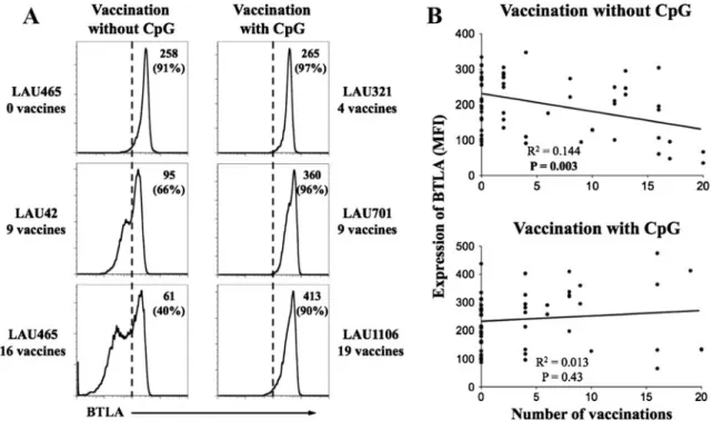
Progressive downregulation of BTLA by B cells in vivo is reversed by vaccination when using CpG as adjuvant CpG can enhance both innate and adaptive immunity and consequently, has a potential utility for immunotherapeutic and vaccine adjuvant applications. CpGs mediate their effects through the triggering of TLR9, which is only expressed by plasmacytoid dendritic cells and B cells in humans [24]. We recently reported that vaccinations of melanoma patients with peptides and CpGs lead to BTLA downregulation on tumor antigen-specific T cells [7]. We therefore addressed whether CpG vaccination altered BTLA expression on B cells from peripheral blood mononuclear cells (PBMC). Patients received serial monthly subcutane-ous vaccinations with Melan-A/MART-1 peptide in conven-tional vaccine formulations, e.g., emulsified in IFA [19]. A second cohort of patients was treated similarly, except that CpG oligodeoxynucleotides were added to the vaccine for-mulation, resulting in a considerably enhanced CD8+ T cell response, as previously reported [20]. Representative anal-yses of BTLA expression by B cells from vaccinated patients are shown in Fig. 1a. We observed a progressive BTLA downregulation in patients vaccinated without CpG
Fig. 1 Sustained BTLA expression upon repeated vaccination with CpG. a Melanoma patients received monthly peptide vaccinations with or without CpG, emulsified in IFA (Montanide ISA-51). Representa-tive histograms of BTLA staining gated on CD20+ B cells (after exclusion of doublets, dead cells, CD14+ monocytes and CD3+ T cells) of PBMCs from five patients, before vaccination (patient LAU465), and after repeated monthly vaccinations, i.e., four
(LAU321), nine (LAU42, LAU701), 16 (LAU465), and 19 (LAU1106) vaccinations. Mean fluorescence intensity (MFI) of BTLA
(and percentage of BTLAhighin parentheses) is indicated. b
Correla-tions between numbers of vaccinaCorrela-tions without CpG (n035) or with
CpG (n026) and the MFI of BTLA expressed by circulating B cells.
(Fig.1a), which significantly correlated with the number of monthly booster vaccinations (Fig.1b). In contrast, patients vaccinated with CpG showed a sustained expression of BTLA by B cells (Fig.1a, b), suggesting that CpG activa-tion of B cells results in maintenance of BTLA expression. Similar results were obtained when data were evaluated in terms of percentage of BTLAhighB cells (data not shown). BTLA is overexpressed on IgM memory B cells
In order to investigate the expression of BTLA on B cells in relation to their differentiation stage, we performed ex vivo multicolor flow cytometry analysis on PBMCs from healthy donors. Using a combination of markers including CD19, immunoglobulin (Ig) D, CD27, and BTLA, we were able to look at BTLA expression on naïve (CD19+IgD+CD27−), classical isotype switched memory (CD19+ IgD−CD27+) and IgM memory B cells (CD19+IgD+ CD27+) [25], as depicted in Fig.2a. We also examined BTLA mRNA ex-pression in B cell subsets from purified naïve IgM memory, switched memory, and transitional B cells from the public GEO datasets GSE22886 [21] and GSE17186 [22] (Supple-mental Fig. 1A). Interestingly, although BTLA was expressed by all B cell subsets, its expression was signifi-cantly lower in naïve and memory B cells subsets compared with IgM memory B cells (Fig.2bupper panel and Supple-mental Fig.1A) and was even higher on transitional (CD27
−IgD+CD24high
and CD38high) circulating B cells (Supple-mental Fig.1A and data not shown) [26], demonstrating that BTLA expression changes during B cell differentiation. Moreover, we also analyzed HVEM expression on B cell differentiation subsets. In contrast to BTLA, we did not find any change in HVEM expression depending on differentia-tion stages (Fig. 2b, lower panel). This was confirmed by HVEM mRNA expression analysis from public GEO data-sets (Supplemental Fig.1B).
CpG induces upregulation of BTLA on BTLAlowB cells Next, we analyzed whether BTLA expression alters while B cells are activated. Thus, purified B cells from healthy individuals were cultured in presence or absence of CpG. Kinetic analysis revealed that, in medium alone (Fig.3a), B cells progressively reduced BTLA expression. Almost half of B cells had a low expression of BTLA at day 5 (Supple-mental Fig. 2). In contrast, B cells stimulated with CpG maintained the expression of BTLA (Fig. 3a and Supple-mental Fig.2). Interestingly, HVEM expression is not mod-ulated upon CpG stimulation (data not shown). These results suggest that CpG-mediated activation of B cells sustain and/or increased BTLA expression. It has recently been shown that germinal center (GC) B cells from lymph nodes do not express BTLA [27], in contrast to B cells from PBMCs (Fig.2) [9]. Therefore, in order to better understand
Fig. 2 Differential expression of BTLA during B cell differentiation. a Gating strategy for the identification of B cell subsets. CD19+ B cells from healthy volunteer PBMCs were analyzed for IgD and CD27 expression to characterize the following subsets: naïve IgD+
CD27−, IgM memory IgD+
CD27+, and memory B cells IgD − CD27+ (upper panel). BTLA expression was analyzed in each subset as shown for a
representative example (lower panel): isotype control (grey), naïve (dotted line), IgM memory (dashed line), and memory (black line). b Statistical assessment of BTLA (upper panel) and HVEM (lower panel) expression on B cell subsets
(n017). Statistical analysis
between medium and CpG point at each time using two-tailed
non-parametric unpaired Mann–
Whitney t test. P<0.01 and P< 0.001 are indicated by ** and ***, respectively
the effect of CpG on BTLA expression, we sorted BTLAneg/
low
and BTLAhigh B cells from normal lymph nodes,
stimulated them with or without CpG, and assessed the expression of BTLA after 5 days of culture (Fig. 3b). When comparing BTLA expression before and after cul-ture in medium alone, it appears that the decrease of BTLA observed in Fig.3aoccurs only in B cells express-ing high levels of BTLA. Interestexpress-ingly, upon CpG stimu-lation, we found a significant upregulation of BTLA expression on both sorted B cells populations, although this augmentation did not reach the expression level be-fore culture (Fig.3c). These data demonstrate that activa-tion with CpGs resulted in enhanced BTLA expression on B cells.
BTLA is recruited to BCR upon CpG activation
Next, we investigated the localization of BTLA. Purified B cells were activated by CpG and, subsequently CD19, used as a surrogate marker of BCR that propagates BCR micro-signalosomes spreading [28], and BTLA distribution were assessed by confocal microcopy. Whereas no clustering was observed without activation (Fig.4a, time 0 min), we found that, 15 min after incubation with CpG, BTLA accumulated and co-localized with the BCR in 55 % of observed B cells (Fig.4a, b). These results suggest that BTLA is translocated to the BCR’s cluster upon CpG activation. Interestingly, the same accumulation of BTLA was obtained when B cells were directly stimulated through the BCR with an anti-IgM antibody (Supplemental Fig.3).
BTLA/HVEM-mediated selective inhibition of proliferation and cytokine production
We evaluated whether BTLA could inhibit B cell functions. First, we analyzed the proliferation of purified B cells from healthy donors by CFSE dilution. After 5 days of CpG stimulation, expansion of B cells was significantly attenuat-ed in the presence of plate-bound or soluble rhHVEM-Fc protein (Fig. 5a, b), which is able to specifically bind to BTLA (Supplemental Fig. 4A). The addition of Fab frag-ment from an anti-BTLA blocking mAb [7,23] restored B cell proliferation (Fig. 5b), showing that BTLA indeed inhibited CpG-mediated B cell proliferation. In addition, B cell survival was not affected by either rhHVEM-Fc or Fab fragment (Supplemental Fig.5). In this experiment, we also assessed B cell activation, as B cell activation by TLR agonists induces expression of CD86 and CD80 [29]. As shown in Fig.5c, Bcells showed significantly lower CD80 and CD86 expression in the presence of rhHVEM-Fc, whereas HLA-DR expression did not change (data not shown). Similar results were obtained when analyzing MFI of CD80 and CD86 (data not shown). Again, this effect was reversed in the presence of blocking BTLA Fab fragment (Fig. 5c). Interestingly, we also observed that the presence
Fig. 3 Maintenance of BTLA expression on B cells upon stimulation with CpG in vitro. a Kinetic analysis of BTLA surface expression during CpG stimulation. Purified B cells from healthy individuals
PBMCs were cultured from 1 to 5 days with or without CpG (2μg/
ml). b Representative experiment depicting BTLA expression by sorted BTLA high and BTLA neg/low CD19+ B cells from normal
lymph nodes (NLN) after 5 days of culture with or without CpG (2μg/
ml). MFI of BTLA (and percentages of BTLAhighin parentheses) is
indicated. c Statistical analysis of this experiment, i.e., of B cells sorted
as“BTLA high” and as “BTLA neg/low” and stimulated with (black
bars) or without CpG (white bars) for 5 days (n05). Gray bars
represent MFI of BTLA in each sorted populations before culture. Comparisons were evaluated using two-tailed non-parametric paired
Wilcoxon’s t test. P<0.05, P<0.01, and P<0.001 are indicated by *,
of rhHVEM-Fc molecules led to the downregulation of BTLA on B cells, independently of the presence of CpG (Fig. 5d). Besides, the possibility of binding competition between rhHVEM-Fc and anti-BTLA mAb was ruled out because labeling of rhHVEM-Fc on 293 T cells expressing BTLA (Supplemental Fig.4A) did not preclude anti-BTLA mAb staining (Supplemental Fig. 4B). All together, these results suggest a regulation of BTLA by its own ligand, as we already showed for CD8+ T cells [7].
In parallel, we measured the secretion of inflammatory cytokines by B cells after 2 days of stimulation with CpG. We found a significant reduction of IL-6, IL-10, and TNFα production when B cells were stimulated in presence of rhHVEM-Fc molecules (Fig.6a). As observed for prolifer-ation and activprolifer-ation marker expression, the addition of blocking anti-BTLA Fab fragment rescued B cell IL-6 se-cretion (data not shown). Interestingly, IL-8 and MIP1-β were not affected by BTLA-mediated inhibition upon CpG stimulation (Fig.6b). Together, these data revealed an in-hibitory role of BTLA in CpG-induced B cell proliferation
and a specific inhibition of cytokine but not chemokine secretion.
Discussion
The co-receptor BTLA is widely expressed on immune cells but has mainly been investigated on T cells where it attenu-ates T cell activation and proliferation [3, 7, 8, 23]. Only little is known on the role and the function of BTLA in B cells. In this study, we described the regulation of BTLA expression in CpG-activated B cells, both ex vivo and in vivo. Our study highlights a mechanism by which the BTLA/HVEM pathway modulates B cell functions.
BTLA is expressed on T cells [2] and B cells (Fig. 2), with a higher expression on the latter [3]. It has been suggested that BTLA does not have a pivotal role in human B cell development in the bone marrow, since only mature bone marrow B cells, compared with precursor B cells, expressed BTLA [10]. In a complementary manner and in
Fig. 4 Co-localization of BTLA and CD19 on CpG-activated B cells. a Representa-tive experiment (out of five) of BTLA and CD19 distribution on resting and CpG-activated purified B cells. Cells were fixed and permeabilized and then stained with anti-BTLA7.1 (red) and anti-CD19 (green) mAbs. Nuclei were finally stained with DAPI (blue). Images were analyzed by con-focal microscopy. Yellow results from the overlay of red and green signals. b Co-localization of CD19 and BTLA on the B-lymphocyte cell surface from images depicted in a at 15 min. The white arrow corresponds to the X-axis of the histogram
contrast to HVEM, our data showing a differential expres-sion of BTLA among circulating B cell subsets, suggest a role of BTLA during B cell differentiation (Fig. 2 and Supplemental Fig.1). We found that circulating transitional and IgM memory B cells express higher levels of BTLA compared with naïve and memory B cells, including switched memory B cells. Transitional B cells correspond to the most immature B cell type in peripheral blood and represent a critical early step, in which BTLA may presum-ably be implicated, in the differentiation and selection of mature B cells [30]. IgM memory B cells are generated in T cell-independent reactions and do not seem to require GC formation [31,32]. These cells are important to elicit im-munoglobulin production in the absence of T cell stimula-tion. Recently, these cells were suggested to recirculate from the spleen marginal zone [33]. Our results suggest that BTLA might be an important regulator of T cell-independent-Ig production. Besides, GC B cells do not express BTLA, in contrast to naïve B cells from the mantle
zone of lymph nodes [27]. These results and ours underlie the putative role of BTLA in the peripheral differentiation of B cells.
BTLA is known to inhibit T cells in mice and humans (reviewed in [34]). However, little is known on the impact of BTLA on B cell functions. Few studies reported that BTLA−/− mice show increased specific antibody responses and autoimmune hepatitis associated with auto-antibodies [2,35], highlighting a potential role of BTLA in regulating B cells autoimmunity. Our data demonstrate that BTLA inhibited CpG-mediated B cell function in humans, suggest-ing that BTLA may compromise B cell-dependent protec-tion from infecprotec-tion. Indeed, we showed for the first time that CpG directly upregulated BTLA on B cells in vitro (Fig.3) and at least sustains its expression in vivo (Fig. 1), in contrast to T cells [7]. Activation of B cells leads to the co-localization and accumulation of BTLA with the BCR (Fig.4), suggesting a link between the signaling pathway of CpG and BCR [36,37]. Furthermore, ligation of BTLA with
Fig. 5 BTLA-mediated inhibition of B cell proliferation. a
CFSE-labeled B cells were activated with CpG (2μg/ml) and cultured for
5 days with Ctrl-Fc or rhHVEM-Fc. In addition, blocking anti-BTLA Fab fragment or isotype control was added into the culture. Histograms depict one representative experiment. b Proliferation of B cells after
CpG stimulation with or without rhHVEM-Fc (n014). The
prolifera-tion ratio was calculated as the percentage of CFSEhighdivided by the
percentage of CFSElow B cells; low ratio implies high level of
proliferating B cells. c Analysis of CD80 and CD86 expression by
CpG-stimulated B cells upon BTLA/HVEM triggering (n07). d BTLA
is downregulated on B cells upon HVEM ligation. Expression of
BTLA by purified B cells after stimulation with CpG (2μg/ml) with
or without rhHVEM-Fc (n08). Statistical analysis was evaluated using
two-tailed non-parametric paired Wilcoxon’s t test. P<0.05, P<0.01
HVEM decreases B cell proliferation (Fig.5a), as described previously [10]. In addition, we show that cytokine secre-tion (IL-6, IL-10, and TNFα) and upregulasecre-tion of co-stimulatory markers (CD80 and CD86) were also dampened by BTLA triggering (Figs.5 and6). Interestingly, the pro-duction of chemokines (IL-8 and MIP1β) was not affected (Fig.6b), underlying a dichotomy in the inhibitory capaci-ties of BTLA, as previously shown for CD8+ T cells [7]. This suggests that the inhibition of B cells by BTLA may
alter their effector functions but not their capacity to attract other immune cells. All together, these results emphasize the importance of BTLA as B cell activation checkpoint. A recent mouse study showed that B cells might contribute to innate immunity against bacterial infection, primarily through TLR4 triggering and GM-CSF secretion. It will be interesting to determine whether BTLA may be involved in the regulation of such B cell populations activated via TLR4 [38].
Recently, CD160 was identified as another co-inhibitory receptor that can also bind HVEM, resulting in the inhibi-tion of CD4+ T cells [39]. However, it is unlikely that this interaction plays a role in B cell physiology, as CD160 is not expressed on B cells [40, 41]. HVEM can bind further proteins, namely LIGHT, lymphotoxinα, and glycoprotein D, but the only ligand present on mature, naive B cells is BTLA [39,40,42], which thus appears as the major inter-acting partner during the initial steps of B cell activation process by CpG. Since B cell tumors express TLR9, CpG can directly affect them, but also indirectly via CpG-activated dendritic cells. Thus, it influences B cell malig-nancy viability (i.e., proliferation, apoptosis) and upregu-lates MHC, co-stimulatory molecules, and other markers such as CD20 and possibly BTLA that serve as targets in immunotherapy approaches [43]. BTLA is one of the few inhibitory receptors expressed by all B cells and also by B cell lymphomas, especially chronic lymphocytic leukemia/ small lymphocytic lymphoma (B-CLL/SLL) [27]. Since we showed that BTLA may dampen B cell functions, the use of agonistic anti-human BTLA mAb in B-CLL/SLL patients may eventually reduce tumor proliferation. Therefore, trig-gering the BTLA inhibitory pathway in B cell malignancies, likely in combination with other agents, may be considered as a new strategy of targeted therapy.
In addition to their role in innate immunity [12], TLRs are critically involved in the initiation and enhancement of adaptive immune response. Enhanced immune response has been reported in mouse tumor models using TLR9 agonists not only as monotherapy but also in combination with other therapies [44]. However, it remains questionable whether these findings are applicable to immunomodulatory strate-gies for patients. According to some recent clinical studies, CpG have anti-tumor activity as single agents [43,44] and enhance the development of anti-tumor-specific T cells responses when used as vaccine adjuvant [20, 44]. More-over, CpG adjuvanted vaccination of melanoma patients lead to BTLA downregulation on tumor-specific human CD8+ T cells, concomitant with restoration of their func-tionality [7]. Here, we show that this type of vaccination additionally sustains BTLA expression on circulating B cells in vivo (Fig. 1), which would most likely decrease their functionality in case of ligation with HVEM. However, HVEM has been recently reported to have co-stimulatory function upon ligation with BTLA [45]. Therefore, we may
Fig. 6 Selective inhibition of cytokine secretion by BTLA. a Analysis of cytokine production after 2 days of stimulation of B cells with CpG
in presence of rhHVEM-Fc or Ctrl-Fc (n06). b Analysis of chemokine
secretion by CpG-stimulated B cells cultured with or without Ctrl-Fc or rhHVEM-Fc. Statistical analysis was evaluated using two-tailed non-parametric paired Wilcoxon’s t test. P<0.05 and P<0.01 are indicated by * and **, respectively
hypothesize that, upon vaccination, a BTLA/HVEM bidi-rectional pathway may occur. BTLA expressed by CpG-activated B cells that present vaccine peptides may trigger HVEM expressed by vaccine-specific T cells, possibly lead-ing to enhanced T cell activation.
In summary, BTLA/HVEM ligation results in the inhibi-tion of activated B cell funcinhibi-tions. Our results underlie the potential role of BTLA/HVEM interactions in vaccination when using an adjuvant that stimulates B cells and provides a baseline for further investigation of targeting BTLA in B cell-related diseases.
Acknowledgments We are obliged to the patients and healthy
vol-unteers for their dedicated collaboration. We thank Dr. Daniel Isnardon and Sébastien Létard for discussion and technical insights. We thank Pfizer and Coley Pharmaceutical Group for pharmaceutical products. This work was supported by grants from Institut National de la Santé et de la Recherche Médicale, the Ludwig Institute for Cancer Research, and the Swiss Cancer League. ML Thibult has been supported by fellowships from Ministère de la Recherche et de l’Enseignement Supérieur and from La Ligue contre le Cancer.
Disclosure of conflict of interest The authors declare no competing
financial interests.
References
1. Peggs KS, Quezada SA, Allison JP (2008) Cell intrinsic mecha-nisms of T-cell inhibition and application to cancer therapy.
Immu-nol Rev 224:141–165
2. Watanabe N, Gavrieli M, Sedy JR, Yang J, Fallarino F, Loftin SK, Hurchla MA, Zimmerman N, Sim J, Zang X et al (2003) BTLA is a lymphocyte inhibitory receptor with similarities to CTLA-4 and PD-1. Nat Immunol 4:670–679
3. Han P, Goularte OD, Rufner K, Wilkinson B, Kaye J (2004) An inhibitory Ig superfamily protein expressed by lymphocytes and APCs is also an early marker of thymocyte positive selection. J
Immunol 172:5931–5939
4. Hurchla MA, Sedy JR, Gavrieli M, Drake CG, Murphy TL, Murphy KM (2005) B and T lymphocyte attenuator exhibits structural and expression polymorphisms and is highly induced in anergic CD4+ T
cells. J Immunol 174:3377–3385
5. Gavrieli M, Watanabe N, Loftin SK, Murphy TL, Murphy KM (2003) Characterization of phosphotyrosine binding motifs in the cytoplasmic domain of B and T lymphocyte attenuator required for association with protein tyrosine phosphatases SHP-1 and SHP-2.
Biochem Biophys Res Commun 312:1236–1243
6. Sedy JR, Gavrieli M, Potter KG, Hurchla MA, Lindsley RC, Hildner K, Scheu S, Pfeffer K, Ware CF, Murphy TL et al (2005) B and T lymphocyte attenuator regulates T cell activation through interaction with herpesvirus entry mediator. Nat Immunol 6:90–98
7. Derre L, Rivals JP, Jandus C, Pastor S, Rimoldi D, Romero P, Michielin O, Olive D, Speiser DE (2010) BTLA mediates inhibi-tion of human tumor-specific CD8+ T cells that can be partially
reversed by vaccination. J Clin Invest 120:157–167
8. Krieg C, Han P, Stone R, Goularte OD, Kaye J (2005) Functional analysis of B and T lymphocyte attenuator engagement on CD4+
and CD8+ T cells. J Immunol 175:6420–6427
9. Otsuki N, Kamimura Y, Hashiguchi M, Azuma M (2006) Expres-sion and function of the B and T lymphocyte attenuator (BTLA/
CD272) on human T cells. Biochem Biophys Res Commun
344:1121–1127
10. Vendel AC, Calemine-Fenaux J, Izrael-Tomasevic A, Chauhan V, Arnott D, Eaton DL (2009) B and T lymphocyte attenuator regu-lates B cell receptor signaling by targeting Syk and BLNK. J Immunol 182:1509–1517
11. Peng SL (2005) Signaling in B cells via Toll-like receptors. Curr Opin Immunol 17:230–236
12. Iwasaki A, Medzhitov R (2004) Toll-like receptor control of the
adaptive immune responses. Nat Immunol 5:987–995
13. Takeda K, Akira S (2005) Toll-like receptors in innate immunity.
Int Immunol 17:1–14
14. Bourke E, Bosisio D, Golay J, Polentarutti N, Mantovani A (2003) The toll-like receptor repertoire of human B lymphocytes: induc-ible and selective expression of TLR9 and TLR10 in normal and
transformed cells. Blood 102:956–963
15. Busconi L, Bauer JW, Tumang JR, Laws A, Perkins-Mesires K, Tabor AS, Lau C, Corley RB, Rothstein TL, Lund FE et al (2007) Functional outcome of B cell activation by chromatin immune complex engagement of the B cell receptor and TLR9. J Immunol
179:7397–7405
16. Hemmi H, Takeuchi O, Kawai T, Kaisho T, Sato S, Sanjo H, Matsumoto M, Hoshino K, Wagner H, Takeda K et al (2000) A Toll-like receptor recognizes bacterial DNA. Nature 408:740–745 17. Leadbetter EA, Rifkin IR, Hohlbaum AM, Beaudette BC, Shlomchik MJ, Marshak-Rothstein A (2002) Chromatin-IgG complexes activate B cells by dual engagement of IgM and
Toll-like receptors. Nature 416:603–607
18. Viglianti GA, Lau CM, Hanley TM, Miko BA, Shlomchik MJ, Marshak-Rothstein A (2003) Activation of autoreactive B cells by
CpG dsDNA. Immunity 19:837–847
19. Lienard D, Rimoldi D, Marchand M, Dietrich PY, van Baren N, Geldhof C, Batard P, Guillaume P, Ayyoub M, Pittet MJ et al (2004) Ex vivo detectable activation of Melan-A-specific T cells correlating with inflammatory skin reactions in melanoma patients vaccinated with peptides in IFA. Cancer Immun 4:4
20. Speiser DE, Lienard D, Rufer N, Rubio-Godoy V, Rimoldi D, Lejeune F, Krieg AM, Cerottini JC, Romero P (2005) Rapid and strong human CD8+ T cell responses to vaccination with peptide, IFA, and CpG oligodeoxynucleotide 7909. J Clin Invest 115:739–746
21. Abbas AR, Baldwin D, Ma Y, Ouyang W, Gurney A, Martin F, Fong S, van Lookeren CM, Godowski P, Williams PM et al (2005) Immune response in silico (IRIS): immune-specific genes identi-fied from a compendium of microarray expression data. Genes
Immun 6:319–331
22. Suryani S, Fulcher DA, Santner-Nanan B, Nanan R, Wong M, Shaw PJ, Gibson J, Williams A, Tangye SG (2010) Differential expression of CD21 identifies developmentally and functionally
distinct subsets of human transitional B cells. Blood 115:519–529
23. Serriari NE, Gondois-Rey F, Guillaume Y, Remmerswaal EB, Pastor S, Messal N, Truneh A, Hirsch I, van Lier RA, Olive D (2010) B and T lymphocyte attenuator is highly expressed on CMV-specific T cells during infection and regulates their function.
J Immunol 185:3140–3148
24. Krieg AM (2002) CpG motifs in bacterial DNA and their immune
effects. Annu Rev Immunol 20:709–760
25. Klein U, Rajewsky K, Kuppers R (1998) Human immunoglobulin (Ig)M+IgD+peripheral blood B cells expressing the CD27 cell surface antigen carry somatically mutated variable region genes: CD27 as a general marker for somatically mutated (memory) B
cells. J Exp Med 188:1679–1689
26. Palanichamy A, Barnard J, Zheng B, Owen T, Quach T, Wei C, Looney RJ, Sanz I, Anolik JH (2009) Novel human transitional B cell populations revealed by B cell depletion therapy. J Immunol
27. M’Hidi H, Thibult ML, Chetaille B, Rey F, Bouadallah R, Nicollas R, Olive D, Xerri L (2009) High expression of the inhibitory receptor BTLA in T-follicular helper cells and in B-cell small lymphocytic lymphoma/chronic lymphocytic leukemia. Am J Clin Pathol 132:589–596
28. Harwood NE, Batista FD (2011) The cytoskeleton coordinates the early events of B-cell activation. Cold Spring Harb, Perspect Biol 3(2) 29. Greenwald RJ, Freeman GJ, Sharpe AH (2005) The B7 family
revisited. Annu Rev Immunol 23:515–548
30. Loder F, Mutschler B, Ray RJ, Paige CJ, Sideras P, Torres R, Lamers MC, Carsetti R (1999) B cell development in the spleen takes place in discrete steps and is determined by the quality of B
cell receptor-derived signals. J Exp Med 190:75–89
31. Carsetti R, Rosado MM, Wardmann H (2004) Peripheral
develop-ment of B cells in mouse and man. Immunol Rev 197:179–191
32. Weller S, Faili A, Garcia C, Braun MC, Le Deist FF, de Saint Basile GG, Hermine O, Fischer A, Reynaud CA, Weill JC (2001) CD40-CD40L independent Ig gene hypermutation suggests a sec-ond B cell diversification pathway in humans. Proc Natl Acad Sci
U S A 98:1166–1170
33. Kruetzmann S, Rosado MM, Weber H, Germing U, Tournilhac O, Peter HH, Berner R, Peters A, Boehm T, Plebani A et al (2003) Human immunoglobulin M memory B cells controlling Streptococcus pneu-moniae infections are generated in the spleen. J Exp Med 197:939–945 34. Murphy TL, Murphy KM (2010) Slow down and survive: enig-matic immunoregulation by BTLA and HVEM. Annu Rev
Immu-nol 28:389–411
35. Oya Y, Watanabe N, Owada T, Oki M, Hirose K, Suto A, Kagami S, Nakajima H, Kishimoto T, Iwamoto I et al (2008) Development of autoimmune hepatitis-like disease and production of autoanti-bodies to nuclear antigens in mice lacking B and T lymphocyte
attenuator. Arthritis Rheum 58:2498–2510
36. Lin YC, Huang DY, Chu CL, Lin WW (2010) Anti-inflammatory actions of Syk inhibitors in macrophages involve non-specific inhibition of Toll-like receptors-mediated JNK signaling pathway.
Mol Immunol 47:1569–1578
37. Sanjuan MA, Rao N, Lai KT, Gu Y, Sun S, Fuchs A, Fung-Leung WP, Colonna M, Karlsson L (2006) CpG-induced tyrosine phos-phorylation occurs via a TLR9-independent mechanism and is
required for cytokine secretion. J Cell Biol 172:1057–1068
38. Rauch PJ, Chudnovskiy A, Robbins CS, Weber GF, Etzrodt M, Hilgendorf I, Tiglao E, Figueiredo JL, Iwamoto Y, Theurl I et al. (2012) Innate response activator B cells protect against microbial sepsis. Science 335:597-601
39. Cai G, Anumanthan A, Brown JA, Greenfield EA, Zhu B, Freeman GJ (2008) CD160 inhibits activation of human CD4+ T cells through interaction with herpesvirus entry mediator.
Nat Immunol 9:176–185
40. Anumanthan A, Bensussan A, Boumsell L, Christ AD, Blumberg RS, Voss SD, Patel AT, Robertson MJ, Nadler LM, Freeman GJ (1998) Cloning of BY55, a novel Ig superfamily member expressed on NK cells, CTL, and intestinal intraepithelial
lympho-cytes. J Immunol 161:2780–2790
41. Maiza H, Leca G, Mansur IG, Schiavon V, Boumsell L, Bensussan A (1993) A novel 80-kD cell surface structure identifies human circulating lymphocytes with natural killer activity. J Exp Med
178:1121–1126
42. Mauri DN, Ebner R, Montgomery RI, Kochel KD, Cheung TC, Yu GL, Ruben S, Murphy M, Eisenberg RJ, Cohen GH et al (1998) LIGHT, a new member of the TNF superfamily, and lymphotoxin alpha are ligands for herpesvirus entry mediator. Immunity 8:21– 30
43. Weiner GJ (2009) CpG oligodeoxynucleotide-based therapy of
lymphoid malignancies. Adv Drug Deliv Rev 61:263–267
44. Vollmer J, Krieg AM (2009) Immunotherapeutic applications of CpG oligodeoxynucleotide TLR9 agonists. Adv Drug Deliv Rev
61:195–204
45. Cheung TC, Steinberg MW, Oborne LM, Macauley MG,
Fukuyama S, Sanjo H, D’Souza C, Norris PS, Pfeffer K, Murphy
KM et al (2009) Unconventional ligand activation of herpesvirus entry mediator signals cell survival. Proc Natl Acad Sci U S A