Introduction
Fractures of the thoracolumbar spine are usually considered to be the result of compression, shear, or torsion stresses, acting either singly or in combination [14, 15, 28]. An ad-ditional mechanism of injury might occur due to flexion-distraction stress. In 1948, G Q Chance was the first to de-scribe a vertebral fracture consecutive to a flexion trauma extending through the posterior spinous process, the pedi-cles, and spreading anteriorly through the vertebral body [5]. Subsequently, other fracture patterns similar to those described by Chance became known as “Chance fractures”. Until a few years ago, this lesion was thought to be ex-tremely rare in children [22, 23, 25]. However, this injury has become more common in children with the increased use of seatbelts by the pediatric population in motor vehi-cles. The use of seatbelts significantly reduces the risk of injury or death in a car crash by preventing ejection from the car, but seatbelts also change the distribution of forces, and may be the cause of other injuries [35]. The present
article describes a previously unreported lumbar spine le-sion, which might be considered as a variant of Chance injury.
Case report
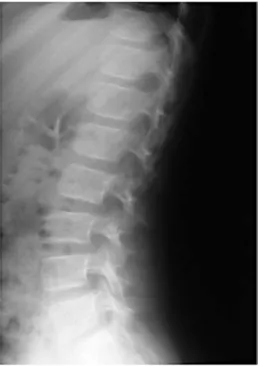
A 7-year-old girl involved in a motor vehicle accident was admit-ted to the emergency department. The child was a rear-seat pas-senger in a car that collided with an embankment head on at high speed. At the time of the accident, she was wearing a seatbelt with a shoulder restraint. On admission, the patient complained of se-vere abdominal and back pain without history of loss of conscious-ness. Abdominal examination revealed a diffuse tenderness and guarding with a transverse abrasion over the lower abdomen. On spinal examination, there was a palpable deformity and swelling in the upper lumbar region. No neurologic deficit was present. Initial radiographs (Fig. 1) showed a discrete widening of the inter-spinous space between the first and the second lumbar vertebrae without visible fracture. On abdominal ultrasonography, the pres-ence of free fluid in the peritoneal cavity was suspect for a visceral injury. Abdominal computed tomography (CT) confirmed the pres-ence of fluid collection in the abdominal cavity, but free air was not noted. No lesion could be detected at the level of the spleen,
Abstract The authors report the
case of a 7-year-old child involved in a motor vehicle accident. She sus-tained an unusual flexion-distraction vertebral injury. This spinal injury was related to seatbelt use and was associated with intra-abdominal le-sions. The spinal lesion consisted of a posterior ligamentous disruption with widening of the posterior inter-vertebral space at two adjacent lum-bar levels. The purpose of this case report is to describe an atypical and perhaps often unrecognized spinal le-sion and to explain our approach to diagnosis and treatment.
Keywords Chance fracture ·
Flexion-distraction · Abdominal lesions · Seatbelt
Received: 20 July 2002 Revised: 14 March 2003 Accepted: 21 March 2003 Published online: 10 July 2003 © Springer-Verlag 2003
D. Ceroni (✉) · M. Mousny · A. Kaelin Paediatric Orthopaedic Unit,
Children’s Hospital, University Hospital Geneva, Rue Willy Donzé 6, 1205 Geneva, Switzerland Tel.: +41-22-3723311, Fax: +41-22-3824783,
e-mail: dimitri.ceroni@hcuge.ch A. Lironi
Paediatric Surgery Unit, Children’s Hospital, University Hospital Geneva, Switzerland
the liver, the kidneys or the pancreas. Despite a hematoma in the retroperitoneal space, the spinal CT scan was interpreted as nor-mal. Emergency laparotomy revealed a mesenteric tear with a le-sion of a superior mesenteric artery branch leading to an important mesenteric hematoma. A jejunal segmental infarction and a more distal small bowel perforation were discovered. Moreover, there were two small seromuscular tears, one in the descending colon and one at the level of the ilio-caecal valvula. Two segmental re-sections of small bowel were performed with immediate restora-tion of the continuity. A magnetic resonance (MR) image (Fig. 2) of the spine showed posterior ligamentous disruption at two levels (L1-L2 and L2-L3). The intervertebral discs appeared fully normal at both levels. Increased height of the intervertebral foramina and widening of the posterior part of the intervertebral space on flexion dynamic X-rays (Fig. 3) confirmed instability at these levels. Ten days after the initial trauma, surgical exploration revealed L1-L2 and L2-L3 instability due to rupture of the supraspinous ligaments, ligamentum flavum and capsules of the posterior joints (Fig. 4). A fracture of the L1 transverse apophysis was discovered during the operative exposure. Reduction was obtained by getting the child lumbar spine into slight lordosis on the operating table, and fixa-tion was performed with two thread loops, one between the spin-ous processes of L1 and L2, the second between those of L2 and L3 (Fig. 5). The child was then immobilized in a body jacket or-thosis for 3 months. Seven months after the trauma, the child was asymptomatic, and conventional dynamic radiographs were inter-preted as normal (Fig. 6).
Discussion
In 1948, Chance first described vertebral fractures that ex-tended horizontally through the posterior spinous process and the neural arch, and ended in the vertebral body [5]. 168
Fig. 1 Widening of the interspinous space between the first and the second and between the second and the third lumbar vertebrae was noted on the thoracolumbar spine radiographs. No fracture
was suspected on these films Fig. 2 Magnetic resonance imaging (MRI): on T2-weighted fat-saturated sequences, there is a hyperintensity signal alteration of the posterior ligamentous elements, evocative of a lesion of these struc-tures
Fig. 3 Dynamic X-rays performed in flexion confirmed instability at two levels (L1-L2 and L2-L3) by the increased height of the in-tervertebral foramina and by a widening of the posterior part of the intervertebral space
The mechanism of the injury was thought to be pure flex-ion. In 1965, Howland et al. associated this particular frac-ture with lap belt use during a motor vehicle accident [16]. Later, Smith and Kaufer hypothesized that Chance frac-tures associated with seatbelt injuries were due to
distrac-tion forces rather than pure flexion stress [31]. These au-thors noted that usually there was not an anterior vertebral compression but rather a failure of the posterior elements in tension, given that the fulcrum of rotation was commonly anterior to the vertebral body. The concept of a flexion-distraction mechanism of injury was put forward by Ren-nie and Mitchell, who considered that compression forces were acting on the anterior portion of the vertebral body, while distraction forces were simultaneously acting on the posterior structures [27]. As seatbelt injuries of the thoraco-lumbar spine were essentially reported in adults [6, 10, 13, 23, 31, 31], it was thought that this lesion was rare in the pediatric population [22, 23, 25]. An increase in the reported frequency of this injury in children has been re-ported [1, 2, 4, 8, 11, 17, 19, 20, 22, 23, 26, 30, 32, 34], and is probably related to the legislation that mandates the use of a seatbelt by children in automobiles. As children have a higher center of gravity due to an increased head-to-body ratio [1, 24], the lever arm movement around the axis is increased, which explains why children are suscep-tible to Chance fracture [1]. Gumley has described the mechanism of action occurring with this injury when the patient slides beneath an improperly placed lap belt [13], resulting in an axis of rotation approximately at the level of the umbilicus [1]. In young children, an ideal place-ment of the lap belt at the level of the hip is difficult to achieve, because of the poorly developed iliac crest and the problem of maintaining an upright posture [30]. This is a particular problem, as children are frequently rear-seat passengers where shoulder restraints are usually absent Fig. 4 Surgical exploration revealed L1-L2 instability due to
rup-ture of the lumbodorsal fascia, supraspinous ligaments, ligamentum flavum and capsules of the posterior joints
Fig. 5 Fixation of the lesion was obtained with two thread loops, one between the spinous processes of L1 and L2, the second between those of L2 and L3
Fig. 6 Final thoracolumbar spine radiograph reveals no residual deformity in standing position
170
[11]. In the case described, the sudden deceleration caused a hyperflexion of the spine around the lap belt. The pat-tern of the lesion is consistent with a mechanism of flex-ion of the spine about an axis anterior to the vertebral body, resulting in a pure distraction separating the poste-rior elements. However, the failure of the posteposte-rior liga-mentous structures at two intervertebral levels makes it unique (Fig. 4).
Chance fracture may be difficult to diagnose on the ini-tial radiographs, and CT scans may fail to detect the injury [32]. In the reported case, MRI and especially dynamic X-rays were the most useful additional investigations. Many types of Chance fracture patterns have been de-scribed. Some classification systems for Chance fractures have been proposed, and all of them are based on the type of the lesion (only osseous, only ligamentous, or a combi-nation of the both) and on the fracture direction [7, 13, 30]. In the case described, lesions through the posterior el-ements occurred at two different levels. This particular case of Chance fracture can therefore not be related to the described classifications.
In adults, neurological damage is uncommon, and com-plete paraplegia has seldom been reported [6, 10, 13, 30, 31]. Rumball and Jarvis have assessed the incidence of paraplegia in children with seatbelt injuries to be 15% [30]. Posterior protrusion of the nucleus pulposus or expulsion of the vertebral apophysis may cause progressive neurologic deficits or spinal canal compromise [31]. As the center of gravity is higher in children than in adults [24], this may re-sult in an increased moment arm, and probably greater dis-traction, contributing to the higher incidence of paraplegia in children. Additionally, child ligaments and bones can tolerate four times more stretch than the spinal cord [3, 12]. This may lead to normal radiographs in a patient with ab-normal neurological findings, known as “spinal cord injury without radiographic abnormality” (SCIWORA) [17, 21]. In our case report, the child showed no neurologic damage. Treatment of a Chance fracture doesn’t only depend on the severity of the injury, but also on the fracture pattern. If the injury is only osseous in all columns, closed reduc-tion and immobilizareduc-tion in an extension brace are appro-priate [29]. If the injury is ligamentous, operative
reduc-tion with fusion is indicated, because ligamentous damage does not heal without instability. However, children are noted to have a better prognosis than adults [9]. Results of Chance fracture bracing in children, including those with ligamentous injury, are better than what would be expected in adults [11, 33]. Glassmann et al. [20] reported that brace treatment failed only in patients with an initial kyphosis of more than 20°. So they advocated immobilization if kypho-sis was less than 20°. In their series, all children with suc-cessful brace treatment had a decrease in kyphosis over time because of the remaining potential for anterior growth. Nevertheless, kyphosis greater than 20° requires surgical stabilization even in children, especially when important ligamentous injuries are present [11], to prevent progres-sion of the kyphosis, instability and chronic pain [18]. In young children, simple interspinous wiring is recommended in order not to disturb the potential of ulterior growth and may be supplemented by postoperative brace immobiliza-tion. In adolescents, standard compression devices can be used to counteract the tensile forces acting on the posterior elements [29]. In our case, we opted for a surgical stabi-lization, given the extended ligamentous injuries. Fixation was performed with two absorbable thread loops, one be-tween the spinous processes of L1 and L2, and the second between those of L2 and L3 (Fig. 5). This treatment was supplemented by postoperative brace immobilization for 3 months.
Conclusion
Chance fractures in a child are potentially devastating in-juries and are largely caused by motor vehicle accidents. They occur when the spine is flexed about an axis anterior to the spine. Associated abdominal injuries are common, but their diagnosis is often delayed. We think that the lesion described in this case report is a variant of Chance frac-ture. This unusual lesion is characterized by a separation of the posterior elements at two levels. MRI and, above all, dynamic conventional radiography are diagnostic. Instabil-ity represents a surgical indication, especially when liga-mentous damage is extensive.
1. Agran PF, Dunkle DE, Winn DG (1987) Injuries to a sample of seat belted children evaluated in a hospital emergency room. J Trauma 27:58–64 2. Anderson PA, Rivara FP, Maier RV,
Drake C (1991) The epidemiology of seatbelt-associated injuries. J Trauma 31:60–67
3. Aufdermaur HR (1974) Spinal injuries in juveniles. Necropsy findings in twelve cases. J Bone Joint Surg Br 56: 513–519
4. Blasier RD, Lamont RL (1985) Chance fracture in a child: a case report with non-operative treatment. J Pediatr Or-thop 5:92–93
5. Chance GQ (1948) Note on a type of flexion fracture of the spine. Br J Ra-diol 21:452–453
6. Dennis R, Allard M, Atlas H, Farkouh E (1983) Changing trends with abdom-inal injury in seatbelt wearers. J Trauma 23:1007–1008
7. Fuentes JM, Bloncourt J, Bourbotte G, et al (1984) La fracture de Chance. Neurochirurgie 30:113–118 8. Gallagher DJ, Heinrich SD (1990)
Pediatric Chance fracture. J Orthop Trauma 4:183–187
(1982) Distraction fractures of the lum-bar spine. J Bone Joint Surg Br 64: 520–525
14. Holdsworth FW (1963) Fractures, dis-locations, and fracture-dislocations of the spine. J Bone Joint Surg Br 45: 6–20
15. Holdsworth FW (1970) Fractures, dis-locations, and fracture-dislocations of the spine. J Bone Joint Surg Am 52: 1534–1551
16. Howland WJ, Curry JL, Buffington CB (1965) Fulcrum fractures of the lumbar spine. JAMA 193:240–241
17. Hubbard DD (1974) Injuries of the spine in children and adolescents. Clin Orthop 100:56–65
induced Chance fracture in an infant. Case report and literature review. Pediatr Radiol 21:575–577
23. Moskowitz A (1989) Lumbar seatbelt injury in a child: case report. J Trauma 29:1279–1282
24. Palmer CE (1928) Center of gravity of the human body during the growth. Am J Phys Anthropol 11:423–455 25. Raney EM, Bennett JT (1992)
Pedi-atric Chance fracture. Spine 17:1522– 1524
26. Reid AB, Letts RM, Black GB (1990) Pediatric Chance fractures: association with intra-abdominal injuries and seat-belt use. J Trauma 30:384–391 27. Rennie W, Mitchell N (1973) Flexion
distraction fractures of the thoracolum-bar spine. J Bone Joint Surg Am 55: 386–390
children: a pitfall in CT diagnosis. AJR 150:1355–1358
33. Tso EL, Beaver BL, Haller JA (1993) Abdominal injuries in restrained pedi-atric passengers. J Pediatr Surg 28: 915–919
34. Voss L, Cole PA, D’Amato C (1996) Pediatric Chance fractures from lap-belts: unique case report of three in one accident. J Orthop Trauma 10:421–428 35. Woelfel G, Moore E, Cogbill T, et al
(1984) Severe thoracic and abdominal injuries associated with lap-harness seatbelts. J Trauma 24:166–167