HAL Id: hal-02458096
https://hal.archives-ouvertes.fr/hal-02458096
Submitted on 28 Jan 2020
HAL is a multi-disciplinary open access archive for the deposit and dissemination of sci-entific research documents, whether they are pub-lished or not. The documents may come from teaching and research institutions in France or abroad, or from public or private research centers.
L’archive ouverte pluridisciplinaire HAL, est destinée au dépôt et à la diffusion de documents scientifiques de niveau recherche, publiés ou non, émanant des établissements d’enseignement et de recherche français ou étrangers, des laboratoires publics ou privés.
Macroevolution of venom apparatus innovations in auger
snails (Gastropoda; Conoidea; Terebridae)
M. Castelin, N. Puillandre, Yu.I. Kantor, M.V. Modica, Y. Terryn, C.
Cruaud, P. Bouchet, M. Holford
To cite this version:
M. Castelin, N. Puillandre, Yu.I. Kantor, M.V. Modica, Y. Terryn, et al.. Macroevolution of venom apparatus innovations in auger snails (Gastropoda; Conoidea; Terebridae). Molecular Phylogenetics and Evolution, Elsevier, 2012, 64 (1), pp.21-44. �10.1016/j.ympev.2012.03.001�. �hal-02458096�
Macroevolution of venom apparatus innovations in auger snails (Gastropoda; Conoidea; Terebridae)
M. Castelina,1b, N. Puillandre1b,c, Yu. I. Kantord, Y. Terryne, C. Cruaudf, P. Bouchetg, M. Holforda*.
a
The City University of New York-Hunter College and The Graduate Center, The American Museum of Natural History NYC, USA.
1b
UMR 7138, Muséum National d’Histoire Naturelle, Departement Systematique et Evolution, 43, Rue Cuvier, 75231 Paris, France
c
Atheris Laboratories, Case postale 314, CH-1233 Bernex-Geneva, Switzerland
d
A.N. Severtsov Institute of Ecology and Evolution, Russian Academy of Sciences, Leninski Prosp. 33, Moscow 119071, Russia
e
NaturalArt, Kapiteinstraat 27, 9000 Gent, Belgium
f
GENOSCOPE, Centre National de Séquencage, 2 rue Gaston Crémieux, CP 5706, 91057 Evry Cedex, France
g
UMR 7138, Muséum National d’Histoire Naturelle, Departement Systematique et Evolution, 55, Rue Buffon, 75231 Paris, France
1
Present address
e-mail address:
M. Castelin : magcastelin@mnhn.fr N. Puillandre : puillandre@mnhn.fr
Yu. I. Kantor : kantor@malaco-sevin.msk.ru Y. Terryn : yves@naturalart.be
C. Cruaud : cruaud@genoscope.cns.fr P. Bouchet : pbouchet@mnhn.fr
M. Holford : mholford@hunter.cuny.edu
* Corresponding author: M. Holford : mholford@hunter.cuny.edu Phone: +001 212-772-5330
Abstract
The Terebridae are a diverse family of tropical and subtropical marine gastropods that use their venom apparatus to produce toxins that capture polychaete and enteropneust preys. The development of a venom apparatus in the Terebridae is a key innovation that can be analyzed within a molecular phylogenetic scaffold to decipher terebrid diversification. Presented here is a molecular phylogeny of 89 terebrid species belonging to 12 of the 15 currently accepted genera, based on Bayesian inference and Maximum Likelihood analyses of fragments of 3 mitochondrial (COI, 16S and 12S) and one nuclear (28S) genes. The evolution of the anatomy of the terebrid venom apparatus was assessed by mapping traits of six related characters, proboscis, venom gland, odontophore, accessory proboscis structure, radula, and salivary glands. A novel result for terebrid phylogeny was the discovery of a previously unrecognized lineage, which includes species of Euterebra and Duplicaria. The non-monophyly of most terebrid genera analyzed indicates that the current genus-level classification of the group is plagued with homoplasy and requires further taxonomic investigations. Foregut anatomy in the family Terebridae reveals an inordinate diversity of features that covers the range of variability within the entire superfamily Conoidea, and a triple origin of the hypodermic radular teeth. These findings illustrate that terebrid venom apparatus evolution is not perfunctory, and involves independent and numerous apparitions of central features in the foregut anatomy. The recurrent terebrid radular origins are presumably associated with variable functionalities, suggesting that terebrids have adapted to dietary changes that may have resulted from predator-prey relationships. The anatomical and phylogenetic results presented serve as a starting point to further investigate the role of predator-prey interactions in the diversification of the Terebridae and of their toxins, which are promising bioactive compounds for biomedical research and therapeutic drug development.
1. Introduction
At the macroevolutionary level, it is hypothesized that the tempo of evolution can be viewed through the lens of key innovations (Sanderson and Donoghue, 1994). Key innovations are biological traits that promote lineage diversification (Heard and Hauser, 1995; Hodges and Arnold, 1995). The development of a venom apparatus in the marine gastropod superfamily Conoidea is a key innovation that can be used as an organizational framework to decipher the evolutionary history of this megadiverse group. Here the evolution of the venom apparatus in auger snails (Neogastropoda; Conoidea; Terebridae) is investigated using a molecular phylogenetic scaffold.
The Terebridae are a diverse family of medium-sized to large (mostly 15-150 mm) marine gastropods distributed throughout most tropical and subtropical oceans. Terebrids use their venom apparatus to capture prey, but perhaps also to defeat competitors or predators. Similar to the peptide toxins produced by cone snails (Neogastropoda; Conoidea; Conidae), the peptide toxins produced by terebrids, teretoxins, are promising bioactive compounds for biomedical research and therapeutic drug development (Puillandre and Holford, 2010). Peptide toxins from a venom source are of increasing interest in the pharmacological industry (Butler, 2008; Casewell et al., 2009; Chin et al., 2006; Hong, 2011; Newman and Cragg, 2007). As recently demonstrated (Fry et al., 2003; Modica and Holford, 2010; Puillandre et al., 2010; Saslis-Lagoudakis et al., 2011), understanding how the organisms that produce these toxins have emerged, and evolved over time, may become central in the process of drug discovery. Specifically, in the case of the Terebridae, not all species have a venom apparatus; therefore identifying the lineages that have a venom apparatus is an effective route to toxin characterization. Currently, the extent of species diversification of the Terebridae is largely underestimated and the evolutionary pathways explored by the terebrid groups, especially regarding the toxins they produce, remains largely unknown. Understanding the evolutionary patterns of venom apparatus evolution in the Terebridae would significantly advance clarifying the phylogeny and systematics of the group, in addition to the characterization of terebrid toxins for therapeutic applications.
Whether used for defense or attack, the diversity of toxins developed by venomous organisms is often attributed to the process of co-evolution in predator-prey relationships (Barlow et al., 2009; Duda, 2008; Kordis and Gubensek, 2000; Kozminsky-Atias et al., 2008; Lynch, 2007). Co-evolutionary predator-prey interactions may lead to the development of specialized adaptations in the predator that are followed by counter-adaptations in the prey, which in turn can lead to further adaptations in the predator, and so on, as dictated by biotic ("Red Queen" hypothesis – Van Valen, 1973) or abiotic ("Court Jester" hypothesis – Barnosky, 2001) pressures. For example, numerous plants produce toxic secondary compounds that influence the behavior, growth, or survival of insects and other herbivores.
In addition, herbivores have developed ways to detoxify, sequester, or render ineffective specific plant poisons (Fowler, 1983; Laycock, 1978; Zangerl et al., 2008). In snakes, it has been demonstrated that venom diversity may result by adaptation toward specific diets (Barlow et al., 2009; Daltry et al., 1996; Wüster et al., 1999). In parallel, some snake prey have developed the ability to inhibit specific venom toxins (Biardi et al., 2005; Heatwole and Poran, 1995).
By its indirect effect on fitness, the predator-prey arms race can represent a driving force of speciation and species diversification in both predators and preys populations. This is referred to as the “escalation/diversification hypothesis” (Ehrlich and Raven, 1964; but see also Berenbaum, 1983; Berenbaum and Feeny, 1981; Vermeij, 1993). Phylogenetic analyses can provide seminal evidence on rates and patterns of predation-traits evolution and species diversification (Farrell et al., 1991). However, the correlation between adaptative changes of predation-traits and species-diversification in predator-prey systems is difficult to study. Such a study requires a good understanding of the biology and the ecology of the species involved and necessitates a thorough taxonomic sampling of both predators and preys taxa. A good alternative, as attempted here with the Terebridae, is to obtain an exhaustive taxonomic sampling of one of the two taxa (predator or prey) and to study the traits or innovations that affect the ability to accomplish or avoid predation. Mapping these innovatons on a phylogenetic tree then reveals patterns that may impact species diversification.
Recent molecular phylogenies (Holford et al., 2009a, 2009b; Puillandre et al., 2011) of the family Terebridae based on samples from Western and Eastern Pacific demonstrated the monophyly of terebrids relative to the other families of conoideans. Also illustrated in these phylogenetic studies is the existence of five distinctive clades, Pellifronia, Oxymeris [= Acus], Terebra, Hastula, and Myurella, numbered clades A to E, respectively, with clade A, containing the recently revised Pellifronia jungi (Terryn and Holford, 2008), as sister species of all the other terebrids. Previous molecular analyses combined with mapping of venom apparatus morphology also indicated that the Terebridae have lost the venom apparatus at least twice during their evolution (in clades B and E). However, these phylogenies were based on a limited number of species (~ 50 for the most complete one, vs the ~ 400 currently described species), and limited to the Pacific. Additionally, only the presence and absence of the venom glands were studied, overlooking other morphological and anatomical innovations potentially linked to the evolution of terebrid predatory skills and toxin diversity. In contrast, the present expanded study of the molecular phylogeny of the family Terebridae almost doubles the number of species from 50 to 89, including 12 out of the 15 accepted genera, almost triples the number of specimens, and increases the geographical area sampled, particularly in the western Indian Ocean. The molecular phylogeny presented is based on the three mitochondrial genes, COI, 12S, 16S,
previously used in conoidean phylogenies, with the addition of one nuclear gene, 28S, shown to be useful in resolving relationships at the genus level in Conoidea and other gastropods (Puillandre et al., 2008; Williams and Ozawa, 2006). The analysis of the venom apparatus, previously reduced to the presence or absence of the venom gland, and thus underestimating the diversity of the evolutionary pathways the terebrids may have explored, is here extended to other anatomical features linked to the venom apparatus. The morphology of the radula, in particular, has been linked to prey capture, and consequently different radula types may correlate to innovations in preadatory behavior, including venom toxin evolution.
2. Material and methods
2.1. Taxon sampling
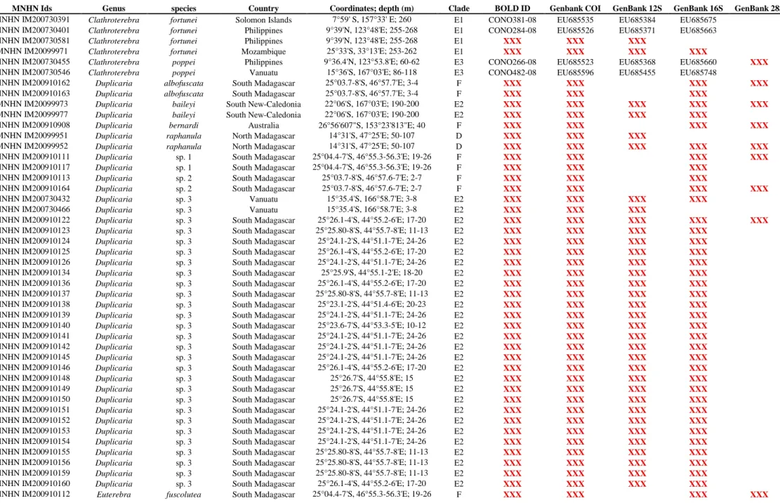
All the material studied herein was collected during several expeditions conducted by the Museum National d’Histoire Naturelle of Paris (MNHN), in partnership with Pro-Natura International (PNI), Instituto Español de Oceanografia (IOE), and Institut de Recherche pour le Développement (IRD), the Natural History Museum of London (NHM), and the Smithonian Tropical Research Institute (STRI) (See Table 1 and acknowledgements). Samples include 406 specimens collected off New Caledonia (4 specimens), Philippine Islands (49), Vanuatu (115), Solomon Islands (12), Australia (4), the Coral Sea (4), Panama (50), Madagascar (87), Mozambique (75), Tahiti (4), New-Zealand (1) and Fiji (1) (Fig. 1). These samples originate from depths ranging from 0 m to ~ 800 m (Table 1). In the field, all sprecimens were specifically fixed for molecular analysis. Shells were kept intact for identification. Vouchers are deposited in MNHN. Taxonomy follows Terryn (2007), with updates in Terryn (Terryn, 2011) (Cinguloterebra synonymized with Triplostephanus, Impages with Hastula, and Acus and
Perirhoe with Oxymeris). Three specimens of the family Turridae (putative sister-group of the
Terebridae – Puillandre et al., 2011), Cochlespiridae (Conoidea) and Conidae (Conoidea) were used as closely related outgroups. Harpa kajiyamai, belonging to another neogastropod family (Harpidae), was used as a distant outgroup to artificially root the tree.
2.2. PCR amplification and DNA sequencing
Total genomic DNA was extracted from muscle tissue using NucleoSpinR 96 Tissues (Macherey-Nagel) and following the manufacturer’s instructions. We amplified fragments of the mitochondrial genes Cytochrome Oxidase I (COI), 16S rRNA and 12S rRNA as well as the nuclear 28S rRNA (Table 2). PCR reactions were performed in 25 µL final volume, containing approximately 3 ng template DNA, 1.5 mM MgCl2, 0.26 mM of each nucleotide, 0.3 µM of each primer, 5% DMSO and 0.75 U of
Taq Polymerase (Qbiogene). Amplification products were generated by an initial denaturation step of 4 min at 94 °C followed by 35 cycles at 94 °C for 40 s, annealing at 50°C for COI, 52°C for 28S, 51°C for 12S rRNA and 16S rRNA for 40 s and by an extension at 72°C for 1 min. PCR products were purified using ExonucleaseI and Phosphatase and sequenced using BigDye Terminator V3.1 kit (Applied biosystem) and the AB3730XL sequencer. All genes were sequenced for both directions to confirm accuracy of each sequence. Chromatograms were edited using CodonCode Aligner version 3.7.1.1. All the sequences were deposited in GenBank and BOLD (Table 1).
2.3. Datasets
Six datasets were analyzed. The first three datasets were analyzed for all taxa listed in Table 1 and consisted of three independent gene analyses performed from COI, 16S and 12S genes. The fourth dataset consisted of a combined data set of COI, 16S, and 12S and is referred to as CD1. To evaluate the robustness of the mitochondrial phylogeny, a fifth dataset corresponding to the nuclear 28S gene set was built, with one representative for most of the species. This reduced dataset was then combined with the three mitochondrial genes and is referred to as CD2.
2.4. Phylogenetic analyses
Sequences were aligned for each gene independently using MUSCLE (Edgar, 2004). The accuracy of automatic alignments was confirmed by eye using BioEdit version 7.0.0.0 (Hall, 1999). Hyper-variable regions of 12S and 16S rRNA genes were excluded from further analyses to avoid ambiguities in the homology hypotheses. Best-fit substitution models were identified for each gene separately and for each combined dataset using Modelgenerator V.85 (Keane et al., 2006). Best-scoring Maximum Likelihood (ML) trees were estimated using RaxML (Stamatakis, 2006) from 100 independant searches each starting from distinct random trees. Robustness of the nodes were assessed using the thorough bootstrapping algorithm (Felsenstein, 1985a) with 1000 replicates. Bayesian Analyses (BA) were performed running two parallel analyses in MrBayes (Huelsenbeck and Ronquist, 2001), consisting each of eight Markov chains of 100,000,000 generations with a sampling frequency of one tree each ten thousand generations. The number of swaps was set to 5, and the chain temperature at 0.02. Convergence of each analysis was evaluated using Tracer 1.4.1 (Rambaut and Drummond, 2007) to check that ESS values were all superior to 200. A consensus tree was then calculated after omitting the first 25% trees as burn-in. For the treatment of combined data using ML and BA, the data were separated into six unlinked partitions: 16S, 12S, 28S and the three codon positions of the COI gene.
Analyses were performed on the Cipres Science Gateway (http://www.phylo.org/portal2), using the RAxML-HPC2 on TG tool for ML and the MrBayes on TG tool for BA.
2.5. Overview of Terebridae anatomy and foregut characters
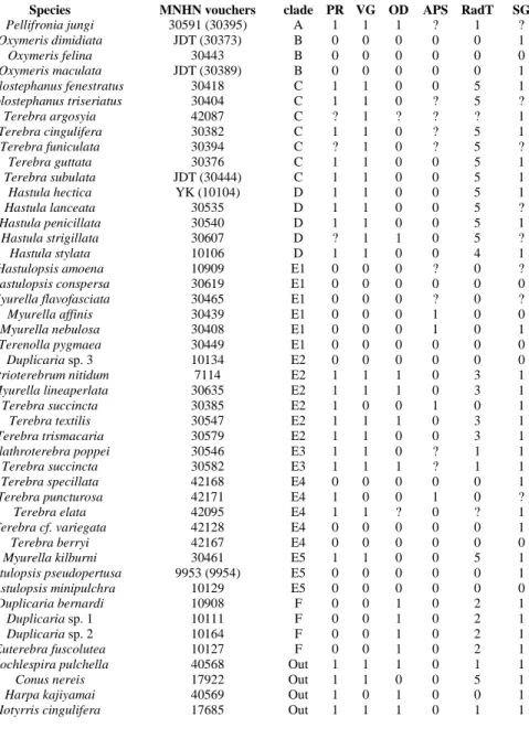
Foregut anatomy was examined by dissecting sequenced specimens. The radulae were cleaned with diluted bleach (1 part of commercially available bleach to 3-4 parts of water), rinsed several times in distilled water, mounted on clear glass cover-slips and air-dried. The cover-slips were glued to stubs, coated with gold and examined by scanning electron microscopy. Terminology previously used for description of the foregut structures in Terebridae was rather inconsistent and confusing (Miller, 1970, 1975, 1979). Here we follow the terminology of Taylor et al. (1993), which is most consistent at the moment and reflects the supposed homologies within the entire Conoidea. Six characters of the foregut were examined and used for tracing evolutionary pathways on the molecular tree (Table 3):
Character 1. Proboscis (PR), very variable in length, from extremely short to very long. In long proboscises, walls often form telescopic folds, while the proboscis can be coiled within the rhynchodaeum. The proboscis contains the buccal tube, i.e., the portion of the alimentrary canal extending between the buccal cavity and the true mouth, which is situated at the distal end of proboscis (Taylor et al., 1993). The buccal tube is absent only in those species where the proboscis is lost. All examined terebrid species possess a more or less long rhynchodeal introvert (also known as labial tube – Miller, 1970). The length of the introvert correlates with the presence of the proboscis: in species without proboscis, the rhynchodeal introvert is much longer than in species with proboscis. The buccal lip is a muscular extension of the anterior walls of the buccal mass, which protrude as a short tube into the lumen of the buccal tube (when a proboscis is present). It was earlier erroneously called “buccal tube” (Holford et al., 2009a; Miller, 1979). A buccal lip is not always present. It is concealed within the proboscis (when there is one) and can be identified usually only on histological sections. When the proboscis is absent, the buccal lip is exposed and protruding into the rhynchodaeum. In species where the proboscis is much reduced (or completely absent) it is sometimes difficult from dissection to distinguish the lip from the reduced proboscis. Usually, even much reduced proboscises have telescopically folded walls, while buccal lip like a straight unfolded tube. The character “buccal lip” was not used in the analysis due to difficulties in its examination in species with a proboscis.
Character 2. Venom gland (VG), sometimes called venom duct, an autapomorphy of Conoidea (Taylor et al., 1993); when present it always has a muscular bulb, also referred to as the venom bulb). The venom gland in Terebridae opens just posterior to the radular sac. Character 3. Odontophore (OD), consisting of subradular cartilages and muscles, usually present in species having a radula with a strong subradular membrane. In Terebridae it can vary from being massive (e.g., Duplicaria bernardii) to being vestigial and hardly recognizable (e.g., Terebra succincta, clade E3).
Character 4. Accessory proboscis structure (APS), an extensible muscular structure that arises from the wall of the rhynchodaeum. It can be branching or club-shaped, distally papillated, or simple, stalk-shaped. A somewhat similar structure, named rhynchodeal outgrowth, is found in other Conoidea – Horaiclavidae and Zemacies (Borsoniidae) (Fedosov and Kantor, 2008).
Character 5. Radula (RadT), which in Terebridae consists only of a pair of marginal teeth per transverse row. Radula was completely lost in several lineages, but when present the marginal teeth exhibit a range of morphological types, and five major types are here recognized: (i) Solid recurved teeth (Fig. 2 F-G) with a broad flatened base, which is attached to the relatively strong subradular membrane. In species with this type of teeth, the radula is short, with only 15-20 rows; (ii) Duplex teeth (Fig. 2 A-C), consisting of a major element (limb), attached to the subradular membrane along most of its length, and an accessory limb, which is the thickened edge of the major element, usually somewhat elevated above the membrane. Here, the radula has about 20-25 rows of teeth; (iii) Flat and simple teeth (Fig. 2 D-E), attached by a narrow base to the subradular membrane. Two, not clearly delimitated, variants - broad triangular (Fig. 2E) and long irregular (Fig. 2D) - are coded as the same radular type in the analysis. The subradular membrane is usually very thin and fragile, and easily tears apart. Radulae with this type of teeth consist of 20 or more rows; (iv)
Semi-enrolled teeth with tooth edges overlaping at the base, forming a loosely Semi-enrolled tube, while
closer to the tip the tooth is trough shape in section. Radulae with this type of teeth are very short, with only about 10 rows; (v) Hypodermic hollow teeth (Fig. 3 A-P), rather similar to the hypodermic teeth present in other Conoidea. Such teeth have a very broad basal opening of the tooth canal, with usually a reflected outward edge of the tooth, forming a collar-like structure; the apical opening can be unarmed or it can have small barb(s) or blade(s). The subradular membrane is usually very thin and vestigial. The number of rows of teeth varies from about 10 (Terebra jenningsi) to about 30 (Hastula hectica and H. penicillata).
Character 6. Salivary glands (SG). These can be paired, but are more often fused, bipartite with paired ducts. In some species, a single gland is present. Accessory salivary gland(s) are present in different species of Terebridae, as well as in some other conoideans. They usually are very small and difficult to find by dissection.
2.6. Evolution of the anatomy
A reduced dataset was built for the 46 species (including the four outgroups) for which anatomical data were available. To minimize the risk of undetected cryptic species, the dissected and sequenced specimens were the same in most cases. However, for Pellifronia jungi and Hastulopsis pseudopertusa (Table 3), the sequencing of the dissected specimens did not work, and a conspecific specimen was used. Four species, Oxymeris dimidiata, O. maculata, Terebra subulata and Hastula hectica, were dissected by YK and John D. Taylor using non-sequenced material, and conspecific specimens were used for sequencing. ML analyses were performed using the method described above. The evolution of the six characters listed in Table 3, and described in the anatomy overview above, was assessed with Mesquite V2.74 (Maddison and Maddison, 2009), using the option ‘‘tracing character history’’ and the parsimony ancestral reconstruction method. The characters PR (proboscis), VG (venom gland), OD (odontophore), and RadT (marginal radular teeth anatomy) were treated as ordered characters (using a stepmatrix), prohibiting some of the transformation sequences, in our case from absent to present, as reapparition of these features is highly unlikely. Other characters were treated as unordered.
3. Results
3.1. Genetic diversity
Of the total of 406 samples of Terebridae used to reconstruct the molecular phylogeny of the family, 389 were sequenced for the COI gene, 400 for the 16S gene and 369 for the 12S gene. For COI, 658 bp were sequenced and no indels were found. After the alignments and the removal of ambiguously-aligned sites, a fragment of 591 and 654 bp in length was obtained for the 16S and 12S genes, respectively. For the 28S gene, a fragment of 761 bp was sequenced for 63 specimens, representing several species in each of the main lineages (see below). For the COI gene, 218 different haplotypes were found, displaying 121 polymorphic sites, 278 parsimony informative sites and a high haplotypic diversity (0.9935). For the 16S gene, 162 different haplotypes were found, displaying 277 polymorphic sites, 235 parsimony informative sites and a haplotypic diversity of 0.9867. For the 12S gene, 164 different haplotypes were found, displaying 412 polymorphic sites, 369 parsimony informative sites and a haplotypic diversity of 0.9866. Representatives of the mitochondiral diversity were also
sequenced for the 28S gene (62 specimens, including 2 outrgoups). Overall, the variability for the 28S gene was less important than for the mitochondrial genes, with 127 polymorphic sites, 94 parsimony informative sites and a haplotypic diversity of 0.9801.
3.2. Phylogenetic analyses: single-gene data sets
Modeltest results indicated that GTR + I + G model was the best-fit model of evolution for the four genes analyzed (COI, 16S, 12S and 28S). For each gene analyzed, no supported conflict was found between the different analyses. In each of the four single gene analyses, the consensus tree showed the Terebridae to be monophyletic; however, the relationships within terebrids were generally poorly resolved, with few well-supported clades. Therefore only the results obtained for the combined datasets CD1 and CD2 are presented.
3.3. Phylogenetic analyses: combined data set 1 (CD1)
Topologies derived from ML analyses of the combined data set 1 (CD1) were congruent with the topology derived from BA analyses. From these combined analyses, the Terebridae were found monophyletic, CD1, Posterior Probabilities PP = 0.99, Bootstraps B = 96 (Fig. 4). Within the Terebridae, the five major clades, Pellifronia, Oxymeris [= Acus], Terebra, Hastula and Myurella (clades A-E, respectively) previously identified in Holford et al. (2009a) were recovered. Each were still strongly supported (PP > 90, B > 70), and the topological relationships among the clades were similar, e.g., clades B-E were grouped together (PP = 0.99, B = 90) (Fig. 4, and see Fig. 2 in Holford et al. (2009a). A sixth clade, hereafter designated as clade F, is novel in the molecular analysis and presented here for the first time. Intra-clade relationships for clades A-F are detailed in Figures 5 and 6, and some shells are illustrated for each clade in Figure 7. Clade F appeared to be the sister group to clades B-E, although the corresponding node is not supported (PP = 0.93, B = 46). It is comprised of six newly-sampled species, four from South Madagascar, one from Australia and one from New-Zealand. The species composition of clade A remained unchanged compared to Holford et al., 2009a and 2009b, still including a single species, and appearing to be the sister group to all the other clades (althgough without statistical support). A newly-sampled species from South Mozambique was added to clade B, now totalling eight species (PP = 0.99, B = 100). Three newly- sequenced species, one from South Madagascar, one from South Mozambique, and one from Philippines and the Solomon Islands, were added to clade C, now comprising nineteen species (PP = 0.99, B = 73). Clade D included eleven species, of which one species, sampled in Madagascar, was new to the taxon set (PP = 1, B = 100). Clade E contained five well-supported subclades (E1-E5), but the relationships among these were in
general poorly resolved. Clade E1 (PP = 1, B = 96) included eleven species of which one, from Vanuatu and Australia, was new to the taxon set. Two newly-sequenced species, one from New Caledonia and one from Vanuatu and South Madagascar, were added to the thirteen species previously included in clade E2 (PP = 1, B = 97). Clade E3 (PP = 0.97, B = 66) included five species of which two, from the Coral Sea and Solomon Islands respectively, were new to the taxon set. Clade E4 (PP = 1, B = 75) was new to the taxon set, with six species from Pacific Panama. Two newly-sampled species from Madagascar were added to clade E5, now comprising eight species (PP = 1, B = 94).
Molecular analyses highlighted several incongruencies at the genus and species levels. With the exception of three genera (Oxymeris – clade B, Pellifronia – clade A and Terenolla – included in clade E1, the two latter represented each by a single species), all the analyzed genera were found to be non-monophyletic. Clade B comprises eight species of the genus Oxymeris. As previously found (Holford et al., 2009a), clade C consists of 6 species of Triplostephanus and 13 of Terebra (s.s.), including Terebra
subulata, the type species of Terebra. Clade D comprises eight species of Hastula and one Duplicaria.
Clade E, the largest clade in terms of number of species, comprises primarily species of the genera
Myurella, Clathroterebra, Terenolla, Hastulopsis, Strioterebrum, and the ‘‘Terebra’’ textilis-group
(Terryn, 2007). However, as shown in Holford et al. (2009a), all these genera (except Terenolla) are polyphyletic, with species of each genus placed in several of the five clades E1-E5. Specifically,
Myurella species were found in E1, E2, E3 and E5, Clathroterebra in E1 and E3, Hastulopsis in E1 and
E5, Strioterebrum in E1 and E2, and species of Terebra (s.s.) are distributed in clades C, E2, E3, E4 and E5. Also, the addition of newly sampled species impacted the generic composition of clade E. For example, clade E2 now includes two species that were attributed to Duplicaria, D. baileyi and a new species D. sp3, and one species currently attributed to Triplostephanus. A newly sampled species, currently attributed to Hastulopsis (H. pseudopertusa), was included in clade E5. The new lineage, clade F, includes both Duplicaria and Euterebra species.
At species level, plumbeum, pertusa, strigilata, succincta and textilis each end up in two distinct clades, revealing cryptic species. Fourteen different lineages (five in the genus Terebra, three in
Strioterebrum, three in Duplicaria, and one each in Myurella, Triplostephanus and Hastula) were not
identified to species and may represent new species. Conversely, two specimens identified as
Triplostephanus cumingii and Terebra punctatostriata (Clade C, Fig. 5) share almost identical
sequences; revealing initial misidentification and/or synonymy of a species in the T. anilis complex.
The combined data set 2 (CD2) included 62 specimens for which at least two mitochondrial genes and the nuclear 28S gene were available. Topologies derived from both ML and BA analyses using CD2 were similar and consistent with the topology derived from analyses of the CD1 data set (Fig. 8). The family Terebridae was confirmed monophyletic (PP = 1, B = 89). The nine clades (A-D, E1, E2, E3, E5 and F) represented in this dataset were also strongly supported, for some of them with PP and/or B superior to the supports obtained in CD1 analysis. Relationships between and within the main clades are generally similar, except for some non-supported ndoes. For example, clade A is sister-group to all the other terebrids in CD1, but in CD2 its position is inverted with clade F.
3.5. Evolution of foregut characters
Reconstruction of the evolution of the proboscis (character 1) clearly demonstrates that it was lost six times in Terebridae: in clades F, B, E1 (all species), and partially in clades E2, E4, and E5 (Fig. 9A). The venom gland (character 2) was lost eight times – in clades F, B, and E1 (all species), and partially in clades E2 (twice), E4 (twice), and E5 (Fig. 9B). In many lineages the odontophore (character 3) is completely absent (including all species having hypodermic marginal radular teeth) (Fig. 9C). Reconstruction of the presence of the odontophore showed that it was lost in most of the clades independently. It is present in clades A and F, and in some species of clades D, E3 and E2. It is vestigial, and hardly discernable in Hastula strigillata, to the extent that its presence was revealed only on serial histological sections (J.D.Taylor, personal communication). It is possible that a rudiment of the odontophore may be present in some other species of Hastula as well. Reconstruction of the presence of accessory proboscis structure (character 4) showed that it appeared independently in clades E1, E2, and E4 (Fig. 9D).
Reconstruction of the presence of the radula and of the morphology of marginal radular teeth (character 5) revealed a complicated evolutionary history of radular transformations (Fig. 9E). The radula was lost several times: in the entire clades B and E1, and in some species of clades E2 and E5. The most parsimonious ancestral state for the Terebridae radular teeth is the duplex type. Duplex teeth are variable in shape: in some species (Terebra succincta, clade E3, and Clathroterebra poppei – Figs. 2 B-C) the limb also has a thickened edge, while in Pellifronia jungi (Fig. 2A) the limb edge is not thickened. Analysis suggests that duplex teeth are the most parsimonious ancestral state for the entire clade E and that flat teeth originated from duplex ones in clade E2. Analysis was not able to resolve a single most parsimonious state for clade D, with duplex and semi-enrolled teeth being equally parsimonious. Solid recurved teeth appeared in the single clade F. Semi-enrolled teeth were found so far in a single of the species examined here, Hastula stylata (Fig. 3Q). Teeth of rather similar shape
were recorded in Hastula bacillus (Taylor and Miller, 1990). Finally, hypodermic teeth appeared independently three times – in clade C, in clade D and in the single species, Myurella kilburni, from clade E5. However, the structure of the hypodermic teeth is slightly different in these three lineages. In the species belonging to clade C (Fig. 3A-G), the teeth are slender, have a constriction at the base, and usually a basal spur, i.e. an anterior projection on the base of the tooth. Another important character for the hypodermic radula of clade C is that the teeth are attached to the subradular membrane only by their bases. In species of clade D (Hastula spp.), the hypodermic teeth are conical, without constriction at the base and without spur. Contrary to the species of clade C, the teeth are attached along most of their length to the subradular membrane. Species in clade D can have a barb or blade at the tip of the tooth. In Hastula hectica the walls of the tooth are penetrated by numerous holes as previously described (Imperial et al., 2007) (Fig. 3J). The only species in clade E5 with hypodermic teeth (Myurella kilburni) has teeth with a peculiar syringe-like shape, with very narrow, attenuated distal end (slightly less than half of tooth length) and broad and probably rather flacid basal part of the tooth. As the specimen examined was badly damaged, it was not possible to examine the radula of the single species of clade E4, Terebra elata, that possesses a venom gland, although the presence of a venom duct was noticed (Holford, personal observation) and the presence of a radula is highly probable.
Although found in several species, such as Triplostephanus fenestratus and Hastula hectica, the presence or absence of the accessory salivary glands cannot be confirmed without histological sections and therefore the character was excluded from the analysis. Reconstruction of the presence and absence of salivary glands (character 6) suggested independent loss in one species of clade B (Oxymeris felina), in most species of clade E1, in one species of clade E5 (Hastulopsis minipulchra) and one species of clade E2 (Duplicaria sp. 3) (Fig. 9F).
4. Discussion
A robust phylogenetic context was used to both clarify the phylogenetic relationships of the Terebridae and to provide a framework to trace the evolution of several anatomical features linked to the venom apparatus, a key innovation of the Conoidea. The molecular phylogeny of the Terebridae presented here was based on an extended dataset compared to the previous large-scale phylogeny of the group (Holford et al., 2009a, 2009b; Puillandre et al., 2011), tripling the number of specimens, doubling the number of species, including fourteen out of the eighteen accepted genera, extending the sampled diversity to the West-Indian Ocean, and including an additional nuclear gene that strengthened the inital phylogeny exclusively based on mitochondrial genes. Analysis of terbrid foregut anatomy for the characters related to the presence of a venom apparatus, namely proboscis, venom gland and radula,
and other characters, such as odontophore, accessory proboscis structure and salivary glands, identified unexpected evolutionary traits within the Terebridae, with implications for the whole superfamily Conoidea. Summarized below are our findings on the taxonmy, venom apparatus evolution, and predators-prey and toxin relationships in the Terebridae.
4.1. Taxonomy
The phylogenetic trees in this analysis confirmed the monophyly of the family Terebridae (Holford et al., 2009a, 2009b) and the existence of five major clades previously identified as Pellifronia, Acus [now Oxymeris], Terebra, Hastula, and Myurella, clades A-E, respectively (Holford et al., 2009a). A novel result for terebrid molecular analysis is the discovery of a new lineage, Clade F, which includes
Euterebra and Duplicaria species, and appears to be the sister group to Clades B-E.
Our results suggest that taxonomic diversity of the family Terebridae is far from being adequately understood. In several cases molecular data suggest the existence of at least two distinct species within what has been identified as a single morphospecies. In three cases (S. plumbeum, H. pertusa and T.
succincta), the two cryptic species identified morphologically as one were collected sympatrically (i.e.
co-occuring in the same region), and sometimes syntopically (i.e. co-occuring at the same sampling station; this is the case for H. pertusa with includes two molecular species sampled at the same station in Santo, Vanuatu). The detection of several new cryptic lineages emphasizes that species diversity in the family Terebridae may be underestimated. Additonally, among the ca. hundred species analyzed in this study, about twenty could not be attributed to a species name according to the taxonomic literature, suggesting that they could represent new species or nominal species currently treated as synonyms.
Increasing the geographic and species diversity of Terebridae analysed in the molecular tree demonstrates that the current genus-level classification of the group is not tenable. Most of the genera recognized in the last working identification guide of the family were found non-monophyletic (10 out of the 12 genera analyzed). For example, the newly sampled genus Duplicaria, represented by six species in our sampling, was found in three distinct clades (D, E2 and F). This was an unanticipated finding since Duplicaria (characterized by a shell axially ribbed, and a well-marked suture doubled on the whorls by an axial sculpture on the subsutural band - Terryn, 2007) is widely accepted in the taxonomy community and was one of the unambiguous genera recognized by Bratcher and Cernhorsky (1987). Similar examples were observed for species of Terebra and Myurella, which were found in five (C, E2-5) and three (E3-5) distinct clades, respectively (see also Clathroterebra, Hastulopsis,
of the shell, used to describe the diversity of terebrids, can be misleading at both genus and species levels, and can lead to an artificial classification of the family.
Despite the extensive sampling efforts deployed to complete the taxonomic coverage, our dataset is still not exhaustive. It covers less than one quarter of the species diversity of the family (with 100 analyzed species out of the ~ 400 currently accepted species, WORMS – www.marinespecies.org), representing 12 out of the 15 currently accepted genera. Further sampling is needed to obtain the missing genera Granuliterebra, Microtrypetes and Pristiterebra. In addition, among the genera analyzed, numerous type-species are not represented. Considering that most terebrid genera are found non-monophyletic, it will also be essential to extend the taxonomic sampling to the numerous synonymised genera. Although further taxonomic investigations are needed to stabilize the classification of the family, the obtained phylogeny provided a robust framework to analyze the evolution of several characters linked to the venom apparatus in the Terebridae.
4.2. Venom apparatus evolution
The apparition of the venom gland and the appearance of the feeding mechanism of Conoidea was the initial key apomorphy of the group (Kantor and Puillandre, in press-a). The unique mechanism of prey envenomation is the most outstanding character of Conoidea and includes use of individual marginal radular teeth (detached from the subradular membrane) at the proboscis tip for stabbing and injecting neurotoxins into prey (Taylor et al., 1993). Teeth of very different morphologies, i.e. not only hypodermic, are used in a similar manner. This was observed directly (e.g., Kohn, 1956) and inferred from serial sectioning of different conoideans (Kantor and Taylor, 1991). Until recently, the Terebridae remained relatively poorly studied anatomically and existing data confirmed a great disparity of anatomy of the foregut, with loss of major organs (including proboscis, venom gland and radula) in many species. Nevertheless, due to the absence of a robust phylogeny, the evolution of the foregut remained largely uncertain, and loss and apparition of novel features were considered anecedotal. Our results illustrate that the evolution of the venom apparatus is not straightforward, as independent and multiple apparitions of key features, together with the loss of various structures of the foregut, appears to be the rule rather than the exception.
Terebridae were always treated as a major independent lineage of Conoidea until the molecular phylogeny of the superfamily (Puillandre et al., 2011), which suggests that Terebridae is a sister group of the family Turridae (s.s.), the component species of which can possess a venom gland, a radula with strong subradular membrane, and have duplex marginal teeth. The discovery of true duplex teeth, and flat teeth, their derivatives, in Terebridae was thus quite unexpected. Prior to our studies only two types
of radula were known in Terebridae – solid recurved teeth and hypodermic teeth. Duplex teeth appeared to be the ancestral state for entire family Terebridae and this is consistent with the Turridae and Terebridae being sister-groups. Clade A, represented at the moment only by Pellifronia jungi and likely the sister clade to all other terebrids, has similar anatomy and radula to that of Turridae.
The reduction and losses of foregut characters in many lineages of the Terebridae are not casual and have a functional explanation. For example, all the species possessing a venom gland have a radula and proboscis. This is explained by the peculiarities of conoidean feeding mechanism, where envenomation of the prey requires the aid of the tooth gripped at the proboscis tip and used for stabbing the prey, or channelling the toxins through the internal lumen of hypodermic teeth. Currently, feeding of radulate terebrids was observed only in different Hastula and Terebra species with hypodermic radular teeth (Marcus and Marcus, 1960; Miller, 1970, 1979; Taylor, 1990; Taylor and Miller, 1990). The observations show that these species feed similarly to other conoideans, with the use of marginal teeth at the proboscis tip. The prey reported were various sedentary polychaetes, mostly spionids. A characteristic feature of terebrid feeding is the well-developed rhynchostomal introvert, which is playing an active role in capturing and engulfing the prey.
Analysis of the anatomical characters revealed that hypodermic teeth originated three times independently in Terebridae, in clades C, D, and in a single species from clade E5, Myurella kilburni. As detailed in the results section, the hypodermic teeth of these three groups appear in fact to be rather different (Fig. 3). Independent apparitions of hypodermic teeth suggest increasing the effectiveness of prey envenomation. Very interesting peculiarity was found in Hastula cinerea and H. hectica - both in clade D - where in most of the specimens examined, a tooth was held at the proboscis tip even when the species was not feeding, concealed within the proboscis with its base resting on the large sphincter (Marcus and Marcus, 1960; Imperial et al., 2007). This can be explained by the presence of a relatively strong subradular membrane and tough attachment of the teeth to the membrane. In Hastula, because the teeth in the radular cecum are still attached to the membrane, they cannot be immediately used for stabbing prey when required. In the process of radular growth, the oldest part of the membrane, situated in the radular cecum, is permanently destroyed and the teeth become dislodged. When the tooth is separated from the membrane, it is transferred to the proboscis tip, where it is presumably held until it is used. This is also assumed for members of the other families of “turrids” that have a strong subradular membrane. In most turrid specimens examined, there was a tooth at the proboscis tip held by the sphincter(s) (Kantor and Taylor, 1991).
Although nothing is known on the feeding of species with duplex/flat teeth, it is reasonable to suppose that they are used on the proboscis tip in a manner similar to other conoideans with
non-hypodermic teeth. In this respect it was interesting to find in Terebra textilis at the proboscis tip flat teeth very similar to those of Terebra trismacaria (Fig. 2D). A group of four teeth attached to the subradular membrane was found in the buccal tube somewhat posterior to the proboscis tip. It is obvious that in this case the teeth cannot be used separately for stabbing the prey, but the mechanism of transport of the teeth from radular sac to the proboscis tip persists in this species. A probable explanation in this case represents an intermediate stage of reduction of radulae and transition to feeding without use of marginal teeth at the proboscis tip.
An odontophore is present in species that have a more or less strong subradular membrane. It is large and powerful in species of clade F, Duplicaria and Euterebra, which lack proboscis and venom gland and therefore do not utilize teeth for stabbing and envenomation of the prey. A well-developed odontophore suggests that the radula is functioning as a whole organ only, probably for transferring the prey from rhynchodaeum to oesophagus. There is no observation on feeding of species of this clade and diet is known for only one species with similar anatomy, Terebra nassoides, feeding on capitellid polychaetes (Taylor, 1990). Similarly to species with hypodermic radulae, an active role of the introvert in prey capture was also shown in Terebra gouldi, a species lacking venom apparatus, radula and proboscis. and that preys on the enteropneust Ptychodera flava, which is swallowed alive.
While reduction of the venom gland provides economy of energy that is otherwise used for producing toxins and constant formation of the radula, the rhynchostomal introvert, which is present and well-developed in all terebrids, may explain the numerous independent losses of the venom gland and associated organs. With the rhynchostomal introvert present, feeding becomes possible without stabbing and envenomation of the prey. In addition, the proboscis also becomes unnecessary, as its primary function, gripping the tooth, does not exist any more. The muscular buccal lip, which is well developed in radular-less species, serves for transferring the swallowed prey further into oesophagus. Although very little is known about diet of terebrids with such foregut anatomy, Miller (1975) suggested that they feed on different hemichordates. The family Raphitomidae is the only other taxon of Conoidea that possesses a developed rhynchostomal introvert. In that family numerous independent reductions and losses of the venom gland and radula were hypothesized (Kantor and Taylor, 2002). It was also suggested that these reductions were connected with the role of introvert in prey capture.
4.3. Predator-prey and toxins
Numerous terebrid lineages have lost the venom apparatus, and by contrast the lineages that kept it each developed novel anatomical features, such as hypodermic marginal radular teeth. The components of the venom apparatus, radular, venom duct, venom bulb, and proboscis, were thought to be so
complicated that they certainly evolved once or twice. However, the Terebridae obviously acquired or lost similar structure several times, with a resulting anatomy sometimes convergent with that of other conoideans. Our finding of a triple origin, in Terebridae alone, of the hypodermic teeth, associated with reduction of the odontophore, is remarkable. Additionally, the detailed anatomy demonstrates not only different origins of the teeth but also suggests differences in functional use. Analysis of radular evolution in entire Conoidea showed, that besides terebrids hypodermic teeth appeared only once in a clade, that unite families Conidae, Conorbidae, Borsoniidae, Clathurellidae, Mitromorphidae, Mangeliidae and Raphitomidae (Kantor and Puillandre, in press-b).
The diversity of foregut anatomy in the single family Terebridae is as large as in the whole superfamily Conoidea, which includes 14 other families. For example, all major types of conoidean radular marginal teeth were recorded in the Terebridae. From prototypic duplex teeth they evolved solid recurved teeth (these appeared independently in some Pseudomelatomidae), flat (appeared from
duplex in some Drilliidae) and hypodermic (appeared independently in common ancestor of a major
clade of Conoidea) (Bouchet et al., 2011; Kantor and Puillandre, in press-a). Moreover, the flat triangular teeth of some Terebridae are unique among Conoidea. Our overview of the foregut anatomy has revealed an inordinate diversity of features in the family Terebridae. These results suggest that predator-prey relationships have played an important role in the evolutionary history of Terebridae. Indeed, repeated innovations in the foregut anatomy of terebrids suggest that they adapted to different diets (e.g., deposit-feeding or carnivorous polychaetes). To date, this hypothesis remains untested as the preys of most of the analyzed terebrid species are unknown. This could be analysed by direct observation, but indirect approaches, such as DNA-barcoding of the gut contents (Garros et al., 2008; Oliverio et al., 2009) or analysis of stable isotopes composition (Fujikura et al., 2009) could also be useful.
Based on the hypothesis that the diversity of foregut structures in the Terebridae is linked to the diversity of feeding types and preys, it could also be argued that the species diversity of the Terebridae could be linked to the prey diversity, and thus to foregut anatomy. However, our results also show that several species may share an apparently identical foregut structure, suggesting that the diversity of the foregut and the prey are not the only factor at the origin of the species diversity in the Terebridae, e.g., reduced dispersion abilities and geographical isolation (Bouchet, 1981; Castelin et al., 2010; Cunha et al., 2005; Cunha et al., 2008; Duda and Palumbi, 1999; Meyer et al., 2005) or differential selection by abiotic factors such as depth (Chase et al., 1998; Quattro et al., 2001; Zardus et al., 2006) and other features of the prey-capture system should be investigated. Given the rate of evolution of the conopeptides in cone snails, it can be proposed that various Terebridae species evolved different toxins
as an answer, or a consequence, of prey adapation. Integrative approaches will be employed to complete the phylogeny of the Terebridae, identify their respective preys, and compare their foregut anatomy and the toxins they produce. An integrated approach is not only a promising way to identify the factors that led to the diversification of the Terebridae and potentially the (co-)evolution of their prey, but is also a step forward in the characterization of novel terebrid toxins with new functions and potentially new therapeutic applications. Terebrids have clearly evolved different responses to the costs and benefits of having a venom apparatus under varying conditions. Using, for example, phylogenetic independent contrasts (Felsenstein, 1985b), the large-scale phylogeny presented here could assist in analysing the potential correlation between the anatomical innovations developed by the Terebridae and various biotic and abiotic parameters.
ACKNOWLEDGMENTS
In addition to the sources enumerated in Holford et al. (2009b), a large part of the material used in the present paper was collected in Mozambique and Madagascar in 2009-2010 during expeditions Mainbaza, Miriky and Atimo Vatae, a cluster of expeditions funded by the Total Foundation, Prince Albert II of Monaco Foundation, and Stavros Niarchos Foundation, and conducted by MNHN and Pro-Natura International (PNI) as part of their "Our Planet Reviewed" programme. The authors also thank Felix Rodriquez, Edwin Diaz, Trinidad Pardo, Moises Bernal, and the crew of the RV-Urraca with collection efforts in Panama. This work was supported by a grant from The Alfred P. Sloan Foundation; by the “Consortium National de Recherche en Génomique” and the “Service de Systématique Moléculaire” (UMS 2700 CNRS-MNHN) as part of agreement 2005/67 between Genoscope and MNHN for the project “Macrophylogeny of life” directed by Guillaume Lecointre; and by grant RFBR 11-04-01284 “Evolution of digestive system of carnivorous gastropods: testing of morphologically-based hypotheses by molecular data” (PI Yu. Kantor). NP was partly funded by CONCO, the cone snail genome project for health, funded by the European Commission: LIFESCIHEALTH-6 Integrated Project LSHB-CT-2007-037592. The phylogenetic analyses were performed on the Cipres Science Gateway. The authors want to thank Dr. John Taylor for sharing unpublished information on terebrid anatomy and providing material from Australia, and Barbara Buge and José Utge for processing and curation of the molecular collection.
Barlow, A., Pook, C.E., Harrison, R.A., Wüster, W., 2009. Coevolution of diet and prey-specific venom activity supports the role of selection in snake venom evolution. Proc. R. Soc. Biol. Sci. Ser. B 276, 2443-2449.
Barnosky, A.D., 2001. Distinguishing the effects of the Red Queen and Court Jester on Miocene mammal evolution in the northern Rocky Mountains. J. Vertebr. Paleontol. 21, 172-185.
Berenbaum, M., 1983. Coumarins and caterpillars: a case for coevolution. Evolution 37, 163-179. Berenbaum, M., Feeny, P., 1981. Toxicity of angular furanocoumarins to swallowtail butterflies:
escalation in a coevolutionary arms race? Science 212, 927-929.
Biardi, J.E., Chien, D.C., Coss, R.G., 2005. California ground squirrel (Spermophilus beecheyi) defenses against rattlesnake venom digestive and hemostatic toxins. J. Chem. Ecol. 31, 2501-2518.
Bouchet, P., 1981. Evolution of larval development in eastern Atlantic Terebridae (Gastropoda), Neogene to Recent. Malacologia 21, 363-369.
Bouchet, P., Kantor, Y.I., Sysoev, A., Puillandre, N., 2011. A new operational classification of the Conoidea (Gastropoda). J. Molluscan Stud. 77, 273-308.
Bratcher, T., Cernohorsky, W.O., 1987. Living terebras of the world: a monograph of the recent Terebridae of the world. American Malacologists, Melbourne.
Butler, M.S., 2008. Natural products to drugs: natural product-derived compounds in clinical trials. Nat. Prod. Rep. 25, 475-516.
Casewell, N., Harrison, R., Wüster, W., Wagstaff, S., 2009. Comparative venom gland transcriptome surveys of the saw-scaled vipers (Viperidae: Echis) reveal substantial intra-family gene diversity and novel venom transcripts. BMC Genomics 10, 564-576.
Castelin, M., Lambourdiere, J., Boisselier, C., Lozouet, P., Couloux, A., Cruaud, C., Samadi, S., 2010. Hidden diversity and endemism on seamounts: focus on poorly dispersive neogastropods. Biol. J. Linn. Soc. 100, 420-438.
Chase, M.R., Etter, R.J., Rex, M.A., Quattro, J.M., 1998. Bathymetric patterns of genetic variation in a deep-sea protobranch bivalve, Deminucula atacellana. Mar. Biol. 131, 301-308.
Chin, Y.W., Balunas, M.J., Chai, H.B., Kinghorn, A.D., 2006. Drug discovery from natural sources. The AAPS Journal 8, 239-253.
Cunha, R.L., Castilho, R., R¸ber, L., Zardoya, R., 2005. Patterns of cladogenesis in the venomous marine gastropod genus Conus from the Cape Verde Islands. Syst. Biol. 54, 634-650.
Cunha, R.L., Tenorio, M.J., Afonso, C., Castilho, R., Zardoya, R., 2008. Replaying the tape: recurring biogeographical patterns in Cape Verde Conus after 12 million years. Mol. Ecol. 17, 885-901.
Daltry, J.C., Wüster, W., Thorpe, R.S., 1996. Diet and snake venom evolution. Nature 379, 537-540. Duda, T.F., 2008. Differentiation of venoms of predatory marine gastropods: divergence of orthologous
toxin genes of closely related Conus species with different dietary specializations. J. Mol. Evol. 67, 315-321.
Duda, T.F., Jr., Palumbi, S.R., 1999. Molecular genetics of ecological diversification: duplication and rapid evolution of toxin genes of the venomous gastropod Conus. Proc. Natl. Acad. Sci. U. S. A. 96, 6820-6823.
Edgar, R.C., 2004. MUSCLE: multiple sequence alignment with high accuracy and high throughput. Nucleic Acids Research 32, 1792-1797.
Ehrlich, P.R., Raven, P.H., 1964. Butterflies and plants: a study in coevolution. Evolution 18, 586-608. Farrell, B.D., Dussourd, D.E., Mitter, C., 1991. Escalation of plant defense: do latex and resin canals
spur plant diversification? Am. Nat. 138, 881-900.
Fedosov, A., Kantor, Y., 2008. Toxoglossan gastropods of the subfamily Crassispirinae (Turridae) lacking a radula, and a discussion of the status of the subfamily Zemaciinae. J. Molluscan Stud. 74, 27.
Felsenstein, J., 1985a. Confidence Limits on Phylogenies: An Approach Using the Bootstrap. Evolution 39, 783-791.
Felsenstein, J., 1985b. Phylogenies and the comparative method. Am. Nat. 125, 1-15.
Folmer, O., Black, M., Hoeh, W., Lutz, R., Vrijenhoek, R., 1994. DNA primers for amplification of mitochondrial cytochrome c oxidase subunit I from diverse metazoan invertebrates. Mol. Mar. Biol. Biotechnol. 3, 294-299.
Fowler, M., 1983. Plant poisoning in free-living wild animals: a review. J. Wildl. Dis. 19, 34-43.
Fry, B.G., Wüster, W., Kini, R.M., Brusic, V., Khan, A., Venkataraman, D., Rooney, A.P., 2003. Molecular evolution and phylogeny of elapid snake venom three-finger toxins. J. Mol. Evol. 57, 110-129.
Fujikura, K., Sasaki, T., Yamanaka, T., Yoshida, T., 2009. Turrids whelk, Phymorhynchus buccinoides feeds on Bathymodiolus mussels at a seep site in Sagami Bay, Japan. Plank. Benth. Res. 4, 23-30.
Garros, C., Ngugi, N., Githeko, A.E., Tuno, N., Yan, G., 2008. Gut content identification of larvae of the Anopheles gambiae complex in western Kenya using a barcoding approach. Mol. Ecol. Res. 8, 512-518.
Hall, T.A., 1999. BioEdit: a user-friendly biological sequence alignment editor and analysis program for Windows 95/98/NT. Nucl. Acid. S. 41, 95-98.
Heard, S.B., Hauser, D.L., 1995. Key evolutionary innovations and their ecological mechanisms. Hist. Biol. 10, 151-173.
Heatwole, H., Poran, N.S., 1995. Resistances of sympatric and allopatric eels to sea snake venoms. Copeia 1, 136-147.
Hodges, S.A., Arnold, M.L., 1995. Spurring plant diversification: Are floral nectar spurs a key innovation? Proc. Biol. Sci., 343-348.
Holford, M., Puillandre, N., Modica, M., Watkins, M., Collin, R., Bermingham, E., Olivera, B., 2009a. Correlating molecular phylogeny with venom apparatus occurrence in Panamic auger snails (Terebridae). PLoS ONE 4, e7667.
Holford, M., Puillandre, N., Terryn, Y., Cruaud, C., Olivera, B., Bouchet, P., 2009b. Evolution of the Toxoglossa Venom Apparatus as Inferred by Molecular Phylogeny of the Terebridae. Mol. Biol. Evol. 26, 15-25.
Hong, J., 2011. Role of natural product diversity in chemical biology. Curr. Opin. Chem. Biol. 15, 350-354.
Huelsenbeck, J.P., Ronquist, F., 2001. MrBayes: a program for the Bayesian inference of phylogeny. Bioinformatics 17, 754–755.
Imperial, J.S., Kantor, Y., Watkins, M., Heralde Iii, F.M., Stevenson, B., Chen, P., Hansson, K., Stenflo, J., Ownby, J.P., Bouchet, P., 2007. Venomous auger snail Hastula (Impages) hectica (Linnaeus, 1758): molecular phylogeny, foregut anatomy and comparative toxinology. J. Exp. Zool. B Mol. Dev. Evol. 308, 744-756.
Jovelin, R., Justine, J.-L., 2001. Phylogenetic relationships within the Polyopisthocotylean monogeneans (Plathyhelminthes) inferred from partial 28S rDNA sequences. Int. J. Parasitol. 31, 393-401.
Kantor, Y., Puillandre, N., in press-a. Evolution of the radular apparatus in Conoidea (Gastropoda: Neogastropoda) as inferred from a molecular phylogeny. Malacologia.
Kantor, Y.I., Puillandre, N., in press-b. Evolution of the radular apparatus in Conoidea (Gastropoda: Neogastropoda) as inferred from a molecular phylogeny. Malacologia.
Kantor, Y.I., Taylor, J.D., 1991. Evolution of the toxoglossan feeding mechanism: new information on the use of the radula. J. Molluscan Stud. 57, 129.
Kantor, Y.I., Taylor, J.D., 2002. Foregut anatomy and relationships of raphitomine gastropods (Gastropoda: Conoidea: Raphitominae). in: Oliverio, M., Chemello, R. (Eds.), Systematics, phylogeny and biology of the Neogastropoda. Bollettino Malacologico, Roma, pp. 161-174. Keane, T., Creevey, C., Pentony, M., Naughton, T., Mclnerney, J., 2006. Assessment of methods for
amino acid matrix selection and their use on empirical data shows that ad hoc assumptions for choice of matrix are not justified. BMC Evol. Biol. 6, 29.
Kohn, A.J., 1956. Piscivorous gastropods of the genus Conus. Proc. Natl. Acad. Sci. U. S. A. 42, 168-171.
Kordis, D., Gubensek, F., 2000. Adaptive evolution of animal toxin multigene families. Gene 261, 43-52.
Kozminsky-Atias, A., Bar-Shalom, A., Mishmar, D., Zilberberg, N., 2008. Assembling an arsenal, the scorpion way. BMC Evol. Biol. 8.
Laycock, W., 1978. Coevolution of poisonous plants and large herbivores on rangelands. J. Range Manag. 31, 335-342.
Lynch, V., 2007. Inventing an arsenal: adaptive evolution and neofunctionalization of snake venom phospholipase A2 genes. BMC Evol. Biol. 7, 2.
Maddison, W.P., Maddison, D., 2009. Mesquite: a modular system for evolutionary analysis. Version 2.74 http://mesquiteproject.org/. 27 Sept 2011.
Marcus, E., Marcus, E., 1960. On Hastula cinerea. Boletim da Faculdade de Filosofía Ciencias e Letras. Universidade de Sao Paulo (Zoologia), 25-66.
Meyer, C.P., Geller, J.B., Paulay, G., 2005. Fine scale endemism on coral reefs: archipelagic differentiation in Turbinid gastropods. Evolution 59, 113-125.
Miller, B., 1970. Studies on the biology of Indo-Pacific Terebra (Ph. D. dissertation). University of New Hampshire, Durham.
Miller, B.A., 1975. The biology of Terebra gouldi Deshayes, 1859, and a discussion of life history similarities among other terebrids of similar proboscis type. Pac. Sci. 29, 227-241.
Miller, B.A., 1979. The biology of Hastula inconstans (Hinds, 1844) and a discussion of life history similarities among other Hastulas of similar proboscis type. Pac. Sci. 33, 289-306.
Modica, M.V., Holford, M., 2010. The Neogastropoda: evolutionary innovations of predatory marine snails with remarkable pharmacological potential. in: Pontarotti, P. (Ed.), Evolutionary biology - concepts, molecular and morphological evolution. Springer, Heidelberg, pp. 249-270.
Newman, D.J., Cragg, G.M., 2007. Natural products as sources of new drugs over the last 25 years. J. Nat. Prod. 70, 461-477.
Oliverio, M., Barco, A., Modica, M., Richter, A., Mariottini, P., 2009. Ecological barcoding of corallivory by second internal transcribed spacer sequences: hosts of coralliophiline gastropods detected by the cnidarian DNA in their stomach. Mol. Ecol. Res. 9, 94-103.
Palumbi, S.R., 1996. Nucleic acids II: the polymerase chain reaction. in: Hillis, D.M., Mable, B.K., Moritz, C. (Eds.), Molecular systematics. Sinauer Associates., Sunderland, pp. 205-247.
Puillandre, N., Holford, M., 2010. The Terebridae and teretoxins: combining phylogeny and anatomy for concerted discovery of bioactive compounds. BMC Chem. Biol. 10, 7.
Puillandre, N., Kantor, Y.I., Sysoev, A., Couloux, A., Meyer, C., Rawlings, T., Todd, J., Bouchet, P., 2011. The dragon tamed? A molecular phylogeny of the Conoidea (Gastropoda). J. Molluscan Stud. 77, 259-272.
Puillandre, N., Samadi, S., Boisselier, M.C., Sysoev, A.V., Kantor, Y.I., Cruaud, C., Couloux, A., Bouchet, P., 2008. Starting to unravel the toxoglossan knot: Molecular phylogeny of the “turrids”(Neogastropoda: Conoidea). Mol. Phylogenet. Evol. 47, 1122-1134.
Puillandre, N., Sysoev, A., Olivera, B., Couloux, A., Bouchet, P., 2010. Loss of planktotrophy, fragmentation and speciation: the deep-water gastropod genus Bathytoma (Gastropoda, Conoidea) in the western Pacific. Syst. Biodivers. 8, 371-394.
Quattro, Chase, Rex, Greig, Etter, 2001. Extreme mitochondrial DNA divergence within populations of the deep-sea gastropod Frigidoalvania brychia. Mar. Biol. 139, 1107-1113.
Rambaut, A., Drummond, A.J., 2007. Tracer. Version 1.4. http://beast.bio.ed.ac.uk/Tracer, 27 Sept 2011.
Sanderson, M.J., Donoghue, M.J., 1994. Shifts in diversification rate with the origin of angiosperms. Science 264, 1590-1593.
Saslis-Lagoudakis, C.H., Klitgaard, B.B., Forest, F., Francis, L., Savolainen, V., Williamson, E.M., Hawkins, J.A., 2011. The use of phylogeny to interpret cross-cultural patterns in plant use and guide medicinal plant discovery: an example from Pterocarpus (Leguminosae). PloS One 6, e22275.
Stamatakis, A., 2006. RAxML-VI-HPC: maximum likelihood-based phylogenetic analyses with thousands of taxa and mixed models. Bioinformatics 22, 2688-2690.
Taylor, J., 1990. The anatomy of the foregut and relationships in the Terebridae. Malacologia 32, 19-34. Taylor, J., Miller, J., 1990. A new type of gastropod proboscis: The foregut of Hastula bacillus
(Gastropoda: Terebridae). J. Zool. 220, 603-617.
Taylor, J.D., Kantor, Y.I., Sysoev, A.V., 1993. Foregut anatomy, feeding mechanisms, relationships and classification of the Conoidea (= Toxoglossa)(Gastropoda). Bull. Natl. Hist. Mus. Zool. Ser. 59, 125-170.
Terryn, Y., 2007. A collectors guide to recent Terebridae (Mollusca: Neogastropoda). ConchBooks/Natural Art, Hackenheim.
Terryn, Y., 2011. Family Terebridae Mörch, 1852. in: Severns, M. (Ed.), Shells of the Hawaiian Islands. The sea shells. Conchbooks, Hackenheim, pp. 370-381.
Terryn, Y., Holford, M., 2008. The Terebridae of the Vanuatu archipelago with a revision of the genus
Granuliterebra Oyama 1961. Visaya Supplement 3, 6-118.
Vermeij, G.J., 1993. Evolution and escalation: an ecological history of life. Princeton University Press, Princeton.
Williams, S.T., Ozawa, T., 2006. Molecular phylogeny suggests polyphyly of both the turban shells (family Turbinidae) and the superfamily Trochoidea (Mollusca: Vetigastropoda). Mol. Phylogenet. Evol. 39, 33-51.
Wüster, W., Daltry, J.C., Thorpe, R.S., 1999. Can diet explain intraspecific venom variation? Reply to Sasa. Toxicon 37, 253-258.
Zangerl, A., Stanley, M., Berenbaum, M., 2008. Selection for chemical trait remixing in an invasive weed after reassociation with a coevolved specialist. Proc. Natl. Acad. Sci. 105, 4547-4552. Zardus, J.D., Etter, R.J., Chase, M.R., Rex, M.A., Boyle, E.E., 2006. Bathymetric and geographic
population structure in the pan Atlantic deep sea bivalve Deminucula atacellana (Schenck, 1939). Mol. Ecol. 15, 639-651.