HAL Id: inserm-00782581
https://www.hal.inserm.fr/inserm-00782581
Submitted on 30 Jan 2013HAL is a multi-disciplinary open access archive for the deposit and dissemination of sci-entific research documents, whether they are pub-lished or not. The documents may come from teaching and research institutions in France or abroad, or from public or private research centers.
L’archive ouverte pluridisciplinaire HAL, est destinée au dépôt et à la diffusion de documents scientifiques de niveau recherche, publiés ou non, émanant des établissements d’enseignement et de recherche français ou étrangers, des laboratoires publics ou privés.
cognitive decline, and dementia.
Marie-Laure Ancelin, Isabelle Carrière, Catherine Helmer, Olivier Rouaud,
Florence Pasquier, Claudine Berr, Isabelle Chaudieu, Karen Ritchie
To cite this version:
Marie-Laure Ancelin, Isabelle Carrière, Catherine Helmer, Olivier Rouaud, Florence Pasquier, et al.. Steroid and nonsteroidal anti-inflammatory drugs, cognitive decline, and dementia.. Neurobiology of Aging, Elsevier, 2012, 33 (9), pp.2082-90. �10.1016/j.neurobiolaging.2011.09.038�. �inserm-00782581�
Neurobiology of Aging, in press
Steroid and nonsteroidal anti-inflammatory drugs, cognitive decline, and dementia
Marie-Laure Ancelin, PhD.a,b,§,*, Isabelle Carrière, PhD.a,b,§, Catherine Helmer, PhD.c, Olivier
Rouaud, MDd, Florence Pasquier, MDe, Claudine Berr, PhD.a,b, Isabelle Chaudieu, PhD.a,b,
Karen Ritchie, PhD.a,b,f.
a
Inserm, U1061, Montpellier, F-34093 France
b
Université Montpellier 1, U1061, Montpellier, France
c
Inserm, U897, Université Bordeaux 2, Bordeaux, France
d
Centre Mémoire Ressources et Recherche, Centre Hospitalier Universitaire, Dijon, France
e
Department of Neurology, Centre Hospitalier Universitaire, Lille, France
f
Faculty of Medicine, Imperial College, London, W12 0NN, U.K.
*
Corresponding author at: Inserm U1061, Hopital La Colombiere, 39, avenue C. Flahault, BP
34493, 34093 Montpellier Cedex 5, France
E-mail address: marie-laure.ancelin@inserm.fr (M.L. Ancelin)
Tel: +33 499 614 562; Fax: +33 499 614 579
§ Joint first authors
marie-laure.ancelin@inserm.fr (M.L.A.); isabelle.carriere@inserm.fr (I.C.);
catherine.helmer@isped.u-bordeaux2.fr (C.H.); olivier.rouaud@chu-dijon.fr (O.R.);
florence.pasquier@chru-lille.fr (F.P.); claudine.berr@inserm.fr (C.B.);
isabelle.chaudieu@inserm.fr (I.Ch.); karen.ritchie@inserm.fr (K.R.)
Paper: 3345 words Abstract: 180 words Title character count: 81 Tables: 5 Figures: 0 Supplementary data: 2 Tables
Abstract
The aim of this study was to evaluate the effects of anti-inflammatory intake on cognitive
function in 7234 community-dwelling elderly persons. Cognitive performance, clinical diagnosis
of dementia, and anti-inflammatory use were evaluated at baseline, and 2, 4, and 7 years later.
Multivariate logistic regression analyses were adjusted for sociodemographic, behavioral,
physical and mental health variables and genetic vulnerability (APOE4). Elderly women taking inhaled corticosteroids were at increased risk for cognitive decline over 7 years in executive
functioning (OR=1.76, 95%CI=1.14-2.71, p=0.04); the effect being increased after continuous
use (OR=3.15, 95%CI=1.29-7.68, p=0.01) and not found after discontinuation of treatment. In
men, no significant associations were observed. Corticosteroid use was not significantly
associated with an increase risk of incident dementia over 7 years. Nonsteroidal
anti-inflammatory drug use was not significantly associated with either dementia incidence or
cognitive decline in both sexes. The association may be related to hypothalamic-pituitary-adrenal
corticotropic axis dysfunctioning rather than a direct anti-inflammatory mechanism. Long term
use of inhaled corticosteroids may constitute a form of reversible cognitive disorder in elderly
women. Physicians should check this possibility before assuming neurodegenerative changes.
Key-words: Corticosteroids; Nonsteroidal anti-inflammatory drugs; Cognitive aging;
1. Introduction
In the absence of effective treatment for neuro-degenerative disorders, research has
focused on the identification of modifiable risk factors to delay cognitive decline and prolong
autonomy (Ritchie et al., 2010). There is some evidence that corticosteroids (CS) could be
associated with reversible cognitive dysfunction.; both experimental and epidemiological
research showing that dysregulation of the hypothalamic-pituitary-adrenal (HPA) corticotropic
axis, a major component of the stress response system, may lead to, or accelerate hippocampal
impairment (Belanoff et al., 2001; Conrad and Bimonte-Nelson, 2010; Lupien et al., 2007). This
may be especially important in the ageing brain, more vulnerable to stress effects and subject to
decrements in cognitive performance due to multiple causes.
Several studies in the elderly have demonstrated a potentially reversible link between
elevated endogenous cortisol levels and decline in memory and frontal-executive abilities
(Greendale et al., 2000; O'Hara et al., 2007; Seeman et al., 1997; Li et al., 2006; Beluche et al.,
2010; Egeland et al., 2005) and dementia (Umegaki et al., 2000; Rasmuson et al., 2002;
Csernansky et al., 2006). Associations between short-term CS administration and cognitive
impairment have been reported in small experimental and clinical studies in adults (Wolkowitz et
al., 2009; Newcomer et al., 1999; Kirschbaum et al., 1996; Lupien et al., 2007; Young et al.,
1999; de Quervain et al., 2003; Keenan et al., 1996). The effect of chronic CS administration
has, however, never been examined in elderly populations who have high rates of both
prescribed and over the counter drug use (Hilmer et al., 2007).
Previous studies have failed to take into account the potential confounding effects on
cognitive decline of pathologies associated with CS use, e.g. chronic pain and respiratory
disorders, or the impact of CS administration mode, oral administration being generally
associated with slower absorption and lower biologically active circulating metabolites than
inhaled CS. Surprisingly, genetic vulnerability has also not been considered despite a possible
2008). Finally, gender differences have not been examined although they have been reported in
relation to both stress response and association between cortisol levels and cognitive decline and
neural activity (Otte et al., 2005; Sauro et al., 2003; Seeman et al., 1997; Wang et al., 2007;
Beluche et al., 2010) as well as in risk profiles for cognitive impairment and progression to
dementia (Artero et al., 2008).
Thus, while there is accumulating evidence to suggest that CS treatment may increase risk
of cognitive decline and dementia in the elderly, this hypothesis remains to be tested within a
large study able to take into account multiple competing causes of cognitive decline. This
prospective study aims to examine the relationship between CS use and cognitive decline and
dementia onset in community-dwelling elderly taking into account gender, genetic vulnerability,
and reasons for CS use. The cognitive effects of other nonsteroidal anti-inflammatory drugs
(NSAID) were also examined.
2. Methods
2.1. Study population
Subjects were recruited as part of a multi-site cohort study of community-dwelling persons
aged 65 years and over from the electoral rolls of three French cities between 1999 and 2001
(The 3C Study Group, 2003). The study protocol was approved by the Ethical Committee of the
University-Hospital of Bicêtre (France). Written informed consent was obtained from each
participant. Participants were administered standardized questionnaires by trained staff and
underwent clinical examinations at baseline and at each 2, 4, and 7year follow-up. Of the 9080
dementia-free participants included, 629 died having only one cognitive evaluation at baseline,
704 were alive at the end of the follow-up but did not have repeated cognitive testing, and 513
had missing data for at least one adjustment variable. The present analyses were conducted on
7234 subjects. The mean (SD) age was 73.6 (5.3) for men and 73.8 (5.2) for women. The
were significantly older, with lower education levels, and worse physical and mental health,
lower baseline cognitive scores, and were more frequently CS users.
2.2. Cognitive measures and dementia
The Isaacs Set Test (Isaacs and Kennie, 1973) provided a measure of verbal fluency or
semantic access. The Benton Visual Retention Test (Benton, 1965) assessed visual memory and
the Trail Making Tests (TMT) A and B psychomotor speed and executive function respectively
(Reitan, 1965). The Mini Mental State Examination (MMSE) was used as a global measure of
cognitive function (Folstein et al., 1975). All tests were administered at baseline, and waves 1, 2,
and 3 of the follow-up, except the TMT which was not given in wave1. Consequently, analyses
relating to these tasks involved only 6085 participants.
A preliminary diagnosis and classification of dementia at each follow-up examination was
made by the study clinical investigators according to DSM-IV revised criteria (American
Psychiatric Association, 1994) and independently validated by a national panel of neurologists
(The 3C Study Group, 2003). The onset of dementia was the date of the follow-up interview
when dementia was diagnosed.
2.3. Corticosteroid and NSAID use
All prescription and over-the-counter drugs used more than once a week over the
preceding month were recorded in a standardized interview. Medical prescriptions and the
medications themselves were checked by the interviewer. Oral and inhaled but not topical
applications were considered in this analysis.
2.4. Socio-demographic and clinical variables
The standardized interview included questions on socio-demographic and lifestyle
blood samples were taken at baseline for lipid and glucose levels and APOE4 genotyping (Ritchie et al., 2007). History of ischemic pathologies (stroke, angina pectoris, myocardial
infarction and cardio-vascular surgery) was established according to standardized questions.
Chronic bronchitis (daily sputum or mucus production or cough for at least 3 consecutive
months/year), other chronic respiratory disorders including wheezing, tachypnea, and asthma
attacks (over the last 12months) were self-reported as well as chronic/regular joint or back pain.
Depressive symptomatology was assessed by the Center for Epidemiological Studies-Depression
Scale (CES-D) (Radloff, 1977) with a 16 cut-off point.
2.5. Statistical analyses
We used logistic regression analyses to determine whether baseline anti-inflammatory use
was associated with odds of cognitive decline. Men and women were examined in separate
analyses as they differed in both anti-inflammatory drug use and profiles of cognitive ability.
The Chi2 test was used to identify gender-related differences. After gender stratification, OR
were adjusted for centre, age, educational level, and baseline cognitive performance (minimally
adjusted model). Multivariate adjusted logistic regression further included covariates associated
with cognitive decline (at p<0.15); depression, diabetes, hypercholesterolemia, caffeine,
smoking, APOE4, ischemic pathologies, chronic joint or back pain, bronchitis, asthma and other chronic respiratory disorders. Due to the skewed distribution of cognitive scores, a
substantial decline in cognitive function over follow-up was defined as the lowest quintile of the
difference between either follow-up visit and baseline score except for response time recorded
for the TMT, for which the highest quintile of the difference was considered (Ritchie et al.,
2007). We also used random-effect models to analyze the association between CS use and 7-year
change on cognitive scores taken as continuous variables. In order to normalize the distribution,
variables were transformed using (15-Benton)1/2, (30-MMSE)1/2 and logTMT (Jacqmin-Gadda et
represents the cross-sectional association between CS and baseline cognitive score. The term
time indicates the linear evolution per year on the cognitive test. The term for interaction
represents the additional annual modification on the selected cognitive tests forCS use. A Cox
model with delayed entry was used in the analysis of dementia incidence taking age as the basic
time scale and birth as the time origin (Commenges et al., 1998). This analysis was undertaken
on 7486 subjects without missing data for baseline adjustment variables but with possibly
missing repeated cognitive testing. Analyses were carried out using SAS software (version 9.1).
3. Results
3.1. Subject characteristics
Within this elderly community-dwelling sample, 352 of the 7234 subjects (4.9%) were
taking CS at baseline, of whom 112 (1.6%) used oral and 240 (3.3%) inhaled preparations. Oral
CS principally consisted of prednisone (16.2% of CS users), prednisolone (4.9%),
methylprednisolone (2.8%) and hydrocortisone (2.3%). The main inhaled CS were
beclometasone (27.4%), budesonide (20.5%), fluticasone (12.1%) and triamcinolone (5.9%)
(table e-1). Fifteen subjects were taking simultaneously inhaled and oral CS. Both oral and
inhaled prednisolone and triamcinolone were used, the former being predominantly by oral
administration and the latter inhaled. Women used more frequently oral (1.7%) and less
frequently inhaled (3.0%) CS than men (1.3% and 3.9%, respectively, p=0.04). Men and women
were found to significantly differ on all other characteristics at baseline except APO4. A higher proportion of women showed low scores on the MMSE, Benton, and TMTA and TMTB but not
on the Isaacs test (data not shown).
For both men and women, CS use was higher in subjects with chronic joint or back pain
and respiratory disorders (p<0.0001) (Table 1). Men with lower education level and low MMSE
and Isaacs scores at baseline were more frequent CS users (p<0.05), whereas women using CS
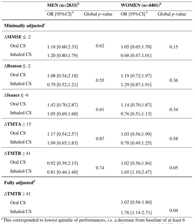
3.2. CS use and cognitive decline
Logistic regression analysis adjusted for age, center, education level and baseline cognitive
performance indicated that women reporting use of inhaled but not oral CS at baseline showed
greater decline over 7years on the TMTB (OR=1.65, 95%CI=1.10-2.47) (Table 2). The
association persisted in the complete model further adjusted for other confounders including
pathologies associated with CS prescription (chronic pain and respiratory disorders, etc.)
(OR=1.76, 95%CI=1.14-2.71). No significant interactions were found for decline on TMTB in
women between CS use and age (p=0.12) or APOE (p=0.38). No significant effect was observed
in men regardless of the cognitive domain. Performing multivariate-adjusted random-effect
linear models with the cognition score as the continuous variable led to the same results, the only
significant association being found between CS use and performance on the TMTB in women
(interaction between time and inhaled CS p=0.003, slope of log(TMTB) increased by 0.008
compared to no CS use). Similar findings were observed when executive function was assessed
using score differences such as time on TMTB minus time on TMTA (data not shown).
3.3. Cognitive decline according to the pattern of CS use during follow-up
Of the 3736 women with a TMT evaluation, 3171 (84.9%) did not report CS use (oral or
inhaled) at baseline and during the 7-year follow-up, 38 (1.0%) reported inhaled CS use only at
baseline or after the first 2-year follow-up, but not at either 4- or 7-year follow-up
(“discontinuing” group), and 22 (0.5%) reported inhaled CS use both at baseline and at least at the 2- and 4-year examination (3 consecutive examinations, “continuing” group). Other subjects
having reported inhaled CS use intermittently during the follow-up or having taken oral CS at
baseline or during the follow-up (n=505) were not considered in the following analyses.
Compared to women who had never used CS, women having continuously used inhaled
95%CI=1.29-7.68, p=0.01) (Table 3). The association was not significant for women in the
“discontinuous” group (OR=1.41, 95%CI=0.69-2.89, p=0.35).
3.4. NSAID and cognitive decline
A total of 180 men (6.4%) and 452 women (10.3%) were taking NSAID at baseline.
Thirty-four (0.5%) were taking both NSAID and CS. NSAID principally consisted of diacerein
(23.1% of users), diclofenac (14.5%), piroxicam (14.2%), ketoprofen (9.9%), and ibuprofen
(8.6%) and 6.8% were taking coxibs (table e-2). There was no significant association between
NSAID use at baseline and cognitive decline in men and women (Table 4). Examining cognitive
decline according to the pattern of NSAID use during follow-up, we only observed a
non-significant association with an increased risk of decline on the Isaacs’ task in the 100 women
having used NSAID continuously [multi-adjusted(OR)=1.50, 95%CI=0.96-2.35, p=0.08].
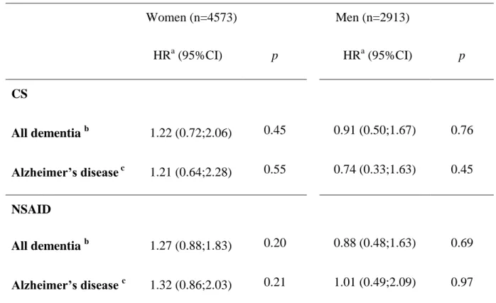
3.5. Dementia incidence
Within the 7486 subjects included in the analysis 527 incident cases were diagnosed during
the 7-year follow-up, of whom 360 had Alzheimer'sdisease (AD). Adjusted Cox models failed to
find a significant association between the incidence of dementia or AD and CS or NSAID use at
baseline (Table 5).
4. Discussion
4.1. CS use and cognitive decline
Our results indicate a 1.8-fold increased risk of decline in a cognitive task sensitive to
alterations in psychomotor speed and frontal executive functioning in women only. The same
results were obtained in the minimally and the fully adjusted model, which was further adjusted
for the pathologies associated with CS treatment (such as chronic pain and respiratory disorders)
models). This underlines the consistency of these associations, in spite of an eventual risk of
over-adjustment and suggests that the cognitive decline was more likely related to the CS
themselves rather than the underlying burden of illness, making unlikely an eventual prescription
bias. This is also supported by the observation that risk of cognitive decline nearly doubled
(OR=3.2, p=0.01) with longer treatment and continuous use, and was non-significant after
discontinuation.
Systemic toxicity is known to be a major concern with long-term use of high-dose
glucocorticoids. In our study, the deleterious effect on cognitive function was only observed for
inhaled CS. This may be linked to differences in the active substances and/or pharmacodynamic
properties related to administration mode; oral administration being generally associated with
slower absorption/distribution, higher metabolism due to hepatic first-pass effect, and lower
biologically active circulating metabolites. Adrenal insufficiency and Cushing’s syndrome have
also been reported in patients treated with inhaled CS (Molimard et al., 2008). In our study,
however, the small number of subjects taking CS using either mode did not allow us to examine
the effect of administration method. We also did not have any information on patient conformity
to prescription; inhaled CS being more likely to be over-dosed.
Only a few small experimental studies in healthy young or adult participants have
examined the link between acute or short-term glucocorticoid administration and cognitive
impairment. Cognitive assessment has principally focused on changes in declarative memory
consistent with deficiencies in hippocampus-dependent activity, although recent studies have
also noted impairments in prefrontal cortex processing (Wolkowitz et al., 2009; Lupien et al.,
2007; Franz et al., 2011); both structures having a relatively high density of gluco- and
mineralo-corticoid receptors. These associations may be transient and dose-dependent (Newcomer et al.,
1999; Kirschbaum et al., 1996; Young et al., 1999; de Quervain et al., 2003; Keenan et al.,
1996). A lower sensitivity of verbal memory skills compared to other cognitive functions has
older adults, in a large study using an extensive cognitive battery; high cortisol levels being
specifically associated with poor frontal-executive functions (Franz et al., 2011). No such studies
have been performed in the elderly.
In our study poor performance is only seen on the TMTB and not on the TMTA trial (the A
trial not involving the cognitive switching task) and similar results were observed when
executive function was assessed using score differences. This thus suggests that executive
performance is affected rather than psychomotor speed. Neuropsychological tests requiring intact
prefrontal cortical activity such as the TMTB may thus be especially vulnerable to chronic
inhaled CS use in elderly women. A “two-factor model of brain aging” which posits that
endocrine-related aging, like normal aging, may primarily involve loss of frontal-striatal circuits
with associated executive function changes, whereas pathological cognitive aging (e.g. AD) is
more strongly associated with hippocampal abnormalities appears to support our observations
but remains speculative (Buckner, 2004; Hedden and Gabrieli, 2004; Franz et al., 2011).
4.2. Gender specificity
We did not observe a significant association between CS use and cognitive decline for men. This
could be due to pharmacodynamic or metabolism differences. In a meta-analysis, Otte et al.
reported a three-fold stronger effect of age on cortisol response in women than in men (Otte et
al., 2005). A gender difference in biological response to stress both at a functional and structural
level may also be possible (Kudielka et al., 2004; Kumsta et al., 2007; Pruessner et al., 2010).
Overall, specific cognitive functions may be affected by cumulative exposure to chronic stress
via glucocorticoids released from the HPA axis, but also from gonadal steroids released from the
hypothalamic-pituitary-gonadal axis (notably estrogens and testosterone which have distinct
cognitive effects in women and men) (Conrad and Bimonte-Nelson, 2010). Interestingly,
hypercortisolism and steroid dementia syndrome previously reported in adult patients after
similarities with the cognitive features observed in this study, including durable executive
dysfunction, higher incidence in females, dose dependency, and reversibility (Egeland et al.,
2005; Wolkowitz et al., 2007; Lewis and Smith, 1983; Sacks and Shulman, 2005; Wolkowitz et
al., 2009).
4.3. CS use and dementia
We observed no significant association between CS use at baseline and risk of developing
dementia over 7-years. To date only one randomized controlled trial has examined the effect of
1-year prednisone treatment on cognitive decline in Alzheimer’s patients (Aisen et al., 2000)
showing no significant difference between CS and placebo treated patients. CS dose and
exposure duration could also be evoked, but the small number of demented subjects taking CS
(n=28) have precluded exploration of this possibility.
4.4. NSAID use, cognitive decline, and dementia
We did not observe any significant associations between NSAID intake at baseline and
cognitive decline and dementia over 7-years. In observational studies, exposure to NSAID were
possibly associated with decreased risk for cognitive decline and AD, depending on class and
dose, longer duration or younger age at intake, and APO4 vulnerability (Szekely et al., 2004; Szekely et al., 2008; Vlad et al., 2008; de Craen et al., 2005; Gorelick, 2010; Szekely and Zandi,
2010). Conversely, the three randomized controlled trials performed using rofecoxib, celecoxib
and naproxen, suggested an increased risk in AD and no consistent association or worsening of
cognitive function with naproxen (in global summary scores and verbal fluency) (Thal et al.,
2005; Lyketsos et al., 2007; Martin et al., 2008).
Inflammatory processes are complex and may have either reparative or detrimental effects
on neurons (Wyss-Coray and Mucke, 2002). In our study, neither CS nor NSAID appeared to be
inhibiting inflammation can reduce neurodegenerative processes in the elderly. The different
patterns of cognitive change observed in our study between CS and NSAID suggests that
deleterious CS effects on executive function may be more likely related to glucocorticoid and
HPA axis functioning rather than an anti-inflammatory effect.
4.5. Limitations and strengths
A limitation of our study was the use of some self-reported covariates with eventual
subsequent recall bias. Bias could also have been introduced through the exclusion of
participants, those lost to follow-up being more likely to have dementia, to be older, and thus
with worse physical and mental health, and using CS more frequently. This may limit the
generalizability of our results, and associations may have thus been underestimated. We did not
consider treatment compliance, which may have caused classification bias. Since we did not
have data on precise duration of medication use, we could not definitively address the question
of whether prolonged use could precipitate non-reversible dementia. Finally, since multiple
analyses have been performed we cannot exclude that some observed associations were due to a
chance finding.
The strengths of this study relate principally to its prospective, community-based design,
large size and extensive information obtained on clinical status. CS and NSAID use was verified
by examining prescriptions and medications, thus minimizing exposure misclassification and
with the advantage, compared to reimbursement data, of including self-medication (Noize et al.,
2009). Finally, we have taken into account a wide range of competing causes of cognitive
dysfunction in the elderly, by controlling for socio-demographic, genetic, health and lifestyle
covariates, thus limiting any potential confounding including prescription bias.
In conclusion, findings from this study suggest that inhaled CS use is associated with
being that medical practitioners should take chronic CS use into account before making a
Disclosure statement
Drs Ancelin, Carrière, Helmer, Rouaud, Pasquier, Berr, and Chaudieu report no
disclosures. Dr. Ritchie serves on scientific advisory boards for the Biomedical Research Centre,
King’s College London, and London and MRC Strategic Steering Committee (Longitudinal Health and Aging Research Unit).
Acknowledgements
The 3C Study is conducted under a partnership agreement between Inserm, the Victor
Segalen – Bordeaux II University and Sanofi-Synthélabo. The Fondation pour la Recherche
Médicale funded the preparation and first phase of the study. The 3C-Study is also supported by
the Caisse Nationale Maladie des Travailleurs Salariés, Direction Générale de la Santé, MGEN,
Institut de la Longévité, Agence Française de Sécurité Sanitaire des Produits de Santé, the
Regional Governments of Aquitaine, Bourgogne and Languedoc-Roussillon and, the Fondation
de France, the Ministry of Research-Inserm Programme “Cohorts and collection of biological
material”. The Lille Génopôle received an unconditional grant from Eisai. Part of this project is financed by two grants from the Agence Nationale de la Recherche (projects 07 LVIE 004 and
06-PNRA-005). We thank the Génopôle of Lille, the Laboratories of Biochemistry of the
University Hospitals of Dijon and Montpellier, the Town Council of Dijon and the Conseil
Général of Côte d'Or. Neither funding source provided scientific input to the study.
Appendix A. Supplementary data
References
Aisen, P.S., Davis, K.L., Berg, J.D., Schafer, K., Campbell, K., Thomas, R.G., Weiner, M.F.,
Farlow, M.R., Sano, M., Grundman, M., Thal, L.J., 2000. A randomized controlled trial of
prednisone in Alzheimer's disease. Alzheimer's Disease Cooperative Study. Neurology 54,
588-593.
American Psychiatric Association, 1994. Diagnostic and Statistical Manual of Mental Disorders
(DSM-IV). American Psychiatric Association, Washington, DC.
Artero, S., Ancelin, M.L., Portet, F., Dupuy, A., Berr, C., Dartigues, J.F., Tzourio, C., Rouaud,
O., Poncet, M., Pasquier, F., Auriacombe, S., Touchon, J., Ritchie, K., 2008. Risk profiles
for mild cognitive impairment and progression to dementia are gender specific. J Neurol
Neurosurg Psychiatry 79, 979-984.
Belanoff, J.K., Gross, K., Yager, A., Schatzberg, A.F., 2001. Corticosteroids and cognition. J
Psychiatr Res 35, 127-145.
Beluche, I., Carriere, I., Ritchie, K., Ancelin, M.L., 2010. A prospective study of diurnal cortisol
and cognitive function in community-dwelling elderly people. Psychol Med 40,
1039-1049.
Benton, A. Manuel pour l'application du test de rétention visuelle. Applications cliniques et
expérimentales. Paris: Centre de Psychologie Appliquée; 1965.
Buckner, R.L., 2004. Memory and executive function in aging and AD: multiple factors that
cause decline and reserve factors that compensate. Neuron 44, 195-208.
Commenges, D., Letenneur, L., Joly, P., Alioum, A., Dartigues, J.F., 1998. Modelling
age-specific risk: application to dementia. Stat Med 17, 1973-1988.
Conrad, C.D., Bimonte-Nelson, H.A., 2010. Impact of the
hypothalamic-pituitary-adrenal/gonadal axes on trajectory of age-related cognitive decline. Prog Brain Res 182,
Csernansky, J.G., Dong, H., Fagan, A.M., Wang, L., Xiong, C., Holtzman, D.M., Morris, J.C.,
2006. Plasma cortisol and progression of dementia in subjects with Alzheimer-type
dementia. Am J Psychiatry 163, 2164-2169.
de Craen, A.J., Gussekloo, J., Vrijsen, B., Westendorp, R.G., 2005. Meta-analysis of
nonsteroidal antiinflammatory drug use and risk of dementia. Am J Epidemiol 161,
114-120.
de Quervain, D.J., Henke, K., Aerni, A., Treyer, V., McGaugh, J.L., Berthold, T., Nitsch, R.M.,
Buck, A., Roozendaal, B., Hock, C., 2003. Glucocorticoid-induced impairment of
declarative memory retrieval is associated with reduced blood flow in the medial temporal
lobe. Eur J Neurosci 17, 1296-1302.
Egeland, J., Lund, A., Landro, N.I., Rund, B.R., Sundet, K., Asbjornsen, A., Mjellem, N.,
Roness, A., Stordal, K.I., 2005. Cortisol level predicts executive and memory function in
depression, symptom level predicts psychomotor speed. Acta Psychiatr Scand 112,
434-441.
Folstein, M.F., Folstein, S.E., McHugh, P.R., 1975. "Mini-mental state". A practical method for
grading the cognitive state of patients for the clinician. J Psychiatr Res 12, 189-198.
Franz, C.E., O'Brien, R.C., Hauger, R.L., Mendoza, S.P., Panizzon, M.S., Prom-Wormley, E.,
Eaves, L.J., Jacobson, K., Lyons, M.J., Lupien, S., Hellhammer, D., Xian, H., Kremen,
W.S., 2011. Cross-sectional and 35-year longitudinal assessment of salivary cortisol and
cognitive functioning: The Vietnam Era Twin Study of Aging. Psychoneuroendocrinology
doi:10.1016/j.psyneuen.2011.01.002.
Gorelick, P.B., 2010. Role of inflammation in cognitive impairment: results of observational
epidemiological studies and clinical trials. Ann N Y Acad Sci 1207, 155-162.
Greendale, G.A., Kritz-Silverstein, D., Seeman, T., Barrett-Connor, E., 2000. Higher basal
cortisol predicts verbal memory loss in postmenopausal women: Rancho Bernardo Study. J
Hedden, T., Gabrieli, J.D., 2004. Insights into the ageing mind: a view from cognitive
neuroscience. Nat Rev Neurosci 5, 87-96.
Hilmer, S.N., McLachlan, A.J., Le Couteur, D.G., 2007. Clinical pharmacology in the geriatric
patient. Fundam Clin Pharmacol 21, 217-230.
Isaacs, B., Kennie, A.T., 1973. The Set test as an aid to the detection of dementia in old people.
Br J Psychiatry 123, 467-470.
Jacqmin-Gadda, H., Fabrigoule, C., Commenges, D., Dartigues, J.F., 1997. A 5-year longitudinal
study of the Mini-Mental State Examination in normal aging. Am J Epidemiol 145,
498-506.
Keenan, P.A., Jacobson, M.W., Soleymani, R.M., Mayes, M.D., Stress, M.E., Yaldoo, D.T.,
1996. The effect on memory of chronic prednisone treatment in patients with systemic
disease. Neurology 47, 1396-1402.
Kirschbaum, C., Wolf, O.T., May, M., Wippich, W., Hellhammer, D.H., 1996. Stress- and
treatment-induced elevations of cortisol levels associated with impaired declarative
memory in healthy adults. Life Sci 58, 1475-1483.
Kudielka, B.M., Buske-Kirschbaum, A., Hellhammer, D.H., Kirschbaum, C., 2004. HPA axis
responses to laboratory psychosocial stress in healthy elderly adults, younger adults, and
children: impact of age and gender. Psychoneuroendocrinology 29, 83-98.
Kumsta, R., Entringer, S., Koper, J.W., van Rossum, E.F., Hellhammer, D.H., Wust, S., 2007.
Sex specific associations between common glucocorticoid receptor gene variants and
hypothalamus-pituitary-adrenal axis responses to psychosocial stress. Biol Psychiatry 62,
863-869.
Lee, B.K., Glass, T.A., Wand, G.S., McAtee, M.J., Bandeen-Roche, K., Bolla, K.I., Schwartz,
B.S., 2008. Apolipoprotein E Genotype, Cortisol, and Cognitive Function in
Lewis, D.A., Smith, R.E., 1983. Steroid-induced psychiatric syndromes. A report of 14 cases and
a review of the literature. J Affect Disord 5, 319-332.
Li, G., Cherrier, M.M., Tsuang, D.W., Petrie, E.C., Colasurdo, E.A., Craft, S., Schellenberg,
G.D., Peskind, E.R., Raskind, M.A., Wilkinson, C.W., 2006. Salivary cortisol and memory
function in human aging. Neurobiol Aging 27, 1705-1714.
Lupien, S.J., Maheu, F., Tu, M., Fiocco, A., Schramek, T.E., 2007. The effects of stress and
stress hormones on human cognition: Implications for the field of brain and cognition.
Brain Cogn 65, 209-237.
Lyketsos, C.G., Breitner, J.C., Green, R.C., Martin, B.K., Meinert, C., Piantadosi, S., Sabbagh,
M., 2007. Naproxen and celecoxib do not prevent AD in early results from a randomized
controlled trial. Neurology 68, 1800-1808.
Martin, B.K., Szekely, C., Brandt, J., Piantadosi, S., Breitner, J.C., Craft, S., Evans, D., Green,
R., Mullan, M., 2008. Cognitive function over time in the Alzheimer's Disease
Anti-inflammatory Prevention Trial (ADAPT): results of a randomized, controlled trial of
naproxen and celecoxib. Arch Neurol 65, 896-905.
Molimard, M., Girodet, P.O., Pollet, C., Fourrier-Reglat, A., Daveluy, A., Haramburu, F., Fayon,
M., Tabarin, A., 2008. Inhaled corticosteroids and adrenal insufficiency: prevalence and
clinical presentation. Drug Saf 31, 769-774.
Newcomer, J.W., Selke, G., Melson, A.K., Hershey, T., Craft, S., Richards, K., Alderson, A.L.,
1999. Decreased memory performance in healthy humans induced by stress-level cortisol
treatment. Arch Gen Psychiatry 56, 527-533.
Noize, P., Bazin, F., Dufouil, C., Lechevallier-Michel, N., Ancelin, M.L., Dartigues, J.F.,
Tzourio, C., Moore, N., Fourrier-Reglat, A., 2009. Comparison of health insurance claims
and patient interviews in assessing drug use: data from the Three-City (3C) Study.
O'Hara, R., Schroder, C.M., Mahadevan, R., Schatzberg, A.F., Lindley, S., Fox, S., Weiner, M.,
Kraemer, H.C., Noda, A., Lin, X., Gray, H.L., Hallmayer, J.F., 2007. Serotonin transporter
polymorphism, memory and hippocampal volume in the elderly: association and
interaction with cortisol. Mol Psychiatry 12, 544-555.
Otte, C., Hart, S., Neylan, T.C., Marmar, C.R., Yaffe, K., Mohr, D.C., 2005. A meta-analysis of
cortisol response to challenge in human aging: importance of gender.
Psychoneuroendocrinology 30, 80-91.
Pruessner, J.C., Dedovic, K., Pruessner, M., Lord, C., Buss, C., Collins, L., Dagher, A., Lupien,
S.J., 2010. Stress regulation in the central nervous system: evidence from structural and
functional neuroimaging studies in human populations. Psychoneuroendocrinology 35,
179-191.
Radloff, L., 1977. The CES-D scale: a self-report depression scale for research in the general
population. Appl Psychol Measurement 1, 385-401.
Rasmuson, S., Nasman, B., Carlstrom, K., Olsson, T., 2002. Increased levels of adrenocortical
and gonadal hormones in mild to moderate Alzheimer's disease. Dement Geriatr Cogn
Disord 13, 74-79.
Reitan, R., 1965. Validity of the Trail Making Test as an indicator of organic brain damage.
Percept Mot Skills 8, 271-276.
Ritchie, K., Carriere, I., de Mendonca, A., Portet, F., Dartigues, J.F., Rouaud, O.,
Barberger-Gateau, P., Ancelin, M.L., 2007. The neuroprotective effects of caffeine: a prospective
population study (the Three City Study). Neurology 69, 536-545.
Ritchie, K., Carriere, I., Ritchie, C.W., Berr, C., Artero, S., Ancelin, M.L., 2010. Designing
prevention programs to reduce dementia incidence. A prospective study of modifiable risk
factors. Br Med J, on line first, Aug 5, 341:c3885.
Sacks, O., Shulman, M., 2005. Steroid dementia: an overlooked diagnosis? Neurology 64,
Sauro, M.D., Jorgensen, R.S., Pedlow, C.T., 2003. Stress, glucocorticoids, and memory: a
meta-analytic review. Stress 6, 235-245.
Seeman, T.E., McEwen, B.S., Singer, B.H., Albert, M.S., Rowe, J.W., 1997. Increase in urinary
cortisol excretion and memory declines: MacArthur studies of successful aging. J Clin
Endocrinol Metab 82, 2458-2465.
Szekely, C.A., Green, R.C., Breitner, J.C., Ostbye, T., Beiser, A.S., Corrada, M.M., Dodge,
H.H., Ganguli, M., Kawas, C.H., Kuller, L.H., Psaty, B.M., Resnick, S.M., Wolf, P.A.,
Zonderman, A.B., Welsh-Bohmer, K.A., Zandi, P.P., 2008. No advantage of A beta
42-lowering NSAIDs for prevention of Alzheimer dementia in six pooled cohort studies.
Neurology 70, 2291-2298.
Szekely, C.A., Thorne, J.E., Zandi, P.P., Ek, M., Messias, E., Breitner, J.C., Goodman, S.N.,
2004. Nonsteroidal anti-inflammatory drugs for the prevention of Alzheimer's disease: a
systematic review. Neuroepidemiology 23, 159-169.
Szekely, C.A., Zandi, P.P., 2010. Non-steroidal anti-inflammatory drugs and Alzheimer's
disease: the epidemiological evidence. CNS Neurol Disord Drug Targets 9, 132-139.
Thal, L.J., Ferris, S.H., Kirby, L., Block, G.A., Lines, C.R., Yuen, E., Assaid, C., Nessly, M.L.,
Norman, B.A., Baranak, C.C., Reines, S.A., 2005. A randomized, double-blind, study of
rofecoxib in patients with mild cognitive impairment. Neuropsychopharmacology 30,
1204-1215.
The 3C Study Group, 2003. Vascular factors and risk of dementia: Design of the three city study
and baseline characteristics of the study population. Neuroepidemiology 22, 316-325.
Umegaki, H., Ikari, H., Nakahata, H., Endo, H., Suzuki, Y., Ogawa, O., Nakamura, A.,
Yamamoto, T., Iguchi, A., 2000. Plasma cortisol levels in elderly female subjects with
Alzheimer's disease: a cross-sectional and longitudinal study. Brain Res 881, 241-243.
Vlad, S.C., Miller, D.R., Kowall, N.W., Felson, D.T., 2008. Protective effects of NSAIDs on the
Wang, J., Korczykowski, M., Rao, H., Fan, Y., Pluta, J., Gur, R.C., McEwen, B.S., Detre, J.A.,
2007. Gender Difference in Neural Response to Psychological Stress. Soc Cogn Affect
Neurosci 2, 227-239.
Wolkowitz, O.M., Burke, H., Epel, E.S., Reus, V.I., 2009. Glucocorticoids. Mood, memory, and
mechanisms. Ann N Y Acad Sci 1179, 19-40.
Wolkowitz, O.M., Lupien, S.J., Bigler, E.D., 2007. The "steroid dementia syndrome": a possible
model of human glucocorticoid neurotoxicity. Neurocase 13, 189-200.
Wyss-Coray, T., Mucke, L., 2002. Inflammation in neurodegenerative disease - a double-edged
sword. Neuron 35, 419-432.
Young, A.H., Sahakian, B.J., Robbins, T.W., Cowen, P.J., 1999. The effects of chronic
administration of hydrocortisone on cognitive function in normal male volunteers.
Table 1. Characteristics of the study population as a function of corticosteroid use at baseline Men Women CS drugs CS drugs Characteristic No (n=2686) % Yes (n=147) % p No (n=4196) % Yes (n=205) % Age 65-69 70-74 75-80 80+ 26.3 35.1 24.5 14.1 21.8 31.3 27.2 19.7 0.17 25.2 32.5 28.1 14.2 20.5 34.1 32.7 12.7 0.30 Education 5 years 9 years 12 years 12 + 21.5 30.0 19.8 28.7 24.5 38.1 11.6 25.8 0.03 25.4 39.9 20.8 13.9 25.4 43.4 21.0 10.2 0.47 Marital status Married Single or divorced Widowed 82.6 7.7 9.7 80.8 11.0 8.2 0.32 45.8 18.9 35.3 41.9 21.0 37.1 0.54 Depressive symptoms (CES-D ≥ 16 a) 13.4 18.4 0.09 28.3 31.2 0.37 Ischemic pathologies b 21.8 25.9 0.25 12.2 15.6 0.14 BMI Normal Overweight Obese 38.0 49.1 12.8 34.3 53.4 12.3 0.59 53.6 33.2 13.2 50.7 31.7 17.6 0.20 Diabetes c 12.6 15.0 0.41 6.7 10.2 0.05 Chronic bronchitis d 3.8 21.1 <0.0001 2.2 12.2 <0.0001 Asthma e 1.5 14.3 <0.0001 1.9 23.9 <0.0001 Hypertension f 59.9 60.5 0.87 53.4 59.5 0.09 Hypercholesterolemia g 83.4 80.3 0.32 68.0 63.4 0.17 Alcohol 0 1-36 g/day > 36g/day 8.0 73.2 18.8 7.5 71.2 21.2 0.75 26.9 71.5 1.6 31.7 67.3 1.0 0.29 Smoking Never Former Current 30.5 61.3 8.2 23.8 69.4 6.8 0.14 81.4 14.9 3.7 79.0 18.6 2.4 0.27 NSAID use 6.2 9.5 0.11 10.3 9.8 0.80 Self-report chronic joint or back pain
Caffeine
consumption per dayh [0-1] unit ]1-3] units > 3 units 27.2 59.5 13.3 35.4 50.3 14.3 0.07 25.1 58.5 16.4 29.8 55.1 15.1 0.32 Carrier of the APOE4 allele 20.8 17.0 0.27 19.6 16.6 0.29 Global cognitive functioning i (MMSE score <26) 11.4 17.7 0.02 15.7 14.6 0.68 Verbal fluency i (Isaacs Set Test score < 39) 19.7 26.5 0.04 20.4 20.0 0.90 Visual memory i (Benton score < 10) 23.3 25.2 0.61 30.6 34.2 0.29 Psychomotor speed i (TMTA score > 70) 16.6 20.7 0.20 21.5 26.9 0.07 Executive function i (TMTB score > 140) 17.9 17.6 0.94 21.0 26.8 0.06 a
The presence of depressive symptoms was assessed using the Center for Epidemiological Studies-Depression Scale (CES-D)(Radloff, 1977) with a cut-off of >16.
b
History of stroke, myocardial infarction, angina pectoris, or arteritis and cardio-vascular surgery c
Diabetes defined as glucose ≥7 mmol/l or treated. d
Chronic bronchitis (with daily sputum or mucus production or cough for at least 3 consecutive months a year).
e
Asthma attacks and other chronic respiratory disorders including wheezing, tachypnea (over the last 12 months).
f
Systolic blood pressure ≥ 160 or diastolic blood pressure ≥ 95 mm Hg or intake of antihypertensive drugs. g Total cholesterol level ≥6.2 mmol/l or treated by lipid lowering agents.
h
one unit = 100 mg caffeine i
The % of subjects with lowest cognitive performances at baseline are reported (lowest quintile except for TMT highest quintile). The numbers of subjects completing the TMT tests were for women, 3573 non CS users and 163 users and for men, 2242 non CS users and 107 users (see Methods for detail).
CS = Corticosteroid; BMI = body mass index; MMSE = Mini Mental State Examination; NSAID = Nonsteroidal anti-inflammatory drug; TMT = Trail Making Test
Table 2. Baseline corticosteroid (CS) use and cognitive declinea over the 7-year follow-up
period
MEN (n=2833)b WOMEN (n=4401)b
OR [95%CI] b Global p-value OR [95%CI] b Global p-value Minimally adjustedc MMSE ≤ -2 Oral CS 1.18 [0.60;2.33] 0.62 1.05 [0.65;1.70] 0.15 Inhaled CS 1.20 [0.80;1.79] 0.68 [0.47;1.01] Benton ≤ -2 Oral CS 1.08 [0.54;2.18] 0.55 1.19 [0.72;1.97] 0.36 Inhaled CS 0.79 [0.52;1.21] 1.29 [0.87;1.91] Isaacs ≤ -6 Oral CS 1.42 [0.70;2.87] 0.61 1.14 [0.70;1.87] 0.34 Inhaled CS 1.05 [0.69;1.60] 0.76 [0.51;1.13] TMTA ≥ 15 Oral CS 1.17 [0.54;2.57] 0.87 1.03 [0.56;1.90] 0.58 Inhaled CS 1.09 [0.65;1.83] 0.78 [0.49;1.25] TMTB ≥ 41 Oral CS 0.92 [0.39;2.15] 0.74 1.02 [0.56;1.84] 0.05 Inhaled CS 0.81 [0.46;1.40] 1.65 [1.10;2.47] Fully adjustedd TMTB ≥ 41 Oral CS 1.02 [0.56-1.86] 0.04 Inhaled CS 1.76 [1.14-2.71] a
This corresponded to lowest quintile of performances, i.e. a decrease from baseline of at least 6 points on the Isaacs total score or at least two points on the Benton test and the MMSE and an increase from baseline of at least 15 (TMTA) or 41 seconds (TMTB).
b
Except for TMTA and TMTB, where n = 2349 for men and 3736 for women. c
Adjusted for centre, age, education, and baseline cognitive performance. d
Adjusted for centre, age, education, baseline cognitive performance, depression, ischemic pathologies, diabetes, hypercholesterolemia, caffeine, smoking, APOE4, chronic joint or back pain, chronic bronchitis, asthma and other chronic respiratory disorders.
MMSE = Mini-Mental State Examination; TMTA = Trail Making Task A; TMTB = Trail Making Task B.
Table 3. Cognitive decline in executive function in women according to pattern of inhaled corticosteroid use during the follow-up period (n=3231a)
Minimally adjustedb Fully adjustedc
OR (95% CI) p OR (95% CI) p ∆TMTB ≥ 41 Noned 1 1 Discontinuing 1.39 [0.69;2.77] 0.36 1.41 [0.69;2.89] 0.35 Continuing 3.01 [1.24;7.26] 0.01 3.15 [1.29;7.68] 0.01 a
The 505 women having taken inhaled CS intermittently during the follow-up or having taken oral CS at baseline or during the follow-up were not considered in this analysis.
b
Adjusted for centre, age, education, and baseline cognitive performance. c
Adjusted for centre, age, education, baseline cognitive performance, depression, ischemic pathologies, diabetes, hypercholesterolemia, caffeine, smoking, APOE4, chronic joint or back pain, chronic bronchitis, asthma and other chronic respiratory disorders.
d.
Nonusers of oral and inhaled corticosteroids during 7 years TMTB = Trail Making Task B
Table 4. Baseline NSAID use and cognitive decline over the 7-year follow-up period MEN (n=2833a) WOMEN (n=4401a) OR [95% CI] b p OR [95% CI] b p ∆MMSE ≤ -2 1.11 [0.81;1.53] 0.52 0.92 [0.75;1.14] 0.45 ∆Benton ≤ -2 1.07 [0.76;1.49] 0.71 0.99 [0.80;1.24] 0.95 ∆Isaacs ≤ -6 0.92 [0.66;1.29] 0.64 1.17 [0.95;1.44] 0.15 ∆TMTA ≥ 15 1.09 [0.74;1.59] 0.67 1.09 [0.86;1.39] 0.47 ∆TMTB ≥ 41 1.04 [0.70;1.54] 0.84 1.12 [0.89;1.42] 0.34 a
Except for TMTA and TMTB, where n=2349 for men and n=3736 from women. b
adjusted for centre, age, education, and baseline cognitive performance.
MMSE = Mini-Mental State Examination; BVRT = Benton Visual Retention Test; TMTA = Trail Making Task A; TMTB = Trail Making Task B.
Table 5. Baseline CS and NSAID use and 7-year incidence of dementia (Cox model with delayed entry) Women (n=4573) Men (n=2913) HRa (95%CI) p HRa (95%CI) p CS All dementia b 1.22 (0.72;2.06) 0.45 0.91 (0.50;1.67) 0.76 Alzheimer’s disease c 1.21 (0.64;2.28) 0.55 0.74 (0.33;1.63) 0.45 NSAID All dementia b 1.27 (0.88;1.83) 0.20 0.88 (0.48;1.63) 0.69 Alzheimer’s disease c 1.32 (0.86;2.03) 0.21 1.01 (0.49;2.09) 0.97 a
Adjusted for gender, centre, age, education, depression, ischemic pathologies, diabetes,
hypercholesterolemia, caffeine, smoking, APOE4, chronic joint or back pain, chronic bronchitis, asthma and other chronic respiratory disorders.
b
320 women and 207 men were diagnosed with incident dementia. c
226 women and 134 men had incident Alzheimer'sdisease. The 94 women and 73 men with other types of dementia were excluded from this analysis.
eTable 1. Frequency of use of CS according to formulation mode among study 7234 participants*
CHEMICAL ACTIVE SUBSTANCE n Frequency (%)
ORAL CS (n=123) BETAMETHASONE 7 1.79 CORTISONE 4 1.03 CORTIVAZOL 3 0.77 DEXAMETHASONE 2 0.51 FLUDROCORTISONE 1 0.26 HYDROCORTISONE 9 2.31 METHYLPREDNISOLONE 11 2.82 PREDNISOLONE 19 4.87 PREDNISONE 63 16.15 TRIAMCINOLONE 4 1.03 INHALED CS (n=267) BECLOMETASONE 107 27.43 BUDESONIDE 80 20.52 FLUNISOLIDE 4 1.03 FLUTICASONE 47 12.06 PREDNISOLONE 1 0.26 TIXOCORTOL 5 1.29 TRIAMCINOLONE 23 5.90
eTable 2. Frequency of use of NSAID (n=675) among study 7234 participants*
CHEMICAL ACTIVE SUBSTANCE n Frequency (%)
ALMINOPROFEN CELECOXIB DIACEREIN DICLOFENAC ETODOLAC FENBUFEN FENOPROFEN FLURBIPROFEN GLUCOSAMINE IBUPROFEN INDOMETHACIN KETOPROFEN MELOXICAM MORNIFLUMATE NABUMETONE NAPROXEN NIFLUMIC ACID NIMESULIDE OXACEPROL PIROXICAM ROFECOXIB SULINDAC TENOXICAM TIAPROFENIC ACID 1 37 156 98 8 1 9 11 1 58 4 67 10 2 8 26 7 16 13 96 9 8 25 2 0.15 5.48 23.11 14.52 1.19 0.15 1.33 1.63 0.15 8.59 0.59 9.93 1.48 0.3 1.19 3.85 1.04 2.37 1.93 14.22 1.33 1.19 3.7 0.3


![Table 4. Baseline NSAID use and cognitive decline over the 7-year follow-up period MEN (n=2833 a ) WOMEN (n=4401 a ) OR [95% CI] b p OR [95% CI] b p ∆MMSE ≤ -2 1.11 [0.81;1.53] 0.52 0.92 [0.75;1.14] 0.45 ∆Benton ≤ -2 1.07 [0.76;1.49] 0.71 0.](https://thumb-eu.123doks.com/thumbv2/123doknet/14648521.550868/28.892.81.686.155.442/table-baseline-nsaid-cognitive-decline-follow-period-benton.webp)