Endothelin Inhibition as a Biologic Target for
Treating Hypertension
Hans R. Brunner
Endothelin, a 21-amino-acid peptide, binds to a specific receptor on vascular smooth muscle cells, thereby inducing vasoconstriction. Although plasma levels are not consistently elevated in hypertension, there is evidence that endothelin has an important role in its pathogenesis.
Administration of endothelin antagonists has lowered blood pressure and reduced end-organ damage in some animal models. It has also reduced the cross-sectional area of neointima due both to hypertension and vascular injury.
Coadministration of endothelin and angiotensin II to rats produced a synergistic hypertensive effect. Similarly, coadministration of an endothelin antagonist with an angiotensin converting enzyme inhibitor resulted in a synergistic lowering of blood pressure. Several preliminary clinical studies have been done. The endothelin antagonist
bosentan has decreased vascular resistance and blood pressure and increased cardiac index in patients with congestive heart failure. Plasma endothelin levels are elevated in the acute phase of myocardial infarction and in chronic heart failure. The magnitude of this increase, measured 3 days after patients experienced myocardial infarction, had a significance at least equal to known risk factors in predicting 1 year survival. Thus, there are reasons to believe that endothelin antagonists may become a useful tool in the management of various cardiovascular disorders. Am J Hypertens 1998;11:103S–109S © 1998 American Journal of Hypertension, Ltd.
KEY WORDS: Endothelin antagonist, hypertension, endothelin.
T
he endothelium plays a role in regulating vascular resistance by releasing substances that dilate or constrict blood vessels. These substances, endothelins, are a family of pep-tides having potent vasoconstrictive effects. There are basically three types of endothelin: endothelin-1, en-dothelin-2, and endothelin-3. They are all formed from precursors (big endothelin) by enzymatic cleavage and are all 21–amino-acid peptides with disulfide bridges (Figure 1). Endothelin-1 and endothelin-2 act on the endothelin-A (ETA) receptor of smooth muscleto produce a prolonged contraction and vasoconstric-tion. Endothelin-3 can also induce vasoconstriction by binding to the endothelin-B (ETB) receptor on smooth muscle, but it also acts on the ETBreceptors of endo-thelial cells, where it can actually have the opposite effect. It has been suggested that, under normal con-ditions, low concentrations of endothelin stimulate ETB receptors, thus contributing to the release of en-dothelium-derived relaxing factors.1 Endothelin-1 is probably the best studied of this family. When admin-istered to animals, endothelin-1 causes severe vaso-constriction.2The contraction of vascular smooth mus-cle produced by endothelin-1 may be mediated by an increase in intracellular calcium, but the details have not been established.3 Infusion of endothelin-1 into healthy human volunteers resulted in a 2.5-fold in-crease in plasma endothelin-1 concentration and a rise From the University of Lausanne, Lausanne, Switzerland.
Address correspondence and reprint requests to Hans R. Brunner, MD, Head, Division of Hypertension and Vascular Medicine, Uni-versity of Lausanne, 1001 Lausanne, Switzerland.
© 1998 by the American Journal of Hypertension, Ltd. 0895-7061/98/$19.00
in blood pressure of approximately 6 mm Hg; in con-trast, infusion of the same molar concentration of en-dothelin-3 raised plasma levels of enen-dothelin-3 2.5-fold, but had no effect on blood pressure.4Like angio-tensin II and norepinephrine, endothelin-1 is now thought to play a role in hemodynamic control. It has not been proven, however, that endothelin normally participates in the control of vascular tone, and its role in hypertension has not been established. Plasma lev-els of endothelin-1 do not increase significantly during hypertension in humans or animals.5,6 However, en-dothelin-1 is increased in the aorta and in mesenteric arteries of deoxycorticosterone acetate (DOCA)-salt hypertensive rats, despite normal blood levels. In con-trast, the vascular tissue of spontaneously hyperten-sive rats does not have a higher endothelin-1 content.7 There is also substantial vascular hypertrophy in DOCA-salt hypertensive rats, compared to limited hy-pertrophy of blood vessels in spontaneously hyperten-sive rats.8 It is therefore possible that endothelin-1, which has powerful hypertrophic and mitogenic prop-erties, could have a causal role in this vascular hyper-trophy. Endothelin-1 is thought to interact with the sympathetic nervous system and the renin-angioten-sin system in the regulation of blood pressure and sodium metabolism, but the mechanisms of these in-teractions are unknown. Endothelin-1 may also help regulate vascular tone by modulating angiotensin con-verting enzyme levels, since it has been reported that endothelin stimulates the release of angiotensin II from mesenteric arteries.9
EFFECTS OF ENDOTHELIN ON BLOOD PRESSURE
Exogenous endothelin-1 alone in sufficiently high con-centrations has been reported to increase blood
pres-sure in both humans and experimental animals. Con-tinuous infusion of endothelin-1 in rats at a rate of 60 mg/kg of body weight/day resulted in a significant increase in the systolic blood pressure, while 6 mg/ kg/day had no significant effect.2Yoshida et al10 stud-ied the effects of endothelin-1 and angiotensin II, sep-arately and in combination, on blood pressure and physiologic parameters of Sprague-Dawley rats. Intra-peritoneal administration of angiotensin II at the rate of 400mg/kg/day did not induce a significant change in systolic blood pressure compared to that of controls (saline infusion). Nor did continuous infusion of 3 mg/kg/day of endothelin-1 affect systolic blood pres-sure significantly. However, the combined infusion of endothelin-1 and angiotensin II at these same concen-trations and rates produced a significant increase in systolic blood pressure (Figure 2). The increase was statistically significant 1 day after the start of the in-fusion and increased thereafter. After 6 days, systolic blood pressure was 148.0, 142.7, and 143.5 mm Hg in rats given angiotensin II, endothelin-1, or saline, re-spectively, whereas it was 189.0 mm Hg in angiotensin II plus endothelin-1–infused rats (P, .05 v all other groups); this is an increase of 32% compared to con-trols. Angiotensin II, endothelin-1, and the combina-tion did not result in significant changes in body weight, fluid retention, urine volume, urinary sodium excretion, or urinary potassium excretion, suggesting that the effect on blood pressure was not due to renal effects. In contrast, in the same study, continuous in-fusion of norepinephrine (360mg/kg/day), endothe-lin-1 (3 mg/kg/day), and the combination norepi-nephrine plus endothelin-1 did not result in any sig-nificant changes in systolic blood pressure. However, sufficiently high infusions of norepinephrine (1.8 mg/ kg/day) have been shown to induce a sustained
in-FIGURE 1. The endothelin system. Big ET-1, precursor of endothelin-1; Big ET-2, precursor of endothelin-2; Big ET-3, precursor of endothelin-3; ET-1, endothelin-1; ET-2, endothelin-2; ET-3, endothelin-3; ECE, endothelin converting enzyme; ETA, endothelin-A
receptor; ETB, endothelin-B receptor; EC, endothelial cell; SMC, smooth muscle cell. Precursors of endothelin-1, -2, and -3 are
crease in systolic blood pressure in rats.11There have been reports that endothelin-1 can amplify the hyper-tensive effect of norepinephrine under certain condi-tions,12 so the interaction of these peptides is still unresolved.
ENDOTHELIN AND NEOINTIMA FORMATION The major problem limiting the long-term effective-ness of percutaneous transluminal coronary angio-plasty (PTCA) is the high incidence of neointima for-mation and vascular restenosis.13,14Endothelin-1 lev-els are elevated after PTCA, suggesting that endothelin-1 is implicated in restenosis. In general, advanced atherosclerotic lesions are characterized by large numbers of smooth muscle cells, macrophages, and T lymphocytes. Angiotensin II and norepineph-rine are thought to be involved in the development of neointima, and endothelin-1, a potent mitogen, may also play a role in the process.
Douglas et al14investigated the effects of exogenous endothelin-1, and of blockage of endothelin-1, on neo-intima formation in both an in vitro model and in vivo. The addition of endothel1 (1 nmol/L) in-creased neointima formation, as evidenced by a nine-fold increase in thymidine incorporation in cultured rat aortic vascular smooth muscle cells. This effect was inhibited in a dose-dependent manner by the simul-taneous addition of the endothelin receptor antagonist SB 209670. Endothelin-1-induced proliferation, as measured by thymidine incorporation, was com-pletely inhibited by 0.1 mmol/L of SB 209670. How-ever, even the maximum concentration of SB 209670 used did not alter cell viability, nor did it change basal proliferation, implying that the effect was not due to a general inhibition. Furthermore, addition of SB 209670
in a concentration that completely blocked endothelin-1-induced proliferation had no effect on proliferation induced by angiotensin II, fibroblast growth factor, or platelet-derived growth factor. As part of the same study, the in vivo effects of endothelin-1 and SB 209670 were determined using balloon angioplasty performed on the left common carotid artery of Sprague-Dawley rats. In one set of experiments, rats were administered endothelin-1 by intraarterial infu-sion over a 30-min period immediately after balloon angioplasty. Endothelin-1 infusion (500 pmol/kg body weight over a 30-min period) significantly in-creased the amount of neointima formation relative to controls. The cross-sectional area of the neointima was 73% greater in the endothelin-1-treated group, and there was a corresponding increase of 53% in the neointima-to-media ratio. This effect was dose depen-dent. Other than size, the morphology of lesions from endothelin-1-treated rats was similar to those treated with saline. The morphology of undamaged contralat-eral carotid arteries was not affected by endothelin-1. In a second experiment, rats were treated either with SB 209670 or saline beginning 3 days before and continuing 2 weeks after angioplasty. The arteries were removed at the end of this period and examined for neointima formation. Treatment with SB 209670 reduced formation of neointima after balloon angio-plasty. It did not affect the tunica media, but de-creased the neointima cross-sectional area by 53% and the ratio of neointima-to-medial cross-sectional area by 47% (Figure 3). All animals treated with SB 209670 appeared healthy on gross physical examination, and there were no differences in body weights between treated rats and controls, indicating that the inhibitory effect of SB 209670 was not due to nonselective cyto-toxicity.14
Several studies have reported that cultured rat and human vascular smooth muscle proliferation is medi-ated by the ETAreceptor.15,16Since endothelin-1 levels
FIGURE 2. Effects of infusion of angiotensin II (AII), endothe-lin-1 (ET-1), and the combination AII plus ET-1 on systolic blood pressure in conscious rats. Control vehicle (physiologic saline) is also shown. Reprinted with permission from Yoshida K et al.10
FIGURE 3. Neointima:media ratio in rats that received either saline or the endothelin receptor antagonist SB 209670. Results are given as mean 6 SEM. *P , .05 compared with control (saline) by ANOVA and Fisher’s protected least-squares differ-ence. Reprinted with permission from Douglas SA et al.14
are sharply elevated in the human coronary sinus after PTCA, it has been suggested that enhanced exposure of smooth muscle to endothelin-1 may stimulate neo-intima formation. Thus, endothelin-1 may be derived from circulating blood or come from damaged endo-thelial or smooth muscle cells in the vicinity of the lesion. While the evidence implicates such a mecha-nism, it has not been definitely established that thelin-1 has a causal role in restenosis. Because endo-thelin-1 induces the release of several growth factors, such as platelet-derived growth factor (PDGF), it may function as an indirect or comitogen. The increase in neointima area in response to endothelin-1 is also interesting considering its short plasma half-life, which is about 1 min.17However, unlike angiotensin II and norepinephrine, the effects of endothelin-1 are prolonged; the half-life for endothelin-1-receptor dis-sociation is over 100 h.18
EFFECTS OF BOSENTAN
Most studies with ETA receptor antagonists, such as BQ-123, found that they produce only moderate low-ering of blood pressure.19 Bosentan is a recently de-veloped, long-acting, nonselective endothelin receptor antagonist. It blocks both ETA and ETB receptors. A study of DOCA-salt hypertensive rats used bosentan to examine whether the increase in vascular endothe-lin contributes to elevated blood pressure and vascu-lar hypertrophy.1 The rats received bosentan (100 mg/kg body weight/day) in their food for 3 weeks. The systolic blood pressure of DOCA-salt hyperten-sive rats increased to 197 mm Hg during the course of the study, whereas blood pressure of bosentan-treated rats increased to only 177 mm Hg (P , .01). The mesenteric resistance arteries of DOCA-salt hyperten-sive rats had a smaller lumen diameter than those in uninephrectomized control animals (230 v 271 mm, P , .01). The arterial lumen diameter in bosentan-treated DOCA rats was 253 mm, which was signifi-cantly larger (P, .05) than the diameter (230mm) in untreated DOCA rats (Figure 4). The media:lumen ratio was significantly greater in DOCA rats than in
the controls (7.6% v 3.9%, P, .01); in bosentan-treated DOCA rats, this parameter was 5.2%, which was sig-nificantly higher than that of uninephrectomized con-trols (P , .05) and untreated DOCA rats (P , .01) (Figure 4). The width of the media and cross-sectional area of the media of resistance arteries were signifi-cantly smaller in bosentan-treated rats compared to untreated DOCA rats. There were no significant dif-ferences in lumen diameter or the cross-sectional area of the media of vessels between bosentan-treated rats and uninephrectomized control rats. The vasoconstric-tor responses of bosentan-treated rats were nearly those of controls. Plasma endothelin-1 was signifi-cantly higher in bosentan-treated rats, possibly indi-cating that it was being produced but not binding to its receptor in these animals. These results strongly suggest that endothelin-1 plays a role in the elevation of blood pressure and in vascular hypertrophy in the DOCA-salt hypertensive rat model.
Further evidence that blockade of endothelin recep-tors by bosentan decreases blood pressure and inhibits vascular hypertrophy came from a study of spontane-ously hypertensive stroke-prone rats (SHRSP).20 SHRSP were administered bosentan in their diet start-ing at 3 months of age. Untreated SHRSP and un-treated Wistar-Kyoto (WKY) rats served as controls. Bosentan-treated rats had lower mean systemic blood pressure, and lower systolic, diastolic, pulse, and mean pressures in arterioles in the cerebrum than did untreated SHRSP. These blood pressures were still significantly higher than those in WKY rats (Table 1). Measurements of first-order arterioles in the cerebrum were made when the animals reached 6 months of age. The cross-sectional area of the vessel wall was signif-icantly larger in SHRSP than in WKY, but in bosentan-treated SHRSP the area was not significantly different from the area in WKY rats (Table 1). That is, treatment with bosentan prevented hypertrophy in cerebral ar-terioles without normalizing either mean systemic pressure or pulse blood pressure. However, bosentan did not have any significant effects on internal or external diameters of arterioles in SHRSP. A number
FIGURE 4. Lumen diameter (mm) and media:lumen ratio (expressed as a percent-age) of resistance arteries in uninephrec-tomized control rats (Uni-Nx), DOCA-salt hypertensive rats (DOCA), and DOCA-salt hypertensive rats adminis-tered bosentan.
of factors have been postulated as contributing to the cerebral vascular hypertrophy and remodeling that occurs during hypertension, including the increased blood pressure itself, angiotensin II and other neuro-hormonal factors, and genetic factors. This study shows that bosentan, an antagonist of both ETAand ETB, prevents hypertrophy of arterioles in the cere-brum in SHRSP, which suggests that endothelin-1 contributes to the hypertrophy of cerebral arteries oc-curring during hypertension. This effect may not ap-ply to all arteries under all conditions. In a study by Li and Schiffrin, endothelin receptor blockade did not significantly affect hypertrophy in small mesenteric, coronary, renal, or femoral arteries.21 This could be due to several differences in the studies. Endothelin-1 may contribute to hypertrophy of cerebral arteries, but not to all other arteries. It is also possible that the effect may only be significant in blood vessels above a cer-tain size, or when blood pressure rises above some threshold.
Bosentan has also been studied in patients with heart failure, in whom plasma endothelin-1 levels are commonly elevated.22 These patients all had conges-tive heart failure of .3 months’ duration. Endothe-lin-1 concentrations were above normal in all patients and correlated with the extent of pulmonary hyper-tension, with left and right heart-filling pressures, with pulmonary vascular resistance, and, inversely with cardiac index. Plasma big endothelin-1 levels were also above normal. This was a randomized, dou-ble-blind study in which patients received two intra-venous infusions of either placebo or bosentan (100 mg followed 60 min later by 200 mg). All patients were previously on regimens consisting of angiotensin converting enzyme inhibitors (24), diuretics (20),
digoxin (15), calcium antagonists (2), low-dose b-blockers (3), long-acting nitrates (9), and antiar-rhythmic drugs, which were withheld prior to infu-sions. Hemodynamics and plasma endothelin-1 con-centrations were determined before and during the 120-min infusion. Bosentan reduced mean pulmonary artery pressure by 13.7% compared to the effects of placebo (Figure 5). It also lowered mean arterial blood pressure by 7.7%, right atrial pressure by 18.2%, and pulmonary artery wedge pressure by 8.6%, all com-pared to results shown by placebo. It increased the cardiac index by 13.6% (Figure 6), but did not change the heart rate. Consequently, calculated systemic vas-cular resistance fell by 16.5% and pulmonary vasvas-cular resistance by 33.2% (P , .01 for both). Most of the effect was seen after the first (100 mg) infusion. Plasma endothelin-1 levels rose significantly in the patients receiving bosentan from a mean of 38.9 pg/mL to 89.6 pg/mL. Big endothelin-1, norepinephrine, angiotensin
RATS AND CONTROLS
Parameter WKY SHRSP-U SHRSP-B Systemic arterial mean pressure, mm Hg 906 3 1836 3* 1526 5*† Pulse pressure, mm Hg 256 3 406 2* 336 2*† Cross-sectional area of vessel wall,mm2 12996 65 16276 173* 12876 78†
Values are mean6 SEM
WKY, Wistar-Kyoto rats; SHRSP-U, spontaneously hypertensive stroke-prone rats, untreated; SHRSP-B, spontaneously hypertensive stroke-stroke-prone rats, bosentan-treated.
* P, .05 v WKY; †P , .05 v untreated SHRSP. Reprinted with permission from Chillon J et al.20
FIGURE 5. Change from baseline in pulmonary arterial pres-sure (mean 6 SEM) after infusion with bosentan or placebo in patients with CHF (congestive heart failure). Reprinted with per-mission from Kiowski W et al.22
FIGURE 6. Change from baseline in cardiac output (mean6 SEM) after infusion with bosentan or placebo in patients with CHF (congestive heart failure). Reprinted with permission from Kiowski W et al.22
II, and renin levels did not change, either in the pla-cebo group or in patients receiving bosentan. These results seem to provide strong evidence that endothe-lin-1 contributes to vascular tone in patients with con-gestive heart failure.
PLASMA ENDOTHELIN AS A PROGNOSTIC INDICATOR
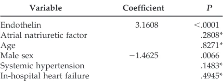
Plasma endothelin levels are elevated in the acute phase of myocardial infarction (MI) and in chronic heart failure. To test whether plasma endothelin con-centration is predictive of subsequent mortality, Om-land et al23 related it to 1-year mortality after docu-mented myocardial infarction. Blood samples from 142 patients in the subacute phase following myocar-dial infarction were assayed for endothelin 3 days after the onset of symptoms. Plasma endothelin was 5.66 0.3 pg/mL (mean 6 SEM) in the patients with myocardial infarction compared to 3.76 0.3 pg/mL in controls (P 5 .05), who were patients admitted to hospital with acute chest pain but without evidence of myocardial necrosis. Of the 142 MI patients, those who had clinical heart failure during their hospitalization had significantly higher plasma endothelin concentra-tions than those without evidence of heart failure (7.2 6 0.8 v 4.9 6 0.2 pg/mL, P , .001). At the conclusion of the 1-year follow-up, the day 3 plasma endothelin level of survivors was found to have been 5.16 0.2 pg/mL (mean 6 SEM). In comparison, pa-tients who died of cardiac causes during this 1-year period had endothelin levels of 9.2 6 1.5 pg/mL, which was significantly higher (P, .001). Analysis by a Cox proportional-hazards model revealed that plasma endothelin level was strongly related to sur-vival (P , .0001). Comparison with variables previ-ously determined to be associated with a poor prog-nosis, eg, age, male sex, and previous history of an-gina, systemic hypertension, or MI, found that, with the exception of male sex, none of these variables provided additional information after introduction of plasma endothelin in a multivariate model (Table 2).
SUMMARY
Endothelins are a family of 21–amino-acid peptides having potent vasoconstrictive effects. They bind to specific receptors, ETA and ETB, on smooth muscle and endothelial cells. Endothelin-1 is the best studied of this family, but its role in hypertension and other pathologies is still not well understood. When admin-istered to animals it causes severe vasoconstriction, but plasma levels are not significantly elevated in hypertension. Recent studies have reported a syner-gistic hypertensive effect of endothelin-1 and angio-tensin II, but not between endothelin-1 and norepi-nephrine. Endothelin receptor blockade has lowered blood pressure and reduced the cross-sectional area of
neointima both in hypertensive rats and after vascular injury. Infusion of the nonspecific endothelin receptor antagonist bosentan in patients with congestive heart failure decreased blood pressure, increased cardiac index, and reduced vascular resistance. Plasma endo-thelin-1 levels were found to have a significance at least equal to known risk factors in predicting the 1-year survival rate of patients after myocardial infarc-tion. While endothelin-1 alone is not thought to be an independent factor in the development of hyperten-sion or associated end-organ damage and pathology, there is increasing evidence that it may play a signif-icant role in such processes and may offer an impor-tant pathway for therapeutic intervention. The re-cently developed endothelin receptor antagonist bosentan appears promising in this regard.
REFERENCES
1. Li J, Lariviere R, Schiffrin E: Effect of a nonselective endothelin antagonist on vascular remodeling in de-oxycorticosterone acetate-salt hypertensive rats. Hy-pertension 1994;24:183–188.
2. Mortensen L, Pawloski C, Kanagy N, et al: Chronic hypertension produced by infusion of endothelin in rats. Hypertension 1990;15:729 –733.
3. Yanagisawa M, Kurihara H, Kimura S, et al: A novel potent vasoconstrictor peptide produced by vascular endothelial cells. Nature 1988;332:411– 415.
4. Kaajager K, Shaw S, Koomans H, et al: Role of endo-thelin receptor subtypes in the systemic and renal re-sponses to endothelin-1 in humans. J Am Soc Nephrol 1997;8:32–39.
5. Schiffrin E, Thibault G: Plasma endothelin in human essential hypertension. Am J Hypertens 1991;4:303–308. 6. Kohno M, Murakawa K, Horio T, et al: Plasma immu-noreactive endothelin-1 in experimental malignant hy-pertension. Hypertension 1991;18:93–100.
7. Lariviere R, Thibault G, Schiffrin E, et al: Increased endothelin-1 content in blood vessels of deoxycortico-sterone acetate-salt hypertensive but not in spontane-ously hypertensive rats. Hypertension 1993;21:294 –300. TABLE 2. MULTIVARIATE RELATION BETWEEN
VARIOUS DEMOGRAPHIC, CLINICAL, AND BIOCHEMICAL VARIABLES AND 1-YEAR MORTALITY AFTER MYOCARDIAL INFARCTION
ACCORDING TO A COX PROPORTIONAL-HAZARDS MODEL
Variable Coefficient P
Endothelin 3.1608 ,.0001
Atrial natriuretic factor .2808*
Age .8271*
Male sex 21.4625 .0066
Systemic hypertension .1483*
In-hospital heart failure .4945*
* Factors not included in the model.
tion. Hypertension 1991;18:543–549.
9. Rakugi H, Tabuchi Y, Nakamaru M, et al: Endothelin activates the vascular renin-angiotensin system in rat mesenteric arteries. Biochem Int 1990;21:867– 872. 10. Yoshida K, Yasujima M, Kohzuki M, et al: Endothelin-1
augments pressor response to angiotensin II infusion in rats. Hypertension 1992;20:292–297.
11. Yasujima, Abe K, Kohzuki M, et al: Atrial natriuretic factor inhibits the hypertension induced by chronic infusion of norepinephrine. Circ Res 1985;57:470 – 474. 12. Tabuchi Y, Nakamaru M, Rakugi H: Effects of
endo-thelin on neuroeffector junction in mesenteric arteries of hypertensive rats. Hypertension 1990;15:739 –743. 13. Libby P, et al: A cascade model for restenosis: a special
case of atherosclerosis progression. Circulation 1992; 86(suppl III):47–52.
14. Douglas S, Louden C, Vickery-Clark L, et al: A role for endogenous endothelin-1 in neointimal formation after rat carotid artery balloon angioplasty. Circ Res 1994;75: 190 –197.
15. Zamora M, Dempsey E, Walchak S, et al: BQ123, an ETA receptor antagonist, inhibits endothelin-1-medi-ated proliferation of human pulmonary artery smooth muscle cells. Am J Respir Cell Mol Biol 1993;9:429 – 433. 16. Ohlstein E, Douglas S, Sung C, et al: The selective ETA
350.
17. A¨ nggård E, et al: The fate of radioiodinated endothe-lin-1 and endothelin-3 in the rat. J Cardiovasc Pharma-col 1989;13(suppl 5):S46 –S49.
18. Waggoner W, Genova S, Rash V: Kinetic analyses dem-onstrate that equilibrium assumption does not apply to [125
I] endothelin-1 binding data. Life Sci 1992;51:1869 – 1876.
19. Bazil M, et al: Pharmacologic characterization of an endothelin-A (ETA) receptor antagonist in conscious rats. J Cardiovasc Pharmacol 1993;20:940 –948.
20. Chillon J, Heistad D, Baumbach G: Effects of endothelin receptor inhibition on cerebral arterioles in hyperten-sive rats. Hypertension 1996;27(part 2):794 –798. 21. Li J, Schiffrin E: Effect of chronic treatment of adult
spontaneously hypertensive rats with an endothelin receptor antagonist. Hypertension 1995;25:495–500. 22. Kiowski W, Sutsch G, Hunziker P, et al: Evidence of
endothelin-1-mediated vasoconstriction in severe chronic heart failure. Lancet 1995;346:732–736. 23. Omland T, Lie R, Aakvaag A, et al: Plasma endothelin
determination as a prognostic indicator of 1-year mor-tality after acute myocardial infarction. Circulation 1994;89:1573–1579.