Cytokine Profiles in Toxoplasmic and Viral Uveitis
Ibtissem Lahmar,1,3Ahmed Abou-Bacar,1Tamer Abdelrahman,1Marie Guinard,1Hamouda Babba,3Salim Ben Yahia,4 Moncef Kairallah,4Claude Speeg-Schatz,2Tristan Bourcier,2Arnaud Sauer,2Odile Villard,1Alexander W. Pfaff,1 Marc Mousli,1Justus G. Garweg,5,aand Ermanno Candolfi1,a1Institut de Parasitologie et de Pathologie Tropicale, Université de Strasbourg, and2Service d’Ophtalmologie, Hôpitaux Universitaires de Strasbourg, Strasbourg, France;3Laboratoire de Parasitologie Mycologie (99-UR/08-05), Faculté de Pharmacie Monastir, and4Département d’Ophtalmologie, Hôpital Universitaire Fattouma Bourguiba, Monastir, Tunisia; and5Clinic for Vitreoretinal Disease, Swiss Eye Institute and Medical Faculty, University of Bern, Bern, Switzerland
Background. Uveitis is a major cause of visual impairment throughout the world. Analysis of cytokine profiles in aqueous humor specimens may provide insight into the physiopathological processes that underly retinal damage in this context.
Methods. Using a multiplex assay, we determined the concentrations of 17 cytokines and chemokines in aqueous humor specimens obtained from patients with ocular toxoplasmosis or viral uveitis and compared these concentra-tions with those in specimens obtained from patients with noninfectious intermediate uveitis or cataract.
Results. Five mediators (interleukin [IL]– 8, monocyte chemoattractant protein–1, tumor necrosis factor–␣,
IL-4, and IL-10) were detected in⬎50% of patients in all groups. In contrast, IL-5 and IL-12 were specific for ocular toxoplasmosis, and granulocyte monocyte colony-stimulating factor and IL-1 were specific for viral uveitis; these mediators could present specific markers for diagnostic purposes. Interferon-␥, IL-6, and macrophage inflammatory protein-1 were common markers of ocular toxoplasmosis and viral uveitis. IL-17 was a common marker of ocular toxoplasmosis and intermediate uveitis.
Conclusions. We found specific cytokine profiles for each type of uveitis, with large interindividual variations and no etiological or clinical correlations. Ocular cytokine mapping contributes to a better understanding of the physio-pathology of specific forms of uveitis and provides guidance for new targeted treatment.
Uveitis is a potentially blinding inflammatory disease that affects individuals of any age. It is the third leading cause of blindness worldwide, accounting for 10%–15% of blindness. Despite huge advances in diagnostic tech-niques, many cases (35%–50%) remain idiopathic. The agents that most frequently cause infectious uveitis are Toxoplasma gondii and herpes simplex virus [1].
Herpes simplex virus is one of the most common in-fectious causes of uveitis, accounting for up to 9% of cases of anterior uveitis in published series. Herpetic uveitis may affect the sclera, cornea, or anterior or
pos-terior uvea and can present as panuveitis [2]. The infec-tion may manifest in an acute, chronic, or recurrent manner. Acute anterior herpetic uveitis is frequently as-sociated with corneal involvement but may also occur without corneal inflammation [3].
Ocular toxoplasmosis is a major cause of visual im-pairment throughout the world. It accounts for 30%– 50% of all cases of posterior uveitis; the estimated incidence rate in the United Kingdom is 0.4 cases per 100,000 persons per year [4]. In southern Brazil, 17.7% of individuals have been found to have retinal lesions compatible with ocular toxoplasmosis [5]. These figures may not represent the entire burden of ocular toxoplasmosis, because ocular toxoplasmosis is often asymptomatic and biological confirmation is not systematically sought. Necrotizing retinocho-roiditis and retinochoroidal scars are the hallmarks of ocular toxoplasmosis and are commonly accompa-nied by mild anterior uveitis and a vitreous inflamma-tory reaction [6]. However, Toxoplasma parasites are rarely detected in aqueous humor specimens from pa-tients with ocular toxoplasmosis [7], which suggests that Received 3 May 2008; accepted 6 November 2008; electronically published 24
March 2009.
Potential conflicts of interest: None reported.
Financial support: Swiss Foundation for the Prevention of Blindness (to J.G.G.) and Hôpitaux Universitaires de Strasbourg (through a Programme Hospitalier de Recherche Clinique program).
aE.C. and J.G.G. contributed equally to this work.
Reprints or correspondence: Dr. Ermanno Candolfi, Institut de Parasitolo-gie et de PatholoParasitolo-gie Tropicale, 3 rue Koeberlé, 67000 Strasbourg, France (ermanno.candolfi@medecine.u-strasbg.fr).
The Journal of Infectious Diseases 2009; 199:1239 – 49
© 2009 by the Infectious Diseases Society of America. All rights reserved. 0022-1899/2009/19908-0021$15.00
DOI: 10.1086/597478
Table 1. Clinical and biological characteristics of patients with ocular toxoplasmosis (OT). Patient Age, years Sex Place of origin Time from first symptom to obtainment of samples, day Severity of inflammation Toxoplasma -specific antibodies PCR result Definitive diagnosis Serum samples Aqueous humor samples IgG level, IU/mL IgM IgG avidity IgG level, IU/mL IgA index a GW coefficient b IgG immunoblot result 1 19 M Bern, Switzerland 21 3 280 Neg CI 290 Neg Pos ND ND RAOT 2 32 M Bern, Switzerland 60 1 35 Neg CI 11 Pos Pos ND Neg RCOT 3 45 M Bern, Switzerland 14 2 21 Neg CI 20 Pos ND ND ND RCOT 4 78 F Bern, Switzerland 120 2 2000 Pos CI 162 Pos Pos ND ND PI 5 58 F Bern, Switzerland 74 1 420 Neg CI 85 Pos Pos ND ND PI 6 45 F Bern, Switzerland 7 3 85 Pos AI 12 Pos Pos ND Neg PI 7 24 M Bern, Switzerland 14 1 118 Neg CI 86 Neg Neg ND Pos RAOT 8 30 M Bern, Switzerland 3 2 620 Pos CI 14 Pos Neg Pos Neg RAOT 9 40 M Bern, Switzerland 25 3 265 ND CI 201 ND Pos ND ND PI 10 18 F Bern, Switzerland 27 1 270 Neg CI 9 Neg Pos Pos ND PI 11 31 M Bern, Switzerland 5 1 7 Neg CI 6 Neg Pos ND ND RCOT 12 28 M Bern, Switzerland 14 3 1340 Neg CI 155 Neg Pos Pos ND PI 13 74 M Bern, Switzerland 20 2 260 Neg CI 74 Neg Pos Pos Neg PI 14 31 F Bern, Switzerland 12 2 26 Neg CI Neg Pos Neg Pos Neg RCOT 15 60 F Bern, Switzerland 25 3 63 Neg CI Neg Pos Neg ND Neg PI 16 60 F Bern, Switzerland 67 0 63 ND CI 11 ND Pos ND Neg PI 17 33 F Bern, Switzerland 10 1 165 Neg CI Neg Pos Neg ND Neg PI 18 17 F Bern, Switzerland 8 1 95 Neg CI 10 Neg Pos ND Neg RCOT 19 61 M Bern, Switzerland 14 2 28 Neg CI 56 Neg Pos Pos Neg RAOT 20 23 F Bern, Switzerland 4 1 17 Neg CI Neg Neg Neg Pos Neg RCOT 21 73 M Bern, Switzerland 8 3 290 Neg CI 71 Pos Neg Pos Neg RAOT 22 33 M Monastir, Tunisia 4 3 59 Neg CI ND ND ND Pos Neg RCOT 23 23 M Monastir, Tunisia 15 2 250 Neg CI ND ND ND Pos Neg RCOT 24 14 F Monastir, Tunisia 7 2 46 Neg CI ND ND ND Pos Neg RCOT 25 49 F Monastir, Tunisia 10 3 604 Neg CI ND ND ND Pos Neg RCOT 26 25 F Strasbourg, France 7 ND 574 Neg CI 1.27 ND Pos Pos ND PI 27 65 M Strasbourg, France 21 ND 110 Neg CI 0.84 ND Pos Pos ND PI NOTE. AI, acute infection; CI, chronic infection; GW, Goldman-Witmer; ND, not determined; PI, primary infection; RAOT, reactivated acquired OT; RCOT, re activated congenital OT. a An index ⬍ 0.5 was considered to be negative, and an index 肁 0.5 was considered to be positive. b A GW coefficient 肁 3 was considered to be positive, and a GW coefficient ⬍ 3 was considered to be negative.
parasite proliferation occurs only during the early phase of in-fection and that retinal damages may be attributable to subse-quent inflammatory processes [8].
Recently, interleukin (IL)–17-producing effector T cells, which are controlled by IL-27, were detected in inflamed brain specimens infected with Toxoplasma [9]. A similar pathway is found in experimental autoimmune uveoretinitis [10], which suggests that an autoimmune inflammatory response might contribute to bystander tissue destruction.
Aqueous humor cytokine and chemokine concentrations are elevated in both infectious and noninfectious uveitis [11], and more specific analysis might reveal the mechanisms of retinal damage in uveitis. However, because of the difficulties involved in obtaining aqueous humor samples and because of the limited available specimen volumes (150 –200L), ELISA can only pro-vide quantitative profiles for 3 or 4 cytokines per sample [12]. Currently, with the advent of the multiplex bioassay (Bio-Plex; Bio-Rad), up to 100 different molecules can be quantified simul-taneously in a sample volume of 20L.
In the present study, we used the multiplex assay to study the profiles of 17 cytokines and chemokines in aqueous humor sam-ples obtained from patients with ocular toxoplasmosis and viral uveitis and compared these profiles with those in aqueous hu-mor samples obtained from control subjects with intermediate uveitis or cataract.
PATIENTS AND METHODS
Patients with ocular toxoplasmosis. Paired aqueous humor and serum samples were obtained from 27 patients (14 female and 13 male patients) aged 14 –78 years (mean age, 40 years) after the patients provided informed consent, in accordance with the Declaration of Helsinki (table 1). The samples were obtained for routine diagnostic purposes from patients with an active retinochoroidal lesion strongly evoking ocular toxoplas-mosis who were seen in ophthalmology departments at 3 loca-tions (Bern, Switzerland; Monastir, Tunisia; and Strasbourg, France). Only 1 patient (patient 13; with ocular toxoplasmosis) had been treated (with corticosteroids) before the aqueous hu-mor sample was obtained. The serum samples were used to com-pare intraocular cytokine levels with such levels in paired serum samples. The diagnosis of ocular toxoplasmosis was confirmed by using at least 1 of the following methods: (1) local specific IgG antibody assay with the Goldmann-Witmer-Desmonts coeffi-cient, the modified Goldmann-Witmer-Desmonts coefficoeffi-cient, or immunoblot of paired aqueous humor and serum samples [7]; (2) local specific IgA assay [13]; or (3) PCR amplification of Toxoplasma DNA in aqueous humor specimens [7].
Patients with viral uveitis. The group of patients with viral uveitis consisted of 14 patients (mean age, 60 years) with
clini-cally typical and polymerase chain reaction– confirmed herpes simplex virus type 1 or varicella-zoster virus uveitis, as described in detail elsewhere [14].
Patients with chronic noninfectious intermediate uveitis.
Thir-teen patients (mean age, 32 years) with chronic cellular vitreal infil-tration of unknown etiology and without chorioretinal lesions (id-iopathic uveitis or sarcoidosis) were included in the group with intermediate uveitis.
Control group. The control group consisted of 22 patients (mean age, 62 years) who had undergone routine cataract sur-gery and had no evidence of any other intraocular pathology on preoperative examination.
Sample collection and processing. Aqueous humor sam-ples (100 –150L) were collected by means of anterior cham-ber paracentesis. Samples were immediately centrifuged at 20,000 g for 3 min, and the supernatant was stored in 25-L aliquots at⫺20°C until analysis. Serum samples were stored at⫺20°C.
Cytokine measurement in aqueous humor and serum samples. The Bio-Plex Human Cytokine 17-Plex Panel as-say (Bio-Rad) was used to measure cytokine and chemokine levels in aqueous humor and serum samples. The immune mediators were classified in 5 categories: (1) proinflamma-tory mediators (IL-1, IL-6, IL-8, monocyte chemoattractant protein [MCP]–1, and macrophage inflammatory protein [MIP]–1), (2) type 1 cytokines (IL-2, IL-7, IL-12, interferon [IFN]-␥, and tumor necrosis factor [TNF]–␣), (3) type 2 cy-tokines (IL-4, IL-5, IL-10, and IL-13), (4) T helper (Th) 17 cytokine (IL-17), and (5) growth factors (granulocyte colony-stimulating factor [G-CSF] and granulocyte monocyte colony-stimulating factor [GM-CSF]).
The cytokine and chemokine assay plate layout consisted of 8 standards in duplicate (1–32,000 pg/mL), 1 blank well (for back-ground fluorescence subtraction), and 50-L duplicates of each aqueous humor or serum sample, diluted 3-fold with Bio-Plex human serum diluent. The Bio-Plex method was performed as recommended by the manufacturer.
Data were analyzed with Bio-Plex Manager software, version 1.1 (Bio-Rad). The index was calculated as the ratio of cytokine concentration in patients with uveitis and the mean concentra-tion plus 3 times the standard error of the mean for each cyto-kine from the 22 patients with cataract. Indexes were calculated for ocular toxoplasmosis, viral uveitis, and intermediate uveitis. An index⬍5 was considered to be low positive, an index of 5–10 was considered to be medium positive, and an index⬎10 was considered to be high.
Statistical analysis. The Kruskal-Wallis test was performed using GraphPad Prism software, version 5, to compare the me-dian titers of immune mediators among the 4 groups of patients. Spearman rank correlation was used to detect correlations among the different mediators.
RESULTS
Comparison of Aqueous Humor and Serum Cytokine Profiles in Patients with Ocular Toxoplasmosis
Paired serum and aqueous humor samples from 27 patients with ocular toxoplasmosis were analyzed. Titers of MIP-1 and IL-7 were significantly lower in aqueous humor samples than in se-rum samples, although IL-2 levels were similar in both types of samples. The titers of the other 14 mediators were significantly higher in aqueous humor samples than in serum samples.
Cytokine and Chemokine Profiles in Aqueous Humor Samples
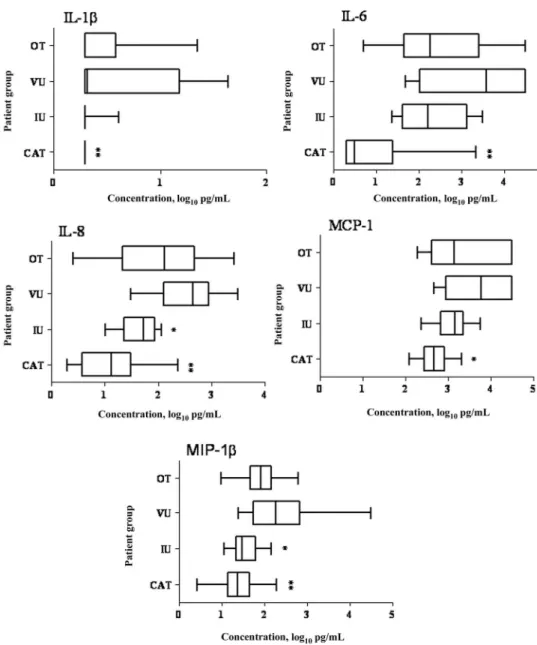
Proinflammatory mediators. IL-1, IL-6, IL-8, MCP-1, and
MIP-1 were detected in aqueous humor samples from 30%, 55%, 63%, 55%, and 55% of patients with ocular toxoplasmosis, respectively (table 2). The levels of all of these mediators were higher in samples from patients with ocular toxoplasmosis than in samples from patients with cataract (P⭐ .002), although IL-8 and MIP-1 levels were higher in samples from patients with ocular toxoplasmosis than in samples from patients with intermediate uveitis (P⭐ .03), and IL-6 and IL-8 levels were lower in samples from patients with ocular toxoplasmosis than in samples from patients with viral uveitis (P⫽ .003) (figure 1).
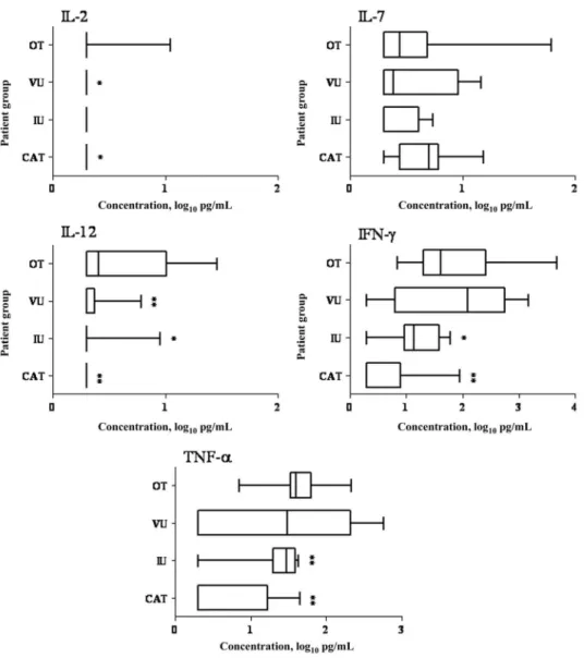
Type 1 cytokines. IL-2, IL-7, IL-12, IFN-␥, and TNF-␣ were
detected in samples from 18%, 15%, 52%, 67%, and 85% of patients with ocular toxoplasmosis, respectively (table 2). Levels of all cytokines, except for IL-7, were significantly higher in sam-ples from patients with ocular toxoplasmosis than in samsam-ples
from patients with cataract (P⭐ .001), although only IL-2 and IL-12 levels were higher in samples from patients with ocular plasmosis than in samples from patients with intermediate uve-itis (P⭐ .01) and in samples from patients with viral uveitis (P⭐ .01). IL-7, IFN-␥, and TNF-␣ levels were similar in sam-ples from patients with ocular toxoplasmosis, samsam-ples from pa-tients with intermediate uveitis, and samples from papa-tients with viral uveitis (figure 2).
Type 2 cytokines. IL-4, IL-5, IL-10, and IL-13 were detected in samples from 63%, 55%, 63%, and 7% of patients with ocular toxoplasmosis, respectively (table 2). All of the type 2 cytokines were up-regulated in patients with ocular toxoplasmosis, com-pared with patients with cataract (P⭐ .02). IL-4, IL-5, and IL-10 levels were higher in samples from patients with ocular toxoplasmosis than in samples from patients with intermediate uveitis (P⭐ .04), and the type 2 cytokine concentrations were similar in samples from patients with ocular toxoplasmosis and in samples from patients with viral uveitis (figure 3).
Growth factors. G-CSF was detected in samples from 44% of patients with ocular toxoplasmosis (table 2). G-CSF levels were higher in samples from patients with ocular toxoplasmosis than in samples from patients with cataract (P⬍ .001). Of in-terest, GM-CSF levels were found to be significantly lower in samples from patients with ocular toxoplasmosis than in sam-ples from patients with cataract (P⬍ .001) and in samples from patients with viral uveitis (P⬍ .001). Levels of both factors were similar in samples from patients with ocular toxoplasmosis and in samples from patients with intermediate uveitis (figure 4A). Table 2. Profiles of immune mediators in different groups of uveitis.
Mediator IU group VU group Index, by patient Patients, no. (%) Index, by patient Patients, no. (%) 1 2 3 4 5 6 7 8 9 10 11 12 13 1 2 3 4 5 6 7 8 9 10 11 12 13 14 IL-1 ⫹⫹⫹ 1 (8) ⫹⫹⫹ ⫹⫹⫹ ⫹⫹⫹ ⫹⫹⫹ ⫹⫹⫹ ⫹⫹⫹ ⫹⫹⫹ 7 (50) IL-6 ⫹ ⫹ ⫹ ⫹ 4 (31) ⫹⫹⫹ ⫹⫹⫹ ⫹⫹⫹ ⫹⫹⫹ ⫹ ⫹⫹⫹ ⫹⫹⫹ 7 (50) IL-8 ⫹ ⫹ ⫹ ⫹ ⫹ 5 (54) ⫹⫹ ⫹ ⫹⫹ ⫹⫹⫹ ⫹ ⫹⫹ ⫹⫹ ⫹⫹ ⫹⫹⫹ ⫹⫹ ⫹ ⫹ 12 (86) MCP-1 ⫹ ⫹ ⫹ ⫹ ⫹ ⫹ ⫹ ⫹⫹⫹ ⫹ 9 (69) ⫹⫹⫹ ⫹⫹⫹ ⫹⫹⫹ ⫹ ⫹ ⫹ ⫹⫹⫹ ⫹⫹⫹ ⫹⫹⫹ ⫹ 10 (71) MIP-1 ⫹ ⫹ ⫹ 3 (23) ⫹⫹⫹ ⫹⫹ ⫹ ⫹ ⫹⫹ ⫹⫹⫹ ⫹⫹⫹ ⫹ ⫹ 9 (64) IL-2 0 0 IL-7 0 ⫹ ⫹ 2 (14) IL-12 ⫹⫹⫹ ⫹⫹⫹ 2 (15) ⫹⫹⫹ ⫹⫹⫹ 2 (14) IFN-␥ ⫹ ⫹ ⫹ ⫹ 4 (31) ⫹⫹ ⫹⫹⫹ ⫹⫹ ⫹⫹⫹ ⫹⫹⫹ ⫹⫹⫹ ⫹⫹⫹ ⫹⫹ 8 (57) TNF-␣ ⫹ ⫹ ⫹ ⫹ ⫹ ⫹ ⫹ ⫹ ⫹ ⫹ 10 (78) ⫹ ⫹⫹⫹ ⫹⫹ ⫹⫹⫹ ⫹⫹ ⫹⫹⫹ ⫹⫹ 7 (50) IL-4 ⫹ ⫹ ⫹ ⫹ ⫹ ⫹ ⫹ 7 (54) ⫹ ⫹⫹ ⫹ ⫹⫹ ⫹⫹ ⫹ ⫹⫹⫹ ⫹⫹ 8 (57) IL-5 ⫹⫹⫹ 1 (8) ⫹⫹⫹ ⫹⫹⫹ ⫹⫹⫹ ⫹⫹⫹ ⫹⫹⫹ 5 (36) IL-10 ⫹⫹⫹ ⫹⫹ ⫹⫹ ⫹⫹ ⫹⫹ ⫹⫹ ⫹⫹ ⫹⫹ 8 (61) ⫹⫹⫹ ⫹ ⫹⫹⫹ ⫹ ⫹⫹⫹ ⫹⫹⫹ ⫹⫹ ⫹⫹ ⫹⫹⫹ ⫹⫹⫹ ⫹⫹⫹ ⫹⫹⫹ 12 (86) IL-13 ⫹ ⫹ 2 (15) ⫹ ⫹ ⫹ ⫹ 4 (28) IL-17 ⫹ ⫹ ⫹ ⫹ ⫹ ⫹ ⫹ 7 (54) ⫹ ⫹⫹ 2 (14) G-CSF ⫹ ⫹ ⫹ ⫹ ⫹ ⫹ ⫹ 7 (54) ⫹⫹ ⫹⫹⫹ ⫹ ⫹⫹⫹ ⫹⫹⫹ ⫹ ⫹⫹⫹ ⫹⫹⫹ 8 (57) GM-CSF 0 ⫹ ⫹ ⫹ ⫹ ⫹ ⫹ ⫹ ⫹ ⫹ ⫹ ⫹ 11 (78)
NOTE. The index was calculated as the ratio of cytokine concentration in patients with uveitis and the mean concentration plus 3 times the standard error of the mean for each cytokine from the patients with cataract. Lower production (⫹) was marked by an index ⬍5, medium production (⫹⫹) was marked by an index of 5–10, and higher production (⫹⫹⫹) was marked by an index ⬎10.
Th17 cytokine. IL-17 was detected in samples from 70% of patients with ocular toxoplasmosis (table 2). IL-17 levels were higher in samples from patients with ocular toxoplasmosis than in samples from patients with cataract (P⬍ .001) and in sam-ples from patients with viral uveitis (P⬍ .001); however, IL-17 levels were not different between samples from patients with ocular toxoplasmosis and samples from patients with interme-diate uveitis (figure 4B).
Individual Variation of Immune Mediators
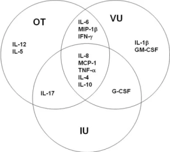
IL-6, IL-8, MCP-1, MIP-1, IL-12, IFN-␥, TNF-␣, IL-4, IL-5, IL-10, and IL-17 were detected in of aqueous humor specimens from⬎50% patients with ocular toxoplasmosis (table 2). Eight patients (30%) with ocular toxoplasmosis had detectable IL-12 and IL-17. These cytokines were also found in samples from 2 patients (14%) with viral uveitis and in a sample from 1 patient with intermediate uveitis (table 2). IL-8, MCP-1, IL-17, TNF-␣, IL-4, IL-10, and G-CSF were up-regulated in samples from ⬎50% of patients with intermediate uveitis; however, only 3 of the 13 patients had detectable levels of all of these mediators (Table 2). IL-1, IL-6, IL-8, MCP-1, MIP-1, IFN-␥, TNF-␣, IL-4, IL-10, and growth factors were detected in samples from ⬎50% of patients with viral uveitis; however, only 3 of the 14 patients had detectable levels of all of these mediators (table 2). Of interest, 5 mediators (8, MCP-1, TNF-␣, 4, and IL-10) were detected in samples from ⬎50% of patients in all groups. In contrast, IL-5 and IL-12 were specific for ocular tox-oplasmosis, and GM-CSF and IL-1 were specific for viral uveitis; thus, these mediators could present specific markers for
diag-nostic purposes. Finally, IFN-␥, IL-6, and MIP-1 were fre-quently detected in samples from patients with ocular toxoplas-mosis and in samples from those with viral uveitis, whereas IL-17 was frequently detected in samples from patients with oc-ular toxoplasmosis and in samples from those with intermediate uveitis (figure 5).
Correlation Between Mediator Levels in Aqueous Humor Samples and Clinical Characteristics of Ocular
Toxoplasmosis
Local cytokine concentrations in patients with ocular toxoplas-mosis did not correlate with age, sex, and region of origin of the patient; time from symptom onset to the obtainment of samples; the degree of uveal inflammation; or the etiology of the infection (primary acquired or congenital).
DISCUSSION
We observed higher concentrations of several cytokines in aque-ous humor samples than in serum samples. This may indicate that these cytokines are locally secreted or that they are not cleared from the eye as quickly as they are from the serum, per-haps because there are fewer immune cells and, therefore, cyto-kine receptors in the eye.
B cells and specific antibodies are abundant in human ocular toxoplasmosis and, in addition to recruited macrophages, are likely to play a protective role. Immunohistochemical studies have shown a predominance of T cells and macrophages among the inflammatory cells found in aqueous humor of fetuses and Table 2. (Continued.) Mediator OT group Index, by patient Patients, no. (%) 1 2 3 4 5 6 7 8 9 10 11 12 13 14 15 16 17 18 19 20 21 22 23 24 25 26 27 IL-1 ⫹⫹⫹ ⫹⫹⫹ ⫹⫹⫹ ⫹⫹⫹ ⫹⫹⫹ ⫹⫹⫹ ⫹⫹⫹ ⫹⫹⫹ 8 (30) IL-6 ⫹⫹ ⫹ ⫹⫹⫹ ⫹ ⫹⫹⫹ ⫹⫹⫹ ⫹⫹⫹ ⫹⫹⫹ ⫹⫹⫹ ⫹⫹ ⫹⫹⫹ ⫹⫹ ⫹⫹⫹ ⫹ ⫹⫹⫹ 15 (55) IL-8 ⫹ ⫹⫹ ⫹ ⫹⫹⫹ ⫹ ⫹⫹⫹ ⫹ ⫹⫹ ⫹ ⫹⫹ ⫹⫹ ⫹ ⫹ ⫹⫹ ⫹⫹⫹ ⫹ ⫹⫹ 17 (63) MCP-1 ⫹⫹ ⫹ ⫹⫹⫹ ⫹ ⫹⫹⫹ ⫹⫹⫹ ⫹⫹⫹ ⫹⫹⫹ ⫹⫹⫹ ⫹⫹ ⫹⫹⫹ ⫹⫹ ⫹⫹⫹ ⫹ ⫹⫹⫹ 15 (55) MIP-1 ⫹ ⫹ ⫹ ⫹⫹ ⫹ ⫹⫹ ⫹ ⫹ ⫹ ⫹ ⫹ ⫹ ⫹ ⫹ ⫹ 15 (55) IL-2 ⫹⫹⫹ ⫹⫹⫹ ⫹⫹⫹ ⫹⫹⫹ ⫹⫹⫹ 5 (18) IL-7 ⫹ ⫹ ⫹ ⫹⫹ 4 (15) IL-12 ⫹⫹⫹ ⫹⫹⫹ ⫹⫹⫹ ⫹⫹⫹ ⫹⫹⫹ ⫹⫹⫹ ⫹⫹⫹ ⫹⫹⫹ ⫹⫹ ⫹⫹⫹ ⫹⫹⫹ ⫹⫹ ⫹⫹⫹ ⫹⫹⫹ 14 (52) IFN-␥ ⫹ ⫹ ⫹⫹ ⫹ ⫹⫹⫹ ⫹⫹ ⫹⫹⫹ ⫹⫹ ⫹⫹ ⫹⫹ ⫹ ⫹⫹⫹ ⫹ ⫹ ⫹⫹ ⫹⫹ ⫹ ⫹⫹⫹ 18 (67) TNF-␣ ⫹ ⫹ ⫹ ⫹ ⫹ ⫹ ⫹⫹ ⫹ ⫹ ⫹ ⫹⫹ ⫹ ⫹ ⫹ ⫹ ⫹ ⫹ ⫹ ⫹⫹ ⫹ ⫹ ⫹ ⫹ 23 (85) IL-4 ⫹ ⫹ ⫹ ⫹ ⫹ ⫹ ⫹ ⫹ ⫹ ⫹ ⫹ ⫹ ⫹ ⫹ ⫹ ⫹ ⫹ 17 (63) IL-5 ⫹⫹⫹ ⫹⫹⫹ ⫹⫹⫹ ⫹⫹⫹ ⫹⫹⫹ ⫹⫹⫹ ⫹⫹⫹ ⫹⫹⫹ ⫹⫹⫹ ⫹⫹⫹ ⫹⫹⫹ ⫹⫹⫹ ⫹⫹⫹ ⫹⫹⫹ ⫹⫹⫹ 15 (55) IL-10 ⫹⫹⫹ ⫹ ⫹⫹ ⫹⫹⫹ ⫹⫹ ⫹⫹⫹ ⫹ ⫹⫹⫹ ⫹ ⫹⫹ ⫹⫹⫹ ⫹⫹⫹ ⫹⫹ ⫹⫹⫹ ⫹⫹⫹ ⫹⫹⫹ ⫹⫹⫹ 17 (63) IL-13 ⫹⫹ ⫹ 2 (7) IL-17 ⫹ ⫹ ⫹ ⫹ ⫹ ⫹ ⫹ ⫹ ⫹ ⫹ ⫹ ⫹ ⫹ ⫹ ⫹ ⫹ ⫹ ⫹ ⫹ 19 (70) G-CSF ⫹ ⫹ ⫹⫹ ⫹⫹⫹ ⫹⫹ ⫹⫹ ⫹⫹ ⫹⫹ ⫹ ⫹⫹ ⫹⫹⫹ ⫹⫹ 12 (44) GM-CSF 0
infants with active disease [15]. The regulatory role of cytokines in this situation is unclear. In mouse models of ocular toxoplas-mosis, IFN-␥, TNF-␣, and nitric oxide play an important role, mainly after primary infection, in the control of acquired ocular toxoplasmosis [16]. The ambiguous role of proinflammatory and immunoregulatory factors in intracellular and, namely, in-traocular pathogen infections is still being debated [17]. Human aqueous humor cytokine mapping may be the first step toward addressing this issue.
Proinflammatory mediator (MCP-1, IL-8, and IL-6) levels were increased in aqueous humor samples from patients with ocular toxoplasmosis, as was reported elsewhere for patients with viral uveitis [11]. During toxoplasmic uveitis, these
proin-flammatory mediators could be produced by Müller cells, retinal pigment epithelial cells, or retinal vascular endothelial cells [18 – 20]. The mediators lead to the recruitment of neutrophils, monocytes, and T lymphocytes, which are essential for clearing the infection. MCP-1 is involved in the recruitment of a partic-ular population of Gr-1⫹monocytes that are capable of lysing T. gondii and other intracellular miccroorganisms [21]. Such in-fected CD11c⫹and CD11b⫹monocytes were shown to dissem-inate the infection to the brain in a mouse model [22]. In a mouse model of retinal detachment [23], MCP-1 was shown to play a central role in photoreceptor cell apoptosis and could, thus, contribute to reduced visual acuity in the context of ocular toxoplasmosis and to the triggering of an autoimmune process. Figure 1. Proinflammatory cytokine levels in aqueous humor samples obtained from 22 patients with cataract (CAT), 13 patients with intermediate uveitis (IU), 14 patients with viral uveitis (VU), and 27 patients with ocular toxoplasmosis (OT). Vertical lines represent median values, boxes represent interquartile ranges, and whiskers represent minimum and maximum values. IL, interleukin; MCP, monocyte chemoattractant protein; MIP, macrophage
inflammatory protein. *P⬍ .05, for OT vs. CAT, OT vs. IU, and OT vs. VU; **P ⬍ .001, for OT vs. CAT, OT vs. IU, and OT vs. VU.
Local production of IL-8 in ocular toxoplasmosis and viral uveitis appears to have a crucial role in neutrophil recruitment to the eye [20]. It is noteworthy that mice lacking IL-8 are more susceptible to T. gondii infection than are mice with detectable IL-8 [24].
IL-6 was detected in the 2 forms of infectious uveitis: acute and chronic intraocular inflammation. Müller cells may be a source of IL-6 [20]. IL-6 is considered to be a strong proinflam-matory mediator in the eye [25]. The high intraocular concen-tration of IL-6 in ocular toxoplasmosis could add to the action of chemokines and aggravate the destructive local inflammatory response within the retina. IL-6 inhibits the activity of trans-forming growth factor (TGF)–, an anti-inflammatory cyto-kine. TGF- may be the key cytokine for maintenance of ante-rior chamber–associated immune deviation, for control of intraocular inflammation, and for maintenance of the integrity
of the eye [26]. On the other hand, IL-6 may also have a protec-tive role against parasite proliferation, because T. gondii–in-fected IL-6⫺/⫺mice have higher parasite loads and more-severe ocular inflammation, compared with IL-6⫹/⫹mice [27].
Elevated local levels of IL-6 and TGF- have been found in murine ocular toxoplasmosis. IL-6 seems to control the ocular parasite burden and inflammatory activity, whereas TGF- (as a regulator of the ocular immune privilege), in association with IL-10 [28], limits inflammatory activity [27].
Type 1 cytokines play an important role in the control of tox-oplasmic infection [29]. We found that ocular IL-12 levels were significantly augmented in ocular toxoplasmosis; this might have been responsible for the local IFN-␥ and TNF-␣ production ob-served in most of our patients. Ocular IFN-␥ and TNF-␣ may have 2 effects in ocular toxoplasmosis: (1) limiting Toxoplasma prolifer-ation [30] and (2) amplifying the local inflammatory reaction [31]. Figure 2. Type 1 cytokine levels in aqueous humor samples obtained from 22 patients with cataract (CAT), 13 patients with intermediate uveitis (IU), 14 patients with viral uveitis (VU), and 27 patients with ocular toxoplasmosis (OT). Vertical lines represent median values, boxes represent interquartile
ranges, and whiskers represent minimum and maximum values. IFN, interferon; IL, interleukin; TNF, tumor necrosis factor. *P⬍ .05, for OT vs. CAT,
This may explain why Toxoplasma DNA was detected in an aqueous humor sample from only 1 of the 17 patients with ocular toxoplas-mosis in our study, possibly because of late presentation after symp-tom onset (mean time from sympsymp-tom onset to presentation, 22.8 days [range, 3–120 days]). Indeed, Toxoplasma undergoes a rapid stage transition in the anterior chamber and is cleared by the immune response [32]. Not surprisingly, IFN-␥ and TNF-␣ lev-els were accordingly augmented in the group of patients with viral uveitis. Studies of mouse models of viral necrotizing retini-tis have revealed that herpes simplex virus type 1 infection leads to a remarkable increase in IFN-␥ and TNF-␣ expression in cho-rioretinal tissue [33]. Together, our data confirm the strong type 1 immune response during retinal infection by intracellular mi-croorganisms [30].
Another pillar of inflammation is IL-17, produced by Th17 lymphocytes, a CD4⫹T cell subset that is considered to be responsible for autoimmune inflammation [34], including autoimmune uveoretinitis [10]. Recently, IL-17 was shown to be involved in murine toxoplasmic encephalitis [9]. We found for the first time, to our knowledge, that IL-17 was up-regulated in 70% of patients with ocular toxoplasmosis. In addition to its anti-Toxoplasma role [35, 36], IL-17 stimulates the production of IL-6 and NO and amplifies the local inflam-matory response in synergy with other mediators, such as IL-1, TNF-␣, and IFN-␥ [34]. Therefore, it is noteworthy that TNF-␣ and IFN-␥ were both strongly up-regulated in our
patients with ocular toxoplasmosis. IL-17 also amplifies local inflammatory responses by recruiting neutrophils and mono-cytes to sites of infection through the production of IL-8, MCP-1, growth-regulated oncogene–␣, and G-CSF [36, 37]. Finally, detection of IL-17 in aqueous humor samples from patients with ocular toxoplasmosis supports the existence of an autoimmune process that results from cytopathic effects to damaged tissues, such as phagocytosis of photoreceptors and retinal pigmented epithelial cells, in the context of ocular tox-oplasmosis [38, 39]. It is highly probable that counterbalanc-ing of IL-17 by retinal IL-27 occurs (not determined in our series) [9, 10].
Type 2 cytokine up-regulation in ocular toxoplasmosis and in viral uveitis, namely up-regulation of IL-4, IL-5, and IL-10, may down-regulate the local inflammatory process by inhibiting IL-12, TNF-␣–producing macrophages, or Tbet⫹Foxp3⫺ lympho-cytes [40 – 42]. Regulation of IL-10 –producing cells (Th1, Th2, or even Th17 cells), found in one-third of the patients with oc-ular toxoplasmosis in our study, seems to be complex, and one of the possible pathways is controlled by IL-27 and TGF- [9, 43, 44].
IL-5 may also have a protective role [45]. It is required for the production of eosinophils and for induction of protective anti-bodies in adaptive immune responses; thus, it is associated with enhancement of the Th2 response. In our study, 55% of patients Figure 3. Type 2 cytokine levels in aqueous humor samples obtained from 22 patients with cataract (CAT), 13 patients with intermediate uveitis (IU), 14 patients with viral uveitis (VU), and 27 patients with ocular toxoplasmosis (OT). Vertical lines represent median values, boxes represent interquartile
ranges, and whiskers represent minimum and maximum values. IL, interleukin. *P⬍ .05, for OT vs. CAT, OT vs. IU, and OT vs. VU; **P ⬍ .001, for
OT vs. CAT, OT vs. IU, and OT vs. VU.
with ocular toxoplasmosis had high levels of IL-5, which was associated with elevated production of IL-12 and IFN-␥. This finding provides a new observation with regard to the role of IL-5 in the regulation of IL-12 and IFN-␥ (which was reported elsewhere [46]). Moreover, IL-4, IL-5, and IL-10 may be respon-sible for local IgG production during Toxoplasma infection [47]. Of interest, GM-CSF seemed to be specifically produced in viral uveitis in our study; however, in a previous clinical study on herpes uveitis, the authors did not find any GM-CSF in aqueous humor samples [11]. In contrast to our findings for GM-CSF, up-regulation of G-CSF occurred in the context of the other forms of uveitis. G-CSF delays neutrophil apoptosis and may thus prolong the inflammatory process in the eye [48].
We found a wide variation of cytokine levels produced among all samples demonstrating uveitis. However, we found no corre-lation between cytokine titers and clinical characteristics in oc-ular toxoplasmosis, which suggests that genetic factors are in-volved in cytokine production [49], as was shown in murine ocular toxoplasmosis [50]. In this context, up-regulation of IL-12 and IL-17 in the absence of GM-CSF seems to be a partic-ularly interesting profile.
In conclusion, we observed wide interindividual variations in local cytokine concentrations among samples obtained from pa-tients with infectious uveitis. However, acute viral ocular infec-tions result mostly in explosive production of chemokines, of which GM-CSF is the most representative. In semichronic or chronic uveitis, ocular toxoplasmosis or intermediate uveitis shared Th1- and Th17-weighted local cytokine profiles; IL-5 and IL-12 were more specific for ocular toxoplasmosis. These data support the hypothesis of a microorganism-induced inflamma-tory response that is probably derived from MCP-1 and IL-8 recruited monocytes, neutrophils, and T cells. Type 2 cytokine response, observed in all 3 types of uveitis, counterbalances the ongoing ocular inflammatory process. The ocular immuno-privileged site thus creates such a counterproductive conse-quence of its ability to specifically exclude infiltrating T cells and macrophages. The resulting imbalance may result in the ob-served retinal damage. Ocular cytokine mapping may, thereby, not only contribute to a closer understanding of the pathophys-iological characteristics underlying uveitis but also provide guid-ance for the detection of new treatment targets, such as GM-CSF, IL-17, IL-6, and even TNF-␣. This information may serve Figure 4. Levels of T helper 17 cytokines (A) and growth factors (B) in aqueous humor samples obtained from 22 patients with cataract (CAT), 13 patients with intermediate uveitis (IU), 14 patients with viral uveitis (VU), and 27 patients with ocular toxoplasmosis (OT). Vertical lines represent median values, boxes represent interquartile ranges, and whiskers represent minimum and maximum values. G-CSF, granulocyte colony-stimulating factor;
GM-CSF, granulocyte monocyte colony-stimulating factor; IL, interleukin.*P⬍ .05, for OT vs. CAT, OT vs. IU, and OT vs. VU; **P ⬍ .001, for OT vs.
as a rational basis to introduce targeted local use of cytokine bioregulators.
Acknowledgments
We thank Gilles Prevost and Daniel Keller for assistance with performing the Bio-Plex assay.
References
1. Hooper C, McCluskey P. Intraocular inflammation: its causes and in-vestigations. Curr Allergy Asthma Rep 2008; 8:331– 8.
2. Miserocchi E, Waheed NK, Dios E, et al. Visual outcome in herpes sim-plex virus and varicella zoster virus uveitis: a clinical evaluation and comparison. Ophthalmology 2002; 109:1532–7.
3. Schacher S, Garweg JG, Russ C, Bohnke M. Diagnosis of herpetic uveitis and keratouveitis [in German]. Klin Monatsbl Augenheilkd 1998; 212: 359 – 62.
4. McCannel CA, Holland GN, Helm CJ, Cornell PJ, Winston JV, Rimmer TG. Causes of uveitis in the general practice of ophthalmology. UCLA Community-Based Uveitis Study Group. Am J Ophthalmol 1996; 121: 35– 46.
5. Glasner PD, Silveira C, Kruszon-Moran D, et al. An unusually high prev-alence of ocular toxoplasmosis in southern Brazil. Am J Ophthalmol
1992; 114:136 – 44.
6. Vallochi AL, da Silva Rios L, Nakamura MV, et al. The involvement of autoimmunity against retinal antigens in determining disease severity in toxoplasmosis. J Autoimmun 2005; 24:25–32.
7. Villard O, Filisetti D, Roch-Deries F, Garweg J, Flament J, Candolfi E. Comparison of enzyme-linked immunosorbent assay, immunoblotting, and PCR for diagnosis of toxoplasmic chorioretinitis. J Clin Microbiol
2003; 41:3537– 41.
8. Garweg J, Candolfi E. Immunopathology in ocular toxoplasmosis: facts and possible clues. Memorias do Instituto Oswaldo Cruz (in press). 9. Stumhofer JS, Laurence A, Wilson EH, et al. Interleukin 27 negatively
regulates the development of interleukin 17–producing T helper cells during chronic inflammation of the central nervous system. Nat Immu-nol 2006; 7:937– 45.
10. Amadi-Obi A, Yu CR, Liu X, et al. TH17 cells contribute to uveitis and scleritis and are expanded by IL-2 and inhibited by IL-27/STAT1. Nat Med 2007; 13:711– 8.
11. Curnow SJ, Falciani F, Durrani OM, et al. Multiplex bead immunoassay analysis of aqueous humor reveals distinct cytokine profiles in uveitis. Invest Ophthalmol Vis Sci 2005; 46:4251–9.
12. Ongkosuwito JV, Feron EJ, van Doornik CE, et al. Analysis of immuno-regulatory cytokines in ocular fluid samples from patients with uveitis. Invest Ophthalmol Vis Sci 1998; 39:2659 – 65.
13. Garweg JG, Jacquier P, Boehnke M. Early aqueous humor analysis in patients with human ocular toxoplasmosis. J Clin Microbiol 2000; 38: 996 –1001.
14. Garweg J, Bohnke M. Varicella-zoster virus is strongly associated with atyp-ical necrotizing herpetic retinopathies. Clin Infect Dis 1997; 24:603– 8. 15. Roberts F, Mets MB, Ferguson DJ, et al. Histopathological features of
ocular toxoplasmosis in the fetus and infant. Arch Ophthalmol 2001; 119:51– 8.
16. Roberts F, Roberts CW, Ferguson DJ, McLeod R. Inhibition of nitric oxide production exacerbates chronic ocular toxoplasmosis. Parasite Immunol 2000; 22:1–5.
17. Gaddi PJ, Yap GS. Cytokine regulation of immunopathology in toxo-plasmosis. Immunol Cell Biol 2007; 85:155–9.
18. Knight BC, Brunton CL, Modi NC, Wallace GR, Stanford MR. The effect of Toxoplasma gondii infection on expression of chemokines by rat ret-inal vascular endothelial cells. J Neuroimmunol 2005; 160:41–7. 19. Nagineni CN, Detrick B, Hooks JJ. Toxoplasma gondii infection induces
gene expression and secretion of interleukin 1 (IL-1), IL-6, granulocyte-macrophage colony-stimulating factor, and intercellular adhesion mol-ecule 1 by human retinal pigment epithelial cells. Infect Immun 2000; 68:407–10.
20. Knight BC, Kissane S, Falciani F, Salmon M, Stanford MR, Wallace GR. Expression analysis of immune response genes of Müller cells infected with Toxoplasma gondii. J Neuroimmunol 2006; 179:126 –31. 21. Robben PM, LaRegina M, Kuziel WA, Sibley LD. Recruitment of Gr-1⫹
monocytes is essential for control of acute toxoplasmosis. J Exp Med
2005; 201:1761–9.
22. Courret N, Darche S, Sonigo P, Milon G, Buzoni-Gatel D, Tardieux I. CD11c- and CD11b-expressing mouse leukocytes transport single
Tox-oplasma gondii tachyzoites to the brain. Blood 2006; 107:309 –16.
23. Nakazawa T, Hisatomi T, Nakazawa C, et al. Monocyte chemoattractant protein 1 mediates retinal detachment–induced photoreceptor apopto-sis. Proc Natl Acad Sci U S A 2007; 104:2425–30.
24. Del Rio L, Bennouna S, Salinas J, Denkers EY. CXCR2 deficiency confers impaired neutrophil recruitment and increased susceptibility during
Toxoplasma gondii infection. J Immunol 2001; 167:6503–9.
25. Hoekzema R, Murray PI, Kijlstra A. Cytokines and intraocular inflam-mation. Curr Eye Res 1990; (Suppl):207–11.
26. Ohta K, Yamagami S, Taylor AW, Streilein JW. IL-6 antagonizes TGF- and abolishes immune privilege in eyes with endotoxin-induced uveitis. Invest Ophthalmol Vis Sci 2000; 41:2591–9.
27. Lyons RE, Anthony JP, Ferguson DJ, et al. Immunological studies of chronic ocular toxoplasmosis: up-regulation of major histocompatibil-ity complex class I and transforming growth factor beta and a protective role for interleukin-6. Infect Immun 2001; 69:2589 –95.
28. D’Orazio TJ, Niederkorn JY. A novel role for TGF- and IL-10 in the induction of immune privilege. J Immunol 1998; 160:2089 –98. 29. Hunter CA, Chizzonite R, Remington JS. IL-1 is required for IL-12 to
induce production of IFN-␥ by NK cells: a role for IL-1  in the T cell–independent mechanism of resistance against intracellular patho-gens. J Immunol 1995; 155:4347–54.
Figure 5. Schematic representation of cytokine profiles of different types of uveitis. Data are from samples obtained from 22 patients with cataract, 13 patients with intermediate uveitis (IU), 14 patients with viral uveitis (VU), and 27 patients with ocular toxoplasmosis (OT). The chemokines and cytokines in
the circles and their intersections were detected in⬎50% of patients in each
group. G-CSF, granulocyte colony-stimulating factor; GM-CSF, granulocyte monocyte colony-stimulating factor; IFN, interferon; IL, interleukin; MCP, monocyte chemoattractant protein; MIP, macrophage inflammatory protein; TNF, tumor necrosis factor.
30. Norose K, Mun HS, Aosai F, et al. IFN-␥–regulated Toxoplasma gondii distribution and load in the murine eye. Invest Ophthalmol Vis Sci 2003; 44:4375– 81.
31. Lu F, Huang S, Hu MS, Kasper LH. Experimental ocular toxoplasmosis in genetically susceptible and resistant mice. Infect Immun 2005; 73: 5160 –5.
32. Hayashi S, Chan CC, Gazzinelli RT, Pham NT, Cheung MK, Roberge FG. Protective role of nitric oxide in ocular toxoplasmosis. Br J Ophthal-mol 1996; 80:644 – 8.
33. Zheng M, Atherton SS. Cytokine profiles and inflammatory cells during HSV-1–induced acute retinal necrosis. Invest Ophthalmol Vis Sci 2005; 46:1356 – 63.
34. Afzali B, Lombardi G, Lechler RI, Lord GM. The role of T helper 17 (Th17) and regulatory T cells (Treg) in human organ transplantation and autoimmune disease. Clin Exp Immunol 2007; 148:32– 46.
35. Ye P, Rodriguez FH, Kanaly S, et al. Requirement of interleukin 17 re-ceptor signaling for lung CXC chemokine and granulocyte colony-stimulating factor expression, neutrophil recruitment, and host defense. J Exp Med 2001; 194:519 –27.
36. Kelly MN, Kolls JK, Happel K, et al. Interleukin-17/interleukin-17 re-ceptor–mediated signaling is important for generation of an optimal polymorphonuclear response against Toxoplasma gondii infection. In-fect Immun 2005; 73:617–21.
37. Miyamoto M, Prause O, Sjostrand M, Laan M, Lotvall J, Linden A. En-dogenous IL-17 as a mediator of neutrophil recruitment caused by en-dotoxin exposure in mouse airways. J Immunol 2003; 170:4665–72. 38. McMenamin PG, Dutton GN, Hay J, Cameron S. The ultrastructural
pathology of congenital murine toxoplasmic retinochoroiditis. I. The localization and morphology of Toxoplasma cysts in the retina. Exp Eye Res 1986; 43:529 – 43.
39. Geiger K, Howes E, Gallina M, Huang XJ, Travis GH, Sarvetnick N. Transgenic mice expressing IFN-␥ in the retina develop inflammation of the eye and photoreceptor loss. Invest Ophthalmol Vis Sci 1994; 35: 2667– 81.
40. Jankovic D, Kullberg MC, Feng CG, et al. Conventional T-bet⫹Foxp3⫺ Th1 cells are the major source of host-protective regulatory IL-10 during intracellular protozoan infection. J Exp Med 2007; 204:273– 83. 41. Horwitz DA, Zheng SG, Gray JD. The role of the combination of IL-2
and TGF-beta or IL-10 in the generation and function of CD4⫹CD25⫹ and CD8⫹regulatory T cell subsets. J Leukoc Biol 2003; 74:471– 8. 42. Fiorentino DF, Zlotnik A, Mosmann TR, Howard M, O’Garra A. IL-10
inhibits cytokine production by activated macrophages. J Immunol
1991; 147:3815–22.
43. Awasthi A, Carrier Y, Peron JP, et al. A dominant function for interleu-kin 27 in generating interleuinterleu-kin 10 –producing anti-inflammatory T cells. Nat Immunol 2007; 8:1380 –9.
44. Kastelein RA, Hunter CA, Cua DJ. Discovery and biology of IL-23 and IL-27: related but functionally distinct regulators of inflammation. Annu Rev Immunol 2007; 25:221– 42.
45. Zhang Y, Denkers EY. Protective role for interleukin-5 during chronic
Toxoplasma gondii infection. Infect Immun 1999; 67:4383–92.
46. Nickdel MB, Roberts F, Brombacher F, Alexander J, Roberts CW. Counter-protective role for interleukin-5 during acute Toxoplasma
gon-dii infection. Infect Immun 2001; 69:1044 –52.
47. Takase H, Futagami Y, Yoshida T, et al. Cytokine profile in aqueous humor and sera of patients with infectious or noninfectious uveitis. In-vest Ophthalmol Vis Sci 2006; 47:1557– 61.
48. Channon JY, Miselis KA, Minns LA, Dutta C, Kasper LH. Toxoplasma
gondii induces granulocyte colony-stimulating factor and
granulocyte-macrophage colony-stimulating factor secretion by human fibroblasts: implications for neutrophil apoptosis. Infect Immun 2002; 70:6048 –57. 49. Strack A, Asensio VC, Campbell IL, Schluter D, Deckert M. Chemokines are differentially expressed by astrocytes, microglia and inflammatory leukocytes in Toxoplasma encephalitis and critically regulated by interferon-␥. Acta Neuropathol 2002; 103:458–68.
50. Lu F, Huang S, Kasper LH. CD4⫹T cells in the pathogenesis of murine ocular toxoplasmosis. Infect Immun 2004; 72:4966 –72.