Case report
Transcatheter stent-valve implantation in a stenotic
pulmonary conduit via a sub-xyphoidian access
Enrico Ferrari
a,*
, Christopher Sulzer
b, Elena Rizzo
c, Ludwig Karl von Segesser
a aDepartment of Cardiovascular Surgery, University Hospital of Lausanne (CHUV), Lausanne, SwitzerlandbDepartment of Anaesthesia, University Hospital of Lausanne (CHUV), Lausanne, Switzerland cDepartment of Radiology, University Hospital of Lausanne (CHUV), Lausanne, Switzerland
Received 31 March 2009; received in revised form 15 April 2009; accepted 27 April 2009; Available online 12 June 2009
Abstract
Patients who develop a severe stenosis in biological pulmonary conduits previously implanted for pulmonary outflow trunk reconstructions are treated either by surgical re-replacement, or by transcatheter stent-valve implantation through a femoral vein access. A catheter-based sub-xyphoidian access through the right ventricle for stent-valve positioning in a pulmonary conduit has rarely been proposed. We describe the case of a 20-year-old man who underwent a pulmonary trunk reconstruction for a congenital pulmonary valve dysplasia and a few years later developed a stenosis in the pulmonary conduit. He was successfully treated with a 23 mm Edwards Sapien#stent-valve implantation in
pulmonary position, through an unusual right ventricular, sub-xyphoidian access and without contrast medium injections and pleura opening.
#2009 European Association for Cardio-Thoracic Surgery. Published by Elsevier B.V. All rights reserved. Keywords: Transcatheter valve replacement; Pulmonary conduit; Valve stenosis
1. Introduction
The stenosis is a severe complication following surgery for pulmonary outflow trunk reconstruction with biological conduits, as human cryopreserved homografts or bovine jugular veins (ContegraW, Medtronic, Minneapolis, USA). The operation for pulmonary conduit replacement under re-sternotomy and cardiopulmonary bypass remains a high-risk procedure with consistent morbidity and mortality[1]. The arrival of new stent-valves has been heralded as an important change in concepts and, recently, the use of transcatheter techniques for pulmonary valve replacement through the femoral vein (MelodyWvalve, Medtronic, Minneapolis, USA) has become routine in specialised cardiac care units[2—4]. In spite of that, there are a few limitations related to the use of this device in the presence of vena cava filters, femoral vein thrombosis/stenosis or very large pulmonary conduits (MelodyW
valve limited to 22 mm). Moreover, the positioning of catheter-based valves requires the injection of contrast medium which is nephro-toxic at high concentration. A sub-xyphoidian access for transcatheter valve implantation
without contrast medium injections, can be a valid alter-native in selected patients.
2. Case report
We hospitalised a 20-year-old man who underwent, 5 years earlier, open heart surgery for pulmonary trunk reconstruction (ContegraW
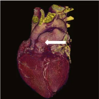
conduit 22 mm, Medtronic, Minneapolis, USA) in a congenital stenotic pulmonary valve dysplasia plus a concomitant interventricular defect closure. He was symptomatic for dyspnoea, fever and fatigue and we made the diagnosis of conduit stenosis plus sub-acute endocarditis; the trans-oesophageal echocardio-gram (TEE) showed a pulmonary peak gradient of 45 mmHg and a thick and dystrophic pulmonary valve with vegeta-tions. The sub-acute endocarditis was treated conserva-tively with intravenous antibiotics and 8 weeks later, the vegetation disappeared, but the patient developed a transitory antibiotic-related renal insufficiency. To decrease the operative risk and prevent an acute renal failure that could have occurred after cardiopulmonary bypass (in re-operation) or contrast medium employment (in trans-femoral approach), we decided for a sub-xyphoidian stent-valve implantation without contrast medium. We performed a pre-operative three-dimensional (3D) computed tomography scan to analyse the pulmonary
www.elsevier.com/locate/ejcts European Journal of Cardio-thoracic Surgery 36 (2009) 595—597
* Corresponding author. Address: Department of Cardiovascular Surgery, Centre Hoˆpitalier Universitaire Vaudois (CHUV), 46, rue du Bugnon, CH-1011 Lausanne, Switzerland. Tel.: +41 79 310 1386; fax: +41 21 314 2278.
E-mail address:enricoferrari@bluewin.ch(E. Ferrari).
1010-7940/$ — see front matter # 2009 European Association for Cardio-Thoracic Surgery. Published by Elsevier B.V. All rights reserved. doi:10.1016/j.ejcts.2009.04.051
conduit. Coronary anomalies were not found (Fig. 1). The operation was carried out under general anaesthesia. Following our protocol for transcatheter valve implanta-tion, a C-arm fluoroscopy, an intra-vascular ultrasound (IVUS) and a TEE were use to identify the landing zone. The surgical access was an uncommon sub-xyphoidian approach: the scar of the previous surgical intervention was extended for about 8 cm, with an incision under the xyphoid process. Then, the diaphragmatic face of the right ventricle was exposed and a pacemaker wire was fixed and tested for rapid pacing. A double 2/0 purse-string suture reinforced with pledgets was prepared in a zone of myocardium without coronary arteries. After heparinisa-tion (100 UI kg 1
), a guide wire and an 11 French sheet were introduced into the right ventricle, towards the pulmonary trunk, and an IVUS probe (nine French) was guided over-the-wire into the right ventricle and across the pulmonary valve. Once the stenosis and the pulmonary bifurcation were identified, we marked the zone, under fluoroscopy, with metal markers, as reported for endovas-cular aorta repairs[5,6]. Then, we performed a pulmonary valvuloplasty and a 23 mm Edwards SapienWvalve implan-tation (Edwards Lifescience, Irvine, CA, USA). The entire procedure was carried out without contrast medium injections and the result was excellent (Fig. 2); the TEE confirmed low gradients and absence of paravalvular leakage. The postoperative recovery was uneventful and the patient was discharged after 10 days.
3. Discussion
Surgery for right ventricular outflow tract reconstruc-tion is necessary in case of pulmonary atresia and Tetralogy of Fallot, common arterial trunk and Rastelli operation in great vessels transposition and ventricular septal defect with pulmonary stenosis. However, although the survival of
biological conduits is improving, re-operations are quite common and they still involve morbidity and mortality. Trans-femoral pulmonary valve replacement is emerging as an alternative option and recent reports have confirmed good short- and mid-term results [3,7—9]. The right ventricular sub-xyphoidian access seems a valid alternative in case of inaccessibility to the iliac/femoral vein axes, but it requires the exclusive use of Edwards SapienW
stent-valves and needs more surgery when compared to the full percutaneous trans-femoral access. However, the SapienW valve is available in bigger diameters (23 mm and 26 mm) and the indication can be extended to more adult patients with larger conduits. Moreover the sub-xyphoidian access is a full intra-pericardial procedure and could be strongly indicated in case of severe respiratory disease. In this report, the patient was suffering from a stenosed pulmonary conduit and we decided on a right ventricular sub-xyphoidian access. The procedure required less than two and a half hours, the pleura were never opened and the patient was extubated immediately after surgery. We did not use a pulmonary pre-stenting (recommended for MelodyW
valve implantation) and we have never used contrast medium: our present policy is to reduce (or avoid) the amount of contrast medium employed during endograft or stent-valve implantation and, as already demonstrated, the IVUS seems the ideal tool for this purpose [5,6]. In
E. Ferrari et al. / European Journal of Cardio-thoracic Surgery 36 (2009) 595—597 596
Fig. 1. A three-dimensional computer tomography scan showing the severe stenosis in the ContegraWpulmonary conduit.
Fig. 2. A control computer tomography scan showing the implanted stent-valve (SapienWvalve, Edwards, Irvine, CA, USA) in the stenotic pulmonary
conclusion, transcatheter pulmonary valve replacement through a sub-xyphoidian access is feasible and can be performed without contrast medium injections and pleura opening.
References
[1] Morales DL, Zafar F, Arrington KA, Gonzalez SM, McKenzie ED, Heinle JS, Fraser Jr CD. Repeat sternotomy in congenital heart surgery: no longer a risk factor. Ann Thorac Surg 2008;86(September (3)):897—902. [2] Bonhoeffer P, Boudjemline Y, Saliba Z, Hausse AO, Aggoun Y, Bonnet D, Sidi
D, Kachaner J. Transcatheter implantation of a bovine valve in pulmonary position: a lamb study. Circulation 2000;102(7):813—6.
[3] Coats L, Tsang V, Khambadkone S, van Doorn C, Cullen S, Deanfield J, de Leval MR, Bonhoeffer P. The potential impact of percutaneous pulmonary valve stent implantation on right ventricular outflow tract re-intervention. Eur J Cardiothorac Surg 2005;27(April (4)):536—43.
[4] Attmann T, Jahnke T, Quaden R, Boening A, Muller-Hulsbeck S, Cremer J, Lutter G. Advances in experimental percutaneous pulmonary valve repla-cement. Ann Thorac Surg 2005;80(September (3)):969—75.
[5] Marty B, Tozzi P, Ruchat P, Haesler E, von Segesser LK. Systematic and exclusive use of intravascular ultrasound for endovascular aneurysm repair—the Lausanne experience. Interact Cardiovasc Thorac Surg 2005;4(June (3)):275—9.
[6] von Segesser LK, Marty B, Ruchat P, Bogen M, Gallino A. Routine use of intravascular ultrasound for endovascular aneurysm repair: angiography is not necessary. Eur J Vasc Endovasc Surg 2002;23(June (6)):537—42. [7] Khambadkone S, Bonhoeffer P. Percutaneous implantation of pulmonary
valves. Expert Rev Cardiovasc Ther 2003;1(November (4)):541—8. [8] Rode´s-Cabau J, Houde C, Perron J, Benson LN, Pibarot P. Delayed
improve-ment in valve hemodynamic performance after percutaneous pulmonary valve implantation. Ann Thorac Surg 2008;85(May (5)):1787—8. [9] Lurz P, Coats L, Khambadkone S, Nordmeyer J, Boudjemline Y, Schievano S,
Muthurangu V, Lee TY, Parenzan G, Derrick G, Cullen S, Walker F, Tsang V, Deanfield J, Taylor AM, Bonhoeffer P. Percutaneous pulmonary valve implantation: impact of evolving technology and learning curve on clinical outcome. Circulation 2008;117(April 15 (15)):1964—72.