Original Article
Foetal echocardiographic assessment of borderline small left
ventricles can predict the need for postnatal intervention
Roland W. Weber,1,2Ricardo Ayala-Arnez,1 Merna Atiyah,1 Yousef Etoom,1 Cedric Manlhiot,1
Brian W. McCrindle,1 Edward J. Hickey,1 Edgar T. Jaeggi,1 Lynne E. Nield1
1
The Hospital for Sick Children, Labatt Family Heart Centre, Toronto, Ontario, Canada; 2Division of Cardiology, University Children’s Hospital, University of Zurich, Zurich, Switzerland
Abstract Background: We sought to prospectively determine foetal echocardiographic factors associated with neonatal interventions in borderline hypoplastic left ventricles. Methods: Foetuses were included who had a left ventricle that was 2–4 standard deviations below normal for length or diameter and had forward flow across the mitral and aortic valves. Factors associated with an intervention in the first month of life or no need for intervention were sought using univariate and multivariate logistic regression models. Results: From 2005 to 2008, 47 foetuses meeting the criteria had an additional diagnosis (1foetal coarctation/1transverse arch hypoplasia): atrioventricular septal defect 7 (12/10), double outlet right ventricle 2 (10/10), Shone’s complex 19 (19/14), and ventricular disproportion 19 (113/111; 4 both). There were seven pregnancies terminated, three foetal demises, and five had compassionate care. There were 32 livebirths that either had a biventricular repair (n 5 20, n 5 2 dead), univentricular palliation (n 5 2, both alive), or no intervention (n 5 9). Overall survival of livebirths to 6 months of age was 79%. Factors associated with early intervention on first foetal echocardiogram were: obstructed or retrograde arch flow (p 5 0.08, odds ratio 3.3), coarctation (p 5 0.05, odds ratio 11.4), and left ventricle outflow obstruction (p 5 0.05, odds ratio 12.5). Neonatal factors included: Shone’s diagnosis (p 5 0.02, odds ratio 4.9), bicuspid aortic valve (p 5 0.005, odds ratio 11.7), and larger tricuspid valve z-score (p 5 0.05, odds ratio 3.6). A neonatal factor associated with no intervention was a larger mitral valve z-score (mean 23.8 versus 24.2 intervention group, p 5 0.04, odds ratio 2.8). Discussion: The need for early intervention in foetuses with borderline hypoplastic left ventricle can be predicted by foetal echocardiography.
Keywords: Borderline hypoplastic left ventricle; coarctation of the aorta; foetal echocardiography; hypoplastic left heart syndrome; left ventricular outflow obstruction
Received: 10 March 2011; Accepted: 25 February 2012; First published online: 5 April 2012
M
ANY FORMS OF CONGENITAL HEART DISEASE CANbe associated with left ventricles that vary
in size from a normal apex-forming
chamber to a slit-like ventricle.1–4 Within that
spectrum, there are left ventricles that appear hypoplastic, but have reasonable sized mitral and aortic valves with forward flow, and have been called borderline small left ventricles.5,6 These lesions do
not fall into the category of ‘‘hypoplastic left heart syndrome’’ but can be prostaglandin dependent at birth and be a challenge to manage postnatally. Despite the presence of a borderline left ventricle, many of these patients can achieve a biventricular repair.7,8 Conversely, it is well known that a foetus with a borderline left ventricle at 18 weeks’ gestation can evolve into hypoplastic left heart syndrome at term.9,10
In the foetus, a common referral reason for a foetal echocardiogram is a discrepancy between the right and left ventricles.10,11This is often associated with left-sided obstructive lesions such as aortic Correspondence to: Dr L. E. Nield, MD, The Hospital for Sick Children, Labatt
Family Heart Centre, 555 University Avenue, Toronto, Ontario, Canada M5G 1X8. Tel: 11 416 813 6141; Fax: 11 416 813 5857; E-mail: lynne.nield@ sickkids.ca
stenosis, coarctation, and Shone’s complex, but can also be associated with a normal heart postnatally. We previously reviewed a cohort of foetuses with atrioventricular septal defect or double outlet right ventricle who had borderline hypoplastic left
ventricles.6 In that retrospective series, we found
that the main predictor of a biventricular repair was the presence of an apex-forming left ventricle.
We sought to prospectively determine foetal echocardiographic factors associated with neonatal outcome in cases of congenital heart disease with a borderline left ventricle. The outcomes of interest were early neonatal surgical or catheter intervention in the first month of life, and those cases that required no intervention and/or had a normal heart postnatally.
Methods
Patient inclusion criteria
This was a prospective cross-sectional study of foetuses diagnosed with a borderline hypoplastic left ventricle at the Hospital for Sick Children, Toronto, Canada from July 1, 2005 to April 30, 2008. Foetuses were included who had a left ventricle that was between two and four standard deviations below normal for length or diameter and had patent left atrioventricular valve and aortic valves with documented forward flow by colour Doppler.5,12Associated cardiac lesions included: complete atrioventricular septal defect, double outlet right ventricle, Shone’s complex, coarctation of the aorta, right to left ventricle size discrepancy, and aortic or left atrioventricular valve stenosis. Shone’s complex was diagnosed when there was one or more significant left-sided obstruction in conjunction with a dysplastic
mitral valve.13 This study was approved from the
hospital research ethics board. Written maternal consent was obtained.
Patient exclusion criteria
Cases were excluded if there was more severe left ventricular hypoplasia (more than four standard deviations or slit-like left ventricle), mitral or left-sided atrioventricular valve or aortic atresia, heterotaxy syndrome, total anomalous pulmonary venous drai-nage, right-sided obstructive lesions, or transposed great arteries. We deliberately excluded patients with more severe left ventricle hypoplasia that would have automatically resulted in a single-ventricle palliation. Demographic data
Demographic data were collected, including maternal age and gestational age at presentation; presence of maternal disease; parity; confirmed chromosomal and/ or extracardiac abnormalities; and foetal and neonatal
outcome including termination of pregnancy, intrau-terine demise, type of surgical or catheter intervention, and clinical status at last follow-up visit. Clinical condition at birth – including cyanosis (oxygen saturation below 85% in room air), low cardiac output (systolic blood pressure below 50 millimetres of mercury), and use of prostaglandins – was recorded. Foetal echocardiogram measurements
Foetal echocardiograms were performed using ATL 5000 CV or Philips iU22 (Philips ATL, Bothell, Washington, United States of America) ultrasound system with 3.5 to 7 megahertz transducers and entailed detailed anatomic and functional assessment of the foetal cardiovascular system. Quantitative measurements of cardiac dimensions were repeated at least three times during the exam (R.W.W., R.A.A., M.A.). The following dimensions were measured: right- and left-sided atrioventricular valve annulus and right and left ventricle end-diastolic dimension in diastole from the four-chamber view; the pulmonary valve annulus in systole in the right outflow tract view; the aortic valve and ascending aorta, distal aortic arch, and the left common carotid artery in the sagittal view in systole. Z-scores were calculated for all valves
and chamber dimensions.12
Direction of Doppler flow was recorded across the foramen ovale, ductal and aortic arches. Valve regurgitation was qualitatively classified as none, mild, moderate, or severe. Foetal heart rate and rhythm, qualitative assessment of right ventricular and left ventricular function, and presence of foetal hydrops were recorded. Mitral and tricuspid inflow patterns were recorded. Pulmonary valve, aortic valve, and aortic arch obstruction were determined by anatomic narrowing or increased flow velocities by Doppler and colour. The size of the left ventricle was determined by the presence or absence of an apex-forming ventricle, and the left ventricular length and diameter were recorded in the four-chamber view in diastole. The presence or absence of endocardial fibroelastosis was recorded and was defined as increased echogenicity of the endocardial surface of the left ventricle, mitral valve papillary muscles in the four-chamber view, and graded qualitatively as absent, focal, or global.
Statistical analysis
The nature of the data analysis was hypothesis generating. The primary end point of interest for this study was the need for surgical or catheter intervention as a neonate (below 31 days of age). Secondary end points included: no intervention after birth, foetal demise, and single ventricle versus biventricular repair. The need for neonatal intervention was chosen as a primary end point because this has important
implications for prenatal, perinatal, and postnatal management planning. The highest morbidity and mortality are in newborn infants with congenital heart disease requiring an intervention. In Ontario, the Hospital for Sick Children receives foetal referrals from across the province. Delivery at the tertiary obstetric centre in Toronto is not always necessary if a neonatal intervention is not anticipated, thus allowing families to remain in their home cities or towns.
Data are described as means with standard deviations, median with minimum and maximum values, and frequencies as appropriate. Comparisons between neo-natal surgery or not were performed using Fisher’s exact chi-square and Student’s t-tests with Satterthwaite estimation of variance as required. These comparisons excluded pregnancy terminations, in utero demise, stillbirths, and patients who had compassionate care only. Multivariable logistic regression models were created to identify independent factors associated with each of those outcomes. Change over time in foetal heart dimensions was modelled in linear or logistic regression models, as appropriate, adjusted for repeated measures with a compound symmetry covariance structure. Patients’ freedom from surgical intervention and survival were modelled in non-parametric Kaplan–Meier survival models. All statistical analyses were performed using the SAS statistical software v9.1 (The SAS Institute, Cary, North Carolina, United States of America).
Results
Patient population
During the study period, a total of 630 pregnancies were diagnosed with congenital heart disease at our institution. In all, 167 foetuses had left ventricular abnormalities: 59 with hypoplastic left heart syndrome,
56 with atrioventricular septal defect, 17 with double outlet right ventricle, 22 with Shone’s complex or mitral valve stenosis, 45 with transverse arch hypoplasia or coarctation, and 13 with aortic stenosis. Of those, 47 foetuses met inclusion criteria for the study and 32 cases were actively managed at birth. Those 32 liveborn infants were analysed for clinical outcomes. Mean gestational age at the first foetal echocardiogram was 25 plus or minus 6 weeks. Patient characteristics and presentation
Foetal cardiac diagnoses are profiled in Table 1, and patient characteristics and extracardiac abnormalities are provided in Table 2. Chromosomal abnormalities were found in 11 foetuses: three with Trisomy 13, one with atrioventricular septal defect and two with right to left ventricle disproportion with coarctation; two with Trisomy 21, two with atrioventricular septal defect and one with coarctation; one with Trisomy 18 – atrioventricular septal defect; one with Turner syndrome – coarctation; one with triple-X syndrome – small mitral valve, coarctation, ventricular septal defect; one with X-linked chromosomal defect – bicuspid aortic valve, coarctation; one with duplicated chromosome 15 – marked right to left ventricular disproportion; and one deletion of chromosome 6q – small left ventricle, ventricular septal defect. Of those, six were actively managed pregnancies, two with Trisomy 21, one with duplicated chromosome 15, one with triple-X syndrome, Turner syndrome, and one other X-chromosomal defect.
Of those, there were seven pregnancy termina-tions and eight non-viable pregnancies including one intrauterine demise; two stillbirths; and five cases of palliative care only. Of the eight non-viable pregnancies, four had major chromosomal or other
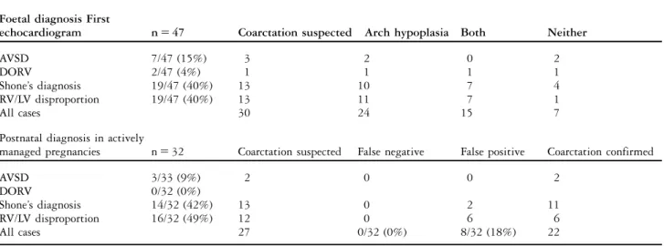
Table 1. Foetal cardiac diagnosis and adjacent anomalies of the aortic arch and isthmus compared with postnatal findings. Foetal diagnosis First
echocardiogram n 5 47 Coarctation suspected Arch hypoplasia Both Neither
AVSD 7/47 (15%) 3 2 0 2
DORV 2/47 (4%) 1 1 1 1
Shone’s diagnosis 19/47 (40%) 13 10 7 4
RV/LV disproportion 19/47 (40%) 13 11 7 1
All cases 30 24 15 7
Postnatal diagnosis in actively
managed pregnancies n 5 32 Coarctation suspected False negative False positive Coarctation confirmed
AVSD 3/33 (9%) 2 0 0 2
DORV 0/32 (0%)
Shone’s diagnosis 14/32 (42%) 13 0 2 11
RV/LV disproportion 16/32 (49%) 12 0 6 6
All cases 27 0/32 (0%) 8/32 (18%) 22
malformations: two cases with Trisomy 13 and one with deletion of chromosome 6q with multiple malformations and one with a large diaphragmatic hernia and severe lung hypoplasia. The majority of actively managed cases received prostaglandins at birth (22 out of 32, 69%), although only three had symptoms of reduced cardiac output. Only five were cyanotic at neonatal presentation. The median follow-up period after birth for actively managed patients was 9 months (1 day–2.5 years).
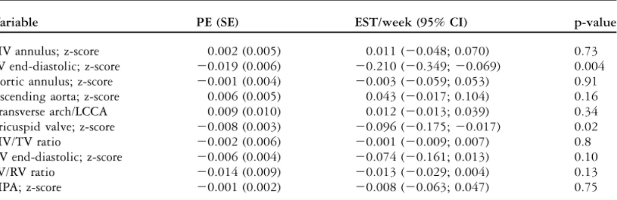
Foetal and neonatal echocardiographic findings A total of 28 cases had more than one foetal echo-cardiogram. Table 3 shows the change in two-dimensional measurements throughout gestation. Both the left ventricular length and end-diastolic
dimension got smaller with gestational age. Other measures such as the right ventricular end-diastolic dimension, main pulmonary artery, aortic and mitral annulus showed normal expected growth with gestation. There were three cases that had left ventricular outflow obstruction at the first foetal echocardiogram and six cases that had abnormal flow in the aortic arch. Only four cases had endocardial fibroelastosis by foetal echocardiogram, which was confirmed postnatally.
In all, 22 liveborn cases had an apex-forming left ventricle and 10 had left ventricle that did not reach the apex of the heart. Of the 10 non-apex-forming left ventricles, two underwent single-ventricle palliation, three had simple coarctation surgery, including one death; one required a mitral valve repair and anomalous left coronary artery from pulmonary artery re-implantation following neona-tal arch repair; two had an unbalanced atrioven-tricular septal defect and underwent bivenatrioven-tricular repair, one following pulmonary artery band; and two needed no intervention. The presence of endocardial fibroelastosis of a non-apex-forming left ventricle was not a predictor of early neonatal intervention.
Patient outcomes
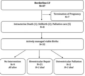
The outcome of foetuses with a borderline left ventricle is depicted in Figure 1. Of the 32 actively managed livebirths, 21 eventually underwent biven-tricular repair, of which 19 underwent a primary complete surgical repair and two had catheter intervention before surgical repair. Only two patients required staged univentricular palliation. No patient underwent a one and a half ventricle repair. Kaplan–Meier freedom from surgical intervention is provided in Figure 2.
Table 2. Clinical characteristics of foetuses with borderline left ventricle.
Value Maternal diagnosis
Age at diagnosis (years) 30.5 6 6.9
Twin pregnancies 5/47 (11%)
Gestational age at diagnosis 24.4 6 5.8
Gestational age at birth 37.7 6 2.6
Fetal extracardiac diagnosis
Kidney 3/47 (6%)
Central nervous system 1/47 (2%)
Bowel 1/47 (2%)
Other 3/47 (6%)
Any extracardiac diagnosis 8/47 (17%)
Chromosome abnormalities 11/47 (23%)
Clinical presentation at birth
Need for prostaglandins 23/32 (71%)
Need for mechanical ventilation 7/32 (22%)
Cyanotic at presentation 6/32 (19%)
Low cardiac output syndrome 6/32 (19%)
Need for inotropic support 2/32 (6%)
Table 3. Change in foetal echocardiogram measurements of 28 foetuses with borderline left ventricle.
Variable PE (SE) EST/week (95% CI) p-value
MV annulus; z-score 0.002 (0.005) 0.011 (20.048; 0.070) 0.73
LV end-diastolic; z-score 20.019 (0.006) 20.210 (20.349; 20.069) 0.004
Aortic annulus; z-score 20.001 (0.004) 20.003 (20.059; 0.053) 0.91
Ascending aorta; z-score 0.006 (0.005) 0.043 (20.017; 0.104) 0.16
Transverse arch/LCCA 0.009 (0.010) 0.012 (20.013; 0.039) 0.34
Tricuspid valve; z-score 20.008 (0.003) 20.096 (20.175; 20.017) 0.02
MV/TV ratio 20.002 (0.006) 20.001 (20.009; 0.007) 0.8
RV end-diastolic; z-score 20.006 (0.004) 20.074 (20.161; 0.013) 0.10
LV/RV ratio 20.014 (0.009) 20.013 (20.029; 0.004) 0.13
MPA; z-score 20.001 (0.002) 20.008 (20.063; 0.047) 0.75
CI 5 confidence interval; EST 5week estimate change of parameter/additional week of gestation; LCCA 5 left common carotid artery; LV 5 left ventricle; MV 5 left-sided atrioventricular valve, respectively, mitral valve; MPA 5 main pulmonary artery; PE 5 parameter estimate; RV 5 right ventricle; SE 5 standard error; TV 5 right-sided AV valve, respectively, tricuspid valve
In all, seven cases required multiple interventions. There were three cases with unbalanced atrioventri-cular septal defect that required initial pulmonary artery banding, and two had additional coarctation repair. Of the three cases, two with an atrioventricular septal defect had a complete repair – one died following mitral valve replacement secondary to coronary complications and endocarditis. The third case eventually underwent a Glenn and Damus–-Kaye–Stansel operation at 9 months of age. Of the remaining four in the multiple intervention group, one had a catheter intervention for critical aortic valve stenosis followed by a coarctation repair; one with Shone’s complex underwent complete biventricular repair but later required anomalous left coronary artery from the pulmonary artery re-implantation and mitral valve repair. The final two patients underwent an initial hybrid procedure – branch pulmonary artery
banding and stenting of the arterial duct – where in one case biventricular repair could be achieved at the age of 10 months. The second case with a hybrid procedure required a Hemi-Fontan operation and Damus–Kaye–Stansel at 7 months of age.
There were two post-operative deaths; one due to endocarditis after mitral valve repair, which is mentioned above. The second death occurred in an infant with Turner’s syndrome, who died because of lymphangiectasia after neonatal coarctation repair. In addition, five palliative patients died with no cardiac intervention. Overall patient survival is depicted in Figure 3.
Factors associated with early intervention
A total of 16 out of 32 (41%) patients had a catheter and/or surgical intervention in their first month of life. This included 11 cases with isolated coarctation repair and two with coarctation plus ventricular septal defect patch repair. There was one patient who had aortic valve dilation and coarctation repair in the first month of life. The remaining two patients had a hybrid procedure with ductal stenting and pulmonary artery branch banding, allowing further cardiac surgery to be delayed until after the neonatal period.
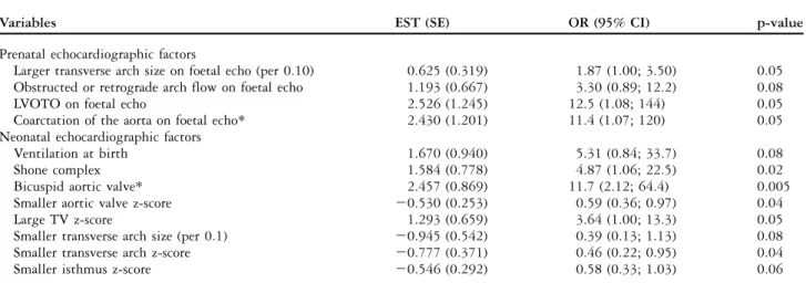
Table 4 lists echocardiographic factors, which were associated with the need for an intervention in the first month of life. The most significant factors were the presence of coarctation of the aorta – diagnosed prenatally and defined as a posterior shelf in the sagittal view – or the presence of left ventricular outflow obstruction or retrograde flow in the arch on the foetal echocardiogram. A bicuspid aortic valve and a larger tricuspid valve z-score from the first postnatal echocardiogram also predicted early intervention. The mean right atrioventricular valve z-score in the early intervention group was 10.04 plus or minus 1.02
Figure 1.
Outcome of foetuses with borderline LV. LV 5 left ventricle.
Figure 2.
Overall survival of foetuses with borderline left ventricle.
Figure 3.
Proportion of patients with borderline left ventricle needing surgical repair.
compared with the mean right atrioventricular valve z-score in the non-intervention group 20.830 plus or minus 0.90 (Fig 4).
Factors associated with no intervention
There were nine cases that did not require any surgical intervention to this date. There were two cases of Shone’s complex and seven with right to left ventricular discrepancy. Of those nine cases, six received prostaglandin after birth. There were no false negative cases. There was one case that had a left superior caval vein with mildly attenuated apex with normal arch and isthmus. There were six cases that had mild transverse aortic arch hypoplasia or isthmal hypoplasia without discrete stenosis. In these six cases, four had additional findings: ventricular septal defect in one, left superior caval vein to coronary sinus with an absent ductus venosus in one, a duplicated chromosome 15 in one, and a foregut duplication cyst in the fourth case. There were two cases that had neither clinical nor echocardiographic signs of coarctation after birth and no follow-up was planned. The other seven cases were followed up clinically until the left-sided structures had normalised for age and body surface area.
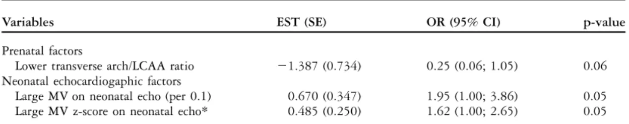
Prenatal echocardiographic factors associated
with non-intervention included a lower transverse aortic arch to left common carotid artery ratio (1.32 6 0.41 versus 1.89 6 0.78 in the intervention group). The most significant predictor of no intervention postnatally was a larger mitral valve z-score on postnatal echocardiogram (Table 5). The mean left-sided atrioventricular valve z-score was – 3.80 plus or minus 1.56 in the non-intervention group compared with a mean left-sided atrioventri-cular valve z-score of 24.18 plus or minus 1.92 in the intervention group. Whereas the postnatal left-sided atrioventricular valve z-score was a significant pre-dictor even in the multivariable model, the prenatal
Table 4. Factors associated with a need for intervention during the first 31 days after birth (n 5 32).
Variables EST (SE) OR (95% CI) p-value
Prenatal echocardiographic factors
Larger transverse arch size on foetal echo (per 0.10) 0.625 (0.319) 1.87 (1.00; 3.50) 0.05 Obstructed or retrograde arch flow on foetal echo 1.193 (0.667) 3.30 (0.89; 12.2) 0.08
LVOTO on foetal echo 2.526 (1.245) 12.5 (1.08; 144) 0.05
Coarctation of the aorta on foetal echo* 2.430 (1.201) 11.4 (1.07; 120) 0.05
Neonatal echocardiographic factors
Ventilation at birth 1.670 (0.940) 5.31 (0.84; 33.7) 0.08
Shone complex 1.584 (0.778) 4.87 (1.06; 22.5) 0.02
Bicuspid aortic valve* 2.457 (0.869) 11.7 (2.12; 64.4) 0.005
Smaller aortic valve z-score 20.530 (0.253) 0.59 (0.36; 0.97) 0.04
Large TV z-score 1.293 (0.659) 3.64 (1.00; 13.3) 0.05
Smaller transverse arch size (per 0.1) 20.945 (0.542) 0.39 (0.13; 1.13) 0.08
Smaller transverse arch z-score 20.777 (0.371) 0.46 (0.22; 0.95) 0.04
Smaller isthmus z-score 20.546 (0.292) 0.58 (0.33; 1.03) 0.06
CI 5 confidence interval; EST 5estimate change in parameter; LVOTO 5 left ventricle outflow obstruction; OR 5 odds ratio; PE 5 parameter estimate; SE 5 standard error; TV 5 right-sided atrioventricular valve, respectively, tricuspid valve
*Remained significant in multivariable model
Figure 4.
Pre- versus postnatal right-sided atrioventricular valve z-scores, respectively, TV as a predictive factor of an early intervention. TV 5 tricuspid valve.
measurements did not reach statistical significance as a predictor for early intervention (Fig 4).
Discussion
The prevalence of a borderline left ventricle among foetal referrals with left ventricular abnormalities to our centre was 28% during the study period. A borderline left ventricle was most frequently seen in the setting of Shone’s complex or right to left ventricular disproportion. This subtype represents a significant proportion of foetuses with left ventricular abnormalities. This prevalence could be even larger depending on the definition of a borderline left ventricle. There is no consensus in the literature as to what constitutes a borderline left ventricle and only scant literature focusing on this topic.6,7,14 Despite this rather high prevalence, the vast majority had a successful biventricular repair, and more interestingly a number required no intervention at all. This was unexpected, as our previous study examined foetuses with a borderline left ventricle associated with atrioventricular septal defect or double outlet right ventricle found that half those cases required a single-ventricle palliation. This perhaps reflects our more stringent criteria for defining a borderline left ventricle, as well as the very small number of cases in this series who in fact had an atrioventricular septal defect or double outlet right ventricle (9 out of 47).
Although overall mid-term survival was reason-able at 79%, the diagnosis of Shone’s complex predicted early intervention. The outcome of this multi-level obstructive lesion hinges on the severity of mitral valve disease, with reported survival at 15 years of only 70%.8,15It is very difficult, if not impossible, to diagnose more complex mitral valve lesions such as a parachute mitral valve, short chordae or deficient mural or anterior leaflets by foetal echocardiogram even in later gestation. However, the left atrioventricular valve annulus dimension can be measured relatively easily in the four-chamber view. Unfortunately, the left atrioventricular valve z-score prenatally was not predictive of outcome; it was only significant as a neonatal echocardiogram measurement.
However, the largest left atrioventricular valve z-scores by foetal echocardiogram were still abnormal. The majority of the left atrioventricular valves had z-scores of between 22 and up to 28, compared with the postnatal measurements, which were between 20.5 and 22 in the non-intervention group. The ‘‘more abnormal’’ left atrioventricular valve z-scores in the foetus may reflect the physiologic conditions in utero, as the left ventricular contribution to cardiac output is less than in the postnatal circulation. It is also of interest that the left ventricular end-diastolic dimen-sion z-score became relatively smaller with later gestational age, which in previous series has been shown to correlate well with the left atrioventricular valve annulus size in infants with left-sided obstruc-tive lesions.16
A very small number of this cohort had endocardial fibroelastosis documented by foetal echo-cardiography (n 5 4). It is well established that the presence of endocardial fibroelastosis in left-sided obstructive lesions is associated with a poor out-come.7,14 Given that the severity of disease in this group was less than cases with hypoplastic left heart syndrome or critical aortic valve stenosis, one would expect that endocardial fibroelastosis would be less frequently identified. Our previous series of atrioven-tricular septal defect and double outlet right ventricle foetuses with a borderline left ventricle found that the most significant predictor of single-ventricle palliation versus biventricular repair was a non-apex-forming left ventricle.6Although this prospective cohort had 10 cases of non-apex-forming left ventricle, we only had two patients undergoing a single-ventricle palliation, both of whom had non-apex-forming left ventricle. Our current study did not find that a non-apex-forming left ventricle predicted neonatal intervention, but this group had a greater number of Shone’s complex or right to left ventricular discrepancy. The presence of a non-apex-forming left ventricle may be more physiologically relevant in foetuses with atrioventricular septal defect or double outlet right ventricle.
Flow disturbances diagnosed by foetal echocar-diography across the left outflow tract and the aortic
Table 5. Factors associated with no intervention.
Variables EST (SE) OR (95% CI) p-value
Prenatal factors
Lower transverse arch/LCAA ratio 21.387 (0.734) 0.25 (0.06; 1.05) 0.06
Neonatal echocardiogaphic factors
Large MV on neonatal echo (per 0.1) 0.670 (0.347) 1.95 (1.00; 3.86) 0.05
Large MV z-score on neonatal echo* 0.485 (0.250) 1.62 (1.00; 2.65) 0.05
CI 5 confidence interval; EST 5estimate change in parameter; LCCA 5 left common carotid artery; MV 5 left-sided AV valve, respectively, mitral valve; OR 5 odds ratio; SE 5 standard error
arch were significant predictors of outcome, as was a prenatal diagnosis of coarctation by posterior shelf. These findings have been noted in previous series looking specifically at foetuses with coarctation and
right to left ventricular disproportion.10,17 These
measurements are relatively simple to obtain and should be included as part of the foetal echocardio-gram for these patients.
A bicuspid aortic valve was associated with an early intervention, but again was a postnatal echo-cardiographic diagnosis. The link between a bicuspid aortic valve and hypoplastic left heart syndrome is well established, and a number of genetic links have been
proposed.18 Additional genetic mutations in animal
models are associated with Shone’s complex.19 As
prenatal genetic testing becomes more sophisticated using microarray techniques, the genetic locus may become identifiable, which may confer intrinsically worse cardiac disease for an individual foetus.
In addition to the importance of the left-sided atrioventricular valve z-score, a larger right-sided atrioventricular valve z-score at the first neonatal echocardiogram predicted a need for intervention, whereas the right ventricular end-diastolic dimen-sion did not. A larger right-sided atrioventricular valve implies a greater right ventricular volume, and has been shown to impact on the left ventricular
compliance and cardiac output.20 That study
demonstrated that a larger right ventricle volume affected the overall left ventricular volume by 25% to 30% in infants with total anomalous pulmonary venous drainage or unbalanced atrioventricular septal defect. A larger right-sided atrioventricular valve in the neonate with critical left heart obstruction is potentially reflective of foetal coarctation physiol-ogy, where the right ventricular cardiac output increases to maintain perfusion to the lower body.21,22 The right-sided atrioventricular valve z-scores possibly reflect better true conditions than right ventricular end-diastolic dimension and pulmonary artery size, as well as overall lung function. Studies comparing the right- versus left-sided atrioventricular valve inflow duration and profile, as well as newer echocardio-graphic modalities such as tissue Doppler imaging and speckle tracking, could provide more insight into right to left ventricular interaction in the foetus with left heart obstruction.
Left ventricular volumes have been shown to be useful predictors of biventricular repair in neonates with borderline left ventricle, when measured by
magnetic resonance imaging.23It has been reported
in earlier studies that a left ventricular volume of more than 20 milliletre per squared metre is a critical parameter when predicting biventricular versus single-ventricle repair in patients with critical aortic stenosis, coarctation, and Shone’s complex.24,25Foetal
magnetic resonance imaging is now feasible and may provide essential left ventricular volume information for postnatal surgical planning.26
Limitations
The definition of a borderline left ventricle is not clearly defined in the literature, and therefore criteria may differ among institutions. We used
the definition described by Tchervenkov et al5
because we felt that it best reflects this subset of hypoplastic left ventricles. It may be that these criteria select out the borderline left ventricles that will go for single-ventricle palliation. Although our study was prospective, we were unable to obtain serial foetal echocardiograms in all patients, mainly because of the logistics of having mothers return on different days. Finally, the overall number of patients in this series is relatively small, reflecting the rarity of a borderline left ventricle, despite the study duration of over 2 years.
Conclusion
Foetuses diagnosed with a borderline left ventricle have a reasonable overall outcome, with pre-selected patients undergoing successful biventricular repair in the neonatal period. Foetuses with evidence of multi-level left heart obstruction, coarctation, or obstructed aortic arch flow were more likely to undergo a neonatal intervention. A subset of these foetuses will not require intervention, which could be predicted by a larger neonatal left-sided atrioventricular valve z-score measured by foetal echocardiography. Future studies focusing on the evolution of left ventricular volumes by magnetic resonance imaging and left-sided atrioventricular valve annular growth may be able to identify the foetuses most at risk for neonatal intervention and single-ventricle palliation.
References
1. Corno AF. Borderline left ventricle. Eur J Cardiothorac Surg 2005; 7: 67–73.
2. Cohen MS, Rychik J. The small left ventricle: How small is too small for biventricular repair? Semin Thorac Cardiovasc Surg Pediatr Card Surg Annu 1999; 2: 189–202.
3. Holzer RJ, Cheatham JP. Therapeutic cardiac catheterisation. In: Moss AJ, Allen HD, Driscoll DJ (eds.). Moss and Adam’s Heart Disease in Infants, Children and Adolescents, 7th edn. Lippincott Williams & Wilkins, Philadelphia, 2008, p 374.
4. Justino H, Pedra C, Freedom RM, Benson LN. Congenital aortic valve stenosis or reurgitation. In: Freedom RM, Yoo SJ, Mikailian H, Williams WG (eds.). The Natural and Modified History of Congenital Heart Disease. Blackwell, New York, 2003, pp 138–141. 5. Tchervenkov CI, Tahta SA, Jutras LC, Be´land MJ. Biventricular repair in neonates with hypoplastic left heart complex. Ann Thorac Surg 1998; 66: 1350–1356.
6. Pitka¨nen OM, Hornberger LK, Miner SE, et al. Borderline left ventricles in prenatally diagnosed atrioventricular septal defect or
double outlet right ventricle: echocardiographic predictors of biventricular repair. Am Heart J 2006; 152: 163.e1–7. 7. Hickey EJ, Caldarone CA, Blackstone EH, et al. Critical left
ventricular outflow tract obstruction: the disproportionate impact of biventricular repair in borderline cases. J Thorac Cardiovasc Surg 2007; 134: 1429–1436.
8. Serraf A, Piot JD, Bonnet N, et al. Biventricular repair approach in ducto-dependent neonates with hypoplastic but morphologically normal left ventricle. J Am Coll Cardiol 1999; 33: 827–834. 9. Hornberger LK, Sanders SP, Rein AJ, Spevak PJ, Parness IA,
Colan SD. Left heart obstructive lesions and left ventricular growth in the mid-trimester fetus: a longitudinal study. Circulation 1995; 15: 1531–1538.
10. Quartermain MD, Cohen MS, Dominguez TE, Tian Z, Donaghue DD, Rychik J. Left ventricle to right ventricle size discrepancy in the fetus: the presence of critical congenital heart disease can be reliably predicted. J Am Soc Echocardiogr 2009; 22: 1296–1301. 11. Brown DL, Durfee SM, Hornberger LK. Ventricular discrepancy as a sonographic sign of coarctation of the fetal aorta: how reliable is it? J Ultrasound Med 1997; 16: 95–99.
12. Schneider C, McCrindle BW, Carvalho JS, Hornberger LK, McCarthy KP, Daubeney PE. Development of Z-scores for fetal cardiac dimensions from echocardiography. Ultrasound Obstet Gynecol 2005; 26: 599–605.
13. Shone JD, Sellers RD, Anderson RC, Adams P Jr, Lillehey CW, Edwards JE. The developmental complex of ‘‘parachute mitral valve,’’ supravalvular ring of left atrium, subaortic stenosis, and coarctation of aorta. Am J Cardiol 1963; 11: 714–725. 14. Lofland GK, McCrindle BW, Williams WG, et al. Critical aortic
stenosis in the neonate: a multi-institutional study of manage-ment, outcomes, and risk factors. Congenital Heart Surgeons Society. J Thorac Cardiovasc Surg 2001; 121: 10–27.
15. Brauner R, Laks H, Drinkwater DC Jr, Shvarts O, Eghbali K, Galindo A. Benefits of early surgical repair in fixed subaortic stenosis. J Am Coll Cardiol 1997; 30: 1835–1842.
16. Schwartz ML, Gauvreau K, Geva T. Predictors of outcome of biventricular repair in infants with multiple left heart obstructive lesions. Circulation 2001; 104: 682–687.
17. Matsui H, Mellander M, Roughton M, Jicinska H, Gardiner HM. Morphological and physiological predictors of fetal aortic coarctation. Circulation 2008; 118(8): 1793–1801.
18. Hinton RB Jr, Martin LJ, Tabangin ME, Mazwi ML, Cripe LH, Benson DW. Hypoplastic left heart syndrome is heritable. J Am Coll Cardiol 2007; 53: 1590–1595.
19. Zhong TP. Zebrafish genetics and formation of embryonic vasculature. Curr Top Dev Biol 2005; 71: 53–81.
20. Phoon CK, Silverman NH. Conditions with right ventricular pressure and volume overload, and a small left ventricle: ‘‘hypoplastic’’ left ventricle or simply a squashed ventricle? J Am CollCardiol 1997; 30(6): 1547–1553.
21. Rudolph AM. Congenital Diseases of the Heart, 3rd edn. Wiley-Blackwell, West Sussex, United Kingdom, 2009, pp 296–297. 22. Allan LD, Chita SK, Anderson RH, Fagg N, Crawford DC, Tynan
MJ. Coarctation of the aorta in prenatal life: an echocardiographic, anatomical, and functional study. Br Heart J 1988; 59: 356–360. 23. Grosse-Wortmann L, Yun TJ, Al Radi O, et al. Borderline
hypoplasia of the left ventricle in neonates: insights for decision-making from functional assessment with magnetic resonance imaging. J Thorac Cardiovasc Surg 2008; 136: 1429–1436. 24. Rhodes LA, Colan SD, Perry SB, Jonas RA, Sanders SP. Predictors
of survival in neonates with critical aortic stenosis. Circulation 1991; 84: 2325–2335.
25. Parsons MK, Moreau GA, Graham TP Jr, Johns JA, Boucek RJ Jr. Echocardiographic estimation of critical left ventricular size in infants with isolated aortic valve stenosis. J Am Coll Cardiol 1991; 18: 1049–1055.
26. Manganaro L, Savelli S, Di Maurizio M, et al. Fetal MRI of the cardiovascular system: role of steady-state free precession sequences for the evaluation of normal and pathological appearances. Radiol Med 2009; 114: 852–870.