Protein Engineering vol.3 no.3 pp. 173-180, 1990
Crystallization and structure solution at 4 A resolution of the
recombinant synthase domain of iV-(5'-phosphoribosyl)anthranilate
isomerase:indole-3-glycerol-phosphate synthase from Escherichia
coli complexed to a substrate analogue
Matthias Wilmanns, Edith Schlagenhauf, Bruno Fol and Johan N.Jansonius1
Department of Structural Biology, Biocentre, University of Basel, Klingelbergstrasse 70, CH^M)56 Basel, Switzerland
'To whom correspondence should be addressed
Hie recombinant synthase domain of the bifunctional enzyme AKS'-phosphoribosyOanthranilate isomerase:indole-3-glycerol-phosphate synthase from Escherichia coli has been crystallized, and the structure has been solved at 4 A resolution. Two closely related crystal forms grown from ammonium sulphate diffract to 2 A resolution. One form (space group R32, a = 163 A, a = 29.5°) contains the unliganded synthase domain; the second crystal form (space group P6£2, a = 144 A, c = 158 A) is co-crystallized with the substrate analogue AK5'-phosphoribit-l-yl)anthranilate. The structure of the synthase-inhibitor complex has been solved by the molecular replacement method. This achievement represents the first successful use of a 03a)g-barrel monomer as a trial model. The recombinant synthase domain associates as a trimer in the crystal, the molecules being related by a pseudo-crystallographic triad. The interface contacts between the three domains are mediated by those residues that are also involved in the domain interface of the bifunctional enzyme. This system provides a model for an interface which is used in both intermolecular and intramolecular domain contacts.
Key words: 03a)8-barrel/crystal structure/IGP synthase/interface
contacts/molecular replacement
Introduction
The monomeric bifunctional enzyme A'-(5'-phosphoribosyl)-anthranilate isomerase:indole-3-glycerol-phosphate synthase (PRALIGPS) from Escherichia coli (EC 4.1.1.48) catalyses two sequential reactions in the biosynthesis of tryptophan. N-(5'-phosphoribosyl)anthranilate (PRA) is converted by PRAI to l-(o-carboxyphenylamino)-l-deoxyribulose 5-phosphate (CdRP) via a practically irreversible Amadori rearrangement (Kirschner
et al., 1987). The succeeding step, the ring-closure reaction from
CdRP to indole-3-glycerol-phosphate (IGP), is catalysed by IGPS. It has been shown by biochemical and genetic studies that the N-terminal half of this bifunctional enzyme is responsible for the IGPS activity, whereas the C-terminal half catalyses the PRAI reaction (Yanofsky etal., 1971; Cohn etal, 1979; Kirschner et al., 1980). The amino acid sequence inferred from the gene sequence consists of 452 amino acid residues (Horowitz
etal., 1983).
The crystal structure of PRAI:IGPS was solved at 2.8 A resolution by the multiple isomorphous replacement method (Priestle et al., 1987). Using oscillation data collected at DESY, Hamburg, the model has been partially refined to a current R-factor of 21% at 2.4 A resolution (M.Wilmanns, unpublished results). The enzyme contains two distinct functional domains,
each folded as a 03a)8-barrel. The domain that catalyses the
IGPS reaction has an additional N-terminal segment of 47 amino acid residues. The active sites have been approximately located using an iodinated substrate analogue and were found to be in depressions on the surface of the domains created by the outward-curving loops between C-termini of the /3-strands and the subsequent a-helices (Priestle etal., 1987).
The two domains of PRAI.IGPS from E.coli were cloned and expressed separately by site-directed mutagenesis by Pflugfelder (1986). TGT, which codes for Cys260, was replaced by the opal-type stop codon TGA into trpC(F) at that position. The IGPS domain (IGPS^^') was expressed in E.coli by this mutated
trpC(F) gene on a plasmid. It has been shown by steady-state
kinetics that IGPS^,,- has synthase activity of the same order of magnitude as in the native bifunctional enzyme (M.Eberhard, personal communication).
Here we describe two crystallographically closely related crystal forms of IGPSmon'. For one of these, a complex between
IGPSnon' and the substrate analogue
Af-(5'-phosphoribit-l-yl)anthranilate (rCdRP), the crystal structure has been determined by molecular replacement methods. Because rCdRP differs from CdRP only by selective reduction of the 2'-carbonyl group to an alcohol group, it can be regarded as a close substrate analogue (Bisswanger et al., 1979). It is a competitive inhibitor for IGPS with K, = 8 nM in 0.1 M potassium phosphate buffer, pH 7.5, at 20°C.
Materials and methods
Purification
IGPSmon' was isolated from a homogenate of E.coli cells that
had been transformed with a recombinant plasmid containing the gene for this domain (Pflugfelder, 1986). This gene trpC codes for residues 1 -259 of the native bifunctional enzyme. The protein purification was accomplished by an existing procedure (Pflugfelder, 1986). The last purification step (gel permeation by Sephacryl S-200) was omitted as the enzyme was at least 95% pure following the preceding steps. The purity of each preparation was checked by PAGE under denaturing conditions (Laemmli, 1970). The specific activity was determined according to Kirschner et al. (1987).
Crystallization
Purified protein was dialysed against 0.05 M potassium phosphate (pH 7.5, 4°C) containing 0.005 M EDTA, 0.002 M 1,4-dithioerythritol and 0.02% (w/v) NaN3 and was
concentrated by centrifugation through a semipermeable membrane (Centricon, TM 10) to 10-15 mg/ml protein. Drops were prepared at 8°C by mixing equal volumes (5 /xl) of freshly concentrated protein solution and reservoir buffer solution containing 0.05 M potassium phosphate (pH 5.0), 1.2 M am-monium sulphate, 0.005 M EDTA and 0.002 M 1,4-dithioerythritol. rCdRP (2 jtl 0.1 M) was added to the drop for the crystallization of inhibited /GPS^*,.. The rCdRP was synthesized according to Bisswanger et al. (1979).
0.05 M potassium phosphate buffer (pH 6.0) for 2 h and then transferred to drops with the same buffer composition but with a reduced protein concentration of 5 —10 mg/ml. The diffraction properties and space group assignments of both crystal forms were determined from precession photographs.
Data collection and processing
Oscillation data of the crystal form II were collected on films (Kodak, NS-5T) using an Arndt-Wonacott rotation camera (Enraf-Nonius) and graphite monochromated Cu-K^ radiation from a rotating anode generator operating at 40 kV and 40 mA. The crystals were mounted in thin-walled glass capillaries with the a axis oriented parallel to the spindle axis, requiring 90° of data to be collected. More efficient data acquisition would have resulted if the 6-fold c axis had been aligned along the spindle, but this was impractical due to the thinness of the crystals.
The X-ray beam was collimated such that the entire crystals were irradiated. The film packs (two films/pack) were placed at a distance of 140 mm from the crystal allowing data collection to 4 A resolution. The crystals were cooled to 5°C in order to prolong their lifetime in the X-ray beam. An oscillation range of 2° per pack was used which required an exposure time of 8 - 1 1 h. Forty-eight film packs with partially overlapping data from six crystals completed a total range of 90°.
The data from the film packs were digitized on a rotating drum scanning densitometer (Optronics Photoscan System, P 1000) using a raster size of 100 /im and a 3.0 optical density scale. The data were processed using a version of the Cambridge oscillation program system (Nyborg and Wonacott, 1977) which incorporates the profile analysis algorithm of Rossmann (1979) as modified by Wilson et al. (1983). The data were scaled using the algorithm of Fox and Holmes (1966) and were refined using the program POSTREF (Winkler et al., 1979) as modified by Greenhough and Helliwell (1982). All negative intensities were set to zero. The final data set included 8536 unique reflections constituting 92.5% of the unique data to 4 A resolution. The final Emerge value for all data (where Rmage = E, | < / , > - / , |/E/,
and < I,- > is the average of /,• over all symmetry equivalents) was 8%.
Structure determination
The structure was solved by the molecular replacement method taking the synthase domain of the partially refined bifunctional enzyme PRAI:IGPS (Priestle etai, 1987; M.Wilmanns, unpublished results) as a trial model. The rotational search was carried out using the program ALMN (written by E.Dodson, York University) which is a version of the fast rotation function of Crowther (1972). A model of the synthase domain (IGPSbi) was positioned in a PI cell with arbitrarily chosen cell axes of
100 A and cell angles of 90°. Structure factors were calculated to 4 A resolution, assuming an overall temperature factor, B, of 25 A2. The Patterson search was carried out using a step size
of 2.5° (in the Eulerian angle system a, /3, 7) for the a and y rotation angles. The step size for the /3 rotation was initially 5° and finally reduced to 0.5° in the vicinity of the solution. The Patterson space chosen was a shell of inner and outer radii of 5 and 30 A. The search was carried out using data from various resolution shells between 4 and 10 A.
The correctly oriented molecule was submitted to a translation search using the T2 translation function of Crowther and Blow
Space group No. of molecules/ asymmetric unit Cell dimensions Rhombohedral Hexagonal Cell volume Rhombohedral Hexagonal R32 1 a = 163 A a = 29.5° a = 83 A c = 465 A 924 000 A3 2 770 000 A3 3 a = 144 A c = 158 A 2 840 000 A3
(1967), implemented in the computer program TFSGEN by Dr I.Tickle (Birkbeck College, London). The observed data between 10 and 4 A resolution were included. In order to obtain an interpretable solution in the translation function a 'sub'-cell was used by selecting reflections with indices (k = h ± 3n), which will be described in detail below (see Results).
A trimer of IGPS molecules was generated from the model which arose from the solution of the translation function. It was refined by the least-squares program CORELS (Sussman et al., 1977) in which each monomeric unit was treated as an independent rigid body. One cycle of scale factor refinement, three cycles including observed data between 10 and 8 A resolu-tion and one final cycle including data between 10 and 6 A resolution were carried out.
Structure improvement
A Sim weighted (2FO-FC) electron density map (Sim, 1959)
was computed using calculated phases and structure factor amplitudes derived from the CORELS refined model. In order to reduce the bias towards this model, maps were limited to small portions of the molecule (typically 5—8%), for which the contributions to the calculated phases and structure factor amplitudes were omitted. A set of 'omit'-maps covering a complete molecule was assembled by an automated procedure, implemented by Dr J.P.Priestle, Basel. In addition, a aa
weighted electron density map (Read, 1986) was computed. All three IGPS^' domains were rebuilt independently on an interactive graphic display system (Evans and Sutherland PS 330) using the FRODO software package (Jones, 1978), further developed at Rice University, Tex. (Pflugrath et al., 1984) and modified by Dr P.Evans (MRC, Cambridge, UK). The atomic positions of the corrected model were refined by the restrained least-squares refinement program package TNT (Tronrud et al., 1987). In order to maintain a favourable ratio between observed data (7689 reflections between 8 and 4 A resolution) and parameters to be refined (2038 non-hydrogen atoms per monomer) only the coordinates of one IGPSn^- domain were refined at a time while the coordinates of the other two monomers were fixed. This procedure was carried out twice for the trimer. Results
Crystal characterization
Two closely related crystal forms of the recombinant IGPS^^,' domain (residues 1—259) from E.coli grow from ammonium sulphate at pH 5.0 (Table I). By reindexing the rhombohedral crystal form I of the unliganded protein to a hexagonal crystal
Structure of the recombinant IGP syntha.se domain
b*
• • #•«• • • : . : • • •••*
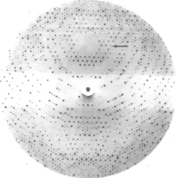
• ••• • •'. :•• ••••• ••• i':*;t'iiFig. 1. Oscillation photograph covering the rotation range <f> = 12° —14° WVito = 0°, 4>hoi = 90"). Experimental details are given in Materials and
methods. The systematic difference between the intensities of the strong reflections (h = k ± 3n) and the remaining reflections (h * k ± 3n) can be seen, for example, in the lune of the reflections which are indexed hk2 (marked by an arrow).
02 .03 .04 .05 .06 .07
.01
Fig. 2. <I>l<a(I)> as a function of resolution for all data to 4 A
resolution. O , data h = k ± 3n; A , data h =£ k ± 3/i; x , all data. system it appears that its c axis (hexagonal) is approximately three times longer than the c axis of the crystal form II which contains the liganded protein. The ab plane (hexagonal) of I is about one-third the area of the ab plane of II. The difference between the cell volumes of I and II is < 3 %. Both crystal forms diffract to - 2 . 0 A resolution and are therefore suitable for X-ray analysis.
Crystal form I grows as hexagonal prisms with a maximum size of 0.6 X 0.6 X 0.3 mm3. Crystal form II, which contains
rCdRP liganded to IGPSn}on; grows with maximum dimensions
of 0.5 x 0.5 X 0.1 mm .These crystals have the shape of an optical lens.
Absorption spectra of crystal form II were recorded with a microspectrophotometer (Zeiss, 03) controlled by a micro-computer (Hewlett-Packard, 9845B) to ascertain whether rCdRP was specifically bound to the crystallized protein. Free rCdRP in solution has an absorption maximum at 327 nm which is shifted
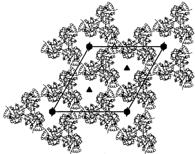
Fig. 3. Relation between the large cell (thick lines, filled symmetry symbols) and the subcell (thin lines, open symmetry symbols). The axes of the large cell are labelled (a,b) and of the subcell (a',b') respectively. The subcell is rotated by 30° around c, relative to the large cell. All additional 2-fold axes and translation components have been omitted for clarity. The local triads of the large cell are located at the positions of the
crystallographic 3-fold axes in the subcell.
by - 4 0 nm to longer wavelengths upon binding to PRALIGPS (Bisswanger et al., 1979, Figure 8A).The absorption spectrum of the crystal form II shows a shift of the peak maximum of ~ 60 run, providing evidence that rCdRP is specifically liganded to IGPS^n'. Crystal form I does not absorb significantly at wavelengths longer than 320 nm.
Analysis of the diffraction pattern
A relation between the two crystal forms is evident from the diffraction pattern of crystal form II. It shows a pattern of strong intensities for reflections with indices (k = h ± 3n) which corresponds to a 'sub'-cell of the same space group with axis
a' = 83 A = aly/3. All other reflections (h * k ± 3n) are
considerably weaker (Figure 1). <I>/<a> is 22.9 for the 2988 reflections (h = k ± 2>ri) to 4 A resolution, whereas this value drops to 7.5 for the remaining 5500 reflections ( < / > is the mean intensity and < a > is the mean standard deviation on intensities). The analysis o f < / > / < a > a s a function of resolution is given in Figure 2. While only 2.6% of the reflections (h = k ± 3n) have negative intensities, this fraction increases to 12.8% for the remaining data (h ± k ± 3 n). No such anomalies were observed in the intensity distribution as function of the index /.
In crystal form I the reflections of every third layer of index / (hexagonal system) are equivalent to reflections which correspond to the subcell of crystal form II. The presence of reflections in the remaining / layers (hexagonal system) requires the assignment of crystal form I to a rhombohedral crystal lattice (Table I).
Packing arrangement
A packing arrangement of the IGPSmon' domains in crystal form
n was considered by taking into account the anomalous intensity distribution of the diffraction pattern. There are three molecules in the asymmetric unit of the real cell, which will be called 'large' cell in the remaining text for ease of discussion. The molecules are related by a local triad which deviates only slightly from crystallographic symmetry. This triad might be distorted by breaking the exact C3 symmetry and/or by a tilt with respect to
the crystallographic 3-fold rotation and 6-fold screw axis. In other words, there is a trimer of IGPSnn,' domains per asymmetric unit related by pseudo-crystallographic symmetry. The triad is located at (x = 1/3, y = 1/3), which is equivalent to the posi-tion of a crystallographic 3-fold axis in crystal form I, since the
crystallographic symmetry by setting the intensities of all reflections with indices (h & k ± 3n) equal to zero. Omission of these data corresponds essentially to use of the subcell, neglecting the fact that the intensities of the strong reflections
(h = k ± 3n) also carry informatidn on deviations from ideal
symmetry. The subcell belongs to the same space group /36322
as the large cell, but does not contain additional non-crystallographic symmetry elements. It is one-third of the volume of the large cell and contains one molecule instead of three in the asymmetric unit. The ab plane of the subcell of II is essentially equivalent to the ab plane of I, highlighting the close relations between both crystal forms (Table I).
These arguments were corroborated by an estimate of the packing density by the method of Matthews (1968). The mol. wt of /GP5mon. as calculated from the amino acid sequence is
Mr = 28 933 (Pflugfelder, 1986). Three molecules per
asymmetric unit result in a VM = 2.7 A3/dalton, corresponding
to a solvent content of 54% which is in the range commonly found for proteins.
Structure solution
The structure of liganded / G P S , ^ ' (crystal form H) was solved by the molecular replacement method using the synthase domain of the bifunctional enzyme (PRALIGPS) as search model. Pure (/3a)g-baiTels have approximate 8-fold symmetry (Lebioda
et al., 19S2), which is reduced to 4-fold symmetry when taking
into account the side chain orientations of the residues which belong to the central /3-barrel (Lesk et al., 1989). Due to this symmetrical appearance of (j3a)8-barrels it was expected that
additional peaks could arise in the rotation function and impede the interpretation. Despite the large number of 03a)8-barrel
structures available (Chothia, 1988), to our knowledge no crystal structure consisting of a monomeric (/3a)g-barrel has been determined by the molecular replacement method. Here the trial model, IGPSH, is a single (/3a)8-barrel. However, it contains a
segment of 47 residues (18% of all residues) at the N-terminus in addition to the (/faVbarrel which diminishes the symmetrical appearance of this structure.
It is possible under most conditions to reveal the nature of local symmetry by using the self-rotation function. However, the failure of the self-rotational search (program ALMN) was not unexpected due to the predicted pseudo-crystallographic arrangement of the local triads. Peaks corresponding to IGPSmon'
domains related by these triads would be disguised by peaks resulting from the crystallographic threefold axes.
The cross-rotation function (program ALMN) gave single peaks of comparable strength for the subcell and the large cell, at positions related by rotations of 30° about the c axis. Three independent peaks would have been anticipated for a trimer in a general orientation, each peak describing the orientation of one monomer relative to the trial model. The appearance of single peak solutions supports the presumed pseudo-crystallographic nature of the local triad.
The peak heights are 5.0 and 4.9 a above the mean for the large cell and the subcell respectively. Since the rotation function is dominated by diffraction data with strong intensities, it becomes clear that, in this case, the systematically weak reflections with indices (h ^ k ± 3n) are de facto neglected. Hence the peaks for both cells arise essentially from the same data.
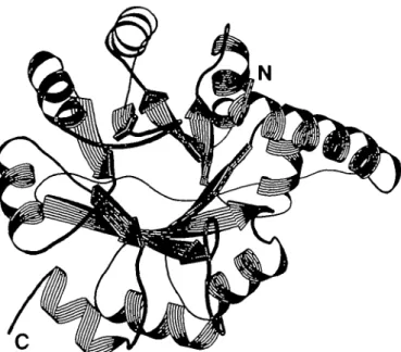
Fig. 4. Schematic drawing of the overall structure of IGPSmon,, created with
the program RIBBON (Priestle, 1988). The view is parallel to the axis of the centra] /3-barrel. Helical ribbons, arrows and ropes represent or-helices, 0-strands and coils respectively. The N-terminus starts in the upper right of the figure with a five-tum a-helix which covers the (/5a)g-barrel
horizontally. The (/Sa)g-barrel begins on the left side of the figure with the first /3-strand (fi\) which is followed by the remaining seven strands in an anticlockwise rotation. Adjacent strands are connected by intervening a-helices shielding the central /3-barrel from solvent. The C-terminus is located at the lower left of the figure. The nomenclature of secondary structure elements used in this publication has been taken from Pnestle
et al. (1987). The assignment of secondary structure elements to the
sequence has been obtained from the partially refined model of IGPSH (M.Wilmanns, unpublished results) and is not identical with the assignment in Figure 2 of Priestle et al. (1987). The present alignment is as follows: 0,, 4 7 - 5 5 ; a,, 7 0 - 8 0 ; fa, 83-88; a2, 97-106; fa, 110-116; a3,
120-128; 04, 133-137; a4, 146-155; fa, 158-164; as, 167-176; 06,
180-184; a6, 196-205; fa, 209-214; a-,, 220-229; fa, 233-236; ag, 245-253.
The best signal-to-noise ratio was obtained by using reflections between 4 and 6 A, the highest resolution available. It has been reported that in cases where pseudo-crystallographic symmetry or special packing arrangements are problematic, the best results were received by using higher resolution data (Dodson, 1985; Driessen and White, 1985).
The translation function (program TFSGEN) was initially applied using the subcell. This essentially replaces the assumed local triad by crystallographic symmetry, thereby simplifying the translational search such that only one peak was expected instead of three. In addition, it was thought that the signal-to-noise ratio would be worse using the large cell, since the search model would then constitute only one-third the scattering matter in the asymmetric unit. One peak (9.8 a above the mean) gave the correct translation in the subcell and was applied to the coor-dinates of IGPS/,,. A trimer was generated by rotation about the presumed local triad at (x = 0, y = 1/3) (cf. Figure 3). A subsequent translational search with this trimer as trial model using the large cell gave the same solution, this time with peak height of 33.8 a above the mean.
It should be noted that it is doubtful whether or not it would have been possible to solve the translation function with one search molecule per asymmetric unit in the large cell without the assumptions concerning the pseudo-crystallographic arrangement of the trimer. We are aware of two examples where knowledge of local symmetry was used for structure 176
Structure of the recombinant IGP synthase domain
Fig. 5. The packing of /G/'5m()n. domains in the crystal form II looking down the crystallographic c axis (same view as in Figure 3). Each IGPSmm' domain
is represented by its Ca backbone. All tnmers of one layer in' the ab plane are included, generated by the symmetry operations (x.yjc), (-yj-y.z),
(y-x, -x,z) and cell translations a and b. The crystallogTaphic 3-fold rotation and 6-fold screw axes are labeled with filled symbols. Both types of local triads
at positions (x = 0, y = 1/3) and (x = 1/3, y = 1/3) are labelled with open symbols (cf. Figure 3). The molecules above and below this layer are rotated by 60° about c relative to the molecules shown. The trimer interface is located near (J: = 1/3, y = 1/3).
determination by molecular replacement. One case was reported by Schirmer et al. (1987) who solved the structure of hexameric C-phycocyanin from Agmenellum quadruplicatum. They deduced the presence of three a(i units per asymmetric unit from the crystal properties and were able to locate the presumed trimer in the cell (space group P321). The structure of y/Ka-crystallin from bovine lens was solved with the molecular replacement method by aligning a local twofold axis parallel to c (White et al.,
1988).
The rigid body refinement program CORELS moved the centres of the three lGPSmon' domains over distances of up to
0.5 A in x, 0.4 A in y and 0.9 A in z. Rotations around the centres of the molecules were < 3 ° . The local triad was preserved although it was not restrained during refinement. The CORELS refined model gave an /?-factor of 48% using all diffraction data between 10 and 4 A resolution.
(2FO-FC) electron density maps allowed unambiguous
positioning of all main chain atoms (except the C-terminal residues 257-259) and 86% of the side chain atoms, eventthough
the current data set is limited to 4 A resolution. From the present model (after refinement by the least-squares program TNT) an ^-factor of 28% was calculated for all data between 10 and 4 A resolution.
Description of the crystal structure
The inhibited /G/)5mon' domain is folded as in the bifunctional
enzyme (Priestle et al., 1987). It contains a central 03a)g-barrel
which was first found for triose phosphate isomerase (Banner
et al., 1975) and is often referred to as the 'TIM-barrer
(Figure 4). The barrel contains 207 residues starting with /3, at Thr48. The preceding 47 residues wrap halfway around the compact (jSa)g-barrel. The N-terminus is located at the
C-terminal side of the central /3-barrel and is followed by a five-turn helix (residues 4-22) which is closely packed against helix a3 of the barrel. A long loop connects the end of this helix with
the start of the (/3a)8-barrel (Thr48).
The packing arrangement of the IGPS,^^' domains in the ab plane is shown in Figure 5. The trimer of IGPS,^' domains in this crystal form is generated by a local triad parallel to c at (x
= 1/3, y = 1/3). Several residues of the (j3a)8-barrel are
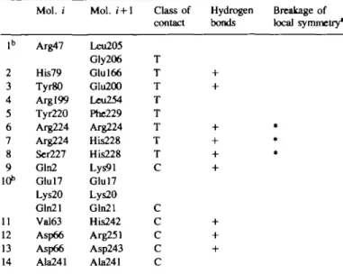
involved in the intermolecular contacts, yielding a tight interface within the trimer (Table 0).
A second type of local triad at (x = 0, y = 1/3) is generated by applying crystallographic symmetry operations on the crystal trimer (Figure 5). The contacts around this local 3-fold axis are formed by residues from the long N-terminal helix. Equivalent residue ranges (17-21) belonging to different trimers of symmetry-related molecules face each other. Unfortunately, the electron density in this contact area is poor, leading to the assumption that this part of the crystal structure of IGPSmon' is
slightly disordered. These interactions between symmetry-related trimers are weaker and less numerous than those within the trimer interface.
is established by identical side chains (Arg224, Ser227, His228) within the trimer interface (Table II). It can be assumed that the guanidinium groups of Arg224 of three /GPS1,^. domains,
Mol. / Mol. i + l Class of Hydrogen Breakage of contact bonds local symmetry* 1° 2 3 4 5 6 7 8 9 10" 11 12 13 14 Arg47 His79 Tyr80 Argl99 Tyr220 Arg224 Arg224 Ser227 Gln2 Glul7 Lys20 Gln21 Val63 Asp66 Asp66 Ala241 Leu205 Gly206 Glul66 Glu200 Leu254 Phe229 Arg224 His228 His228 Lys91 Glul7 Lys20 Gln21 His242 Arg251 Asp243 Ala241 T T T T T T T T C C C C C C
Contacts between IGPSmon' domains within the crystal trimer are labelled T;
contacts between crystallographic symmetry-related trimers are labelled C. aIf the pattern of contacts between molecule i and molecule i + 1 is different for the three monomers, the contact is labelled *.
bThe electron density in this contact area is poor, hence a detailed interpretation has not yet been possible.
A (Fo—FJ map shows strong electron density for the
sub-strate analogue rCdRP at the presumed active site (Priestle et al., 1987), between residues Glu53, Glu214, Ser215 and Ser237. These residues are partially conserved in 11 sequences of IGPS (Priestle et al., 1987). However, due to the limited resolution of the current data, an unambiguous fit of the rCdRP molecule to this electron density has not yet been possible.
Discussion
Structure comparison
In order to discuss global structural differences between
IGPSmon' and IGPS^ the overall errors of the atomic positions
were estimated by plotting i?-factors against resolution (Luzatti, 1952). This resulted in an error estimate of 0.5 A in the atomic positions of the current model.
The r.m.s. deviations of all 256 Ca atoms of the current
model in the three pairs of superimposed /GPS,,^. domains are 0.63, 0.75 and 0.79 A. If the superpositions are restricted to the Ca atoms which belong to the /3-strands of the barrel
34 atoms), the r.m.s. deviations drop to 0.29, 0.30 and 0.34 . The local triad is exactly parallel to c, and adjacent molecules in the trimer are related by rotations of 120 ± 2° about c. There is a small but significant translation component between each pair of IGPSna,' domains along the local triad. The values of the translations are+0.4, +0.4 and —0.8 A, with an error of ~0.1 A. This is one manifestation of the broken Cj symmetry of the local triad in crystal form II.
Another set of comparisons was carried out by superimposing each individual IGPSmn-' molecule of the trimer onto the,
2.0
n nn nnnn nnnn nnnn n
25 50 75 100 125 150 175
Residue Number
200 225 250
Fig. 6. Deviations in the Ca atom positions between pairs of superimposed IGPSmai, and IGPS^ molecules in comparison to the deviations in the Ca atom
positions between superimposed pairs of the three IGPSmm, monomers. Heavy line: averaged differences between Ca atoms (IGPSmonJIGPSbi). The values of
residues 141-145 exceed the area of the graph; the maximum difference is 4.1 A (Aspl42). Thin line: averaged differences between CQ atoms of the three pairs (JGPSmon,IIGPSmtm). The curves have been smoothed by computing the value per residue (i) and averaging it over the neighbouring five residues [(i
-2) • • • ( ' + -2)]. The assignments of the secondary structure elements are given at the bottom of the figure (cf. Figure 4). The N4erminal helix is labelled OQ. Loops are named in the text according to the secondary structure elements which connect them, e.g. ^ l a ^ The last three residues at the C-terminus (residues 257-259) are not included in the current model.
Structure of tbe recombtnant IGP synthase domain domain from the bifunctional enzyme. The overall r.m.s. values
for all Ca atoms were 1.08, 1.10 and 1.17 A. There are some
segments where the average differences between Ca atoms
(IGPSmonUGPSbi) are considerably larger than those between Ca
atoms (/GP5mon://G/>5'mon.), especially in the barrel loops /32a2
and P4CX4 (Figure 6). Since both loops contribute to the active site of IGPS and contain several invariant residues (Priestle et al.,
1987), these deviations may arise from binding of the substrate analogue rCdRP to the active site of IGPS. Further significant differences were observed for the carboxyl end of strand /35
(Glul63 is invariant) as well as for the loops a5/36, / S ^ and
helix a-j. For the N-terminal part of the structure, the carboxyl end of helix org and loop Offij, the differences between Ca atoms
are generally high in both types of superpositions,
IGPSmonIIGPSmm. and IGPS^/IGPSt,. The electron density of
a (2FO—FJ map is generally weak in these areas so that these
segments are considered to be rather flexible in the crystal structure of
Intermolecular contacts
The interface between the IGPS domain and the PRAI domain of the partially refined model of the bifunctional enzyme was examined for intramolecular domain—domain contacts. The following IGPS residues are closer than 4 A to the PRAI domain: 4 7 - 5 0 , 220, 223-224, 247 and 250-254. The interface includes seven hydrogen bonds plus a number of hydrophobic contacts.
All of these residues except Ala247 are involved in intermolecular contacts within the trimer interface of IGPSm^' molecules (Figure 7). In contrast to this, of ah" the residues (92, 155, 157, 186, 193, 228) within IGPS^ that contribute to crystal contacts of the bifunctional enzyme, only one (His228) makes intermolecular contacts between IGPSno,,' molecules. We conclude that the residues involved in contacts between the two domains of IGPSb, also form the crystal trimer interface of the
IGPSnon' molecules. It can be regarded as a quasi-subunit
interface. Solution studies by gel permeation chromatography
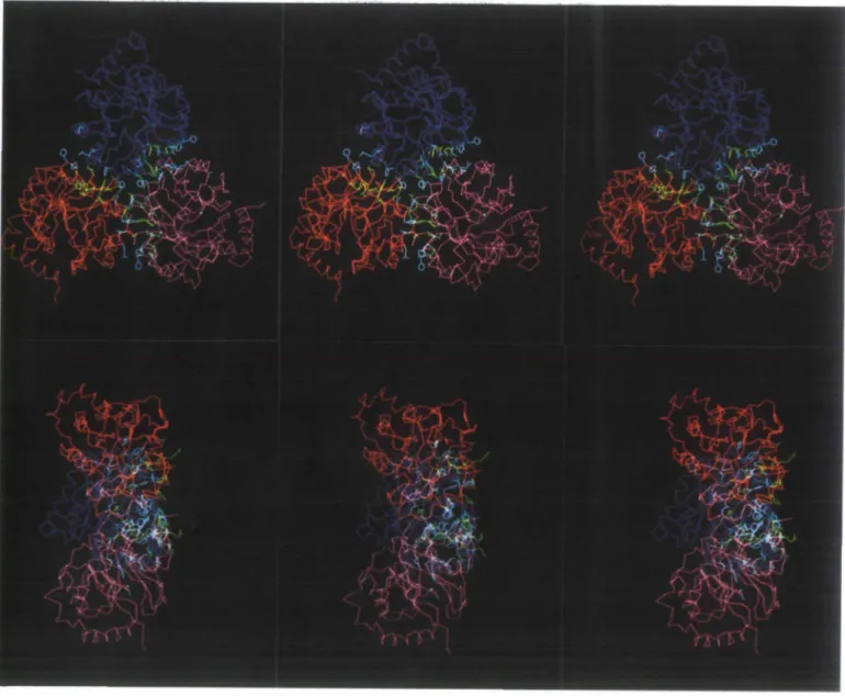
Fig. 7. Stereo presentation of the trimeric association of JGPS^,,,' highlighting the interface between the three monomers. The Ca backbones of lGPSmon, (1),
IGPS^x,, (2) and lGPSmoB, (3) are in red, pink and blue respectively. Side chains of trimer residues which are involved in the interface of IGPSH a r e shown
in yellow. Side chains of residues which make further contacts in the trimer interface are shown in white. (A) Stereo image of the trimer viewed down the crystallographic c axis; (B) stereo image of the trimer with the c axis horizontally oriented. The contacts are concentrated on one side (at the right in this picture), resulting in an overall tripod-like shape of the trimer.
These findings support the ideas of Argos (1988) and Miller
et al. (1987), who have systematically studied the nature and
composition of domain interfaces of monomeric enzymes and subunit interfaces of oligonmeric enzymes. These authors conclude that both types of interfaces have about the same amino acid compositions, which can be considered as lying between the two compositional extremes provided by interior and exterior amino acids. It is energetically unfavourable for large surfaces containing mainly hydrophobic residues to be exposed to an aqueous environment. In this paper we present an example where the domain interface contacts of a two-domain enzyme PRALIGPS can be directly compared to the intermolecular interface contacts of one of the two domains (IGPSnon), which is trimeric in the present crystal structure. The fact that almost all residues contributing to interdomain contacts in IGPS^ are also involved in the interface between IGPSnm' molecules is consistent with the statistical conclusions of Argos and Miller
et al. It would be energetically favourable for the domain
interface of IGPS^ to participate in IGPSno,,' as a quasi-subunit interface rather than being exposed to solvent. A comparison of both structures at high resolution should allow a more quantitative analysis of these interfaces.
Acknowledgements
We thank Professor K.Kirschner for providing us with the E. coli strain KK8 and the plasmid containing the trpC gene. We are grateful to M.Eberhard for advice on protein purification. Drs J.P.Pnestle and R.A.Pauptit are thanked for helpful discussions concerning the structure solution. This work was supported by grants from the Swiss National Science Foundation (3.098-85 and 31-25712.88 to J.NJ.). References
Argos.P. (1988) Protein Engng, 2, 101-113.
Banner.D.W., Bloomer.A.C, Petsko.G.A., Phillips.D.C, Pogson.C.I., Wilson.I.A., Corran.P.H., Furth,AJ., MilmanJ.D., Offord.R.E., PrkkOeJ.D. and Waley.S.G. (1975) Nature, 255, 609-614.
Bisswanger.H., Kirschner.K., Cohn.W., Hager.V. and Hansson.E. (1979)
Biochemistry, 18, 5946-5953.
Chothia.C. (1988) Nature, 333, 598-599.
Cohn.W., Kirschner.K. and Paul.C. (1979) Biochemistry, 18, 5953-5959. Crowther.R.A. (1972) In Rossmann.M.G. (ed.), The Molecular Replacement
Method. Gordon and Breach, New York, pp. 173-178.
Crowther.R.A. and Blow.D.M. (1967) Acta Crystallogr., 23, 544-548. Dodson.E.J. (1985) In Machin.P.A. (ed.), Molecular Replacement. Daresbury
Laboratory, pp. 3 3 - 4 5 .
Driessen.H.P.C. and White.H. (1985) In Machin.P.A. (ed.), Molecular
Replacement. Daresbury Laboratory, pp. 27—32.
Fox.G.C. and Holmes.K.C. (1966) Acta Crystallogr., 20, 886-891. Greenhough.T.J. and HelliwelU.R. (1982)/. Appl. Crystallogr., 15, 493-508. Horowitz.R, van ArsdellJ. and Platt.T. (1983) 7. Mol. Biol., 169, 775-797. Joncs.T.A. (1978)7. Appl. Crystallogr., 11, 268-272.
Kirschncr.K., Szadkowski,H., Henschen,A. and Lottspeich.F. (1980) J. Mol.
Biol., 143, 395-409.
Kirschner.K., Szadkowski.H., Jardetzky.T.S. and Hager.V. (1987) Methods
Enzymol., 142, 386-397.
Laemmli.U.K. (1970) Nature, 111, 680-685.
Lebwda.L., Hatada,M.H., Tulinsky.A. and Mavridis.I.M. (1982)7. MoL Biol.
162, 445-458.
Lesk.A.M., Branden.C.I. and Chothia.C. (1989) Proteins, 5, 139-148. Luzatti,V. (1952) Acta Crystallogr., 5, 802-810.
Matthews.B.W. (1968)7. MoL Biol., 33, 491-497.
MUler.S., JaninJ., Lesk.A.M. and Chothia.C. (1987) 7. Mol. Biol., 196, 641-656.
Nyborg,J. and Wonacott.A.J. (1977) In Amdt.U.W. and Wonacott.A.J. (eds),
The Rotation Method in Crystallography. North-Holland, New York,
pp. 139-152.
PriestleJ.P., Griitter,M.G., White.J.L., Vincent.M.G., Kania.M., Wilson.E., Jardetzky.T.S., Kirschner.K. and JansonhisJ.N. (1987) Proc. Natl. Acad. Sri.
USA, 84, 5690-5694.
Read.R.J. (1986) Acta Crystallogr., A42, 140-149. Rossmann.M.G. (1979) 7. Appl. Crystallogr., 12, 225-238.
Schirmer.T., Huber.R., Schneider.M., Bode.W., Muler.M. and Hackert.M.L. (1986) 7. Mol. Biol., 188, 651-676.
Sim.G.A. (1959) Acta Crystallogr., 12, 813-815.
Sussman^l.L., Holbrook.S.R., Church.G.M. and Kim,S.-H. (1977) Acta
Crystallogr., A33, 800-804.
Thalkr.C, Weaver.L.H., Eichele.G., Wilson.E., Karlsson.R. and JansoniusJ.N. (1981) 7. Mol. Biol., 147, 465-469.
Tronrud.D.E., Ten Eyck.L.F. and Matthews.B.W. (1987) Acta Crystallogr.,
A43, 489-501.
White.H.E., Driessen.H.P.C., Slingsby.C, Moss.D.S., TumeU.W.G. and Lindley.P.F. (1988) Acta Crystallogr., B44, 172-178.
Wilson.K.S., Stura.E.A., Wild.D.L., Todd.R.J., Stuart.D.L, Babu.Y.S., JenkinsJ.A., Standing.T.S., Johnson.L.N., Fourme,R., Kahn,R., Gadet.A., Bartels.K.S. and Bartumk,H.D. (1983) 7. Appl. Crystallogr., 16, 2 8 - 4 1 . Winkler.F.K., Schutt.C.E. and Harnson.S.C. (1979) Acta Crystallogr., A35,
901-911.
Yanofsky.C, Hom.V., Bonner.M. and Stasiowski.S. (1971) Genetics, 69, 409-433.