2008/84
MRI-based inverse finite element approach for the
mechanical assessment of patellar articular cartilage from
static compression test
MRT-basierter Finite-Elemente-Ansatz zur mechanischen Beurteilung von
patellarem Gelenkknorpel aus statischen Kompressionsversuchen
Sven Knecht1,*, Roger Luechinger2, Peter
Boesiger2and Edgar Stu¨ssi1
1Institute for Biomechanics, ETH Zurich, Switzerland 2Institute for Biomedical Engineering, University and
ETH Zurich, Switzerland
Abstract
The mechanical property of articular cartilage determines to a great extent the functionality of diarthrodial joints. Consequently, the early detection of mechanical and, thus, functional changes of cartilage is crucial for pre-ventive measures to maintain the mobility and the quality of life of individuals. An alternative to conventional mechanical testing is the inverse finite element approach, enabling non-destructive testing of the tissue. We evalu-ated a method for the assessment of the equilibrium material properties of the patellar cartilage based on magnetic resonance imaging during patellofemoral com-pression. We performed ex vivo testing of two equine patellas with healthy cartilage, one with superficial defects, and one with synthetically degenerated cartilage to simulate a pre-osteoarthritic stage. Static compression with 400 N for 2 h resulted in morphological changes comparable to physiological in vivo deformations in humans. We observed a decrease of the equilibrium Young’s modulus of the degenerated cartilage by -59%, which was in the range of the results from indentation (-74%) and confined compression tests (-58%). With the reported accuracy of magnetic resonance imaging and its reproducibility, the results indicate the potential to measure differences in Young’s modulus with regard to cartilage degeneration and consequently to distinguish between healthy and pre-osteoarthritic cartilage.
Keywords: articular cartilage; assessment;
biomecha-nics; osteoarthritis.
Zusammenfassung
Die Funktionalita¨t von diarthrodialen Gelenken wird in großem Maße von den mechanischen Eigenschaften des Gelenkknorpels bestimmt. Um die Mobilita¨t und die
*Corresponding author: Sven Knecht, Institute for Biomechanics, ETH Zurich, Wolfgang-Pauli Strasse 10, 8093 Zurich, Switzerland
Phone: q41-44-6336859 Fax: q41-44-6331124 E-mail: [email protected]
Lebensqualita¨t eines Menschen zu erhalten, ist es darum wichtig, fru¨hzeitig Vera¨nderungen der mechanischen Eigenschaften und somit der Funktionalita¨t des Gelenk-knorpels zu detektieren. Eine vielversprechende Alterna-tive zu konventionellen mechanischen Tests bietet der inverse Finite-Elemente-Ansatz, mit dem zersto¨rungsfrei-es Pru¨fen dzersto¨rungsfrei-es Gewebzersto¨rungsfrei-es ermo¨glicht wird. Wir haben dizersto¨rungsfrei-ese Methode in Kombination mit der Magnetresonanztomo-graphie wa¨hrend Kompressionstests an patellofemoralen Gelenken zur Beurteilung der mechanischen Eigenschaf-ten von Gelenkknorpel evaluiert. Wir fu¨hrEigenschaf-ten Ex-vivo-Tests an zwei Pferdekniescheiben mit gesundem Knorpel, einer mit defekter Knorpeloberfla¨che und einer mit ku¨nstlich degeneriertem Knorpel durch, um das pra¨arthrotische Stadium zu simulieren. Statische Kom-pressionstests mit 400 N fu¨r 2 h fu¨hren zu a¨hnlichen mor-phologischen Vera¨nderungen wie physiologische
In-vivo-Deformationen beim Menschen. Wir
beobachte-ten eine Abnahme des Young-Moduls beim degenerier-ten Knorpel um -59%, die in der Gro¨ßenordnung der Ergebnisse aus den Indentationstests (-74%) und Kom-pressionstests (-58%) liegt. Diese Ergebnisse zeigen, dass aufgrund der Genauigkeit der Magnetresonanzto-mographie und der Reproduzierbarkeit diese Methode das Potenzial zur Ermittlung der Unterschiede des Young-Moduls hat, die mit der Degeneration von Knorpel einhergehen. Somit kann hiermit zwischen gesundem und pra¨arthrotischem Knorpel unterschieden werden.
Schlu¨sselwo¨rter: Arthrose; Beurteilung; Biomechanik;
Gelenkknorpel.
Introduction
Articular cartilage (AC) plays a fundamental role for the functionality of diarthrodial joint. It protects the subjacent bone from high stresses and enables in combination with the synovial fluid a nearly frictionless movement over the whole lifespan of a person. The early detection of func-tional changes with regard to degenerative joint diseases, such as osteoarthritis (OA), is essential to prevent or reduce long-term disability w2, 5x. Since the functional behavior of AC is determined by the cartilage morphol-ogy and the mechanical properties, both parameters are appropriate to assess cartilage tissue. Magnetic reso-nance imaging (MRI) combined with state-of-the-art post-processing methods has shown its applicability to obtain accurate and highly reproducible quantitative data
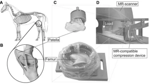
Figure 1 Sketch of the sample preparation from equine patellofemoral joints (A, B).
The femoral counterpart of the patella was cut at the dashed line (B) and fixed with acrylic resin in a PE chamber (C, bottom). Both components were attached to the MR-compatible compression device (D) and positioned in the MR scanner.
of the morphology in healthy w6x and progressed osteo-arthritic cartilage w8x. However, the biomechanical pro-perties of AC seem to be more sensitive to pathological changes of the tissue as alterations of the structural and biochemical properties, determining the mechanical behavior, are one of the first events in AC degeneration w3x. As shown by Knecht et al. w14x, measuring the car-tilage static Young’s modulus has the potential to identify early degenerative changes in AC. However, its reliable assessment in vivo fails up to now due to the lack of appropriate measurement techniques. Commonly used methods demand the excision of a well-defined cartilage sample w4x or invasive testing in situ w15x. Herberhold et al. w10x showed ex vivo that patellofemoral compression of human cadaveric knee with 1.5 times body weight (BW) for 3.5 h results in a local maximal deformation of the patellar cartilage thickness of 44% and a mean vol-umetric change of 29%. However, they did not assess the mechanical properties. In a preliminary study, we tested the in vivo applicability of an MR-compatible patellofemoral static compression test and showed a decrease of the global mean thickness of approximately 5% after loading with approximately 0.5 BW for 60 min w21x. This deformation from static compression was sim-ilar to dynamic in vivo results after knee bending w7x. The aim of the present study was to explore an MR-controlled patellofemoral compression test combined with an inverse finite element (FE) approach for the assessment of the equilibrium material properties of patellar AC.
Materials and methods
Patellofemoral compression test in vitro
Four equine joints were frozen and stored under -208C within 8 h after death. They were defrosted overnight and dissected. The femoral counterpart of the patella was cut off (Figure 1B) and both parts were fixed with acrylic resin (Beracryl, Suter-Swiss composite, Fulenbach, Switzer-land) in MR-compatible PE chambers (Figure 1C). The patellar sample was mounted onto the wooden lever of
the compression device (Figure 1D). All tests were per-formed in 0.1Mphosphate-buffered saline (PBS).
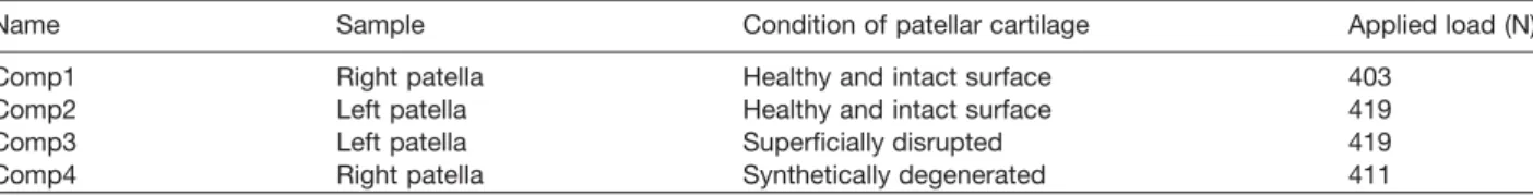
We explored this approach using two healthy and visu-ally intact, one superficivisu-ally disrupted and one syntheti-cally degenerated patellar cartilages (Table 1). Artificial degeneration was performed by treating the cartilage with a diluted solution of 1 mg/ml trypsin and 0.152 mg/ml EDTA=4 Na in Hanks’ balanced salt (Gibco, Invi-trogen, Basel, Switzerland) solution for 20 min at 378C to simulate osteoarthritic cartilage w17x. Prior to the patello-femoral loading with 400 N for 2 h, all samples were equilibrated for 1 h in PBS.
Cartilage imaging was performed in the transversal plane using a 1.5-T MR scanner (Gyroscan Intera, Philips Medical System, Best, The Netherlands) and a surface microscopy coil (diameter 47 mm) with a spoiled 3D gra-dient echo sequence with water selective excitation (TR: 30 ms, TE: 7.4 ms, Flip angle: 208, number of averages: 2). A 320=320 scan resolution with a field of view of 80 mm and a slice thickness of 1.4 mm results in a scan time of 7 min 42 s. Images were acquired before contact between the patellar and femoral cartilage was estab-lished (_pre), after load application (_00), after 2 h of creep test (_end), and 8 min after load removal (_post) (Figure 2).
Conventional mechanical assessment
The femoral and patellar samples were equilibrated after the patellofemoral compression test for 2 h in PBS to enable full recovery. Stress-relaxation indentation tests were performed at three positions on the medial and three on the lateral facet of the patella within the patel-lofemoral contact area. The patella was attached to a mechanical testing system (EnduraTec ELF 3200, Bose, Minnetonka, MN, USA) with a 22-N force cell (Honeywell Sensotec, Columbus, OH, USA). After a pre-load of 0.015 N for 60 s with a plane-ended circular indenter (diameter 0.99 mm), five displacement steps of 50mm with a veloc-ity of 5 mm/s up to a maximal deformation of 250 mm were conducted, and the resulting force was recorded for each of the six measurement sites. Cartilage thickness
Table 1 Overview of the tested patellar cartilage samples.
Name Sample Condition of patellar cartilage Applied load (N)
Comp1 Right patella Healthy and intact surface 403
Comp2 Left patella Healthy and intact surface 419
Comp3 Left patella Superficially disrupted 419
Comp4 Right patella Synthetically degenerated 411
was determined using a needle probe technique w12x. The equilibrium Young’s modulus E was determined by a linear curve fit of the equilibrium stress-strain curve w20x with a Poisson’s ratio of 0.1.
For confined compression tests, cartilage plugs were punched from locations next to the indentation testing site using a disposable skin biopsy punch (Stiefel Labo-ratorium, Offenbach, Germany) and removed from the subchondral bone. The plugs were tested in creep using the above-described testing system. Specimens were transferred into a smooth confining chamber (dia-meter 3.62 mm) and loaded with a porous sintered filter (diameter 3.54 mm, pore size 45 mm, porosity 45%; Schunk Sintermetalltechnik, Heuchelheim, Germany) in 0.1MPBS. Strains of 5%, 10% and 15% were applied in a stepwise manner at a rate of 1 mm/s with 30 min relaxation time after each step. The aggregate modulus
HAwas calculated by a linear curve fit of the equilibrium stress-strain curve. To determine the hydraulic permea-bility k, the experimental stress-relaxation curve for 5% strain was fitted with the theoretical solution by Soltz et al. w19x using a least squares algorithm in MATLAB (MathWorks, Inc., Natick, MA, USA).
Post-processing
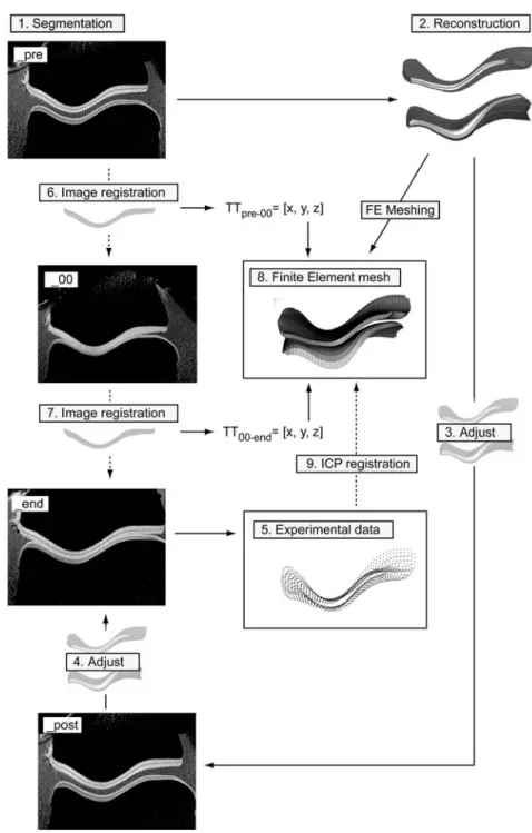
Segmentation of the undeformed patellar and femoral cartilage and 3D reconstruction was performed using a custom-written software. For the reproducibility test, the data were segmented three times over a defined number of images with 1 week between the procedures. The polygonal surface models were imported into a com-mercial software package (Raindrop Geomagic, Geo-magic Inc., Research Triangle Park, NC, USA) to fit non-uniform rational B-splines (NURBS) to the cartilage surface and the bone cartilage interface. The NURBS surfaces of the 3D geometries were imported into a com-mercial pre-processor (Patran 2005, MSC Software Cor-poration, Santa Ana, CA, USA) and meshed with linear 3D continuum elements (Figure 2, (8)). After registration of the images from the end and after load application with the undeformed data based on the bone cartilage interface (Figure 2, (3, 4)), the deformed cartilage geom-etries were segmented using the undeformed geomgeom-etries as drafts. Translation and rotation of the geometries while establishing contact (_pre-_00) and during compression test (_00-_end) were determined by registration and implemented into the FE model to obtain precise models of the contact conditions at the beginning and the end of the compression test (Figure 2, (6, 7)). The point cloud of the patellar surface nodes at equilibrium was regis-tered based on the undeformed bone cartilage interface to the initially undeformed FE mesh of the patellar
carti-lage using an iterative closest point (ICP) algorithm (Fig-ure 2, (9)), programmed in MATLAB.
Quantitative data
Global morphological data, such as mean thickness, maximal thickness, and volume were calculated for the samples before load application, at the end of the com-pression and 8 min after load removal. Contact of the patellofemoral joint was defined when the distance between patellar and femoral surface was smaller than the in-plane resolution. Multiplication of the length of the contact with the slice thickness and their summation over all slices results in the total contact area w9x. Simplistic assumption that the volumetric deformation DV only occurs at the contact area A, divided by the undeformed mean thickness hmeanresults in a mean strain´mean
DV
´means (1)
A*hmean
This allows for calculation of a mean compressive modulus Emeanwith the applied load P by
P*hmean
Emeans (2)
DV
Three-dimensional deviations of the undeformed and deformed patellar cartilage were determined using Geomagic Qualify (Geomagic Inc.) and displayed gray-scale-coded.
Inverse finite element approach
Large displacement contact analysis with frictionless small sliding was used with ABAQUS 6.5.4 (ABAQUS Inc., Pawtucket, RI, USA). The femoral bone cartilage interface nodes were fixed in all six directions, whereas the patellar bone cartilage interface nodes were kine-matically coupled to a reference node outside the object. The load was applied linearly in one step within 2 s onto this reference node. AC was modeled as isotropic linear elastic material. This simplification is valid since the fluid flow was ceased in the tissue for the herein performed equilibrium test. Calculations were performed on a dual processor PC (Xeon CPU 3.4 GHz, 2 GB RAM). The dif-ference between the two surfaces, defined as the dis-tance between each predefined cartilage surface node of the FE calculation and its closest node on the surface of the experimental point cloud, was minimized using a non-linear least squares optimization algorithm in MAT-LAB. The robustness of the optimization algorithm and the sensitivity of the segmentation, registration and load-ing error were performed for one representative patellar
Figure 2 Flowchart of the entire pre-processing step to obtain the experimental data (5) and generate the FE model (8).
Unloaded patellar and femoral cartilage were segmented (1), reconstructed using NURBS (2), meshed and imported into a FE software (8). The segmented geometry was used to segment the patellar cartilage after load removal (_post) and at the end of the compression test (_end). The point cloud of the patellar surface nodes at the end of the test (5) was registered based on the undeformable bone-cartilage interface to the undeformed FE mesh using an ICP algorithm (9).
geometry with an EFE value of 2 MPa for six randomly generated starting values.
Statistical analysis
Values of the quantitative data were displayed as mean"standard deviation (SD). The coefficient of varia-tion (CV%) was calculated as standard deviavaria-tion=100 divided by the average value for the reproducibility test of the segmentation process and divided by the exact value for the assessment of the inverse FE approach. The mechanical properties from conventional mechanical
testing were analyzed statistically using a two-way analy-sis of variance in Systat (Systat Software Inc., San Jose, CA, USA) with a significance level of ps0.05.
Results
Conventional mechanical assessment
The values of the equilibrium properties were independ-ent of the side (medial/lateral) for both healthy and degenerated cartilage. For healthy patellar cartilage, the
Table 2 Summary of the calculated mechanical properties of patellar articular cartilage for the four samples.
Sample Method Confined Patellofemoral compression
indentation compression
Inverse FE Volumetric
E (MPa) HA(MPa)
E (MPa) Emean(MPa)
Comp1 1.669"0.496 n.d.a. 5.541 4.7
Comp2 1.374"0.305 0.568"0.130 5.627 4.8
Comp3 1.604"0.199 0.522"0.199 4.636 4.9
Comp4 0.400"0.053 0.228"0.094 2.176 1.8
All material properties were calculated using the linear elastic homogeneous isotropic material model and are displayed as mean"SD of all six measurements (three medial and three lateral). n.d.a., no data available.
Figure 3 Exemplary views of the cartilage deformation of test Comp1 (intact) and Comp4 (synthetically degenerated).
Deformation after 2 h of static compression with a load of approximately 400 N is displayed grayscale-coded. Dashed line represents the patellofemoral contact area. Circles represent the locations of the conventional mechanical test.
equilibrium Young’s modulus varied between 1.37 MPa and 1.67 MPa, and the mean aggregate modulus was around 0.53 MPa (Table 2). The Young’s and the aggre-gate moduli of the degenerated cartilage were evidently different from the healthy cartilage, whereas the hydraulic permeability was not different.
Reproducibility of the segmentation
The mean thickness for a reconstructed patellar cartilage was 2.03"0.02 mm, the maximal thickness was 2.94"0.05 mm, and the volume was 4559"56 mm3. The
CV% of these global morphological parameters was 0.84% for the mean thickness, 1.67% for the maximal thickness and 1.23% for the cartilage volume.
Deformational behavior
The mean thickness of the segmented patellar geometry decreased with compression between -3.67% and -5.30% for the healthy and by around -9% for the degen-erated cartilage. Within 8 min after load removal, the mean thickness increased to the initial thickness for Comp3. Maximal thickness showed neither a decreasing nor an increasing trend during compression or after load removal. Cartilage volume decreases by around 190 mm3
for healthy and 477 mm3 for the degenerated cartilage.
As for mean thickness, the cartilage volume increased to
the initial volume for the superficially disrupted cartilage sample. The approximately calculated mean compres-sive modulus Emeanwas around 4.8 MPa for the healthy and 1.8 MPa for the degenerated patellas (Table 2).
3D comparison of the undeformed and the deformed patellar cartilage revealed a maximal local deformation at the cartilage surface of around -0.5 mm for healthy and -0.8 mm for degenerated AC (Figure 3).
Inverse finite element approach
The entire calculation lasts approximately 160 min for a mean number of 60 forward FE calculations in ABAQUS, each lasting approximately 100 s. The CV% of the cal-culated Young’s modulus EFEfor different starting values was less than 2.2%.
Sensitivity analysis
Segmentation error showed a maximal error in the cal-culated Young’s modulus of 3.1%. Realistic out-of-plane registration error of 0.2 mm resulted in a maximal error of 2.7%. An experimental load error of -5% (-20 N) result-ed in an error for EFE of less than 7%. Poisson’s ratio error was around 10% for all analyses. Worst-case cal-culations implementing the segmentation error, registra-tion errors of approximately 0.2 mm and an error in the
applied load of approximately 4 N resulted in a slight overestimation of the Young’s modulus of ;3%.
Linear elastic material behavior
The Young’s moduli EFE and the Poisson’s ratio are between 4.5 and 5.5 MPa for the healthy and around 2.2 MPa for the degenerated patella. The mean of EFE and the Poisson’s ratio of healthy cartilage were 5.268"0.549 MPa and 0.103"0.022, respectively (Table 2).
Discussion
The detection of cartilage changes, e.g., in the early degenerative stage OA is increasingly important to pre-vent or reduce incidence of long-term disability. We eval-uated a novel approach for the assessment of the mechanical properties of the patellar AC, which shows a prevalence of OA similar to that in the tibiofemoral joint (35% and 45%, respectively) w16x.
The static patellofemoral compression test with human physiological loads of 0.5 BW (;400 N) on excised joints resulted in obvious and physiological deformation of the cartilage surface (Figure 3). The mean thickness defor-mation was in the range of patellar cartilage in young individuals after 30 deep knee bends w11x and after 80 min of in vivo patellofemoral compression w21x. The volumetric changes for the intact patellar cartilage are well comparable to physiological deformation in vivo after 50 knee bends w7x. Furthermore, the detected mechani-cal parameters are in the range of the human cartilage properties w1x just as the morphological parameter w10, 11x. Thus, the used equine animal model was adequate to simulate static loading on human patellofemoral joints with physiological human loads. In accordance with Set-ton et al. w18x, we observed that superficial defects might be detected based on relaxation behavior.
The mean compressive modulus from the quantitative morphological parameter was in the range of the moduli from the inverse FE approach. Analyzing the morphologi-cal deformational data of six human cadaveric knee joints from reference w10x with Eq. (2) resulted in comparable mean compressive moduli between 1.6 and 3.0 MPa.
FE calculations with a difference in the Young’s moduli of 30% and the consecutive 3D comparison of the deformed geometries revealed deviations of the cartilage deformation by more than 100mm for all healthy samples and by more than 125mm for the degenerated sample. As the position for the cartilage boundary can be located from MR images within a 0.5-pixel size w13x, the herein used in-plane image resolution of 0.25=0.25 mm2allows
to distinguish between differences of the Young’s moduli by 30%.
The sensitivity analysis of the presented inverse FE approach revealed that the Young’s modulus was only slightly dependent on the applied load, the segmentation and the registration process. We suppose that the low sensitivity of the Young’s modulus to the applied load allows the use of this method for an in vivo application even with a more vague loading condition than in the herein performed idealized ex vivo setup.
The Young’s moduli from indentation tests are higher than in other published studies. However, we observed with all performed tests a decrease of the mechanical properties of the synthetically degenerated cartilage compared to the pooled healthy data (Es-74%, HAs -58%, EFEs-59%, Emeans-62%). Thus, all mechanical tests and analyses seem to be applicable to detect dif-ferences in the mechanical properties between healthy and degenerated cartilage. To verify this assumption statistically, a higher number of specimens have to be tested.
Due to the large individual differences in Young’s mod-ulus between subjects, this method might be used for longitudinal studies to detect time-dependent degener-ative or adaptive processes. It might be applicable to detect adaptive processes that come with physiothera-peutic treatments or changes in the loading regime. How-ever, for further in vivo application, the loading apparatus has to be improved to allow for a more comfortable load-ing of the patellofemoral joint for at least 1 h.
In conclusion, we explored the sensitivity and accuracy of a novel MR-controlled patellofemoral compression test to detect mechanical changes in situ. The results indicate that this method may have potential to distinguish between patellar cartilage with differences in the Young’s moduli between 20% and 30%, and consequently to dis-tinguish between healthy and moderately degenerated AC in the early stage of the osteoarthritis process w14x. Furthermore, solely considering the morphological pro-perties, statements about the intactness of the superficial zone and about an approximate estimate of the modulus of the cartilage might be possible. The adaptation of this method for the in vivo application may provide a novel approach to determine functional changes of AC non-invasively and quantitatively for the first time.
Acknowledgements
The authors thank Mr. Ivo Telley for his help and the International Society of Biomechanics (ISB) for financial support.
References
w1x Armstrong CG, Mow VC. Variations in the intrinsic mechanical properties of human articular cartilage with age, degeneration, and water content. J Bone Joint Surg Am 1982; 64: 88–94.
w2x Bjorklund L. ‘‘The bone and joint decade 2000–2010. Inau-gural meeting 17 and 18 April 1998, Lund, Sweden’’. Acta Orthop Scand Suppl 1998; 281: 67–80.
w3x Buckwalter JA, Mankin HJ. Articular cartilage: degenera-tion and osteoarthritis, repair, regeneradegenera-tion, and transplan-tation. Instr Course Lect 1998: 47: 487–504.
w4x Buschmann MD, Soulhat J, Shirazi-Adl A, Jurvelin JS, Hunziker EB. Confined compression of articular cartilage: linearity in ramp and sinusoidal tests and the importance of interdigitation and incomplete confinement. J Biomech 1998; 31: 171–178.
w5x Cicuttini FM, Jones G, Forbes A, Wluka AE. Rate of car-tilage loss at two years predicts subsequent total knee arthroplasty: a prospective study. Ann Rheum Dis 2004; 63: 1124–1127.
w6x Eckstein F, Gavazzeni A, Sittek H, et al. Determination of knee joint cartilage thickness using three-dimensional magnetic resonance chondro-crassometry (3D MR-CCM). Magn Reson Med 1996; 36: 256–265.
w7x Eckstein F, Tieschky M, Faber S, Englmeier KH, Reiser M. Functional analysis of articular cartilage deformation, recovery, and fluid flow following dynamic exercise in vivo. Anat Embryol (Berl) 1999; 200: 419–424.
w8x Eckstein F, Mosher T, Hunter D. Imaging of knee osteo-arthritis: data beyond the beauty. Curr Opin Rheumatol 2007; 19: 435–443.
w9x Heino-Brechter J, Powers CM. Patellofemoral stress dur-ing stair ascent and descent in persons with and without patellofemoral pain. Gait Posture 2002; 16: 115–123. w10x Herberhold C, Faber S, Stammberger T, et al. In situ
meas-urement of articular cartilage deformation in intact femo-ropatellar joints under static loading. J Biomech 1999; 32: 1287–1295.
w11x Hudelmaier M, Glaser C, Hohe J, et al. Age-related chang-es in the morphology and deformational behavior of knee joint cartilage. Arthritis Rheum 2001; 44: 2556–2561. w12x Jurvelin JS, Rasanen T, Kolmonen P, Lyyra T. Comparison
of optical, needle probe and ultrasonic techniques for the measurement of articular cartilage thickness. J Biomech 1995; 28: 231–235.
w13x Kauffmann C, Gravel P, Godbout B, et al. Computer-aided method for quantification of cartilage thickness and vol-ume changes using MRI: validation study using a synthetic model. IEEE Trans Biomed Eng 2003; 50: 978–988. w14x Knecht S, Vanwanseele B, Stuessi E. A review on the
mechanical quality of articular cartilage – implications for
the diagnosis of osteoarthritis. Clin Biomech 2006; 21: 999–1012.
w15x Korhonen RK, Wong M, Arokoski J, et al. Importance of the superficial tissue layer for the indentation stiffness of articular cartilage. Med Eng Phys 2002; 24: 99–108. w16x McAllindon T, Zhang Y, Hannan M, et al. Are risk factors
for patellofemoral and tibiofemoral knee osteoarthritis dif-ferent? J Rheumatol 1996; 23: 332–337.
w17x Niederauer GG, Niederauer GM, Cullen LC, Athanasiou KA, Thomas JB, Niederauer MQ. Correlation of cartilage stiffness to thickness and level of degeneration using a handheld indentation probe. Ann Biomed Eng 2004; 32: 352–359.
w18x Setton LA, Zhu W, Mow VC. The biphasic poroviscoelastic behavior of articular cartilage: role of the surface zone in governing the compressive behavior. J Biomechan 1993; 26: 581–593.
w19x Soltz MA, Ateshian GA. Experimental verification and the-oretical prediction of cartilage interstitial fluid pressuriza-tion at an impermeable contact interface in confined compression. J Biomech 1998; 31: 927–934.
w20x Toyras J, Rieppo J, Nieminen MT, Helminen HJ, Jurvelin JS. Characterization of enzymatically induced degradation of articular cartilage using high frequency ultrasound. Phys Med Biol 1999; 44: 2723–2733.
w21x Vanwanseele B. Quantification of the influence of the absence of normal joint loading and movement on the arti-cular cartilage in the joints of spinal cord injured patients. PhD thesis, ETH Zurich, Switzerland 2003.