doi:10.1093/brain/awl045 Brain (2006), 129, 1027–1030
The optic nerve: a new window into cerebrospinal
fluid composition?
H. E. Killer,
1,2G. P. Jaggi,
2J. Flammer,
1N. R. Miller
4and A. R. Huber
31
University of Basel, Eye Institute,2Department of Ophthalmology and3Department of Laboratory Medicine, Kantonsspital Aarau, Switzerland and4Wilmer Ophthalmological Institute, Johns Hopkins Hospital, Baltimore, MD, USA
Correspondence to: PD Dr. H.E. Killer, Kantonsspital Aarau, CH-5001 Aarau, Switzerland E-mail: [email protected]
Cerebrospinal fluid (CSF) pressure and composition are generally thought to be homogeneous within small limits throughout all CSF compartments. CSF sampled during lumbar puncture therefore should be representative for all CSF compartments. On the basis of clinical findings, histology and biochemical markers, we present for the first time strong evidence that the subarachnoid spaces (SAS) of the optic nerve (ON) can become separated from other CSF compartments in certain ON disorders, thus leading to an ON sheath compartment syndrome. This may result in an abnormal concentration gradient of CSF molecular markers determined in locally sampled CSF compared with CSF taken during lumbar puncture.
Keywords: beta trace protein; cerebrospinal fluid; optic nerve; subarachnoid space
Abbreviations: CSF = cerebrospinal fluid; ON = optic nerve; SAS = subarachnoid space; ONSF = optic nerve sheath fenestration; MRI = magnetic resonance imaging; PGD = prostaglandin D; NSE = neuron-specific enolase
Received November 28, 2005. Revised January 24, 2006. Accepted January 27, 2006. Advance Access publication February 27, 2006.
Introduction
Cerebrospinal fluid (CSF) is mainly produced by the choroid plexus epithelium in the ventricles, from where it flows into interconnecting chambers, namely, the cisterns and the sub-arachnoid spaces (SAS), including the SAS of the optic nerves (ONs) (Dichiro, 1964; Bito and Vavson, 1966; Milohart, 1972; Wood, 1982). Recent findings demonstrate that CSF dysfunction might be caused by changes of molecular flux and CSF flow rate (Reiber, 2001), which in turn could result in accumulation of toxic components. For example, abnor-mal flow rates are thought to be the main cause for the pathological protein concentrations seen in neurological dis-orders such as Alzheimer’s disease (Felgenhauer, 1980; Reiber and Felgenhauer, 1987; Tumani et al., 1998; Reiber, 2001). Furthermore, recent studies have allowed for description of theoretical models of CSF flux that are well supported by empirically measured protein concentrations in the CSF (Reiber, 1994, 2001). The CSF contains a variety of proteins, of which 85% are derived from the blood, for example, albu-min, immunoglobulin (Ig) G, whereas approximately 15% are synthesized primarily in the brain, for example, beta-trace protein, tau protein, S100 B protein and neuron-specific enolase (NSE) (Reiber, 2001). Beta-trace protein (molecular mass 25 kDa) is the most abundant synthesized protein in the CSF. On the basis of amino acid sequencing it has been
identified as prostaglandin D (PGD) synthase and therefore has biological active properties (Hoffman et al., 1993).
In contrast to all other cranial nerves, the ON (a white matter tract of the central nervous system) is covered by the meninges and surrounded by CSF throughout its entire length. A rise in intracranial pressure thus may cause impair-ment of axoplasmic transport leading to papilloedema (Hayreh, 1964). Because CSF is assumed to communicate freely among the different CSF compartments, it has been concluded that there also exists a homogeneous pressure and homogeneous concentration of CSF components. However, to date, no data on measurements of actual protein concen-trations in the perioptic CSF space (a subarachnoid space) have been reported. Certainly, the fact that most patients with papilloedema have more or less symmetric swelling of the optic discs supports the assumption of free communication among the CSF compartments; however, cases of asymmetric and even completely unilateral papilloedema have been reported (Killer et al., 1999, 2003a, b; Killer and Flammer, 2001), and a satisfactory pathophysiological explanation for these phenomena is lacking. As the pressure gradient of CSF is directed from intracranial towards the SAS of the ON, it is difficult to understand how CSF can recycle from there, as a change of direction of fluid in order to return to the site of
resorption at the arachnoid villi is unlikely because of the volume gradient of CSF. A CSF outflow pathway from the SAS of the ON is therefore of physiological relevance to prevent fluid accumulation. The recent discovery of lympha-tic vessels in the dura of the human ON—and their ability to drain CSF as demonstrated with a marker substance—may offer a CSF outflow route from the perioptic space (Killer et al., 1999, 2003). The results of experimental studies in animals describe CSF drainage into lymphatic vessels (Boulton et al., 1998; Johnston, 2000, 2003; Zakharov et al., 2003). In cats CSF outflow from cisternal infusion to the distal portion of the ON has been demonstrated (Lu¨demann et al., 2005). In addition, the anatomy and the arrangement of trabecula and septae in the SAS of the ON offer the possibility that a one-way valve mechanism explains local CSF entrapment as well as reduced influx of CSF into the SAS of the ON (Killer et al., 1999, 2003) (Fig. 1). Unilateral and highly asymmetric dilation of the SAS of the ON can partly be explained by reduced local drainage on the side of the more pronounced papilloedema or by a bigger resistance to influx of CSF on the side of the less marked papilloedema. Owing to the small size of the SAS of the ON, local pressure measurements are difficult and to date have been performed only in cadavers (Liu and Kahn, 1993).
Material and methods
We performed ON sheath fenestration (ONSF) (Galbraith and Sullivan, 1973) in six patients after ophthalmological and neurolo-gical examination including lumbar puncture in five patients. In five patients MRI imaging of the brain and orbit was performed. One patient underwent high-resolution computed tomography. In one patient ONSF was performed on both ON. All patients were operated at the same institution (Kantonsspital Aarau) by the same surgeon (H. E. Killer). Three had unilateral optic disc swelling: one from anterior ischaemic optic neuropathy, one from ON sheath meningioma and one idiopathic optic disc swelling (presumed from a local vascular cause). The other three patients had highly asymmetric papilloedema: one from pachymeningitis, one from idiopathic intracranial hypertension and one following a cerebral haemorrhage.
Marked dilation of the perioptic CSF spaces was present on MR imaging in all three patients with papilloedema. These MRI findings were first described by Brodsky et al. (1998). Less-marked dilation was present in the three patients with unilateral optic disc swelling. CSF was collected in all patients from the SAS of the ON after the ‘CSF gush’ following the incision of the dura and from the lumbar SAS at the time of the lumbar puncture. The CSF from both com-partments was examined for the content of albumin, IgG and beta-trace protein. For the determination of serum and CSF albumin, IgG and beta-trace proteins, nephelometric methods were employed using polyclonal antibodies and a nephelometer (BNII) from Dade Behring, Marburg, Germany.
Results
Beta-trace concentrations of CSF obtained from the SAS of the ON was 79.84 6 27.14 mg/l, whereas the CSF : serum
ratio of albumin and IgG was 0.24 6 0.17 and 0.21 6 0.15, respectively. Further, beta-trace concentrations and albumin and IgG ratio in CSF obtained from a spinal tap were 17.53 6 3.06, and 0.02 6 0.02 and 0.01 6 0.01 mg/l, respectively. Reference values for beta-trace protein are 15.3 mg/l (Link and Olsson, 1972; Rubenstein, 1998) (Figs 2 and 3).
Discussion
This paper describes six cases with unilateral optic disc swelling and asymmetric papilloedema with distension of the perioptic nerve sheath on orbital MRI or CT scan.
The CSF findings strongly suggest that the ON can develop into a separate CSF compartment under certain pathological conditions. In addition, despite different aetiologies of optic disc swelling, the CSF obtained from the SAS of the ON in all of these cases demonstrated markedly different biomarker concentrations compared with those in the CSF obtained from the corresponding lumbar SAS. These findings are con-sistent with the compartmentalization of the SAS of the ON. In addition, they suggest that additional mechanical damage (e.g. elevated local pressure) as well as biochemical injury through an accumulation of biologically active molecules may contribute to disease progression and tissue damage to
Fig. 1 Histological studies of the meninges of the human ON. (Courtesy of P. Groscurth, Department of Anatomy, University of Zu¨rich, Switzerland). (A) Scanning electron microscopy of the retrobulbar portion of the SAS of the ON. Note the complex network of trabeculae bridging the SAS. (B) Transmission electron microscopy of arachnoid trabeculae. Note the thin layer of meningoepithelial cells surrounding each trabeculum and the cytoplasmic bridges connecting adjacent trabeculae. The anatomical arrangement of trabeculae and cytoplasmatic bridges allow for local CSF entrapment in the SAS causing an ON sheath compartment syndrome. (C) Lymphatic capillary in the dura of the human ON sheath. Marker particles are visible in the lumen of the vessel (asterisk). (D) Arachnoid layer with pore for CSF outflow into the dura. Note the macrophage entering the pore.
blood vessels, axons and neurons (Link and Olsson, 1972; Logdberg and Wester, 2000; Urade and Hayaishi, 2000; Reiber, 2001; Fujitani et al., 2002; Taniike et al., 2002; Ragolia et al., 2003; Zakharov et al., 2003).
Although elevated concentrations of IgG and albumin, seen in patients with blood-brain barrier dysfunction, might have been caused by the surgical procedure, the increased concentration of CSF beta-trace protein in the SAS of the ON can only be due to abnormal local synthesis or pathological flow dynamics (reduced beta-trace clearing) in the occluded compartment. Whether or not this markedly increased concentration of beta-trace further aggravates compartmentalization is not known at this time. Ours is the first report to provide information concerning the biochemical constituents of the CSF in the SAS of the ON and the difference between the concentrations of these constituents in the perioptic SAS versus the lumbar SAS.
Furthermore, the results provide strong evidence for compartmentalization of the SAS of the ON, thus leading under certain circumstances to the development of an
ON sheath compartment syndrome. Several studies have demonstrated apoptosis-inducing properties of beta-trace in neuronal tissues (Link and Olsson, 1972; Logdberg and Wester, 2000; Urade and Hayaishi, 2000; Fujitani et al., 2002; Taniike et al., 2002; Ragolia et al., 2003). Given the biological potential of some molecules in the CSF, it could be postulated that the accumulation of these biologically active molecules (e.g. beta-trace) in blocked compartments of the CSF space would cause significant, possibly devastating, damage to the tissues in the vicinity of the affected area. A relationship between an impaired CSF circulatory system, impaired clearance of noxious substances in the CSF and dementias of the aged has recently been proposed (Rubenstein, 1998; Silverberg et al., 2003). Following this line of thought, our findings may prove helpful to shed a new light onto the pathophysiology of progressive anterior and posterior ischaemic optic neuropathy or more common ON disorders, such as chronic open-angle and normal-tension glaucoma, which in many cases show progressive visual field loss despite well-regulated intraocular pressure, 0 10 20 30 40 50 60 70 80 90 100 110 120 LP ON LP ON LP ON 0.00 0.01 0.10 1.00 A B C
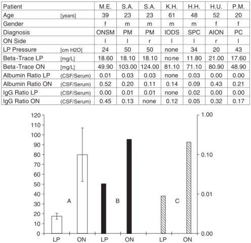
Patient M.E. S.A. S.A. K.H. H.H. H.U. P.M.
Age [years] 39 23 23 61 48 52 20
Gender f m m m m f f
Diagnosis ONSM PM PM IODS SPC AION PC
ON Side l l r l l r l
LP Pressure [cm H2O] 24 50 50 none 34 20 43 Beta -Trace LP [mg/L] 18.60 18.10 18.10 no ne 11.80 21.00 17.60 Beta -Trace ON [mg/L] 49.90 103.00 124.00 81.10 71.10 80.90 48.90 Albumin Ratio LP (CSF/Serum) 0.01 0.03 0.03 none 0.03 0.00 0.00 Albumin Ratio ON (CSF/Serum) 0.52 0.20 0.11 0.14 0.09 0.43 0.21 IgG Ratio LP (CSF/Serum) 0.00 0.01 0.01 none 0.02 0.00 0.00 IgG Ratio ON (CSF/Serum) 0.45 0.13 none 0.12 0.05 0.32 0.17
Fig. 2 Table and graph of CSF data from six patients. Table: The CSF samples are taken from seven SAS of the left (l) or/and the right (r) ON from three male (m) (mean age 44.0 6 19.3 years) and three female (f ) (mean age 37.0 6 16.0 years) patients after informed consent. Clinical diagnosis: pseudotumour cerebri (PC) and secondary pseudotumour cerebri (SPC), anterior ischaemic optic neuropathy (AION), pachymeningitis (PM), idiopathic optic disc swelling (IODS) and ON sheath meningioma (ONSM). Lumbar
puncture (LP) was performed in five patients and markers determined as described. Graph: Beta trace (A; light bars) in (mg/l) 6 SD, CSF/serum ratio of albumin (B, dark bars), and of IgG (C, hatched bars) of mean 6 SD and logarithmic from six patients
obtained by spinal tap (LP) and puncture of the SAS of the ON.
suggesting additional pathophysiological components (Gliklich et al., 1989).
Acknowledgement
We would like to thank Ronald M. Burde—former head of Department of Ophthalmology, Albert Einstein College of Medicine, New York—for continued inspiration and friendship.
Conflict of interest statement. None declared. References
Bito LZ, Vavson H. Local variations in cerebrospinal fluid composition and its relationship to the composition of the extracellular fluid of the cortex. Exp Neurol 1966; 14: 264–80.
Boulton M, Armstrong D, Flessner M, et al. Raised intracranial pressure increases CSF drainage through arachnoid villi and extracranial lymphatics. Am J Physiol 1998; 275: 889–96.
Brodsky M, Vaphiades M. Magnetic resonance imaging in pseudotumor cerebri. Ophthalmology 1998; 105: 1686–93.
Dichiro G. Movement of the cerebrospinal fluid in human beings. Nature 1964; 204: 290–1.
Felgenhauer K. Protein filtration and secretion at human body fluid barriers. Pflugers Arch 1980; 384: 9–17.
Fujitani Y, Kanaoka Y, Aritake K, et al. Pronounced eosinophilic lung inflammation and Th2 cytokine release in human lipocalin-type prostaglandin D synthase transgenic mice. J Immunol 2002; 168: 443–9. Galbraith JEK, Sullivan JH. Decompression of the perioptic meninges for
relief of papilledema. Am J Ophthalmol 1973; 76: 687–92.
Gliklich RE, Steinmann WC, Spaeth GL. Visual field change in low-tension glaucoma over a five-year follow-up. Ophthalmology 1989; 96: 316–20.
Hayreh SS. Pathogenesis of oedema of the optic disc (papilloedema). A preliminary report. Br J Ophthalmol 1964; 48: 522–43.
Hoffman A, Conradt HD, Gross G, et al. Purification and chemical characterization of beta-trace protein from human cerebrospinal fluid: its identification as prostaglandin D synthase. J Neurochem 1993; 61: 451–6.
Johnston M. Relationship between cerebrospinal fluid and extracranial lymph. Lymphology 2000; 33: 1–3.
Johnston M. The importance of lymphatics in cerebrospinal fluid transport. Lymphat Res Biol 2003; 1: 41–4.
Killer HE, Flammer J. Unilateral papilledema caused by a fronto-temporoparietal arachnoid cyst. Am J Ophthalmol 2001; 132: 589–91. Killer HE, Laeng HR, Groscurth P. Lymphatic capillaries in the meninges of
the human optic nerve. J Neuroophthalmol 1999; 19: 222–8.
Killer HE, Laeng HR, Flammer J, et al. Architecture of arachnoid trabeculae, pillars, and septa in the subarachnoid space of the human optic nerve: anatomy and clinical considerations. Br J Ophthalmol 2003a; 87: 777–81.
Killer HE, Mironov A, Flammer J. Optic neuritis with marked distension of the optic nerve sheath due to local fluid congestion. Br J Ophthamol 2003b; 87: 249.
Link H, Olsson JE. Beta-trace protein concentration in CSF in neurological disorders. Acta Neurol Scand 1972; 48: 57–68.
Liu D, Kahn M. Measurement and relationship of subarachnoidal pressure of the optic nerve to intracranial pressures in fresh cadavers. Am J Ophthal-mol 1993; 116: 548–56.
Logdberg L, Wester L. Immunocalins: a lipocalin subfamily that modulates immune and inflammatory responses. Biochim Biophys Acta 2000; 1482: 284–97.
Lu¨demann W, Berens von Rautenfeld D, Samii M, et al. Ultrastructure of the cerebrospinal fluid outflow along the optic nerve into the lymphatic system. Childs Nerv Syst 2005; 21: 96–103.
Milohart TH. Hydrocephalus and the cerebrospinal fluid. Baltimore: Williams and Wilkins; 1972.
Ragolia L, Palaia T, Paric E, et al. Elevated L-PGDS activity contributes to PMA induced apoptosis concomitant with downregulation of PI3-K. Am J Physiol Cell Physiol 2003; 284: C119–26.
Reiber H. Dynamics of brain-derived proteins in cerebrospinal fluid. Clin Chim Acta 2001; 310: 173–86.
Reiber H. Flow rate of cerebrospinal fluid (CSF)—a concept common to normal blood-CSF barrier function and to dysfunction in neurological diseases. J Neurol Sci 1994; 122: 189–203.
Reiber H, Felgenhauer K. Protein transfer at the blood cerebrospinal fluid barrier and the quantitation of the humoral immune response within the central nervous system. Clin Chim Acta 1987; 163: 319–28. Rubenstein E. Relationship of senescence of cerebrospinal fluid circulatory
system to dementias of the aged. Lancet 1998; 351: 283–5.
Silverberg GD, Mayo M, Saul T, et al. Alzheimer’s disease, normal-pressure hydrocephalus, and senescent changes in CSF circulatory physiology: a hypothesis. Lancet Neurol 2003; 2: 506–11.
Taniike M, Mohri I, Educhi N, et al. Perineuronal oligodendrocytes protect against neuronal apoptosis through the production of lipocalin-type prostaglandin D synthase in a genetic demyelinating model. J Neurosci 2002; 22: 4885–96.
Tumani H, Nau R, Felgenhauer K. Beta-trace protein in cerebrospinal fluid: a blood-CSF barrier-related evaluation in neurological diseases. Ann Neurol 1998; 44: 882–9.
Urade Y, Hayaishi O. Prostaglandin D synthase: structure and function. Vitam Horm 2000; 58: 89–120.
Wood JH. Neurobiology of cerebrospinal fluid. 2nd ed. New York and London: Plenum Press; 1982.
Zakharov A, Papaiconomou C, Djenic J, et al. Lymphatic cerebrospinal fluid absorption pathway in neonatal sheep revealed by subarachnoid injection of microfil. Neuropathol Appl Neurobiol 2003; 29: 563–73.
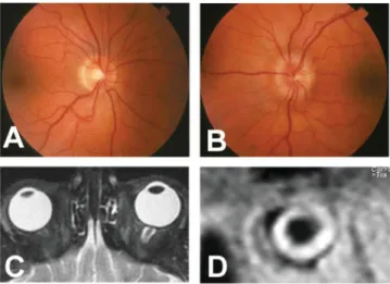
Fig. 3 Fundus photography and MRI studies of a patient with unilateral papilloedema (due to nerve sheath meningioma). (A) Unremarkable optic disc on the right. (B) Unilateral optic disc swelling due to left ON sheath meningioma. (C) Distended perioptic nerve sheath on the left with accumulation of CSF (white on T2-weighted magnetic resonance image, axial view). These radiological features meet the criteria for papilloedema as described previously by Brodsky and Vaphiades (1998). (D) Coronal view of the left orbit. Note marked distension of the SAS and accumulation of CSF.